Contents
What is claw hand
Claw hand is a condition that causes curved or bent fingers. Claw hand is a hand characterized by curved or bent fingers, making the hand appear claw-like. This makes the hand appear like the claw of an animal.
Causes of claw hand can also be due to anything that may lead to ulnar nerve palsy. Someone can be born with claw hand (congenital), or they can develop it because of certain disorders, such as ulnar nerve injury. Ulnar nerve palsy can arise from a laceration anywhere along its course. Proximal injuries to the medial cord of the brachial plexus may also present with sensory loss distally. Ulnar nerve palsies can also be due to cubital tunnel syndrome and ulnar tunnel syndrome. These are compression neuropathies at the elbow and wrist. Another cause of ulnar nerve palsy may be due to a failure to splint the hand in an intrinsic-plus posture following a crush injury. There are a few systemic diseases which may also lead to ulnar nerve palsy. These include leprosy, syringomyelia, and Charcot-Marie-Tooth disease. However, these systemic diseases usually involve more than one nerve.
The brachial plexus
The roots (anterior rami) of spinal nerves C5–C8 and T1 form the brachial plexus, which extends inferiorly and laterally on either side of the last four cervical and first thoracic vertebrae (Figures 1 and 2). The brachial plexus passes above the first rib posterior to the clavicle and then enters the axilla (armpit).
The brachial plexus provides almost the entire nerve supply of the shoulders and upper limbs (Figures 1 and 2). Five large terminal branches arise from the brachial plexus:
- (1) The axillary nerve supplies the deltoid and teres minor muscles.
- (2) The musculocutaneous nerve supplies the anterior muscles of the arm.
- (3) The radial nerve supplies the muscles on the posterior aspect of the arm and forearm.
- (4) The median nerve supplies most of the muscles of the anterior forearm and some of the muscles of the hand.
- (5) The ulnar nerve supplies the anteromedial muscles of the forearm and most of the muscles of the hand.
Figure 1. Brachial plexus
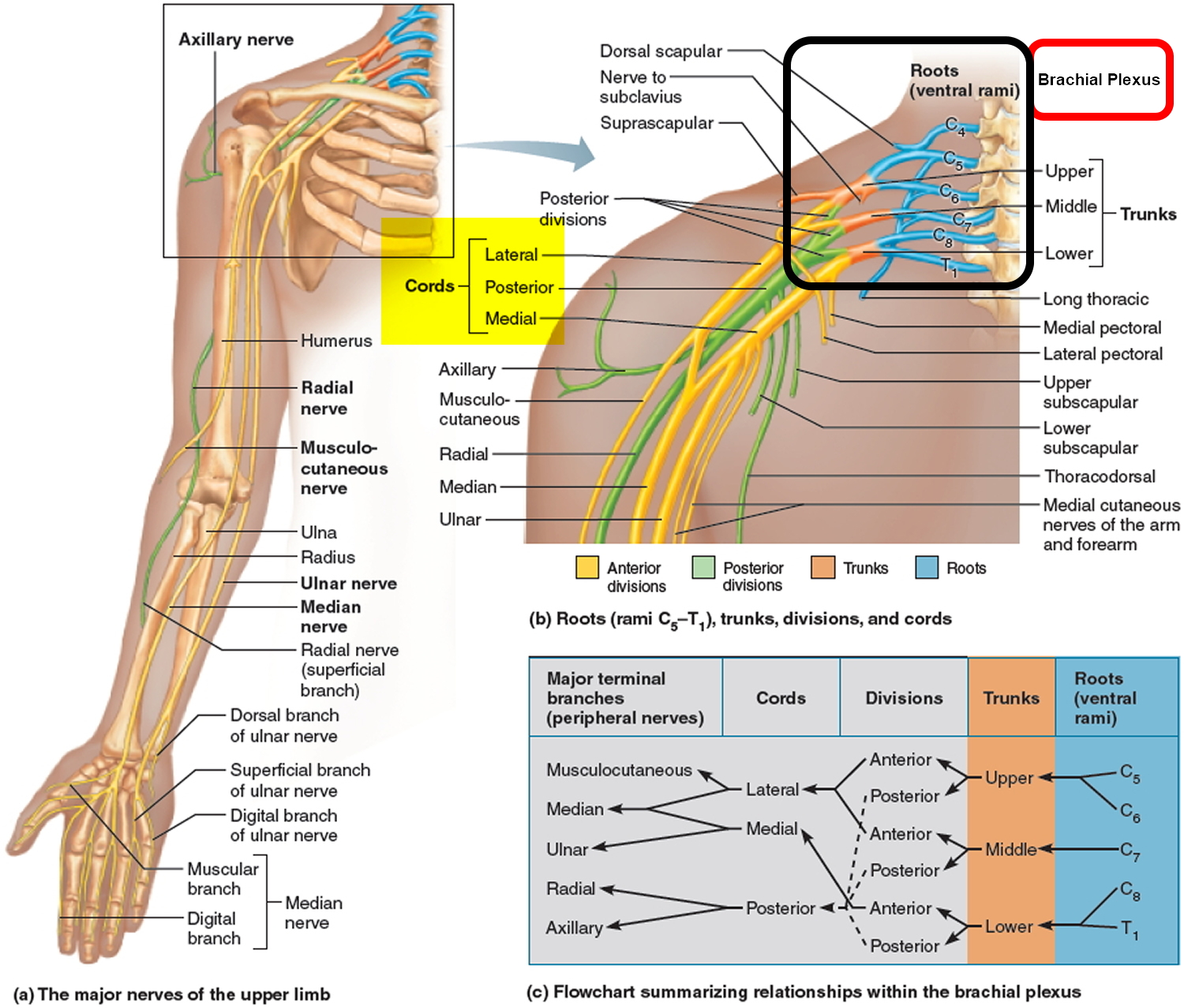
Figure 2. Brachial plexus motor and sensory innervation of the upper limb
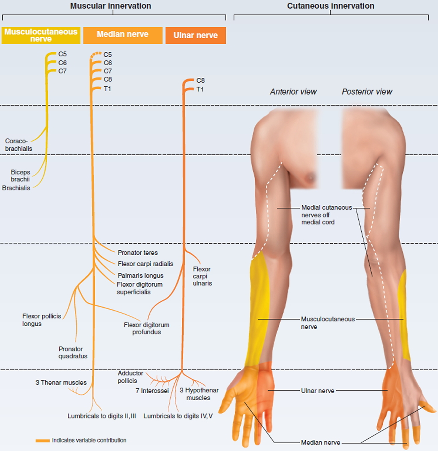
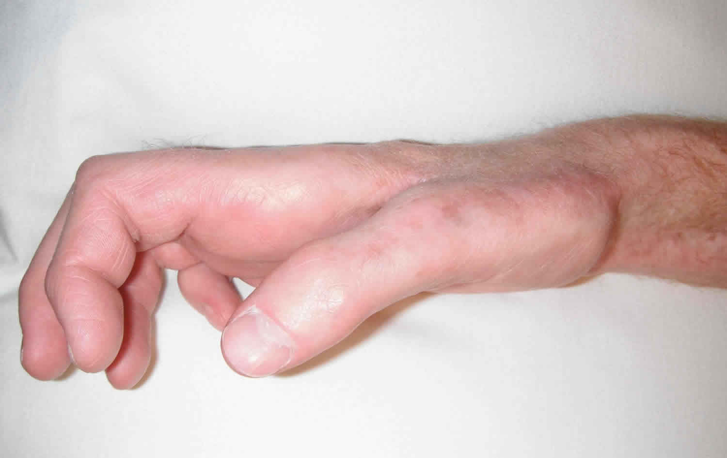
Figure 3. Klumpke’s paralysis (C8-T1 nerve damage) claw hand (Ulnar claw hand)
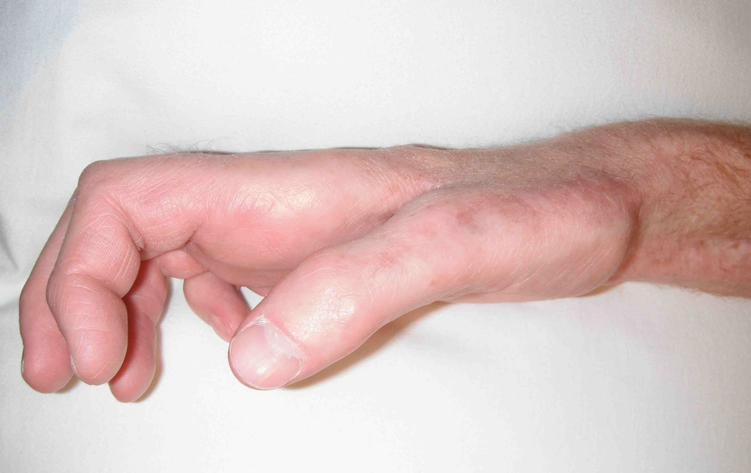
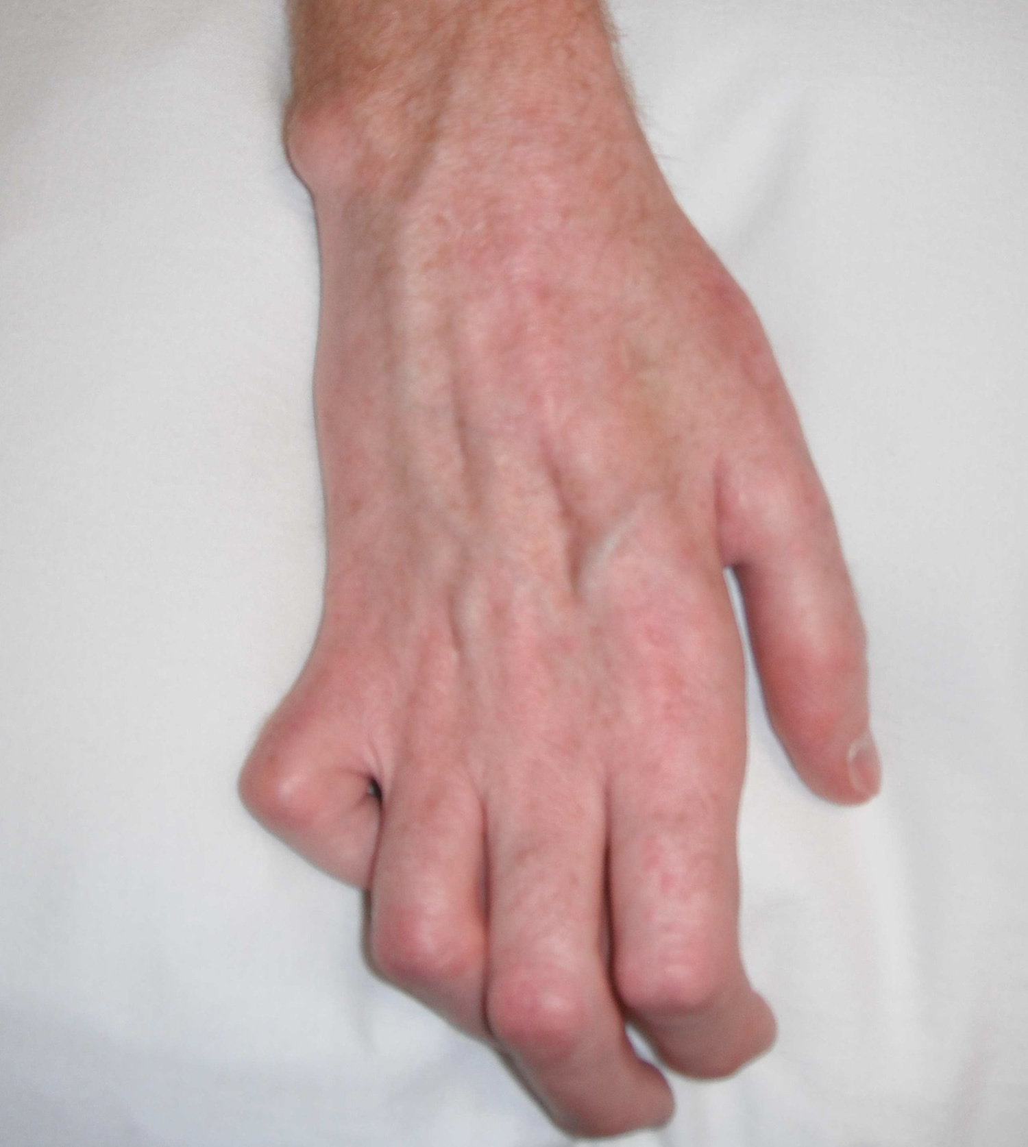
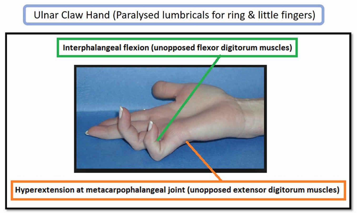
Figure 4. Ulnar claw hand
Claw hand causes
Causes claw hand may include:
- Congenital abnormality, such as from Charcot-Marie-Tooth disease
- Ulnar nerve damage in the arm
- Scarring after a severe burn of the hand or forearm
- Rare infections, such as leprosy
Ulnar nerve palsy is the loss of sensory and motor function. This can occur after injury to any portion of the ulnar nerve. The ulnar nerve is the terminal branch of the brachial plexus medial cord (C8, T1) (see Figures 1 and 2). The ulnar nerve innervates the flexor carpi ulnaris after it passes through the cubital tunnel.
Other muscles innervated by the ulnar nerve are the flexor digitorum profundus of the ring and small fingers and the following hand muscles:
- Abductor digiti minimi
- Flexor digiti minimi
- Opponens digiti minimi
- Ring
- Small finger lumbricals
- Dorsal and palmar interosseous muscles
- Adductor pollicis
- Deep head of flexor pollicis brevis
- The first dorsal interosseous
When the ulnar nerve is injured, the muscles innervated by the nerve begin to weaken. This leads to an imbalance between the strong extrinsic muscles (i.e., extensor digitorum communis) and the weakened intrinsic muscles (i.e., interosseous and lumbricals). This imbalance is characterized clinically by metacarpophalangeal (MCP) hyperextension and proximal interphalangeal (PIP) and distal interphalangeal (DIP) flexion (see Figures 3 and 4).
Claw hand diagnosis
Initial presentation will include a decrease in normal hand function.
The metacarpophalangeal (MCP) joints will be hyperextended, and the interphalangeal joints flexed.
The second and third digits will not be as involved as the fourth and fifth digits with a true ulnar nerve palsy. This is because the median nerve innervates the lumbricals involving the second and third digits, and the ulnar nerve innervates the lumbricals involving the fourth and fifth digits.
The patient may also exhibit functional weakness while attempting a grasp, grip or pinch.
A provocative test for claw hand is bringing the metacarpophalangeal (MCP) joints into flexion. This will correct the distal interphalangeal (DIP) and proximal interphalangeal (PIP) joint deformities.
Several other specific tests for ulnar nerve palsy include:
- Froment sign: Hyperflexion of the thumb interphalangeal joint while attempting to grab. This indicates a substitution of flexor pollicis longus (innervated by median nerve) for adductor pollicis (innervated by ulnar nerve).
- Jeanne sign: Reciprocal hyperextension of the thumb metacarpophalangeal joint indicating substitution of flexor pollicis longus for adductor pollicis.
- Wartenberg sign: Abduction of the small finger at metacarpophalangeal joint indicating deficient palmar intrinsic muscle (innervated by ulnar nerve) with abduction from extensor digiti minimi (innervated by radial nerve).
- Duchenne sign: Clawing of the ring and small fingers, hyperextension of metacarpophalangeal joints and flexion of proximal interphalangeal joints indicating deficient interosseous and lumbrical muscles of the ring and small fingers.
Tests
The following tests may be done to evaluate the ulnar nerve damage and to rule out other diagnoses:
- Electromyography (EMG) to check the health of the muscles and the nerves that control the muscles
- Nerve conduction studies to check how fast electrical signals move through a nerve
Claw hand treatment
Treatment depends on the cause of claw hand. It may include:
- Splinting
- Surgery to fix problems that may be contributing to the claw hand, such as nerve or tendon problems, joint contractures, or scar tissue
- Therapy to straighten the fingers
Nonoperative management is applied if a fixed flexion contracture of more than 45 degrees occurs at the proximal interphalangeal joint. A strenuous hand therapy program is utilized involving serial casting.
The majority of cases will need operative management in the form of contracture release and passive tenodesis versus active tendon transfer 1). This treatment is reserved for those patients with a progressive deformity that is affecting their quality of life. The goal is to prevent lasting metacarpophalangeal joint hyperextension.
- Surgery is usually in the form of tendon transfers. This addresses issues including the lack of thumb adduction and lateral pinch, the claw deformity of the fingers that impairs object acquisition and the loss of ring and small finger flexion.
- The extensor carpi radialis brevis or the flexor digitorum superficialis are the most commonly used transfers to restore thumb adduction. The brachioradialis can be used if the extensor carpi radialis brevis is required for an intrinsic reconstruction of the fingers.
- To correct the claw deformity of the fingers include static procedures or dynamic transfers. A dynamic transfer uses the flexor digitorum superficialis, extensor carpi radialis longus, extensor carpi radialis brevis, or flexor carpi radialis as a donor muscle.
- To restore the ring and small finger extrinsic muscle function, a transfer of flexor digitorum profundus ring and small to flexor digitorum profundus middle is performed.
Claw hand surgery complications
- More complications occur after intrinsic muscle transfers than adductorplasty because of the unique balance of the extensor hood mechanism.
- The transfer may not be suitable if the chosen muscle has insufficient strength or excursion. Elongation is also a problem with sewing the transfer into the lateral bands of the extensor hood.
- Tendon transfers that are not strong enough can be treated with a therapy program to strengthen the muscle but often require surgical revision.
- The transfer may also not be suitable if the chosen muscle has too much strength or too short of excursion. When the transfer is sewn too tightly into the lateral band, it can produce a swan-neck deformity of the digit.
- Tendon transfers that are too tight or too strong can be treated with a passive range of motion therapy to allow stretching to occur.
Claw hand surgery postoperative and rehabilitation care
A very experienced hand therapist plays a vital role in the postoperative care of tendon transfers for ulnar nerve palsy. Protecting the transfers with custom splints while mobilizing uninvolved joints requires strict adherence to postoperative protocols. Following most procedures, the hand is immobilized for 3 to 4 weeks, followed by a blocking splint to allow motion within the restraints of the splint for the next 3 to 4 weeks. Passive exercises are started at 6 weeks and strengthening at 8 weeks for the adductorplasty and 10 to 12 weeks for the intrinsic tendon transfers.
References [ + ]


