Contents
What is color blind
Color blindness or its correct name color vision deficiency, is the inability to see certain colors in a normal way. There are color-sensing pigments in the nerve cells of the eye that pick up red, blue, or green light. People with color blindness lack some or all of these pigments. If just one pigment is missing, it may be difficult to see the difference between red and green or between blue and yellow.
Color vision deficiency can range from mild to severe, depending on the cause. It affects both eyes if it is inherited and usually just one if it is caused by injury or illness. Most people with color blindness (color vision deficiency) aren’t aware of differences among colors that are obvious to the rest of us. The most severe form of color blindness is achromatopsia (total color blindness).
There are three main kinds of color blindness, based on photopigment defects in the three different kinds of cones that respond to blue, green, and red light.
- Red-Green color blindness are the most common. Red-green color blindness is congenital or inherited. It’s far more common in males than females, but still very rare. Red-green color blindness affects 5 to 8 percent of males and 0.5 percent of females. For people with red-green color blindness, reds and greens look similar to each other as a kind of brownish, muted tone.
- The other major types are Blue-Yellow color blindness. Blue-yellow color vision defects affect males and females equally. This condition occurs in fewer than 1 in 10,000 people worldwide.
- A complete absence of color vision (achromatopsia) —total color blindness – is rare. A person with achromatopsia cannot see any color, so everything is in shades of gray.
People who don’t have the more severe types of color blindness may not even be aware of their condition unless they’re tested in a clinic or laboratory.
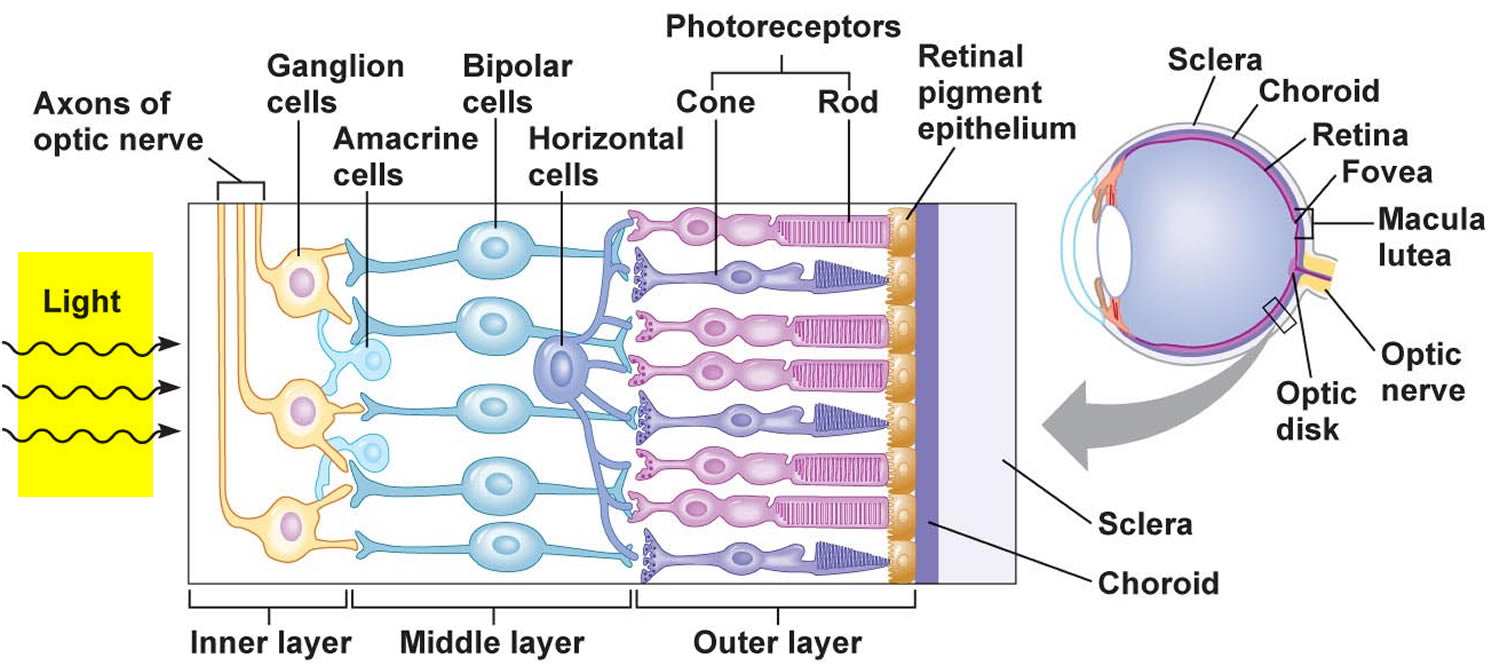
Inherited color blindness is caused by abnormal photopigments. These color-detecting molecules are located in cone-shaped cells within the retina, called cone cells (see Figure 2). In humans, several genes are needed for the body to make photopigments, and defects in these genes can lead to color blindness.
To understand what causes color blindness, you need to know about the cones in your eyes (see Figure 2). These cones are cells on your retina, an area the size of a postage stamp that’s at the back of your eye. You have “red,” “blue,” and “green” cones, which are sensitive to those colors and combinations of them. You need all three types to see colors properly. Color blindness usually affects both eyes equally and remains stable throughout life.
Color blindness can happen when one or more of the color cone cells are absent, not working, or detect a different color than normal. When your cones don’t work properly, or you don’t have the right combination, your brain doesn’t get the right message about which colors you’re seeing. To someone who’s color blind, a green leaf might look tan or gray.
- Most of the time, color blindness is genetic. There is no treatment, but most people adjust and the condition doesn’t limit their activities. If you have color vision deficiency, it is possible for you to learn to recognize color by other means. Some people learn to tell colors apart by brightness or location. Also, there are specially tinted eyeglasses that may help you to tell certain colors apart.
- Sometimes color blindness can be caused by physical or chemical damage to the eye, the optic nerve, or parts of the brain that process color information. These nonhereditary conditions are described as acquired color vision deficiencies. Color vision can also decline with age, most often because of cataract – a clouding and yellowing of the eye’s lens.
Early diagnosis can help children with color blindness from low self-esteem in primary school. If your child has a color vision deficiency, be sure to tell their teachers so they can design lesson plans accordingly. It is helpful to label crayons, markers, or colored pencils. Make sure reading materials are printed with black ink on white paper, as colored paper and ink can cause problems. Teach color blind students the colors of common items so they will have a frame of reference when people are discussing colors.
If you suspect you have problems distinguishing certain colors or your color vision changes, see an eye doctor for testing. It’s important that children get comprehensive eye exams, including color vision testing, before starting school.
There’s no cure for inherited poor color vision, but if illness or eye disease is the cause, treatment may improve color vision.
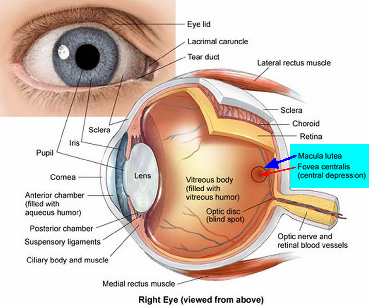
Figure 1. Structures of the eye
Inner Layer of the Eye
The inner layer of the wall of the eye consists of the retina, which contains the visual receptor cells (photoreceptors). The retina is a nearly transparent sheet of tissue continuous with the optic nerve in the back of the eye and extending forward as the inner lining of the eyeball. The retina ends just behind the margin of the ciliary body.
The retina is thin and delicate, but its structure is quite complex. It has a number of distinct layers, as Figure 2 illustrates. In the central region of the retina is a yellowish spot called the macula lutea. A depression in its center, called the fovea centralis, is in the region of the retina that produces the sharpest vision (see Figure 1).
Just medial to the fovea centralis is an area called the optic disc. Here, nerve fibers from the retina leave the eye and form the optic nerve. Because the optic disc region does not have photoreceptors, it is commonly known as the blind spot of the eye. A central artery and vein also pass through the optic disc. These vessels are continuous with the capillary networks of the retina. Along with vessels in the underlying choroid coat, they supply blood to the cells of the inner layer.
Photoreceptors are modified neurons of two distinct kinds – rods and cones, as Figure 2 illustrates. There is only one type of rod, but there are three types of cones.
- One group of photoreceptors, called rods, have long, thin projections at their ends, and rods detect only light and dark and are very sensitive to low light levels. Rods are more responsive to dim light, which makes them useful for night vision.
- The other group of photoreceptors, called cones, have short, blunt projections, and provide color vision. Cones are more responsive to bright light, such as in the daytime when light is plentiful. Cone cells detect color and are concentrated near the center of your vision. There are three types of cones that see color: red, green and blue. The brain uses input from these cone cells to determine our color perception.
- A human eye has 125 million rods and 7 million cones. Rods and cones are in a deep part of the retina. They are closely associated with an adjacent layer of retinal pigment epithelium (RPE) that absorbs light the photoreceptors do not absorb. With the pigment of the choroid coat, the retinal pigment epithelium keeps light from reflecting off surfaces inside the eye.
Another important difference is that all rods contain only one photopigment, while cones contain one of three different photopigments. This makes cones sensitive to long (red), medium (green), or short (blue) wavelengths of light. The presence of three types of photopigments, each sensitive to a different part of the visual spectrum, is what gives us our rich color vision.
Humans are unusual among mammals for our trichromatic vision – named for the three different types of photopigments we have. Most mammals, including dogs, have just two photopigment types. Other creatures, such as butterflies, have more than three. They may be able to see colors we can only imagine.
Most of us have a full set of the three different cone photopigments and so we share a very similar color vision experience, but because the human eye and brain together translate light into color, each of us sees colors differently. The differences may be slight. Your blue may be more blue than someone else’s, or in the case of color blindness, your red and green may be someone else’s brown.
Photoreceptors are stimulated only when light reaches them. A light image focused on an area of the retina stimulates some photoreceptors, which results in impulses traveling to the brain. However, the impulses leaving in response to each activated photoreceptor deliver only a fragment of the information required for the brain to interpret a complete scene.
Rods and cones contribute to different aspects of vision. Rods are hundreds of times more sensitive to light than cones and therefore can provide vision in dim light, but without color. Cones detect color but are less sensitive and do not respond in dim light.
Rods and cones also differ in the sharpness of the perceived images, or visual acuity. Cones provide sharp images, and rods provide more general outlines of objects. Rods give less precise images because axon branches from rods undergo convergence. Because of this, impulses from a group of rods are conducted to the brain on a single nerve fiber. Thus, if a point of light stimulates a rod, the brain cannot tell which one of many receptors has been stimulated. Convergence of impulses is less common among cones. When a cone is stimulated, the brain can pinpoint the stimulation more accurately.
The fovea centralis, the area of sharpest vision, does not have rods but contains densely packed cones with few or no converging fibers. Also in the fovea centralis, the overlying layers of the retina and the retinal blood vessels are displaced to the sides, more fully exposing photoreceptors to incoming light. Consequently, to view something in detail, a person moves the eyes so that the important part of an image falls on the fovea centralis.
Both rods and cones contain light-sensitive pigments that decompose when they absorb light energy. The light-sensitive biochemical in rods is called rhodopsin or visual purple. In the presence of light, rhodopsin molecules are broken down into a colorless protein called opsin and a yellowish substance called retinal (retinene) that is synthesized from vitamin A.
Decomposition of rhodopsin molecules activates an enzyme that initiates a series of reactions altering the permeability of the rod cell membrane. As a result, a complex pattern of nervous stimulation originates in the retina. The impulses are conducted away from the retina along the optic nerve into the brain, where they are interpreted as vision. In bright light, nearly all of the rhodopsin in the rods of the retina decomposes, greatly reducing rod sensitivity. In dim light, however, regeneration of rhodopsin from opsin and retinal is faster than rhodopsin breakdown. ATP provides the energy required for this regeneration.
The light-sensitive pigments in cones, as in the rods, are composed of retinal and protein. In cones, however, three types of opsin proteins, different from the protein portion of rhodopsin found in rods, combine with retinal to form the three cone pigments. The three types of cones each contain one of these three photopigments.
Poor vision in dim light, called nightblindness, results from vitamin A deficiency. Lack of the vitamin reduces the supply of retinal, rhodopsin production falls, and rod sensitivity is low. Nightblindness is treated by supplementing the diet with vitamin A.
The wavelength of light determines the color that the brain perceives from it. For example, the shortest wavelengths of visible light are perceived as violet, and the longest are perceived as red. One type of cone pigment (erythrolabe) is most sensitive to red light, another (chlorolabe) to green light, and the third (cyanolabe) to blue light. The color a person perceives depends on which cones the light in a given image stimulates. If all three sets of cones are stimulated with equal intensity, the person senses the light as white, and if none are stimulated, the person senses black. Different forms of colorblindness result from the body’s inability to produce different types of cone pigments.
Figure 2. The retina (inside human eye)
What is the most common type of color blindness?
The most common type of color blindness is red-green color blindness. It is often due to a genetic problem that is more common in men (X-linked recessive inheritance pattern). About 1 in 12 men have some form of color blindness.
Can color blindness affect my child’s future?
Color blindness is not a severe limitation as they learn to adapt by looking for other cues, such as brightness or location. Color blindness can make certain jobs more difficult, such as electricians, fashion designers, painters, cooks, and military pilots.
Types of color blindness
The most common types of color blindness are inherited. They are the result of defects in the genes that contain the instructions for making the photopigments found in cones. Some defects alter the photopigment’s sensitivity to color, for example, it might be slightly more sensitive to deeper red and less sensitive to green. Other defects can result in the total loss of a photopigment. Depending on the type of defect and the cone that is affected problems can arise with red, green, or blue color vision.
Red-Green Color Blindness
The most common types of hereditary color blindness are due to the loss or limited function of red cone (known as protan) or green cone (deutran) photopigments. This kind of color blindness is commonly referred to as red-green color blindness.
Red-green color vision defects and blue cone monochromacy are inherited in an X-linked recessive pattern. The OPN1LW and OPN1MW genes are located on the X chromosome, which is one of the two sex chromosomes. In males (who have only one X chromosome), one genetic change in each cell is sufficient to cause the condition. Males are affected by X-linked recessive disorders much more frequently than females because in females (who have two X chromosomes), a genetic change would have to occur on both copies of the chromosome to cause the disorder. A characteristic of X-linked inheritance is that fathers cannot pass X-linked traits to their sons.
- Protanomaly: In males with protanomaly, the red cone photopigment is abnormal. Red, orange, and yellow appear greener and colors are not as bright. This condition is mild and doesn’t usually interfere with daily living. Protanomaly is an X-linked disorder estimated to affect 1 percent of males.
- Protanopia: In males with protanopia, there are no working red cone cells. Red appears as black. Certain shades of orange, yellow, and green all appear as yellow. Protanopia is an X-linked disorder that is estimated to affect 1 percent of males.
- Deuteranomaly: In males with deuteranomaly, the green cone photopigment is abnormal. Yellow and green appear redder and it is difficult to tell violet from blue. This condition is mild and doesn’t interfere with daily living. Deuteranomaly is the most common form of color blindness and is an X-linked disorder affecting 5 percent of males.
- Deuteranopia: In males with deuteranopia, there are no working green cone cells. They tend to see reds as brownish-yellow and greens as beige. Deuteranopia is an X-linked disorder that affects about 1 percent of males.
Blue-Yellow Color Blindness
Blue-yellow color vision defects are inherited in an autosomal dominant pattern, which means one copy of the altered OPN1SW gene in each cell is sufficient to cause the condition. In many cases, an affected person inherits the condition from an affected parent.
Blue-yellow color blindness is rarer than red-green color blindness. Blue-cone (tritan) photopigments are either missing or have limited function.
- Tritanomaly: People with tritanomaly have functionally limited blue cone cells. Blue appears greener and it can be difficult to tell yellow and red from pink. Tritanomaly is extremely rare. It is an autosomal dominant disorder affecting males and females equally.
- Tritanopia: People with tritanopia, also known as blue-yellow color blindness, lack blue cone cells. Blue appears green and yellow appears violet or light grey. Tritanopia is an extremely rare autosomal recessive disorder affecting males and females equally.
Complete color blindness
People with complete color blindness (monochromacy) don’t experience color at all and the clearness of their vision (visual acuity) may also be affected.
There are two types of monochromacy:
- Cone monochromacy: This rare form of color blindness results from a failure of two of the three cone cell photopigments to work. There is red cone monochromacy, green cone monochromacy, and blue cone monochromacy. People with cone monochromacy have trouble distinguishing colors because the brain needs to compare the signals from different types of cones in order to see color. When only one type of cone works, this comparison isn’t possible. People with blue cone monochromacy, may also have reduced visual acuity, near-sightedness, and uncontrollable eye movements, a condition known as nystagmus. Cone monochromacy is an autosomal recessive disorder.
- Rod monochromacy or achromatopsia: This type of monochromacy is rare and is the most severe form of color blindness. It is present at birth. None of the cone cells have functional photopigments. Lacking all cone vision, people with rod monochromacy see the world in black, white, and gray. And since rods respond to dim light, people with rod monochromacy tend to be photophobic – very uncomfortable in bright environments. They also experience nystagmus. Rod monochromacy is an autosomal recessive disorder.
Achromatopsia
Achromatopsia is a condition characterized by a partial or total absence of color vision. People with complete achromatopsia cannot perceive any colors; they see only black, white, and shades of gray. Incomplete achromatopsia is a milder form of the condition that allows some color discrimination.
Achromatopsia also involves other problems with vision, including an increased sensitivity to light and glare (photophobia), involuntary back-and-forth eye movements (nystagmus), and significantly reduced sharpness of vision (low visual acuity). Affected individuals can also have farsightedness (hyperopia) or, less commonly, nearsightedness (myopia). These vision problems develop in the first few months of life.
Achromatopsia is different from the more common forms of color vision deficiency (also called color blindness), in which people can perceive color but have difficulty distinguishing between certain colors, such as red and green.
Achromatopsia affects an estimated 1 in 30,000 people worldwide. Complete achromatopsia is more common than incomplete achromatopsia.
Complete achromatopsia occurs frequently among Pingelapese islanders, who live on one of the Eastern Caroline Islands of Micronesia. Between 4 and 10 percent of people in this population have a total absence of color vision.
Genetic Changes
Achromatopsia results from changes in one of several genes: CNGA3, CNGB3, GNAT2, PDE6C, or PDE6H. A particular CNGB3 gene mutation underlies the condition in Pingelapese islanders.
Achromatopsia is a disorder of the retina, which is the light-sensitive tissue at the back of the eye. The retina contains two types of light receptor cells, called rods and cones. These cells transmit visual signals from the eye to the brain through a process called phototransduction. Rods provide vision in low light (night vision). Cones provide vision in bright light (daylight vision), including color vision.
Mutations in any of the genes listed above prevent cones from reacting appropriately to light, which interferes with phototransduction. In people with complete achromatopsia, cones are nonfunctional, and vision depends entirely on the activity of rods. The loss of cone function leads to a total lack of color vision and causes the other vision problems. People with incomplete achromatopsia retain some cone function. These individuals have limited color vision, and their other vision problems tend to be less severe.
Some people with achromatopsia do not have identified mutations in any of the known genes. In these individuals, the cause of the disorder is unknown. Other genetic factors that have not been identified likely contribute to this condition.
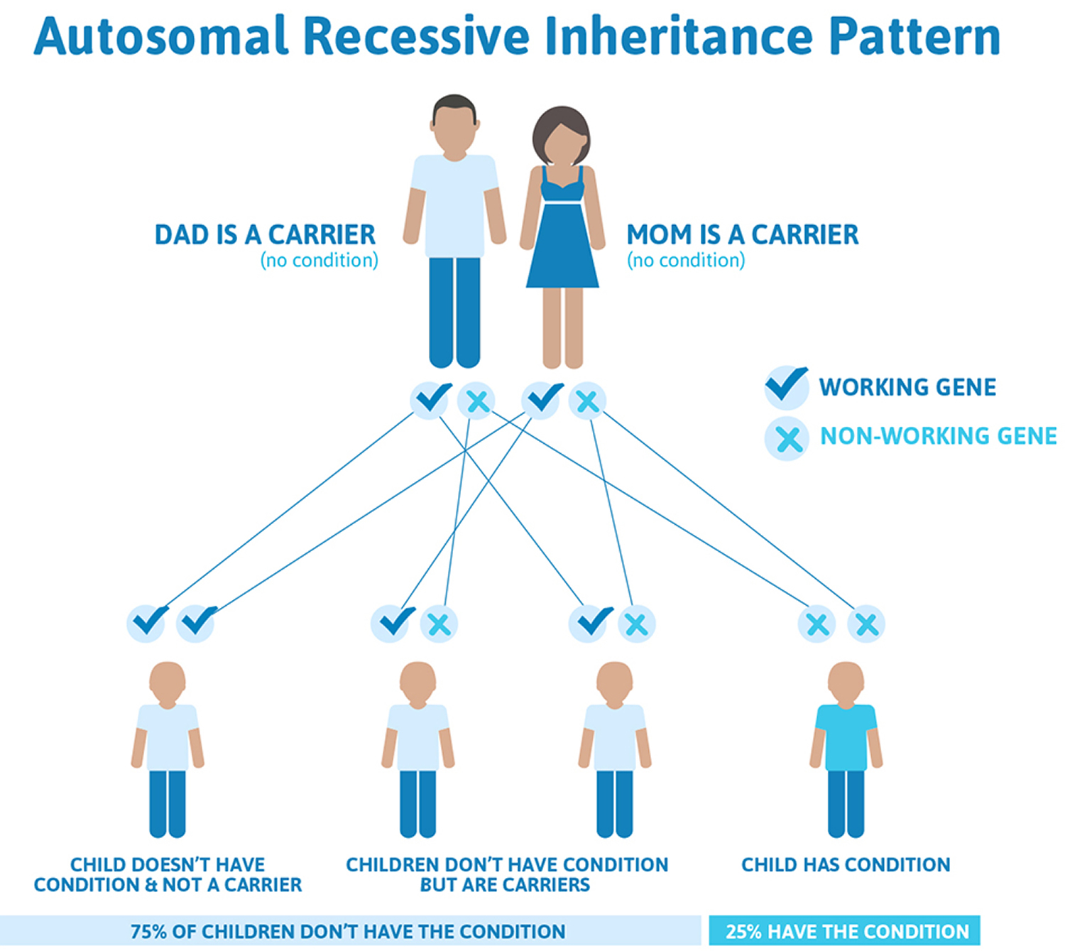
This condition is inherited in an autosomal recessive pattern, which means both copies of the gene in each cell have mutations. The parents of an individual with an autosomal recessive condition each carry one copy of the mutated gene, but they typically do not show signs and symptoms of the condition.
Autosomal refers to the fact that whatever gene is involved is found on one of the first 22 chromosomes (called the autosomes) and not on the X or Y chromosome (the sex chromosomes).
Recessive refers to the fact that you have two copies of each gene, one from mom and one from dad, and in order to have a recessive condition a person has to have BOTH copies of a gene not working. The copy they inherit from mom is not working AND the copy they inherit from dad is not working, resulting in zero functioning copies of that gene. With recessive conditions, if you have one copy of the non-working gene (called a carrier), you do not have the condition and typically do not have any related symptoms. If both partners are a carrier for the same condition, there is only a 25% chance that they will both pass down the non-working copy of the gene to the baby, thus causing a genetic condition. This chance is the same with each pregnancy, no matter how many children they have with or without the condition. That means also there’s a 50% chance the baby would be a carrier just like it’s parents.
How will achromatopsia affect my child’s schooling?
With adequate help from teachers for the visually impaired, children with achromatopsia are usually able to attend mainstream schools. Front seat placement, large print books, and magnifying devices can be very helpful. A low vision evaluation will be necessary before school begins. More severely affected individuals may benefit from services available in schools specifically designated for the visually impaired.
Figure 3. Achromatopsia autosomal recessive inheritance pattern
How is the achromatopsia diagnosis made?
The diagnosis will be made by your ophthalmologist. Initially, symptoms such as light sensitivity and reduced vision will provide clues essential to the diagnosis. The retinal examination may in fact be normal. The color vision tests commonly performed in the clinic are the Ishihara pseudoisochromatic tests, H-R-R tests and the City University tests. The diagnosis can be confirmed by a specialized test called electroretinography (ERG) which usually confirms the diagnosis by showing specific waveforms shape. Genetic testing can be performed to test for the mutations in the most common genes. Also, new advances in retinal imaging gives Ophthalmologists better tools to evaluate the condition.
What treatment is available for Achromatopsia?
Currently there is no cure for achromatopsia. Research on gene therapy is ongoing and may lead to clinical treatments in the future. Animal models of achromatopsia in dogs and mice have shown promising results in restoring some cone function in the retina. Children should be checked for refractive errors (need for glasses). Prescribing glasses to correct refractive conditions such as far-sightedness (hyperopia), near-sightedness (myopia) and astigmatism can improve the vision somewhat but will not restore normal levels of vision. Red colored lenses help reduce the sensitivity to light and thus enhance visual functioning. NoIR (injection-molded) plastic wrap-around glasses have a top ‘shield’ that covers the top of a prescription frame as well as broad side shields which is important since stray light can be disabling. Examples are Corning Lenses: CPF 550 lenses (5% transmission, darkened) and CPF 550XD lenses (4% transmission, darkened). A newer device known as an eyeborg can help people with no color vision to perceive color through sound waves. Artist Neil Harbisson who suffered from achromatopsia was one of the first to use this device.
Color blindness causes
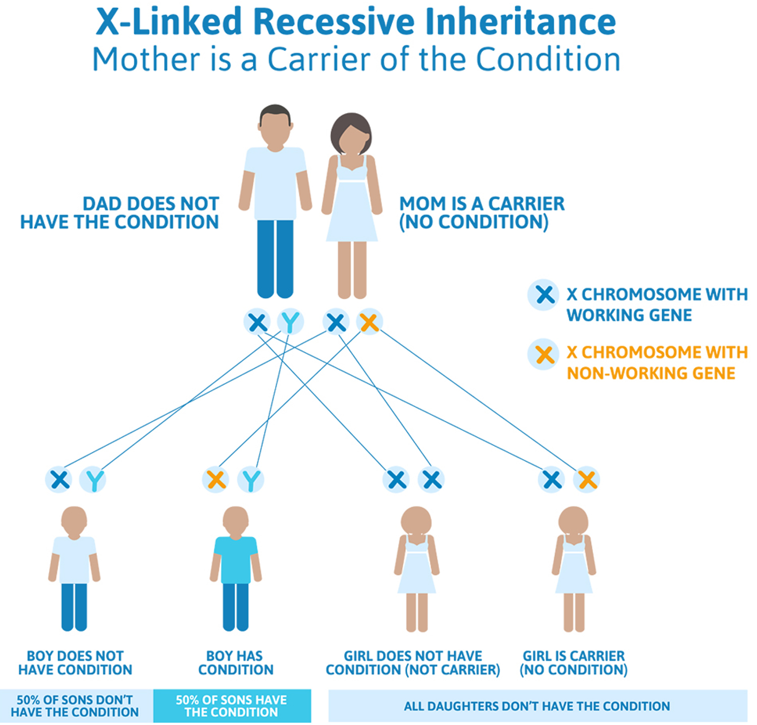
Most people with color blindness are born with it. This is called a congenital color blindness. Congenital red-green color vision defects usually pass from mother to son (X-linked recessive inheritance pattern).
Genes are bundled together on structures called chromosomes. One copy of each chromosome is passed by a parent at conception through egg and sperm cells. The X and Y chromosomes, known as sex chromosomes, determine whether a person is born female (XX) or male (XY) and also carry other traits not related to gender.
X-linked recessive means different things for men and women. Because women have two X chromosomes, if they have one broken copy of a gene on the X chromosome, they usually have back-up copy on their other X chromosome (called carriers). Because of this, often times women who are carriers for X-linked recessive conditions do not show any symptoms, although there are exceptions to this. Women generally would need to have inherited non-working genes on both of their X chromosomes to be affected with the genetic condition.
Men, however, only have the one X chromosome. If a gene on their X chromosome is not working, they do not have another X chromosome to provide a back-up (instead of a second X chromosome, they have a Y chromosome). Therefore, if an X-linked recessive gene is passed down to a male, they will be affected with the associated genetic condition.
These defects are due to partial or complete lack of cones in the retina. Cones help you to distinguish the colors red, green, and blue.
Figure 4. Red-Green Color Blindness (X-linked recessive inheritance pattern)
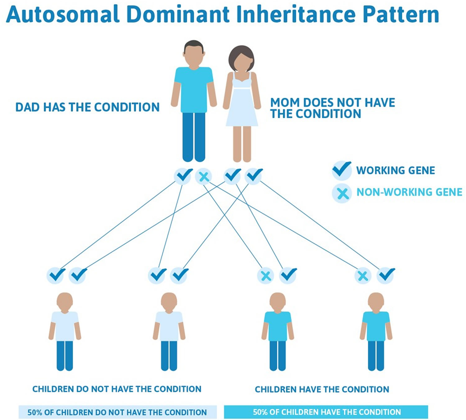
Blue-Yellow Color Blindness autosomal dominant inheritance pattern
Autosomal refers to the fact that whatever gene is involved is found on one of the first 22 chromosomes (called the autosomes) and not on the X or Y chromosome (the sex chromosomes).
Dominant refers to the above explanation that we have two copies of each gene, one from mom and one from dad, and in order to have an autosomal dominant condition, a person only has to have one copy of the gene not working. They can inherit this copy from mom or dad, who may also have the condition.
In cases where the autosomal dominant condition does run in the family, the chance for an affected person to have a child with the same condition is 50% regardless of whether it is a boy or a girl. It is the same 50% chance for each pregnancy.
Figure 5. Blue-Yellow Color Blindness (autosomal dominant inheritance pattern)
Most color vision problems that occur later in life are a result of:
- disease
- trauma
- toxic effects from drugs
- metabolic disease, or
- vascular disease
Color vision defects from disease are less understood than congenital color vision problems. Disease-specific color blindness often affects both eyes differently. Color vision defect caused by disease usually gets worse over time. Acquired color vision loss can be the result of damage to the retina or optic nerve.
Who is at risk for color blindness?
Men are at much higher risk for being born with color blindness than women, who seldom have the problem. An estimated one in ten males has some form of color deficiency. Color blindness is more common among men of Northern European descent.
Having certain conditions may increase your risk for acquired color deficiency, including:
- Glaucoma
- Diabetes
- Macular degeneration
- Alzheimer’s disease
- Parkinson’s disease
- Chronic alcoholism
- Leukemia, and
- Sickle cell anemia
Certain drugs may also increase your risk for acquiring color blindness. The drug hydroxychloroquine (Plaquenil) can cause color blindness. It is used to treat rheumatoid arthritis, among other conditions.
Color blind symptoms
The symptoms of color blindness can range from mild to severe. Symptoms can vary by the amount of pigment that is missing. Many people have such mild symptoms that they are unaware that they have a color deficiency. Parents may only notice a problem with a child when he is learning his colors. Often there is an inability to tell the difference between shades of the same or similar colors. Colors appear washed out and are easily confused with other colors. A child may color things “wrong” by making the sky purple, the grass orange, or trees yellow. Since most people with color blindness can see some colors, they often don’t know they see color differently than others.
The color blind symptoms include:
- trouble seeing colors and the brightness of colors in the usual way;
- inability to tell the difference between shades of the same or similar colors. This happens most with red and green, or blue and yellow.
Except in the most severe form, color blindness does not affect the sharpness of vision. The inability to see any color at all and to see everything only in shades of gray is called achromatopsia. This rare condition is often associated with:
- amblyopia
- nystagmus
- light sensitivity, and
- poor vision
Color blind diagnosis
Eye care professionals use a variety of tests to diagnose color blindness. These tests can quickly diagnose specific types of color blindness. The most common test uses a book with several patterns of colored dots. People with color deficiency will not see certain patterns.
The Ishihara Color Test is the most common test for red-green color blindness (see Figure 5 below). The test consists of a series of colored circles, called Ishihara plates, each of which contains a collection of dots in different colors and sizes. Within the circle are dots that form a shape clearly visible to those with normal color vision, but invisible or difficult to see for those with red-green color blindness.
If you do not have a color deficiency, you will be able to see numbers and shapes among the dots. If you are color blind, you will have a hard time finding the number or shape in the pattern. You may not see anything in the pattern at all.
The newer Cambridge Color Test uses a visual array similar to the Ishihara plates, except displayed on a computer monitor. The goal is to identify a C shape that is different in color from the background. The “C” is presented randomly in one of four orientations. When test-takers see the “C,” they are asked to press one of four keys that correspond to the orientation.
The anomaloscope uses a test in which two different light sources have to be matched in color. Looking through the eyepiece, the viewer sees a circle. The upper half is a yellow light that can be adjusted in brightness. The lower half is a combination of red and green lights that can be mixed in variable proportions. The viewer uses one knob to adjust the brightness of the top half, and another to adjust the color of the lower half. The goal is to make the upper and lower halves the same brightness and color.
The HRR Pseudoisochromatic Color Test is another red-green color blindness test that uses color plates to test for color blindness.
The Farnsworth-Munsell 100 Hue Test uses a set of blocks or pegs that are roughly the same color but in different hues (shades of the color). The goal is to arrange them in a line in order of hue. This test measures the ability to discriminate subtle color changes. It is used by industries that depend on the accurate color perception of its employees, such as graphic design, photography, and food quality inspection.
The Farnsworth Lantern Test is used by the U.S. military to determine the severity of color blindness. Those with mild forms pass the test and are allowed to serve in the armed forces.
There is no treatment for congenital color blindness. It usually does not cause any significant disability. However, there are special contact lenses and glasses that may help.
Your ophthalmologist can treat acquired forms of color blindness. He or she will address the underlying condition or drug that caused the problem.
Color blind test
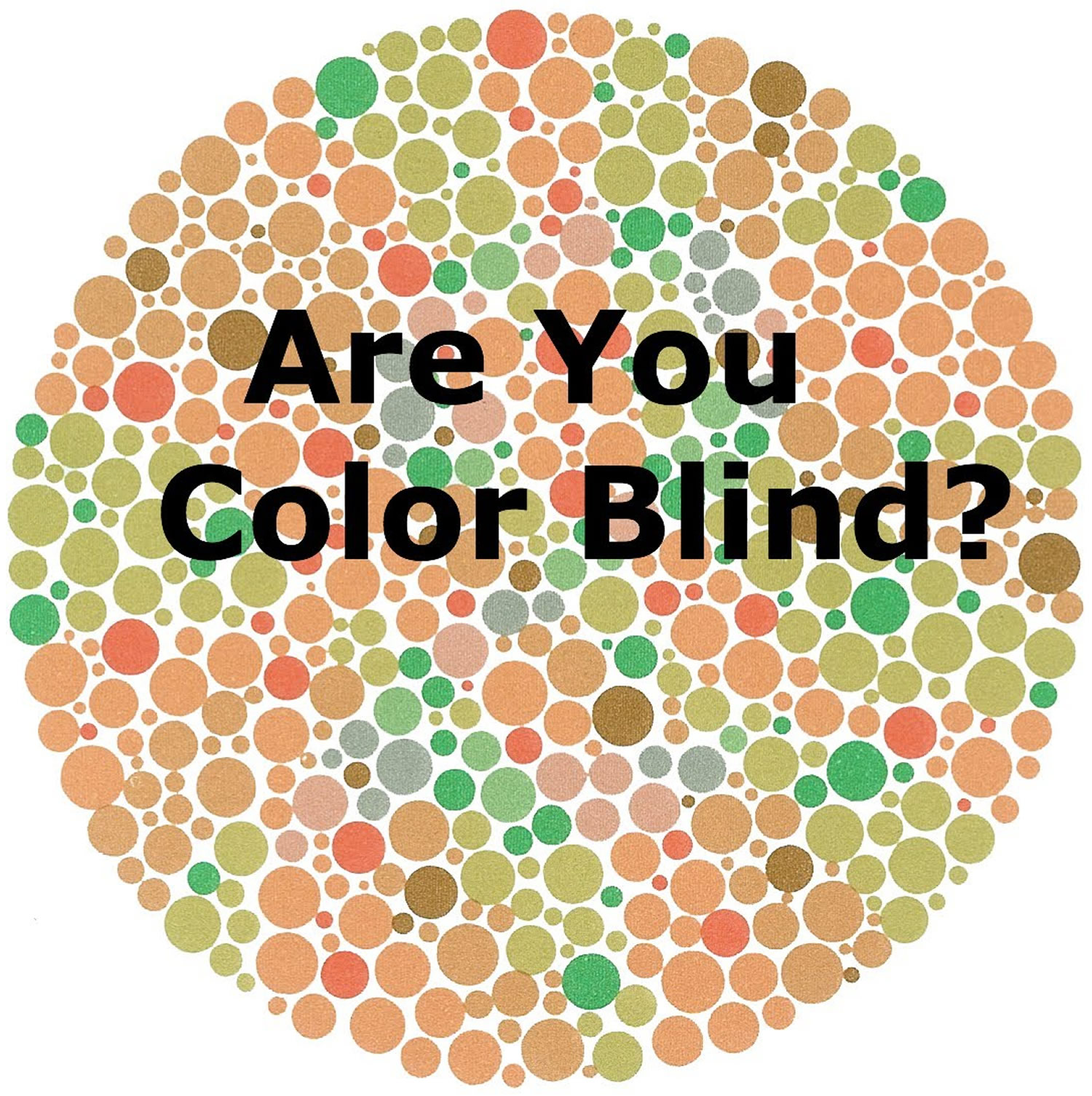
It’s easy to test whether you’re color blind. You don’t even need to go to a doctor. A set of images called the Ishihara color plates is one of the most common and reliable tests.
Simply look at the images, which have numbers embedded in dots of color. The numbers are a different color than the background. If you can’t see the numbers, you’re probably colorblind. School kids often take the Ishihara test as a classroom activity. It’s that easy. And can be fun. The Ishihara test is named for Japanese ophthalmologist Ishihara Shinobu, a professor at the University of Tokyo who developed the screening in 1918 for the military.
But for anyone taking this test, it’s important to understand what it means to be color blind. The condition is uncommon and it’s rarely serious. Testing positive is no reason to panic. But if you’re having noticeable vision problems that have come on suddenly or are getting worse and affect your color vision see your ophthalmologist for a check-up right away.
The American Academy of Ophthalmology doesn’t have formal recommendations for color blindness testing. You can have red/green color blindness and function well without even knowing it.
Figure 6. Ishihara color blindness test chart
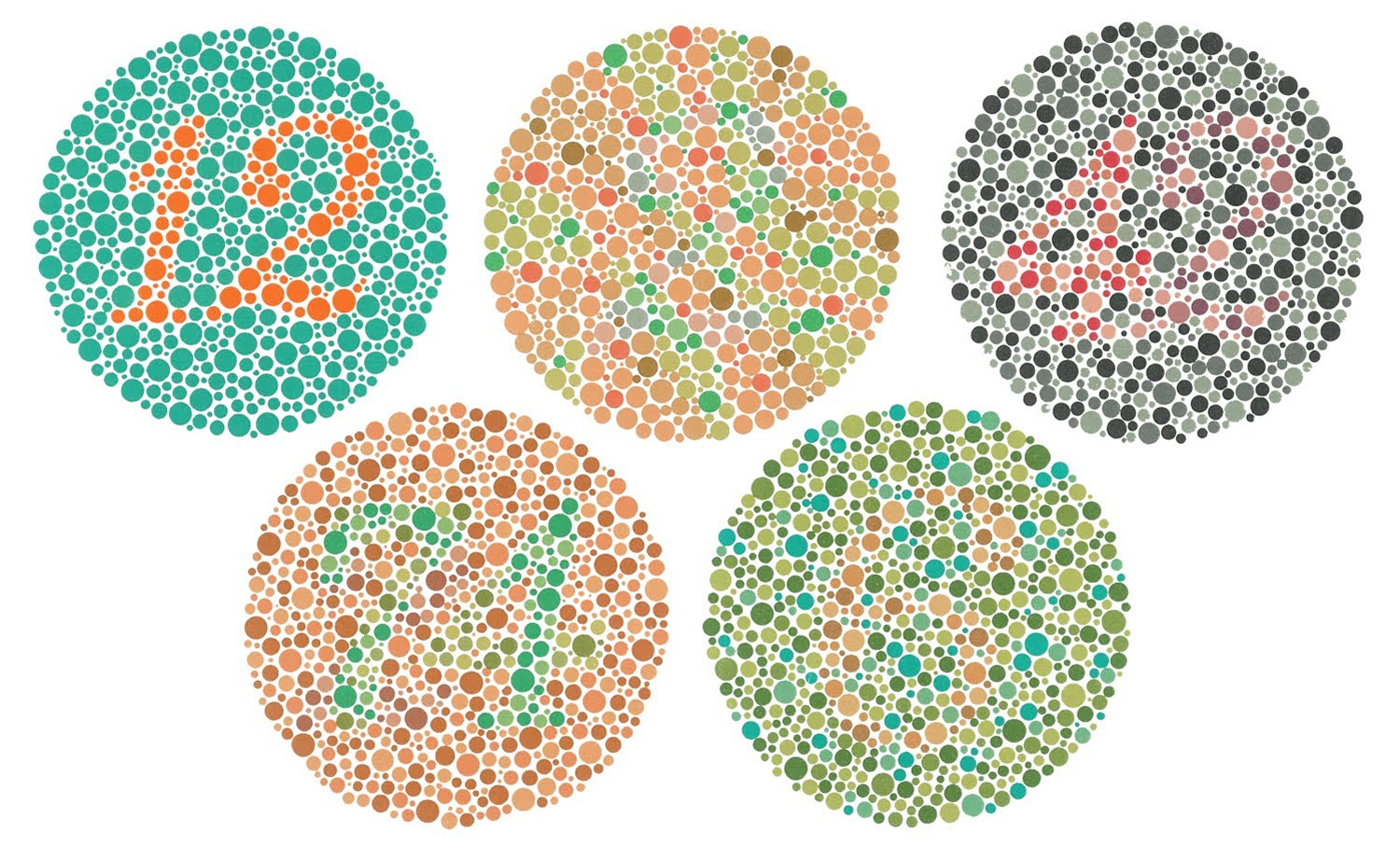 Note: The numbers from top left to right are 12, 2 and 42. And bottom left to right are 74 and 6
Color blind treatment
Inherited color blindness is something you are born with and is a lifelong condition. There is no cure for congenital color blindness. However, people with red-green color blindness may be able to use a special set of lenses to help them perceive colors more accurately. These lenses can only be used outdoors under bright lighting conditions. Visual aids have also been developed to help people cope with color blindness. There are iPhone and iPad apps, for example, that help people with color blindness discriminate among colors. Some of these apps allow users to snap a photo and tap it anywhere on the image to see the color of that area. More sophisticated apps allow users to find out both color and shades of color. These kinds of apps can be helpful in selecting ripe fruits such as bananas, or finding complementary colors when picking out clothing.
Wearing a colored filter over eyeglasses or a colored contact lens may enhance your perception of contrast between colors. But such lenses won’t improve your ability to see all colors.
Some rare retinal disorders associated with color deficiency could possibly be modified with gene replacement techniques. These treatments are under study and might become available in the future.
Try the following tips to help you work around your poor color vision.
- Memorize the order of colored objects. If it’s important to know individual colors, such as with traffic lights, memorize the order of the colors.
- Label colored items that you want to match with other items. Have someone with good color vision help you sort and label your clothing. Arrange your clothes in your closet or drawers so that colors that can be worn together are near each other.
Living with color blindness
Color blindness can make it difficult to read color-coded information such as bar graphs and pie charts. This can be particularly troubling for children who aren’t yet diagnosed with color blindness, since educational materials are often color-coded. Children with red-green color blindness may also have difficulty reading a green chalkboard when yellow chalk is used. Art classes, which require selecting appropriate colors of paint or crayons, may be challenging.
Color blindness can go undetected for some time since children will often try to hide their disorder. It’s important to have children tested, particularly boys, if there is a family history of color blindness. Many school systems offer vision screening tests that include color blindness testing. Once a child is diagnosed, he or she can learn to ask for help with tasks that require color recognition.
Simple everyday tasks like cooking meat to the desired color or selecting ripe produce can be a challenge for adults. Children might find food without bright color as less appetizing. Traffic lights pose challenges, since they have to be read by the position of the light. Since most lights are vertical, with green on bottom and red on top, if a light is positioned horizontally, a color blind person has to do a quick mental rotation to read it. Reading maps or buying clothes that match colors can also be difficult. However, these are relatively minor inconveniences and most people with color blindness learn to adapt.


