Contents
What is coma
A coma is a deep state of unconsciousness – where a person is unresponsive and cannot be woken 1. A coma, sometimes also called persistent vegetative state, is a profound or deep state of unconsciousness. Persistent vegetative state is not brain-death. An individual in a coma is alive but unable to move or respond to his or her environment and and has minimal brain activity. It is not possible to wake a coma patient using physical or auditory stimulation.
Consciousness requires both wakefulness and awareness.
- Wakefulness is the ability to open your eyes and have basic reflexes such as coughing, swallowing and sucking.
- Awareness is associated with more complex thought processes and is more difficult to assess. Currently, the assessment of awareness relies on physical responses being detected during an examination.
The coma person’s eyes will be closed and they’ll appear to be unresponsive to their environment. They won’t normally respond to sound or pain, or be able to communicate or move voluntarily. Additionally a person in a coma fails to respond normally to painful stimuli, light, or sound; lacks a normal sleep-wake cycle and, does not initiate voluntary actions, being unable to consciously feel, speak, hear, or move.
Someone in a coma will also have very reduced basic reflexes such as coughing and swallowing. They may be able to breathe on their own, although some people require a machine to help them breathe.
Over time, the person may start to gradually regain consciousness and become more aware. Some people will wake up after a few weeks, a coma rarely lasts more than 2 to 4 weeks, while others may go into a Vegetative State (Unresponsive Wakefulness Syndrome) or Minimally Conscious State. Coma patients can exhibit different levels of unconsciousness and unresponsiveness depending on which brain regions have been damaged and how much or how little of the brain is functioning.
People in a coma generally recover but some don’t. It can be very difficult to predict recovery when a person is in a coma. Sometimes a coma is followed by a persistent vegetative state.
Coma can occur as a complication of an illness, exposure to a drug (e.g, drug abuse, overdose or misuse of medications), alcohol, toxin, metabolic abnormalities, central nervous system diseases, acute neurologic injuries (e.g, stroke, hernia, hypoxia, hypothermia) or as a result of injuries, such as head trauma caused by falls or vehicle collisions etc. A coma is a medical emergency. However, in some instances, coma may be deliberately induced using pharmaceutical agents in order to preserve higher brain functions following brain trauma, or to save the patient from extreme pain during healing of injuries or diseases.
The outcome for coma depends on the cause, severity, and site of the damage. People may come out of a coma with physical, intellectual, and psychological problems. Some people may remain in a coma for years or even decades. For those people, the most common cause of death is infection, such as pneumonia.
Levels of coma
There are different levels of coma, ranging from very deep, where the patient shows no response or awareness at all, to shallower levels, where the patient responds to stimulation by movement or opening eyes. Still shallower levels can occur, where the patient is able to make some response to speech. Level of coma is usually initially assessed by the Glasgow Coma Scale (GCS).
The Glasgow Coma Scale (GCS) is a very simple, easy to administer technique which is used to rate the severity of coma. It assesses the patient’s ability to open their eyes, move and speak.
Caring for and monitoring a person in a coma
Doctors assess a person’s level of consciousness using a tool called the Glasgow Coma Scale (see below). This level is monitored constantly for signs of improvement or deterioration.
The Glasgow Coma Scale (GCS) assesses three things:
- Eye opening – a score of one means no eye opening, and four means opens eyes spontaneously
- Verbal response to a command – a score of one means no response, and five means alert and talking
- Voluntary movements in response to a command – a score of one means no response, and six means obeys commands
Most people in a coma will have a total score of eight or less. A lower score means someone may have experienced more severe brain damage and could be less likely to recover.
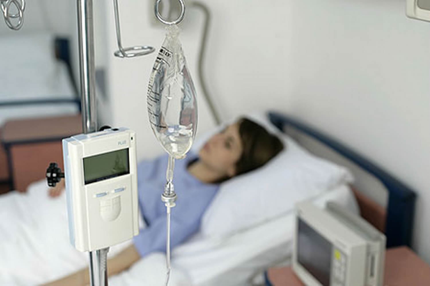
In the short term, a person in a coma will normally be looked after in an intensive care unit (ICU). Treatment involves ensuring their condition is stable and their body functions, such as breathing and blood pressure, are supported while the underlying cause is treated.
In the longer term, healthcare staff will give supportive treatment on a hospital ward. This can involve providing nutrition, trying to prevent infections, moving the person regularly so they don’t develop bedsores, and gently exercising their joints to stop them becoming tight.
What you can do as a visitor
The experience of being in a coma differs from person to person. Some people feel they can remember events that happened around them while they were in a coma, while others don’t.
Some people have reported feeling enormous reassurance from the presence of a loved one when coming out of a coma.
When visiting a friend or loved one in a coma, you may find the advice below helpful:
- when you arrive, announce who you are
- talk to them about your day as you normally would – be aware that everything you say in front of them might be heard
- show them your love and support – even just sitting and holding their hand or stroking their skin can be a great comfort
Research has also suggested that stimulating the main senses – touch, hearing, vision and smell – could potentially help a person recover from a coma.
As well as talking to the person and holding their hand, you might want to try playing them their favorite music through headphones, putting flowers in their room or spraying a favorite perfume.
The Glasgow Coma Scale
The treatment team will use the Glasgow Coma Scale (GCS) to evaluate a person’s level of consciousness (LOC) and the severity of brain injury by attempting to elicit body movements (M), opening of the eyes (E), and verbal responses (V).
Clinicians use the three values separately and collectively to make medical decisions and monitor the patient’s progress. This scale is typically used very early in the treatment process.
Table 1. Glasgow Coma Scale
| (M) Motor Response | (E) Eye Opening | (V) Verbal Response |
|---|---|---|
| (6) Follows commands | (4) Natural | (5) Oriented & converses |
| (5) Localizes to pain | (3) To voice | (4) Disoriented & converses |
| (4) Withdrawal to pain | (2) To pain | (3) Inappropriate words |
| (3) Decorticate | (1) No response | (2) Incomprehensible sounds |
| (2) Decerebrate | (1) No response | |
| (1) No response |
Your score for each is added up to give a total. A slightly different version of the Glasgow Coma Scale (GCS) is used for children under five years of age.
Table 2. Glasgow Coma Scale scores range from 15 to 3
| GCS Total Score | Level of Brain Injury |
|---|---|
| 13 to 15 | Mild Brain Injury |
| 9 to 12 | Moderate Brain Injury |
| 8 to 3 | Severe Brain Injury |
Someone with a Glasgow Coma Scale (GCS) score of 3 (the lowest possible score) will be in a coma (an unconsciousness state where a person is unresponsive and can’t be woken).
Glasgow Outcome Scale (GOS)
The Glasgow Outcome Scale (GOS) is a brief, one-item descriptive assessment utilized by the treatment healthcare team following brain injury. The Glasgow Outcome Scale (GOS) is helpful in determining next steps in the individual’s care, but is not useful in detecting small, gradual improvements.
There are five possible descriptive measures in the Glasgow Outcome Scale (GOS):
- Dead (Severe injury or death without recovery of consciousness)
- Vegetative (Severe damage with prolonged state of unresponsiveness and a lack of higher mental functions)
- Severely Disabled (Severe injury with permanent need for help with daily living)
- Moderately Disabled (No need for assistance in everyday life; Employment is possible but may require special equipment)
- Good Recovery (Light damage with minor neurological and psychological deficits)
In addition to the Glasgow Coma Scale (GCS), the treatment team will likely utilize other assessments to understand and classify the severity of brain injury.
Glasgow Outcome Scale – Extended (GOS-E)
The Glasgow Outcome Scale – Extended (GOS-E) is an extension of the Glasgow Outcome Scale (GOS) designed to address the original scale’s insensitivity to changes or progress over time. The Glasgow Outcome Scale – Extended (GOS-E) extends the original five categories in the Glasgow Outcome Scale (GOS) to eight:
- Dead
- Vegetative
- Lower Severe Disability
- Upper Severe Disability
- Lower Moderate Disability
- Upper Moderate Disability
- Lower Good Recovery
- Upper Good Recovery
Abbreviated Injury Scale (AIS)
The Abbreviated Injury Scale (AIS) is a one-time tool the treatment team will use to measure the severity of brain injury. The Abbreviated Injury Scale (AIS) differs from other measures in that it represents the threat a brain injury presents to an individual’s life rather than assessing the severity of the injury.
Table 3. Abbreviated Injury Scale (AIS)
| Score | Injury |
|---|---|
| 1 | Minor |
| 2 | Moderate |
| 3 | Serious |
| 4 | Severe |
| 5 | Critical |
| 6 | Not Survivable |
JFK Coma Recovery Scale – Revised (JFK CRS-R)
The JFK Coma Recovery Scale – Revised (JFK CRS-R) is a measure often used by the treatment team to assess individuals who are in a minimally conscious or vegetative state. It is especially useful when measuring emerging consciousness. The JFK Coma Recovery Scale – Revised (JFK CRS-R) measures 23 items addressing auditory, visual, motor, and automatic body functions. It also measures communication and arousal functions.
Galveston Orientation & Amnesia Test (GOAT)
The Galveston Orientation and Amnesia Test (GOAT) is one of several instruments used by the treatment team to measure post-traumatic amnesia. The test features ten questions that assess the patient’s temporal and spatial orientation (Where Am I?), biographical recall (Who Am I?), and memory (What Happened?).
Points are awarded for responses to each question, with a total of 100 points possible. A score of 78 or greater over three consecutive days indicates the patient is emerging from post-traumatic amnesia.
Younger patients (those under the age of fifteen) are given a modified version of the test, known as the Children’s Orientation and Attention Test (COAT).
Orientation Log (O-LOG)
The Orientation Log (OLOG) is one of several instruments used by the treatment team to measure post-traumatic amnesia. The OLOG measures the individual’s orientation to time, place, and circumstance over a period of time.
Westmead Post-Traumatic Amnesia Scale (WPTAS)
The Westmead Post-Traumatic Amnesia Scale (WPTAS) is a brief test that is often administered at the individual’s bedside by a member of his or her treatment team. The WPTAS measures the length of post-traumatic amnesia in patients who have sustained a brain injury.
Coma outlook
Comas can last from days to weeks while some severe cases have lasted several years. Recovery from a coma depends, to a considerable extent, on the original cause of the coma and on the severity of any brain damage. Some patients (e.g. patients in a diabetic coma) will make a complete recovery while others, particularly those who have suffered a head trauma, may have some physical, intellectual or psychological impairment that will require further treatment. They may need physiotherapy, occupational therapy, psychological assessment and support during a period of rehabilitation and may need care for the rest of their lives.
The chances of someone recovering from a coma largely depend on the severity and cause of their brain injury, their age and how long they’ve been in a coma. It’s impossible to accurately predict whether the person will eventually recover, how long the coma will last and whether they’ll have any long-term problems.
Patients can gradually come out of the coma, some progress to a Vegetative State (also known as Unresponsive Wakefulness Syndrome) and others die. Some patients who have entered a vegetative state go on to regain a degree of awareness (Minimally Conscious State). The likelihood of significant functional improvement for coma patients diminishes over time.
The outlook for coma and persistent vegetative state depends on the cause, severity, and site of neurological damage. Individuals may emerge from coma with a combination of physical, intellectual, and psychological difficulties that need special attention. Recovery usually occurs gradually, with some acquiring more and more ability to respond. Some individuals never progress beyond very basic responses, but many recover full awareness. Individuals recovering from coma require close medical supervision. A coma rarely lasts more than 2 to 4 weeks. Some patients may regain a degree of awareness after persistent vegetative state. Others may remain in that state for years or even decades. The most common cause of death for someone in a persistent vegetative state is infection, such as pneumonia.
Recovering from a coma
A coma usually only lasts a few weeks, during which time the person may start to gradually wake up and gain consciousness, or progress into a different state of unconsciousness called a vegetative state or minimally conscious state.
- a Vegetative State – where a person is awake but shows no signs of being aware of their surroundings or themselves
- a Minimally Conscious State – where a person has limited awareness that comes and goes
Recovery from coma is a gradual process, starting with the person’s eyes opening, then responding to pain, and then responding to speech. People do not just wake up from a coma, and say, ‘Where am I?’ as is sometimes portrayed in films. The length of coma is one of the most accurate predictors of the severity of long-term symptoms. The longer the coma, the greater the likelihood of residual symptoms, particularly physical disabilities, although this is only a guide and some people can make good recoveries after an extended period in a coma.
Some people may recover from these states gradually, while others may not improve for years, if at all.
People who do wake up from a coma usually come round gradually. They may be very agitated and confused to begin with.
Some people will make a full recovery and be completely unaffected by the coma. Others will have disabilities caused by the damage to their brain. They may need physiotherapy, occupational therapy and psychological assessment and support during a period of rehabilitation, and may need care for the rest of their lives.
The chances of someone recovering from a coma largely depend on the severity and cause of their brain injury, their age and how long they’ve been in a coma. But it’s impossible to accurately predict whether the person will eventually recover, how long the coma will last and whether they’ll have any long-term problems.
What is Vegetative State (Unresponsive Wakefulness Syndrome)
A vegetative state is when a person is awake but showing no signs of awareness 2. On recovery from the coma state, Vegetative State/Unresponsive Wakefulness Syndrome is characterized by the return of arousal without signs of awareness. In contrast, a coma is a state that lacks both awareness and wakefulness. Absence of awareness can only be inferred by lack of responsiveness to the environment and not as lack of consciousness that scientists may not be able to detect by behavioral measures. For this reason, many authors have suggested that the term ‘Unresponsive Wakefulness Syndrome’ 3 or ‘post-coma unresponsiveness’ are more accurate descriptive terms for vegetative state.
A person in a vegetative state may open their eyes, wake up and fall asleep at regular intervals and have basic reflexes, such as blinking when they’re startled by a loud noise, or withdrawing their hand when it’s squeezed hard. They’re also able to regulate their heartbeat and breathing without assistance.
However, a person in a vegetative state doesn’t show any meaningful responses, such as following an object with their eyes or responding to voices. They also show no signs of experiencing emotions nor of cognitive function.
Vegetative State/Unresponsive Wakefulness Syndrome patients’ eyes might be in a relatively fixed position, may track moving objects (visual pursuit), or move in a completely unsynchronized manner. Sleep-wake cycles may resume or patients may appear to be in a state of chronic wakefulness. They may grind their teeth, swallow, smile, shed tears, grunt, moan, or scream without any apparent external stimulus. Vegetative State/Unresponsive Wakefulness Syndrome patients do not respond to sound, hunger, or pain. Patients cannot obey verbal commands and lack local motor responses. Additionally Vegetative State/Unresponsive Wakefulness Syndrome patients cannot talk in comprehensible terms and may become noisy, restless, and hypermobile.
One of the most challenging tasks facing clinicians is that of differentiating Vegetative State/Unresponsive Wakefulness Syndrome from Minimally Conscious States.
Whilst neuro imaging such as MRI is widely used in assessing brain damage and functional abilities, behavioral assessment has, until now, been the “gold standard” for detecting signs of consciousness and thereby for determining diagnosis.
If a person is in a vegetative state for a long time, it may be considered to be:
- a continuing vegetative state – when it’s been longer than four weeks
- a permanent vegetative state – when it’s been more than six months if caused by a non-traumatic brain injury, or more than 12 months if caused by a traumatic brain injury
If a person is diagnosed as being in a permanent vegetative state, recovery is extremely unlikely but not impossible.
Criteria for a vegetative state
A vegetative state is when a person is awake but showing no signs of awareness. Doctors are particularly careful when diagnosing a permanent vegetative state, as there’s a risk of misdiagnosis.
A confident diagnosis can only be made if the following criteria have been met:
- the cause of the brain injury has been established – for example, if a case of meningitis is suspected, a diagnosis can be confirmed by testing the fluid that surrounds the brain for infection
- it’s been confirmed drugs or medication aren’t responsible for the symptoms
- it’s been confirmed treatable problems with the body’s chemistry (a metabolic disorder) aren’t responsible for the symptoms of loss of awareness – an
- example of a metabolic disorder is a diabetic coma, where people lose consciousness because their blood sugar levels are either dangerously high or dangerously low
- the possibility of a treatable cause in the brain, such as a brain tumor, has been ruled out by brain imaging scans
- examinations have been carried out by a trained assessor experienced in prolonged disorders of consciousness
For a permanent vegetative state to be confirmed, the above criteria must apply and either:
- six months must have passed since the start of symptoms after a non-traumatic brain injury
- 12 months must have passed since the start of symptoms after a traumatic brain injury.
Vegetative State/Unresponsive Wakefulness Syndrome Outlook (Prognosis)
Many patients emerge spontaneously from Vegetative State/Unresponsive Wakefulness Syndrome within a few weeks. Some people improve gradually, whereas others stay in a state of impaired consciousness for years. Many people never recover consciousness.
The chances of recovery depend on the extent of injury to the brain and age, with younger patients having a better chance of recovery than older patients. Generally, adults have about a 50 percent chance and children a 60 percent chance of recovering consciousness from Vegetative State/Unresponsive Wakefulness Syndrome within the first 6 months in the case of traumatic brain injury. For non-traumatic injuries such as strokes, the recovery rate falls within the first year. After this period the chances that Vegetative State/Unresponsive Wakefulness Syndrome patient will regain consciousness are very low and, of those patients who do recover consciousness, most experience significant disability. The longer a patient is in Vegetative State/Unresponsive Wakefulness Syndrome the more severe the resulting disabilities are likely to be.
Some patients who have entered a vegetative state go on to regain a degree of awareness (see Minimally Conscious State). The likelihood of significant functional improvement for Vegetative State/Unresponsive Wakefulness Syndrome patients diminishes over time. There are only isolated cases of people recovering consciousness after several years. The few people who do regain consciousness after this time often have severe disabilities caused by the damage to their brain.
Vegetative State/Unresponsive Wakefulness Syndrome Treatment
Careful, ongoing assessment of the patient, using empirically validated assessment tools (e.g, the Coma Recovery Scale-Revised) is essential in order to evaluate and measure signs of progress, improvement or deterioration. Treatment is addressed at presenting symptoms and the patient’s needs; Vegetative State/Unresponsive Wakefulness Syndrome patients require constant monitoring and assistance with feeding, hydration, hygiene, assisted movement and physical therapies (to help prevent ulcers and blood clots in the legs), and elimination of waste products.
Currently no treatment for Vegetative State/Unresponsive Wakefulness Syndrome exists that would satisfy the efficacy criteria of evidence-based medicine. Pharmacological methods, surgery, physical therapy, and various stimulation techniques have been suggested. Pharmacological therapy mainly uses activating substances such as tricyclic antidepressants or methylphenidate. Surgical methods (e.g., deep brain stimulation) are used infrequently due to the invasiveness of the procedures. Stimulation techniques include sensory stimulation, sensory regulation, music and musicokinetic therapy, social-tactile interaction, etc.
Treatment can’t ensure recovery from a state of impaired consciousness, however supportive treatment is used to give the best chance of natural improvement.
This can involve:
- providing nutrition through a feeding tube
- making sure the person is moved regularly so they don’t develop pressure ulcers
- gently exercising their joints to prevent them becoming tight
- keeping their skin clean
- managing their bowel and bladder – for example, using a catheter to drain the bladder
- keeping their teeth and mouth clean
- efforts should be made to establish functional communication and environmental interaction when possible. Offering opportunities for periods of meaningful activity – such as listening to music or watching television, being shown pictures or hearing family members talking
Sensory stimulation;
- visual – showing photos of friends and family, or a favorite film
- hearing – talking or playing a favorite song
- smell – putting flowers in the room or spraying a favorite perfume
- touch – holding their hand or stroking their skin with different fabrics
While not empirically validated, families have reported benefits from arousal regimes, such as those implemented by Dr Ted Freeman (e.g, Coma Arousal Therapy). This intensive therapy involves family members taking the patient through a regimen of controlled auditory, visual and physical stimulation for up to six hours a day every day.
What is Minimally Conscious State
The minimally conscious state is a defined as severely altered consciousness in which minimal but definite, sustained and/or reproducible behavioral evidence of awareness of self or environment is demonstrated.
The person may have periods where they can communicate or respond to commands, such as moving a finger when asked.
A person may enter a minimally conscious state after being in a coma or vegetative state. In some cases a minimally conscious state is a stage on the route to recovery, but in others it’s permanent.
A continuing minimally conscious state means it has lasted longer than four weeks. However, it’s more difficult to diagnose a permanent minimally conscious state because it depends on things such as:
- the type of brain injury
- how severe the injury is
- how responsive the person is
In most cases, a minimally conscious state isn’t usually considered to be permanent until it’s lasted several years.
To make the diagnosis of Minimally Conscious State, limited but clearly discernible evidence of self or of environmental awareness must be demonstrated on a reproducible or sustained basis by one or more of the following behaviors:
- Follow simple commands (e.g, touch your nose, look up).
- Gestural or verbal yes/no responses (regardless of accuracy).
- Intelligible verbalization or communicate in some intelligible way
- Purposeful behavior that is not due to reflexive activity. For example, appropriate smiling or crying in response to emotional but not to neutral topics or stimuli – reaching for objects demonstrating a clear relationship between object location and direction of reach – touching or holding objects appropriately in relation to the size and shape of the object – pursuit eye movement or sustained fixation that occurs in direct response to moving or salient stimuli.
Minimally Conscious State Outlook (Prognosis)
To date, the natural history and long-term outcome of Minimally Conscious State has not been adequately investigated. Minimally Conscious State may occur in a variety of neurologic conditions, such as traumatic brain injury, stroke, progressive degenerative disorders, tumors, neurometabolic diseases, and congenital or developmental disorders. Following an acute injury, Minimally Conscious State can be a transitional or permanent state. Although it is not known how many patients will emerge from Minimally Conscious State twelve months or more after injury, most patients in Minimally Conscious State for this length of time remain severely disabled. The likelihood of significant functional improvement for Minimally Conscious State patients diminishes over time.
Minimally Conscious State Treatment
Treatment can’t ensure recovery from a state of impaired consciousness, however supportive treatment is used to give the best chance of natural improvement.
This can involve:
- providing nutrition through a feeding tube
- making sure the person is moved regularly so they don’t develop pressure ulcers
- gently exercising their joints to prevent them becoming tight
- keeping their skin clean
- managing their bowel and bladder – for example, using a catheter to drain the bladder
- keeping their teeth and mouth clean
- efforts should be made to establish functional communication and environmental interaction when possible. Offering opportunities for periods of
- meaningful activity – such as listening to music or watching television, being shown pictures or hearing family members talking
Sensory stimulation;
- visual – showing photos of friends and family, or a favorite film
- hearing – talking or playing a favorite song
- smell – putting flowers in the room or spraying a favorite perfume
- touch – holding their hand or stroking their skin with different fabrics
In all circumstances, the patient should be treated with dignity, and caregivers should be cognizant of the patient’s potential for understanding and perception of pain. In early Minimally Conscious State, prevention of complications and maintenance of bodily integrity should be emphasized because of the likelihood of further improvement. While not empirically validated, families have reported benefits from arousal regimes, such as those implemented by Dr Ted Freeman (e.g, Coma Arousal Therapy). The therapy involves family members taking the coma patient through a regimen of controlled auditory, visual and physical stimulation for up to six hours a day every day.
Staying in environments with limited exposure to sensory stimuli, such as intensive care units (ICU), increases the risk of sensory deprivation in patients and have some effects like perceptual, cognitive and emotional impairments on patients 4. One of the steps that can be done to prevent sensory deprivation in coma patients is using the sensory stimulation through sensory stimulation programs. Sensory stimulation programs for coma patients through stimulating the reticular activating system and increasing the level of cognitive functioning stimulate the brain and cause the patients come out of coma as soon as possible and achieve to optimal performance levels 5. Onset of sensory stimulation on the first 72 hours after brain injury has a great importance in saving the patient’s life, improving quality of life, and prognosis of the disease 6. In this regard Hyunsoon and Whasook 7 wrote: To facilitate the healing process and prevent the sensory deprivation in comatose patients with traumatic brain injury using the organized sensory stimulation programs in the early stages after brain injury is essential. Hosseinzadeh et al., 8 in their study concluded that patients receiving auditory stimulation back into consciousness by the nurse’s voice earlier than other patients. Among the various sensory stimulations, auditory stimulation more attention has taken into consideration because hearing is the last sense goes in coma patients, and unlike the other senses, there is no obstacle to stimulate this sense 4.
A person with experience in neurologic assessment of patients with impaired consciousness should be primarily responsible for establishing the diagnosis and prognosis and for coordinating clinical management. An additional opinion of a physician or other professional with particular expertise in the evaluation, diagnosis, and prognosis of patients in Minimally Conscious State is recommended when the assessment will impact critical management decisions.
Coma causes
Disorders of consciousness can occur if the parts of the brain involved with consciousness are damaged.
These types of brain injury can be divided into:
- Traumatic brain injury – the result of a severe head injury, such as an injury sustained during a car accident or a fall from a great height
- Non-traumatic brain injury – where the injury to the brain is caused by a health condition, such as a stroke
- Progressive brain damage – where the brain is gradually damaged over time; for example, because of Alzheimer’s disease
Non-traumatic brain injury
Non-traumatic brain damage is usually caused by a health condition, such as:
- a condition that deprives the brain of oxygen – without a continuous supply of oxygen, brain tissue begins to die
- a condition that directly attacks brain tissue
Specific causes of non-traumatic brain injury include:
- strokes
- heart attacks
- severe brain infections – such as meningitis (an infection of the outer layer of the brain) or encephalitis (an infection of the brain itself)
- drug overdoses
- poisoning
- almost drowning or other types of suffocation, such as smoke inhalation
- a blood vessel bursting, leading to bleeding inside the brain – the medical term for this is a ruptured aneurysm
Progressive brain damage
In some cases brain damage can gradually occur over time. Examples of conditions that cause progressive brain damage include:
- Alzheimer’s disease
- Parkinson’s disease
- a brain tumor
Traumatic brain injury
Traumatic brain injury occurs when an object or outside force causes severe trauma to the brain. This is most often caused by:
- falls
- traffic accidents
- violent assault
Severe head injuries require immediate medical attention because there’s a risk of serious brain damage.
Severe head injury complications
Severe head injuries can cause serious complications, mainly because the brain can be damaged, sometimes permanently.
A particularly severe head injury can be fatal, so a person with this type of injury will be closely monitored in hospital, so that any complications that arise can be dealt with promptly and effectively.
Infection
If your skull is fractured during a head injury, you may have a greater risk of developing an infection. Skull fractures can occasionally tear the membrane (the thin layer of cells) that surrounds the brain. If this happens, bacteria can enter the wound and cause an infection.
It’s important that any external wounds on your head are kept clean so they don’t become infected. You may also be prescribed antibiotics.
Post-concussion syndrome
Some people may experience long-term symptoms after sustaining concussion from a head injury. This could be post-concussion syndrome.
The symptoms and effects of post-concussion syndrome can include:
- difficulty looking after yourself
- not being able to work
- a persistent headache
- dizziness
- feeling weak
- tinnitus (hearing sounds that come from inside the body, rather than from an outside source)
- nausea
- feeling very tired and problems sleeping
- memory problems
- difficulty understanding others
- poor concentration
These symptoms usually clear up in around three months, but you may need to be referred for further assessment by your doctor. You may be seen by a neurologist, who specializes in problems of the nervous system (brain, spinal cord and nerves), or a psychiatrist (mental health specialist).
Impaired consciousness
Some people who sustain a severe head injury enter a state of impaired consciousness, such as a coma, vegetative state or minimally conscious state.
These disorders of consciousness affect wakefulness (the ability to open your eyes and have basic reflexes) and awareness (more complex thoughts and actions, such as following instructions, remembering and communicating).
These states sometimes only last a few weeks, after which time a person may wake up or progress into a different state of impaired consciousness. However, they can last years and some people will never regain consciousness.
Brain injury
A severe head injury can damage the brain in several ways. For example, brain damage can occur as a result of increased pressure on the brain caused by a blood clot between the skull and the surface of the brain (subdural haematoma), or bleeding in and around the brain (subarachnoid haemorrhage).
After a brain injury, there’s also an increased risk of epilepsy. A person who develops epilepsy following a head injury may need medication for a period of time or for life.
Brain injuries can also lead to a number of other problems, which can be temporary or permanent. The effect of a brain injury will depend on:
- the exact location of the injury
- the type of injury – for example, if the skull is fractured
- the severity of the injury – for example, if surgery is required
The different effects of a brain injury are described below.
Physical effects
Physical effects of a brain injury can include difficulty moving or keeping your balance and loss of co-ordination. You may also experience headaches or increased tiredness.
Hormonal effects
Some head injuries can damage the pituitary gland (a small gland that sits at the base of the brain and regulates the thyroid). If the pituitary gland is damaged, it may lead to reduced hormone production and problems such as an underactive thyroid (hypothyroidism).
Sensory effects
You may lose your sense of taste and smell. You may also notice blind spots in your vision, or you may not be able to control your body temperature as well as before, so that you feel too hot or too cold.
Cognitive effects
Following a head injury, you may find it difficult to think, process information and solve problems. You may also experience memory problems, particularly with your short-term memory, and have difficulty with speech and communication skills.
Emotional or behavioral effects
After a severe head injury, you may experience changes to your feelings and behavior. For example, you may be angrier or more easily irritated than before.
You may be less sensitive to other people’s feelings, or lose your inhibitions and behave in a way that other people consider inappropriate. You may also laugh or cry more than you did before the injury.
Severe head injury signs and symptoms
Symptoms of a severe head injury can include:
- unconsciousness – where a person has collapsed and is unresponsive, even for a brief period of time
- concussion – a sudden but short-lived loss of mental function that occurs after a blow or other injury to the head; a person with concussion may have a glazed look or appear confused, but won’t necessarily be unconscious
- fits or seizures
- difficulty speaking or staying awake
- problems with the senses – such as hearing loss or double vision
- repeated episodes of vomiting
- blood or clear fluid coming from the ears or nose
- memory loss (amnesia)
- sudden swelling or bruising around both eyes or behind the ear
- difficulty with walking or co-ordination
Dial your local emergency number immediately to request an ambulance if you’re with someone who experiences any of these symptoms after sustaining a head injury.
You should also go to hospital if someone has injured their head and:
- the injury was caused by a forceful blow to the head at speed, such as being hit by a car or falling one meter or more
- the person has had previous brain surgery
- the person has had previous problems with uncontrollable bleeding or a blood clotting disorder, or is taking medication that may cause bleeding problems,
- such as warfarin
- the person has been drinking alcohol or has taken drugs
- the injury wasn’t accidental – for example, you deliberately hurt yourself or someone else hurt you on purpose
Diagnosing a severe head injury
A person with a severe head injury should always be seen in the hospital an accident and emergency department.
If you’ve had a severe head injury and there’s a chance you may have a brain injury, you’ll have a computerized tomography (CT) scan to assess the seriousness of the injury.
A Computerised tomography (CT) scan can help determine the extent of your injury and assess your risk of developing complications of a severe head injury.
The CT scan produces a detailed image of the inside of your head and shows whether there’s any bleeding or swelling in your brain.
Depending on your scan results, you may be allowed to go home. However, you’ll usually be kept in hospital for a short period of time to make sure that your injury hasn’t caused any serious problems.
The Glasgow Coma Scale (GCS) is often used to assess head injuries. This is a scale from 3 to 15 that identifies how serious your head injury is, based on your symptoms and whether the brain has been damaged (with 3 being most severe and 15 the least severe).
A Glasgow Coma Scale (GCS) score of 13 or above would indicate a minor head injury. A score of 9 to 12 would be a moderate head injury. If a person has a severe head injury, they’ll have a score of 8 or less.
Some people with significant head injuries have a high Glasgow Coma Scale (GCS) score initially, but their score decreases when they’re reassessed at a later stage.
If you have a severe head injury, you’ll be closely monitored and frequently reassessed to check your condition.
Someone with a Glasgow Coma Scale (GCS) score of 3 (the lowest possible score) will be in a coma (an unconsciousness state where a person is unresponsive and can’t be woken). Their chances of survival will be small.
Based on your assessment, you may be allowed to go home or you may be referred for further testing and treatment in hospital. You may also need to have follow-up appointments at your local neurological center or head injury clinic.
Going home
After a severe head injury, you’ll only be allowed to go home if the results of your CT scan show that you don’t have a brain injury and the person in charge of your care (a neurosurgeon or an accident and emergency consultant) thinks you have a low risk of developing one.
You’ll need someone to take you home, because you won’t be allowed to drive until you’ve completely recovered. If possible, you’ll also need someone to stay with you for the first 24 hours after your injury to keep an eye out for problems.
Before leaving hospital, you’ll be advised about what to do and what not to do in the weeks following your injury.
Admission to hospital
You may need to be admitted to hospital for observation following a severe head injury. This may be because:
- scans have identified a problem
- you have persistent symptoms of a possible neurological problem (a problem with the nervous system)
- your GCS score hasn’t returned to 15
- you have other injuries, such as broken bones or internal bleeding
- you’re under the influence of alcohol or drugs
- there’s no one at home to look after you
Severe head injury treatment
A severe head injury must always be treated in hospital to minimize the risk of complications.
Initial treatment
The healthcare professionals treating you will prioritize any potentially life-threatening injuries.
For example, they may:
- check your airway is clear
- check your breathing and start cardiopulmonary resuscitation (CPR or mouth-to-mouth)
- stabilize your neck and spine – for example, by using a neck brace
- stop any severe bleeding
- provide pain relief if you’re in a lot of pain
- splint any fractured or broken bones (strapping them into the correct position)
Once your condition is stable, you’ll have a computerized tomography scan (CT) scan to help determine the severity of your injury.
Observation
If you need to stay in hospital for observation, the healthcare professionals treating you will regularly check:
- your level of consciousness and how alert you are
- the size of your pupils and how well they react to light
- how well you can move your arms and legs
- your breathing, heart rate, blood pressure, temperature and the level of oxygen in your blood
These checks will be made every half an hour until it’s clear you know who and where you are, you can speak and move as requested, and your eyes are open. After this, checks will be less frequent.
If your CT scan results show bleeding or swelling inside your skull, a small device called an intracranial pressure (ICP) monitor may be fitted. A thin wire will be inserted into the space between your skull and the brain, through a small hole drilled into the skull. The wire is attached to an electronic device that will alert hospital staff to any changes in the pressure inside your skull.
Cuts and grazes
Any external cuts or grazes to your head will be cleaned and treated to prevent further bleeding or infection. If there are foreign bodies in the wound, such as broken glass, they’ll need to be removed.
Deep or large cuts may need to be closed with stitches until they heal. Local anesthetic may be used to numb the area around the cut so you don’t feel any pain.
Neurosurgery
Neurosurgery is any type of surgery used to treat nervous system problems (problems with the brain, spinal cord and nerves). In cases of severe head injury, neurosurgery is usually carried out on the brain.
Possible reasons for neurosurgery include:
- a hemorrhage – severe bleeding inside your head, such as a subarachnoid hemorrhage, which puts pressure on the brain and may result in brain injury and, in severe cases, death
- a hematoma – a blood clot inside your head, such as a subdural hematoma, which can also put pressure on the brain
- cerebral contusions – bruises on the brain, which can develop into blood clots
- skull fracture
These problems will be identified during tests and a CT scan. If surgery is needed, a neurosurgeon (an expert in brain and nervous system surgery) may come and speak to you or your family about it.
However, as the problems listed above can be serious and may require urgent treatment, there may not be time to discuss surgery before it’s carried out. In such cases, your surgeon will take the time to discuss the details of the surgery with both you and your family after the operation.
Craniotomy
A craniotomy is one of the main types of surgery used to treat severe head injuries.
During a craniotomy, a hole is made in the skull so that the surgeon can access your brain. The procedure will be carried out under general anaesthetic, so you’ll be unconscious and unable to feel any pain or discomfort.
The surgeon will remove any blood clots that may have formed in your brain and repair any damaged blood vessels. Once any bleeding inside your brain has stopped, the removed piece of skull bone will be replaced and reattached using small metal screws.
Skull fractures
Your skull may be fractured during a head injury. The CT scan will help determine the extent of the injury.
There are different types of skull fractures, including:
- simple (closed) fracture – where the skin hasn’t broken and the surrounding tissue isn’t damaged
- compound (open) fracture – where the skin and tissue is broken and the brain is exposed
- linear fracture – where the break in the bone looks like a straight line
- depressed fracture – where part of the skull is crushed inwards
- basal fracture – a fracture to the base of the skull
Open fractures are often serious because there’s a higher risk of bacterial infection if the skin is broken. Depressed fractures can also be very serious because small pieces of bone can press inwards against the brain.
Treating skull fractures
Most skull fractures will heal by themselves, particularly if they’re simple, linear fractures. The healing process can take many months, although any pain will usually disappear in around 5 to 10 days.
If you have an open fracture, antibiotics may be prescribed to prevent an infection developing.
If you have a severe or depressed fracture, surgery may be needed to help prevent brain damage. This will usually be carried out under general anaesthetic.
During surgery, any pieces of bone that have been pressed inwards can be removed and returned to their correct position. If necessary, metal wire or mesh may be used to reconnect the pieces of your skull.
Once the bone is back in place, it should heal naturally. Your surgeon will be able to explain the procedure you’re having in more detail.
After surgery
Depending on the seriousness of your operation, you may need to recover in an intensive care unit (ICU). This is a small, specialized ward where you’ll be constantly monitored.
In an ICU, you may be placed on a ventilator, which is an artificial breathing machine that moves oxygen-enriched air in and out of your lungs.
Once you’re well enough, you’ll be moved to a high-dependency unit (HDU) or another ward and your condition will continue to be monitored until you’re well enough to leave hospital.
Severe head injury recovery
When you’re discharged from hospital, you’ll be given information and advice to help your recovery at home.
Your recovery program will depend on the exact nature of your injury, your individual needs and general health.
Advice for adults
If you’re recovering from a severe head injury you may be advised to:
- have someone stay with you for the first 24 hours and keep a phone to hand, in case any problems arise and you need medical help
- get plenty of rest and avoid stressful situations
- avoid drinking alcohol or taking illegal drugs
- avoid taking sleeping pills, sedatives or tranquillisers (unless prescribed by your doctor)
- take paracetamol if you have a headache, but avoid non-steroidal anti-inflammatory drugs (NSAIDs), such as ibuprofen and aspirin (unless prescribed by a doctor)
- avoid playing contact sports, such as football or rugby, for at least three weeks, and speak to your doctor before starting to play these sports again
- not return to work or school until you’ve completely recovered and feel well enough to do so
- not drive a car or motorbike, ride a bicycle or operate machinery until you’ve completely recovered and it’s safe and legal to do so
When to seek medical attention
You should seek immediate medical attention if you develop any further symptoms of a severe head injury while recovering at home.
Advice for children
If your child is recovering from a severe head injury, you may be advised to:
- give them paracetamol if they have a headache, but avoid NSAIDs, such as ibuprofen and aspirin (aspirin should never be given to children under the age of 16)
- only give them light meals for the first day or two
- avoid getting them too excited
- avoid having too many visitors when they return home
- not let them play contact sports until a doctor says it’s safe to do so
- not let them play roughly for a few days
Seek immediate medical attention if your child develops any further symptoms of a severe head injury while recovering at home.
Follow-up appointments and rehabilitation
You may be advised to see your doctor the week after you’re discharged from hospital, so they can check how you’re coping.
You may also have a number of follow-up appointments at a head injury clinic. These will usually be with a specialist, such as a neurologist (an expert in the brain and nervous system).
Depending on how your head injury has affected you, you may need various types of treatment to help with your recovery, such as:
- physiotherapy – to help with physical problems such as weakness, stiffness or poor co-ordination
- occupational therapy – to help you make changes in your home or workplace if you’re struggling with everyday tasks
- speech therapy
- psychological therapy – such as cognitive behavioral therapy (CBT) to help you cope better if your injury has affected your mental wellbeing.
Coma symptoms
Someone who is in a coma is unconscious and has minimal brain activity. They’re alive, but can’t be woken up and show no signs of being aware.
The person’s eyes will be closed and they’ll appear to be unresponsive to their environment. They won’t normally respond to sound or pain, or be able to communicate or move voluntarily.
Someone in a coma will also have very reduced basic reflexes such as coughing and swallowing. They may be able to breathe on their own, although some people require a machine to help them breathe.
The signs and symptoms of a coma commonly include:
- Closed eyes
- Depressed brainstem reflexes, such as pupils not responding to light
- No responses of limbs, except for reflex movements
- No response to painful stimuli, except for reflex movements
- Irregular breathing
Coma diagnosis
Because people in a coma can’t express themselves, doctors must rely on physical clues and information provided by families and friends. Be prepared to provide information about the affected person, including:
- Events leading up to the coma, such as vomiting or headaches
- Details about how the affected person lost consciousness, including whether it occurred suddenly or over time
- Any noticeable signs or symptoms prior to losing consciousness
- The affected person’s medical history, including other conditions he or she may have had in the past, such as a stroke or transient ischemic attacks
- Recent changes in the affected person’s health or behavior
- The affected person’s drug use, including prescription and over-the-counter medications as well as unapproved medications or illegal, recreational drugs
It takes extensive testing to assess levels of wakefulness and awareness before a disorder of consciousness can be confirmed.
These examinations need to be carried out by someone experienced in disorders of consciousness, although the views of other healthcare professionals and family members should also be taken into consideration.
For some states of impaired consciousness, such as vegetative state and minimally conscious state, there are recommended criteria to help confirm a diagnosis.
This may involve tests such as brain scans, but is largely based on the specific features a person displays, such as whether they can respond to commands.
Physical exam
In a physical exam, doctors will check the affected person’s movements and reflexes, response to painful stimuli, and pupil size. Doctors will observe breathing patterns to help diagnose the cause of the coma. Doctors also may check the skin for signs of any bruises due to trauma.
To determine the affected person’s level of consciousness, doctors may speak loudly or press on the angle of the jaw or nail bed. Doctors will watch for signs of arousal, such as vocal noises, eyes opening or movement.
Doctors will test reflexive eye movements. These tests can help determine the cause of the coma and the location of brain damage.
Doctors also may squirt ice-cold or warm water into the affected person’s ear canals and observe eye reactions.
The Glasgow Coma Scale
Doctors can score a person’s level of consciousness using a tool called the Glasgow Coma Scale. This assesses three things:
- eye opening – a score of 1 means the eyes don’t open at all, and 4 means opens eyes spontaneously
- verbal response to a command – 1 means no response, and 5 means a person is alert and talking
- voluntary movements in response to a command – 1 means no response, and 6 means a person can follow commands
A lower score indicates a more severely impaired consciousness, such as a coma, although this level will be monitored regularly to look for any changes.
Other scoring systems
There are also more specific scoring systems, which are based on more detailed observations of a person’s behaviour.
One example is known as the JFK Coma Recovery Scale-Revised (CRS-R). This system uses 23 different items, each with individual scales to assess how a person is responding.
Laboratory tests
Blood samples will be taken to check for:
- Complete blood count
- Electrolytes, glucose, thyroid, kidney and liver function
- Carbon monoxide poisoning
- Drug or alcohol overdose
A spinal tap (lumbar puncture) can check for signs of infections in the nervous system. During a spinal tap, a doctor or specialist inserts a needle into the spinal canal and collects a small amount of fluid for analysis.
Brain scans
Brain scans are used to help assess the level of brain damage in someone with impaired consciousness. They can also check for signs of any complications, such as hydrocephalus, a build-up of fluid on the brain.
There are several types of scans that can assess brain structure. A CT scan or MRI scan are used if the person is able to tolerate it.
Research brain scans
There are separate scans that can show areas of brain activity as well as brain damage. These are only used in research settings at the moment, but may be useful if a person can’t move or speak.
One example is a functional magnetic resonance (fMRI) scan. A fMRI scan is able to show changes if the brain is responding to lights and sound.
However, they don’t necessarily show awareness because the brain is able to respond to stimulation even without the person actually being aware of it. Research is being carried out into ways brain scans can be used to show true awareness.
Electroencephalography (EEG)
An EEG measures the electrical activity inside the brain. Doctors attach small electrodes to the scalp. Doctors send a low electrical current through the electrodes. The brain’s electrical impulses are then recorded. This test can determine if seizures may be the cause of a coma.
Coma treatment
Initial treatment will depend on the cause of the coma and will be directed at preventing further damage to the brain. In the short term, a person in a coma will normally be looked after in an intensive care unit (ICU). If the patient is having difficulty breathing, he or she may be placed on a respirator while the underlying cause is treated. After a head injury, surgery may be required to stop bleeding and/or reduce swelling. If there is an underlying illness, or poisoning, then treatment will be directed at the underlying cause. The patient’s respiration and circulation must be monitored and maintained, using intubation and ventilation. Intravenous fluids or blood and other supportive care must be provided as needed.
Once stable and no longer in immediate danger then further treatment will be directed at maintaining the patient’s physical condition and preventing complications. For example, this could involve providing adequate nutrition, and preventing infection (e.g. pneumonia, bed sores). In the longer term, healthcare staff will give supportive treatment on a hospital ward. This can involve providing nutrition, trying to prevent infections, moving the person regularly so they don’t develop bedsores, and gently exercising their joints to stop them becoming tight.
A person in a coma may become restless, requiring care to prevent them from hurting themselves or attempting to pull on tubes or dressings. In these instances, medicine may be given to calm the patient. Side rails on the bed should be kept up to prevent the patient from falling. While not empirically validated, families have reported benefits from arousal regimes, such as those implemented by Dr Ted Freeman (e.g, Coma Arousal Therapy). The therapy involves family members taking the patient through a regimen of controlled auditory, visual and physical stimulation for up to six hours a day every day.
Supportive treatment is used to give the best chance of natural improvement. This can involve:
- providing nutrition through a feeding tube
- preventing pneumonia
- making sure the person is moved regularly so they don’t develop pressure ulcers or bedsores
- gently exercising their joints to prevent them becoming tight
- keeping their skin clean
- managing their bowel and bladder – for example, using a tube known as a catheter to drain the bladder
- keeping their teeth and mouth clean
- offering opportunities for periods of meaningful activity – such as listening to music or watching television, being shown pictures or hearing family members talking
- physical therapy may also be used to prevent contractures (permanent muscular contractions) and deformities of the bones, joints, and muscles that would limit recovery for those who emerge from coma.
Sensory stimulation
In some cases, a treatment called sensory stimulation may be used in an attempt to increase responsiveness. This involves stimulating the main senses, such as vision, hearing and smell.
It’s usually carried out by a trained specialist, but family members are often encouraged to be involved.
Some examples of sensory stimulation include:
- visual – showing photos of friends and family, or a favorite film
- hearing – talking or playing a favorite song
- smell – putting flowers in the room or spraying a favorite perfume
- touch – holding their hand or stroking their skin with different fabrics
It’s not entirely clear how effective sensory stimulation is, but it’s sometimes considered worthwhile.
- Mandeep PK. Effectiveness of early intervention of coma arousal therapy in traumatic head injury patients. Int J Head Neck Surg. 2012;3:137–42.[↩]
- Vegetative State (Unresponsive Wakefulness Syndrome). http://brainfoundation.org.au/disorders/vegetative-state[↩]
- Laureys S, Celesia GG, Cohadon F, et al. Unresponsive wakefulness syndrome: a new name for the vegetative state or apallic syndrome. BMC Medicine. 2010;8:68. doi:10.1186/1741-7015-8-68. https://www.ncbi.nlm.nih.gov/pmc/articles/PMC2987895/[↩]
- Taylor C, Lillis C, LeMone P. Philadelphia: Lippincott Williams and Wilkins; 2005. Fundamentals of nursing: The art and science of nursing care; pp. 907–20.[↩][↩]
- Goudarzi F, Bassampoor S, Zakaeri Moghadam M, Faghih Zadeh S, Rezayi F, Mohamadzade F. Changes in the level of consciousness during coma for 14 days with the familiar sound of auditory stimulation. J Nurs Midwifery Iran Univ Med Sci. 2003;23:43–50.[↩]
- Hasanzadeh F, Hoseini Azizi T, Esmaily H, Ehsaee M. The impact of familiar sensory stimulation on level of Consciousness in patients with head injury in ICU. J North Khorasan Univ Med Sci. 2012;4:121–33.[↩]
- Oh H, Seo W. Sensory stimulation programme to improve recovery in comatose patients. J Clin Nurs. 2003;12:394–404. https://www.ncbi.nlm.nih.gov/pubmed/12709114[↩]
- Hossein Zadeh E, Mahmoodi Shan G, Vakili M, Kazem Nejad K, Mohammadi M, Taziki M, et al. The effect of voice auditory stimulation on the consciousness of the coma patients suffering from head injury. J Res Dev Nurs Midwifery. 2013;10:1–9.[↩]


