Contents
What is erythroblastosis fetalis
Erythroblastosis fetalis also called immune hydrops or Rh disease during pregnancy, that occurs when Rh-negative mother’s immune system attacks the blood cells of the Rh-positive baby. For example, a mother who has an Rh-negative blood type who is carrying a baby with an Rh-positive blood type may have an immune response that attacks and destroys the Rh-positive blood cells of the baby. This leaves the baby anemic or with low blood count. As the baby tries to make more blood cells to replace those being destroyed, organs that help make blood become enlarged and begin to fail. These include the liver, kidneys and adrenal gland. The baby’s heart is also affected because the low blood count causes it to have to work harder and it can eventually fail.
The blood cells produced in these other organs are usually immature and are not able to do the work of mature red blood cells and are referred to as erythroblasts. This causes erythroblastosis fetalis. In the newborn, the resulting condition is called hemolytic disease of the newborn (HDN).
Babies affected by erythroblastosis fetalis are usually in a mother’s second or higher pregnancy, after she has become sensitized with a first baby. Erythroblastosis fetalis due to Rh incompatibility is about three times more likely in Caucasian babies than African-American babies.
In a first pregnancy, Rh sensitization is not likely. Usually Rh disease only becomes a problem in a future pregnancy with another Rh positive baby. During that pregnancy, the mother’s antibodies cross the placenta to fight the Rh positive cells in the baby’s body. As the antibodies destroy the red blood cells, the baby can become anemic. The anemia can lead to other complications including jaundice, heart failure, and organ enlargement.
Some of the more common complications of Rh disease for the fetus and newborn baby include the following:
- Anemia (in some cases, the anemia is severe with enlargement of the liver and spleen)
- Jaundice. This is a yellowing of the skin, eyes, and mucous membranes.
- Hydrops fetalis. This occurs as the fetal organs are unable to handle the anemia. The heart begins to fail and large amounts of fluid build up in the fetal tissues and organs. A fetus with hydrops fetalis is at great risk of being stillborn.
After birth, the red blood cell destruction may continue. Problems may include the following:
- Severe jaundice. The baby’s liver is unable to handle the large amount of a substance called bilirubin that results from red blood cell breakdown. The baby’s liver is enlarged and anemia continues.
- Kernicterus. The most severe form of too much bilirubin and results from the build up of bilirubin in the brain. This can cause seizures, brain damage, deafness, and death.
Most cases of Rh incompatibility (Rh negative mother and Rh positive baby) will result in a mild to moderate hemolytic (blood) problem. However, in 20 to 25 percent of cases, a more severe form develops that leads to hydrops fetalis.
The incidence of erythroblastosis fetalis has decreased greatly since the introduction of RhoGAM in the 1960’s. RhoGAM is a shot given to the mother during pregnancy to keep her body from attacking her baby’s body.
Erythroblastosis fetalis cause
Every person has a blood type (O, A, B, or AB) and an Rh factor, either positive (Rh+) or negative (Rh-). The blood type and the Rh factor simply mean that a person’s blood has certain specific characteristics. The blood type is found as proteins on red blood cells and in body fluids. The Rh factor is a protein that is found on the covering of the red blood cells. If the Rh factor protein is present on the cells, the person is Rh positive. If there is no Rh factor protein, the person is Rh negative.
Rh factors are genetically determined. A baby may have the blood type and Rh factor of either parent, or a combination of both parents. Rh factors follow a common pattern of genetic inheritance. The Rh positive gene is dominant (stronger) and even when paired with an Rh negative gene, the positive gene takes over. For example:
- If a person has the genes + +, the Rh factor in the blood will be positive.
- If a person has the genes + -, the Rh factor will be positive.
- If a person has the genes – -, the Rh factor will be negative.
- A baby receives one gene from the father and one from the mother.
If a father’s Rh factor genes are + + Rh positive and the mother’s are + + Rh positive, the baby will have one + from the father and one + gene from the mother.
- The baby will be: + + Rh positive
If a father’s Rh factor genes are + + Rh positive, and the mother’s are – – Rh negative, the baby will have one + from the father and one – gene from the mother.
- The baby will be: + – Rh positive
If the father’s genes are + – Rh positive, and the mother’s are + – Rh positive.
- The baby can be:
- + + Rh positive
- + – Rh positive
- – – Rh negative
If the father’s genes are – – Rh negative, and the mother’s are + – Rh positive.
- The baby can be:
- + – Rh positive
- – – Rh negative
If the father’s genes are – – Rh negative and the mother’s are – – Rh negative.
- The baby will be:
- – – Rh negative
Erythroblastosis fetalis most frequently occurs when an Rh negative mother has a baby with an Rh positive. When the baby’s Rh factor is positive, like the father’s, problems can develop if the baby’s red blood cells cross to the Rh negative mother. This usually happens at delivery when the placenta detaches. However, it may also happen anytime blood cells of the two circulations mix, such as during a miscarriage or abortion, with a fall, or during an invasive prenatal testing procedure (such as an amniocentesis or chorionic villus sampling).
The mother’s immune system sees the baby’s Rh positive red blood cells as “foreign.” Just as when bacteria invade the body, the immune system responds by developing antibodies to fight and destroy these foreign cells. The mother’s immune system then keeps the antibodies in case the foreign cells appear again, even in a future pregnancy. The mother is now “Rh sensitized.”
In a first pregnancy, Rh sensitization is not likely. Usually, erythroblastosis fetalis only becomes a problem in a future pregnancy with another Rh positive baby. During that pregnancy, the mother’s antibodies cross the placenta to fight the Rh positive cells in the baby’s body. As the antibodies destroy the red blood cells, the baby can become sick. This is called erythroblastosis fetalis during pregnancy. In the newborn, the condition is called hemolytic disease of the newborn.
Erythroblastosis fetalis symptoms
When the mother’s antibodies attack baby’s red blood cells, they are broken down and destroyed (hemolysis). This makes the baby anemic. Anemia is dangerous because it limits the ability of the blood to carry oxygen to the baby’s organs and tissues. As a result:
- The baby’s body responds to the hemolysis by trying to make more red blood cells very quickly in the bone marrow and the liver and spleen. This causes these organs to get bigger. The new red blood cells, called erythroblasts, are often immature and are not able to do the work of mature red blood cells.
- As the red blood cells break down, a substance called bilirubin is formed. Babies are not easily able to get rid of the bilirubin and it can build up in the blood and other tissues and fluids of the baby’s body. This is called hyperbilirubinemia. Because bilirubin has a pigment or coloring, it causes a yellowing of the baby’s skin and tissues. This is called jaundice.
Complications of hemolytic disease of the newborn can range from mild to severe. The following are some of the problems that can result:
Erythroblastosis fetalis symptoms
The following are the most common symptoms of erythroblastosis fetalis. However, each baby may experience symptoms differently. During pregnancy symptoms may include:
- With amniocentesis, the amniotic fluid may have a yellow coloring and contain bilirubin.
- Ultrasound of the fetus shows enlarged liver, spleen, or heart and fluid buildup in the fetus’s abdomen, around the lungs, or in the scalp.
The symptoms of Rh disease may resemble other conditions or medical problems. Always consult your doctor for a diagnosis.
After birth, symptoms may include:
- A pale coloring may be evident, due to anemia.
- Jaundice, or yellow coloring of amniotic fluid, umbilical cord, skin, and eyes may be present. The baby may not look yellow immediately after birth, but jaundice can develop quickly, usually within 24 to 36 hours.
- The newborn may have an enlarged liver and spleen.
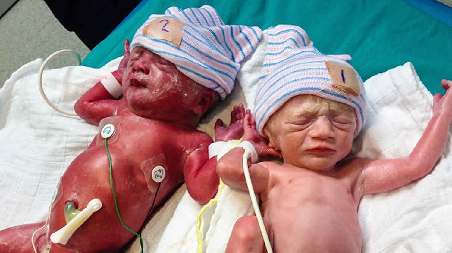
- Babies with hydrops fetalis have severe edema (swelling) of the entire body and are extremely pale. They often have difficulty breathing.
During pregnancy
- Mild anemia, hyperbilirubinemia, and jaundice. The placenta helps rid some of the bilirubin, but not all.
- Severe anemia with enlargement of the liver and spleen. When these organs and the bone marrow cannot compensate for the fast destruction of red blood cells, severe anemia results and other organs are affected.
- Hydrops fetalis. This occurs as the baby’s organs are unable to handle the anemia. The heart begins to fail and large amounts of fluid build up in the baby’s tissues and organs. A fetus with hydrops is at great risk of being stillborn.
After birth
- Severe hyperbilirubinemia and jaundice. The baby’s liver is unable to handle the large amount of bilirubin that results from red blood cell breakdown. The baby’s liver is enlarged and anemia continues.
- Kernicterus. Kernicterus is the most severe form of hyperbilirubinemia and results from the buildup of bilirubin in the brain. This can cause seizures, brain damage, deafness, and death.
Erythroblastosis fetalis prevention
Fortunately, erythroblastosis fetalis is a very preventable disease. Because of the advances in prenatal care, nearly all women with Rh negative blood are identified in early pregnancy by blood testing. If a mother is Rh negative and has not been sensitized, she is usually given a drug called Rh immunoglobulin (RhIg), also known as RhoGAM. This is a specially developed blood product that can prevent an Rh negative mother’s antibodies from being able to react to Rh positive cells. Many women are given RhoGAM around the 28th week of pregnancy. After the baby is born, a woman should receive a second dose of the drug within 72 hours, if her baby is Rh positive. If her baby is Rh negative, she does not need another dose.
RhoGAM destroys any anti-Rh antibodies that enter in the mother’s circulation before her immune system becomes sensitized. This helps protect a future Rh positive baby.
Erythroblastosis fetalis diagnosis
Because anemia, hyperbilirubinemia, and hydrops fetalis can occur with other diseases and conditions, the accurate diagnosis of erythroblastosis fetalis depends on determining if there is a blood group or blood type incompatibility. Sometimes, the diagnosis can be made during pregnancy based on information from the following tests:
- Testing for the presence of Rh positive antibodies in the mother’s blood
- Ultrasound – to detect organ enlargement or fluid buildup in the fetus. Ultrasound is a diagnostic imaging technique which uses high-frequency sound waves and a computer to create images of blood vessels, tissues, and organs. Ultrasound is used to view internal organs as they function, and to assess blood flow through various vessels.
- Amniocentesis – to measure the amount of bilirubin in the amniotic fluid. Amniocentesis is a test performed to determine chromosomal and genetic disorders and certain birth defects. The test involves inserting a needle through the abdominal and uterine wall into the amniotic sac to retrieve a sample of amniotic fluid.
- Sampling of some of the blood from the fetal umbilical cord during pregnancy to check for antibodies, bilirubin, and anemia in the fetus.
Once a baby is born, diagnostic tests for erythroblastosis fetalis may include the following:
- Testing of the baby’s umbilical cord blood for blood group, Rh factor, red blood cell count, and antibodies
- Testing of the baby’s blood for bilirubin levels
Erythroblastosis fetalis treatment
Once erythroblastosis fetalis is diagnosed, treatment may be needed. Specific treatment for erythroblastosis fetalis will be determined by your baby’s doctor based on:
- Your baby’s gestational age, overall health, and medical history
- Extent of the erythroblastosis fetalis disease
- Your baby’s tolerance for specific medications, procedures, or therapies
- Expectations for the course of the disease
- Your opinion or preference
During pregnancy, treatment for erythroblastosis fetalis may include:
- Intrauterine blood transfusion of red blood cells into the baby’s circulation. This is done by placing a needle through the mother’s uterus and into the abdominal cavity of the fetus or directly into the vein in the umbilical cord. It may be necessary to give a sedative medication to keep the baby from moving. Intrauterine transfusions may need to be repeated.
- Early delivery if the fetus develops complications. If the fetus has mature lungs, labor and delivery may be induced to prevent worsening of erythroblastosis fetalis.
After birth, treatment may include:
- Blood transfusions (for severe anemia)
- Intravenous fluids (for low blood pressure)
- Help for respiratory distress using oxygen, surfactant, or a mechanical breathing machine
- Exchange transfusion to replace the baby’s damaged blood with fresh blood. The exchange transfusion helps increase the red blood cell count and lower the levels of bilirubin. An exchange transfusion is done by alternating giving and withdrawing blood in small amounts through a vein or artery. Exchange transfusions may need to be repeated if the bilirubin levels remain high.
- Intravenous immunoglobin (IVIG). Intravenous immunoglobin (IVIG) is a solution made from blood plasma that contains antibodies to help the baby’s immune system. Intravenous immunoglobin (IVIG) may help reduce the breakdown of red blood cells and lower bilirubin levels.


