Contents
What is morphea
Morphea is also known as localised scleroderma, is a rare skin condition that causes painless, discolored patches on your skin. Morphea is characterized by an area of inflammation and fibrosis (thickening and hardening) of the skin due to increased collagen deposition. Typically, the morphea skin changes appear on the abdomen, chest or back. But morphea might also appear on your face, arms or legs. Morphea tends to affect only the outermost layers of your skin. But some forms of the condition also restrict movement in the joints.
Subtypes of morphea vary according to the location of involved skin. Any subtype of morphea can also result in deep or subdermal involvement of the underlying fat, fascia, muscle or bone.
Unlike systemic scleroderma, morphea does not cause:
- Skin thickening of the fingers and toes (sclerodactyly)
- Specific autoantibodies in the blood (such as anti-centromere antibody or anti-Scl70)
- Abnormal small blood vessels in the fingers (nail fold capillaries)
- Fibrosis and/or vascular damage of internal organs.
Signs and symptoms of morphea vary, depending on the type and stage of the condition. They include:
- Reddish or purplish oval patches of skin, often on the abdomen, chest or back
- Patches that gradually develop a lighter or whitish center
- Linear patches, especially when on the arms or legs
- A gradual change in the affected skin, which becomes hard, thickened, dry and shiny
- Loss of hair and sweat glands in the affected area over time
Morphea usually affects only the skin and underlying tissue and, rarely, bone. The condition generally lasts several years and then disappears on its own. But it usually leaves some patches of darkened or discolored skin.
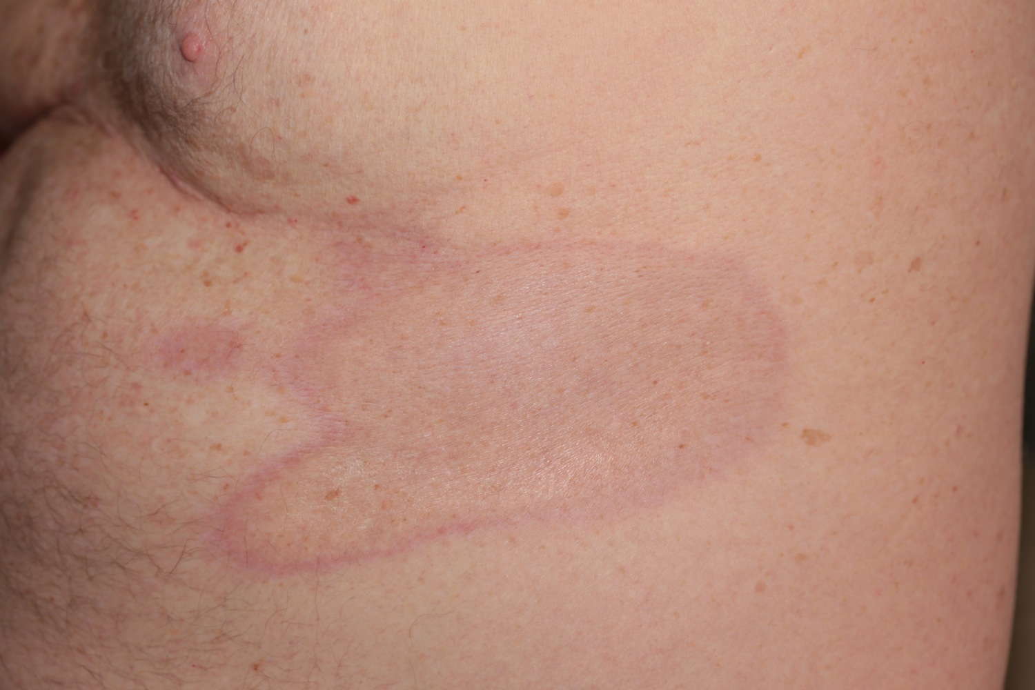
Figure 1. Morphea skin – note the classic violaceous border
Morphea is rare, and is estimated to have an incidence of 1–3 per 100,000 children. It is three times more common in adult women compared to males and often begins in childhood. Various antibodies may be found including antinuclear antibodies (ANA), anti-single stranded DNA, anti-histone antibodies (AHA) and rheumatoid factor. Anti-histone antibodies (AHA) were found in 87% of patients with generalized morphea vs. 42% of patients with localized disease. 80% of patients with morphea have a positive antinuclear antibodies (ANA). Although not hereditary, certain HLA subtypes (HLA-DRB1*04:04 and HLA-B*37) are associated with an increased risk of morphea.
Morphea can follow a protracted course, which can be relapsing and remitting, or chronically active. Milder forms of the disease tend to become inactive within 3–5 years. Very rarely, patients may go on to develop systemic sclerosis.
Relapse can occur after successful treatment, especially in morphea that begins in childhood.
Extended courses of systemic treatments of 4 to 5 years or more may be required to minimize the risk of relapse.
In the meantime, medications and therapies are available to help treat the skin discoloration and other effects.
If you notice reddish patches of hardening or thickening skin, see your doctor. Early diagnosis and treatment may help slow the development of new patches and allow your doctor to identify and treat complications before they worsen.
Morphea subtypes and clinical features
The specific appearance of morphea varies, and may include:
- An inflammatory phase: pink, purple or bruise-like patches
- Sclerosis: the skin becomes hard, thickened, bound down, shiny, ivory white and may have a purple border of surrounding inflammation
- Dyspigmention and atrophy. This develops after a variable time period (usually months or years); hyperpigmentation is more common than hypopigmentation. Superficial (dermal) atrophy is seen as shiny skin, often with visible blood vessels. Deeper atrophy affecting fat, muscle or bone presents as concave indentations or discrepancies of limb circumference.
Whilst morphea classically passes through each of these morphological stages, this is not always the case.
Scleroderma Variants
| Disease | Features |
|---|---|
| Morphea (Localized) | Indurated, sclerotic plaques, usually on the trunk. |
| Generalized Morphea | Two or more body regions involved. May have muscle atrophy, but no systemic involvement. |
| Superficial morphea | Multiple hypopigmented to hyperpigmented patches and plaques. They usually involve intertriginous sites symmetrically and lack significant induration, contractures, or atrophy. |
| Progressive Systemic Sclerosis | Raynaud’s, woody edema of hands, sclerodactyly, internal organ involvement, e.g., pulmonary fibrosis. |
| CREST Syndrome | Calcinosis, Raynaud’s, esophageal dysmotility, sclerodactyly, and telangiectasias. |
| Linear Scleroderma | Linear indurated lesions. May follow lines of Blaschko. |
| Morphea Profunda | Process involves the deep subcutaneous tissue including the fascia. |
| Pansclerotic Morphea | Widespread involvement of the deeper tissue including the fat fascia, muscle, and bone. |
| Atrophoderma of Pasini and Pierini (APP) | Brown, atrophic depressed lesions, usually on the trunk of women. |
| Linear Atrophoderma of Moulin | Similar to Atrophoderma of Pasini and Pierini but linear and may follow lines of Blaschko. |
| En Coup de Sabre | Linear atrophic, indurated lesion running along scalp and forehead. |
| Parry Romberg | Atrophy of one side of the facial skin with possible involvement of the muscle, fat, cartilage, bone, and brain. |
When describing morphea, consider three features.
- Anatomical distribution: location and whether limited, linear, generalized or mixed
- Morphology: inflammatory, sclerotic, dypsigmented and/or atrophic
- Depth of tissue involvement: skin, fat, fascia, muscle, bone
Anatomical distribution
- Subtypes of morphea are limited, linear, generalized or mixed.
Limited plaque morphea
- The most common type in adults
- Oval shaped patches of 1 to 20cm in diameter
- Two or fewer body sites.
Rare types of limited plaque morphea include guttate morphea and atrophoderma of Pasini-Pierini.
Linear morphea (Linear scleroderma)
Linear morphea (linear scleroderma) is a form of localized scleroderma characterized by sclerotic lesions distributed in a linear, band-like pattern.
A band-like linear induration, often with hypo or hyperpigmented areas, linear morphea most commonly seen on the leg but also arm and forehead (en coup de sabre) is characteristic. A deep component with fixation to underlying structures may be present. Joint pain is quite common and joint contractures due to skin and tissue involvement may occur. Twenty percent of patients have onset by age 10; 75% by age 40. An underlying sclerosing bone dysplasia called melorheostosis can occur with the X-ray appearance said to resemble dripping candle wax. ANA (antinuclear antibodies) may be strongly positive but often the diagnosis is made clinically. It is more common in children but occurs in adults as well.
- Most common subtype in children
- Found on the limbs, trunk or head (craniofacial)
- Follows lines and swirls called Blaschko lines
- Usually unilateral, but may be bilateral and widespread in severe cases.
Linear atrophoderma of moulin is a rare subtype of atrophic and pigmented linear morphea.
Craniofacial linear morphea was previously sub-classified as:
- En coup de sabre (linear atrophic band +/- sclerosis, classically on the forehead or scalp)
- Parry Romberg / progressive hemifacial atrophy (atrophy of the fat, muscle and bone +/- dyspigmented +/- sclerotic changes, affecting one side of the face).
These types of craniofacial linear morphea occur together, and vary based on morphology and depth of tissue involvement.
Figure 2. Linear morphea
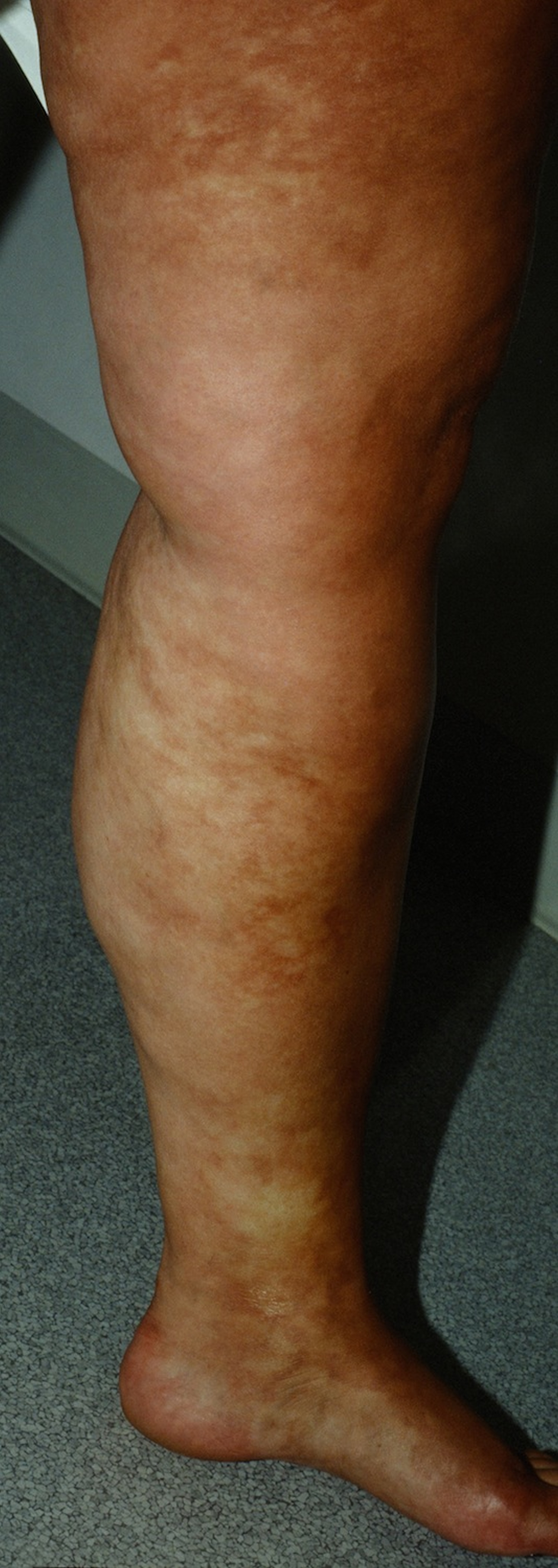
Figure 3. Linear morphea scalp
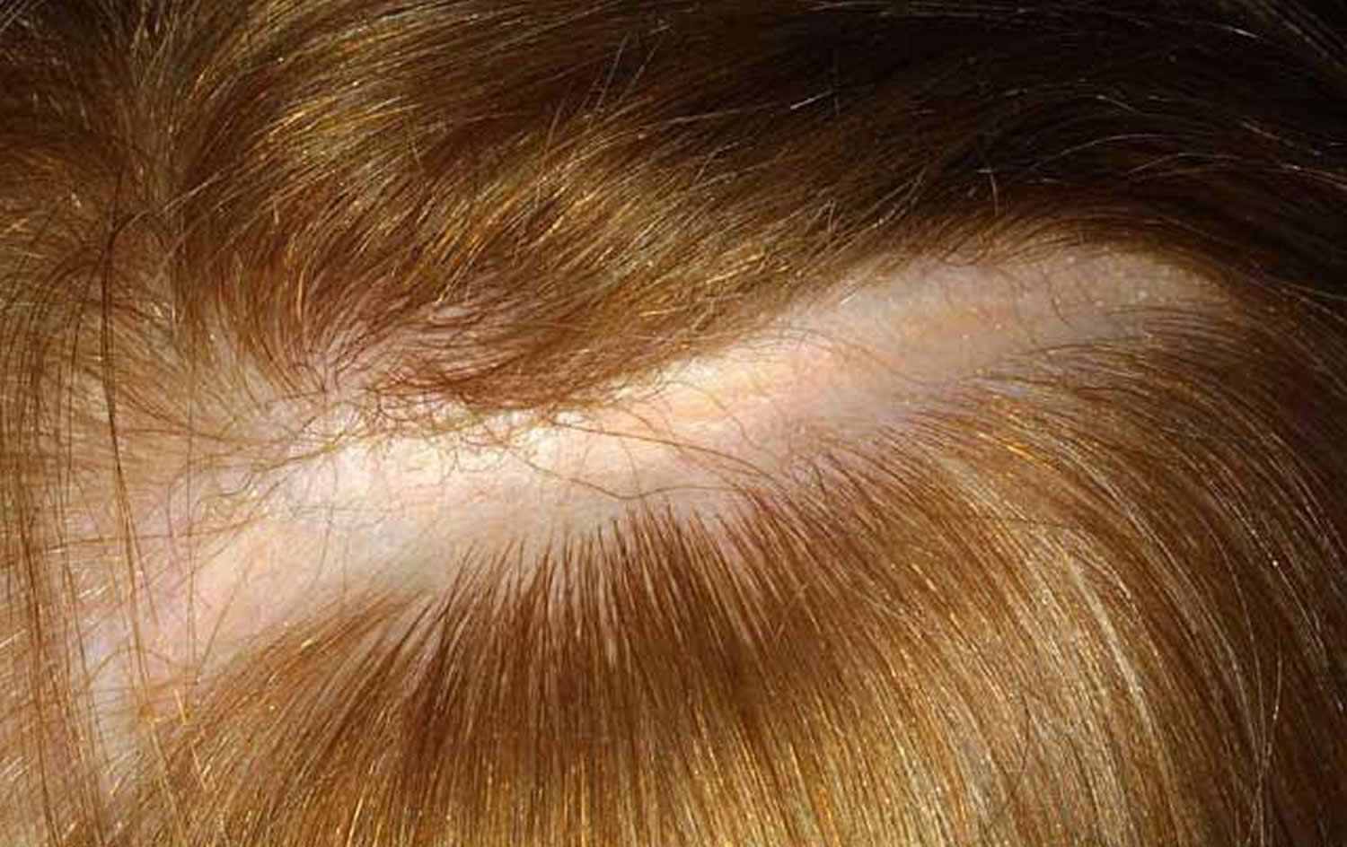
Figure 4. Morphea skin in children
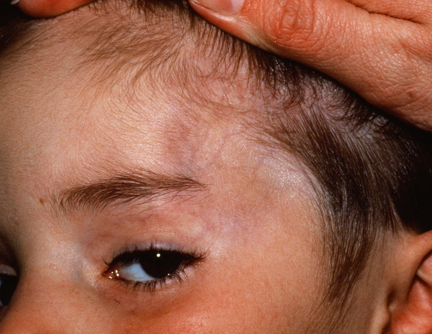
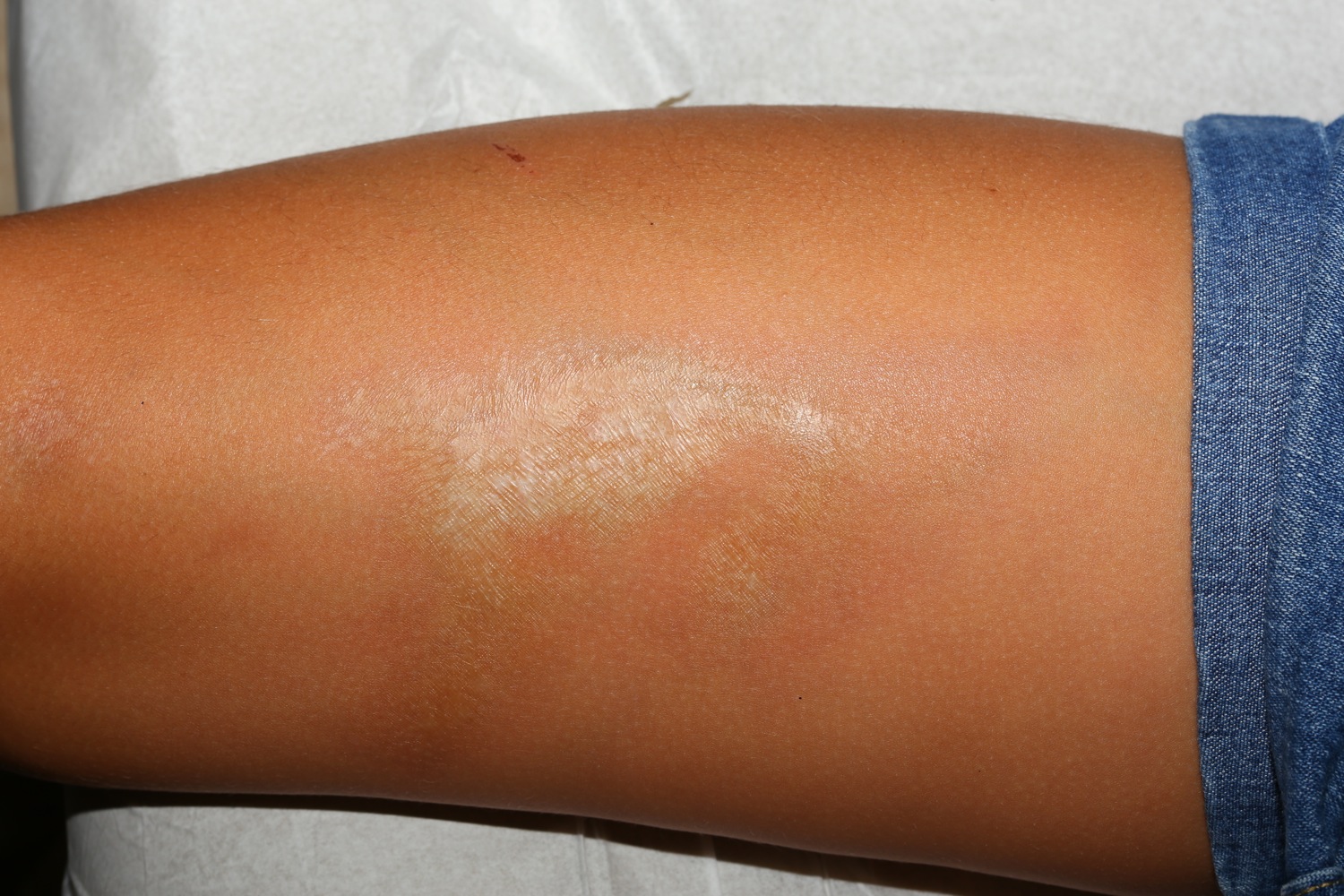
Figure 5. Morphea scleroderma with central lichen sclerosus on the anterior thigh of a young girl
Generalized morphea
Generalized morphea affects three or more body sites. There are two major patterns;
- Disseminated plaque morphea: scattered plaques with intervening unaffected skin. May be isomorphic (occurring at sites of friction such as the waistband, bra line and inguinal creases) or non-isomorphic (scattered plaques at any site).
- Pansclerotic morhpea: circumferential, confluent sclerosis, which is usually rapidly progressive and affects most of the body surface area. This occurs in childhood (disabling pansclerotic morphea of childhood), and adulthood (when it overlaps with eosinophilic fasciitis).
- Generalized morphea spares the digits and periareolar skin.
Mixed pattern morphea
- Mixed pattern morphea occurs when more than one anatomical subtype is present.
- The most common mixed pattern is linear morphea of a limb, with limited plaque morphea on the trunk.
Limited morphea is considered mild, while linear morphea and generalised morphea are more severe subtypes. The depth of tissue involvement is also important when assessing disease severity; subdermal involvement implies more severe disease.
Deep tissue involvement in morphea
Any subtype of morphea can affect connective tissue under the skin, such as the fat, fascia, muscle, bone, joints or rarely, brain (in craniofacial linear morphea). Deep tissue involvement is a marker of severe disease.
When the fascia — the connective tissue under the fat — is involved:
- The skin can look puckered, with a cobblestone or orange peel appearance (peau d’orange)
- There may be guttering along blood vessels (the groove sign).
Involvement of bones or joints results in loss of bony tissue, pain and/or restricted joint movement (contractures).
Eosinophilic fasciitis is a form of pansclerotic morphea with deep fascial involvement. Circumferential inflammation and sclerosis of the fascia (the connective tissue beneath the fat) frequently co-exists with overlying dermal sclerosis or other anatomical subtypes of morphea indicating it is debatable whether it should be considered a separate condition.
Morphea causes
The causes of morphea are unknown. It may be due in part to an unusual reaction of the immune system.
- Localized genetic factors appear to play a role; for example, cutaneous mosaicism may important in linear morphea, which follows Blaschko lines of epidermal development.
- Up to 40% of patients with severe forms of morphea have a personal or family history of autoimmune disease (e.g, thyroid disease, vitiligo) or rheumatologic disease (eg, rheumatoid arthritis).
For unknown reasons, morphoea often develops after an external trigger such as:
- Radiation therapy
- Repeated trauma to the affected area
- A recent infection, such as measles or chickenpox
- Insect bite or tick bite (the role of Borellia burgdorferi, cause of Lyme disease is controversial)
- Injection (e.g., bleomycin, silicone) or vaccination
- Repeated friction
- Surgery
The condition isn’t contagious. Trauma-related morphoea may occur at the affected site, or at unrelated distant sites.
Risk factors for developing morphea
Certain factors may affect your risk of developing morphea, including:
- Your sex and age. Females are more likely to develop morphea than are males. The condition can affect people at any age. It usually appears between the ages of 2 and 14 or in the mid-40s.
- Your race. Morphea is more prevalent among Caucasians.
Morphea complications
Morphea can cause a number of complications, including:
- Self-esteem issues. Morphea can have a negative effect on your self-esteem and body image, particularly if discolored patches of skin appear on your arms, legs or face. Because morphea affects your appearance, it can be an especially difficult condition to live with. You may also be concerned that it will get worse before it goes away.
- Movement problems. Morphea that affects the arms or legs can impair joint mobility.
- Widespread areas of hardened, discolored skin. Numerous new patches of hard, discolored skin may seem to join together, a condition known as generalized morphea.
- Eye damage. Children with head and neck morphea may experience unnoticeable, permanent eye damage.
Morphea symptoms
Morphoea may be asymptomatic, or symptoms may arise from the skin or deeper tissues, or they may due to extracutaneous manifestations.
Morphea skin disease symptoms
- Some patients experience itch and/or pain from actively inflamed areas of morphea.
- Adnexal structures are destroyed by sclerosis. This is of particular relevance in linear morphea of the scalp, which results in permanent hair loss.
- The sclerosis can entrap superficial nerves and result in pain, tingling and sometimes mild weakness.
Morphea profunda symptoms
- Deep involvement over joints causes joint pain or limited joint movement (contractures).
- Constriction of the chest wall in generalized pansclerotic morphea can result in shortness of breath.
- Teeth and jaw involvement in craniofacial linear morphea can cause oral and dental problems. Temporomandibular joint involvement causes difficulty chewing, jaw locking or pain.
- Skull and/or brain involvement can cause headaches or in some rare cases, neurological symptoms such as nerve palsies or seizures.
Morphea systemic symptoms
Up to 30% of patients with more severe types of linear or generalized morphea can have extracutaneous non-specific inflammatory symptoms. These include:
- Fatigue, lethargy
- Non-specific joint pain and/or inflammation (arthralgia, arthritis)
- Muscle pain
- Reflux/heartburn
- Raynaud phenomenon (cold hands with red/white/blue colour changes)
- Eye dryness, irritation or blurred vision due to ocular involvement (most commonly episcleritis, anterior uveitis, keratitis) – related or unrelated to the site of morphea
These extracutaneous manifestations imply that morphea is a systemic inflammatory condition. In contrast, systemic sclerosis results in direct damage and fibrosis of the lungs, heart, kidneys and/or gastrointestinal tract — which do not occur in morphea.
Morphea diagnosis
Your doctor may diagnosis morphea by examining the affected skin and asking you about your signs and symptoms. He or she may take a small sample of the affected skin (skin biopsy) for examination in the laboratory. This may reveal changes in your skin, such as thickening of the collagen in the second layer of skin (dermis). Collagen is a protein that makes up your connective tissues, including your skin. It helps make your skin elastic and resilient.
It’s important to distinguish morphea from systemic scleroderma, so if you have morphea, your doctor will likely refer you to a specialist in skin disorders (dermatologist) or diseases of the joints, bones and muscles (rheumatologist).
If your child has head and neck morphea, take him or her in for regular comprehensive eye exams, as morphea may cause unnoticeable yet irreversible eye damage.
Ultrasound and magnetic resonance imaging (MRI) may be useful in monitoring disease progression and how it is responding to treatment.
Skin biopsy
A skin biopsy enables the skin, subcutaneous fat (and sometimes fascia) to be examined under the microscope. Classic findings in morphea include:
- Sharply squared-off biopsy (‘cookie cutter’ sign)
- Atrophic epidermis
- Thickened, hyalinised collagen, with possible loss of appendageal structures (such as sweat glands and hair follicles)
- Fat loss / entrapment
- Inflammatory cell infiltrate (lymphocytes +/- plasma cells).
Blood tests
- Eosinophil count (may be raised)
- Inflammatory markers (ESR or CRP may be raised)
- Anti-nuclear antibody (may be raised; but systemic sclerosis specific ENA antibodies such as anti-Sc70 are negative)
- Thyroid function tests (may be abnormal; consider if at risk of associated autoimmune thyroid disease)
- Rheumatoid factor or anti-CCP antibodies if arthralgia/arthritis (may be raised)
Imaging
Magnetic resonance imaging (MRI) may be performed to assess how deep the morphea extends beyond the skin, for example in generalized morphea, linear morphea crossing joints, and craniofacial linear morphea.
Morphea treatment
Morphea usually goes away without treatment, though it may leave scars or areas of discolored skin. Until your condition clears up, you may want to pursue treatment that helps control your signs and symptoms.
There is no cure for morphoea. Treatment is aimed at halting ongoing disease activity and progression.
Treatment options include:
- Light therapy. A special treatment that uses ultraviolet light (phototherapy) may improve your skin’s appearance, especially when used soon after skin changes appear.
- Drugs that fight inflammation. Your doctor may prescribe an immunosuppressive medication, such as oral methotrexate (Rheumatrex, Trexall). This may be used in combination with corticosteroid pills for the first few months. Each of these drugs has potentially serious side effects.
- A form of vitamin D. The prescription cream calcipotriene is a synthetic form of vitamin D. It may help soften the skin patches caused by morphea. Skin generally begins to improve during the first months of treatment. Possible side effects include burning, stinging and a rash.
- Physical therapy. This type of treatment uses exercise to prevent joint deformity and maintain movement.
Topical therapy
Topical therapy may help limited and superficial forms of morphea, and can reduce itch.
- Topical steroids
- Tacrolimus ointment
- Calcipotriol ointment or cream
- Imiquimod cream
Phototherapy
Phototherapy can soften morphea and has anti-inflammatory effects.
Different wavelengths of ultraviolet radiation can be used.
- UVA1 penetrates most deeply and is hence useful when deeper subcutaneous tissues are involved, but is rarely available.
- Topical or systemic photochemotherapy (PUVA, ie, psoralen + UVA) may be helpful.
- Narrowband UVB is less effective, as it only penetrates superficially.
Systemic treatment
Systemic treatment is intended to prevent progression and switch off the active disease process. It is usually needed for at least 2 to 4 years, but relapse can occur.
- Methotrexate
- Systemic corticosteroids
- Mycophenolate mofetil
Specific treatment decisions in morphea are guided by the subtype of morphea and its severity.
Superficial limited morphea — mild
- Topical therapy
- Phototherapy (UVA1, nbUVB, PUVA)
Superficial generalized or adult onset linear morphea — moderate
- Topical therapies AND phototherapy
- Systemic therapy
Deep forms of morphea and paediatric onset linear morphea — severe
- Methotrexate +/- systemic corticosteroids (IV pulsed &/or oral)
- Mycophenolate mofetil +/- corticosteroids (IV pulsed &/or oral)
- Combination therapies
- Combine 1st and 2nd line treatments
- Add hydroxychloroquine
- Add phototherapy (UVA1 or PUVA if available)
- Other options
- Ciclosporin
- Abatacept
- Tocilizumab
- Extracorporeal photopheresis
- Other experimental and reported therapies
Physiotherapy to improve joint mobility should be undertaken with caution in active morphea, as the resultant trauma may potentially act as an ongoing disease trigger.
In some cases, surgery may be of benefit, such as autologous fat transfer, to improve atrophy.
Monitoring treatment response
Disease progression and treatment response can be monitored using photographs, the localized scleroderma cutaneous assessment tool and other highly specialized tests, such as infrared thermography.


