
Contents
Jaundice
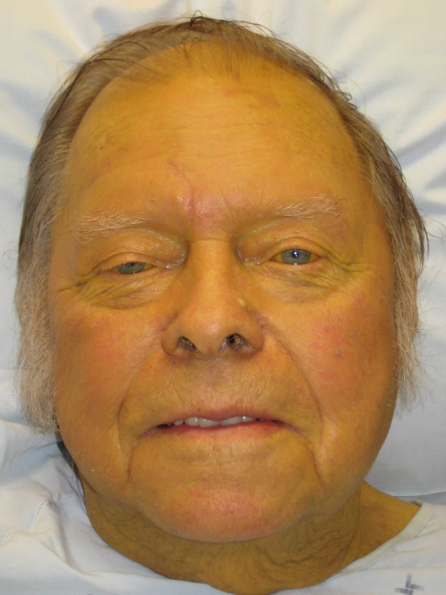
Jaundice also known as icterus, is a medical term to describe to the yellow discoloration of your skin, mucus membranes and eyes resulting from the accumulation of excess bilirubin. The yellow coloring comes from bilirubin, a byproduct of old red blood cells. Jaundice can occur when too much bilirubin builds up in your blood, a condition known as hyperbilirubinemia. The normal serum levels of bilirubin are less than 1 milligram per deciliter (mg/dL). The clinical presentation of jaundice with peripheral yellowing of the eye sclera, also called scleral icterus, is best appreciated when serum bilirubin levels exceed 3 mg/dL (51.3 μmol per L) (Figure 1) 1. With further increase in serum bilirubin levels, the skin will progressively discolor ranging from lemon yellow to apple green, especially if the process is long-standing; the green color is due to biliverdin (Figure 2) 1. You may also have itchy skin, darker urine and paler stool (poop) than usual.
Bilirubin is a orange-yellow pigment, a waste product primarily produced by the normal breakdown of heme. Heme is a component of hemoglobin (the substance that carries oxygen in your red blood cells), which is found in red blood cells (RBCs). As red blood cells break down, your body builds new red blood cells to replace them. The old ones are processed by the liver. If the liver cannot handle the blood cells as they break down, bilirubin builds up in your body and your skin may look yellow.
Many healthy babies have some jaundice during the first week of life. It usually goes away. However, jaundice can happen at any age and may be a sign of a problem. Jaundice can happen for many reasons, such as:
- Blood diseases
- Genetic syndromes such as Rotor syndrome, Dubin-Johnson syndrome and Crigler-Najjar syndrome and birth defects such as biliary atresia. Biliary atresia must be quickly detected and treated, usually with surgery, to prevent serious liver damage that may require liver transplantation within the first few years of life. Some children may require liver transplantation despite early surgical treatment.
- Liver diseases, such as hepatitis or cirrhosis
- Blockage of bile ducts due to gallstones, liver cancer, bile duct cancer or pancreatic cancer
- Infections, most commonly viral hepatitis
- Use of certain drugs. Drugs that can decrease total bilirubin include barbiturates, caffeine, penicillin, and high doses of salicylates. The drug atazanavir increases unconjugated (indirect) bilirubin.
- Other medical conditions
Note: If your skin is yellow and the whites of your eyes are not yellow, you may not have jaundice. Your skin can turn a yellow-to-orange color if you eat a lot of beta carotene, the orange pigment in carrots. Skin color changes can occur in conditions other than hyperbilirubinemia, such as Addison’s disease, anorexia nervosa or use of spray-tanning products 2.
If you have jaundice, you should see your doctor as soon as possible.
In order to find out what is causing your jaundice, your doctor may ask about your lifestyle, your symptoms and medical history. Your doctor will perform a physical exam. The physical examination will show jaundice and possibly liver swelling. Specific tests vary but may include liver function and bilirubin blood tests to determine how well your liver is working.
Your doctor may order blood and urine tests. These allow your doctor to check your level of bilirubin and assess the health of your liver.
Your doctor may also order an ultrasound, MRI or CT scan to check for blockages. These can also be used to check for signs of liver and pancreatic disease. In some cases, your doctor may request a liver biopsy to confirm liver disease.
Other tests may include:
- Hepatitis virus panel to look for infection of the liver
- Liver function tests to determine how well the liver is working
- Complete blood count to check for low blood count or anemia
- Abdominal ultrasound
- Abdominal CT scan
- Magnetic resonance cholangiopancreatography (MRCP)
- Endoscopic retrograde cholangiopancreatography (ERCP)
- Percutaneous transhepatic cholangiogram (PTCA)
- Liver biopsy
- Cholesterol level
- Prothrombin time
Jaundice treatment depends on the underlying cause of your jaundice.
Figure 1. Scleral icterus (jaundice eyes)
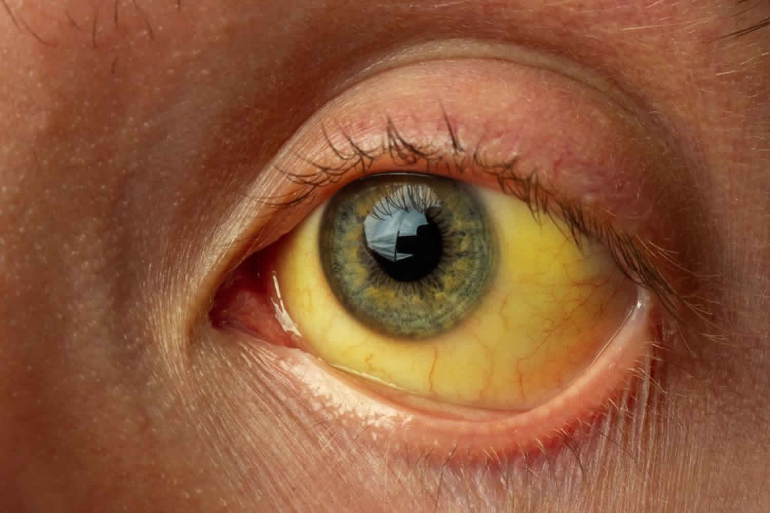
Figure 2. Jaundice of the skin
Are some people more at genetic risk of abnormal bilirubin levels?
Several inherited chronic conditions increase bilirubin levels in the blood and include Gilbert syndrome, Dubin-Johnson syndrome, Rotor syndrome, and Crigler-Najjar syndrome. The first three are usually mild, chronic conditions that can be aggravated under certain conditions but in general cause no significant health problems. For example, Gilbert syndrome is very common; about 1 in every 6 people has this genetic abnormality, but usually people with Gilbert syndrome do not have elevated bilirubin. Crigler-Najjar syndrome is the most serious inherited condition listed; this disorder is relatively rare, and some people with it may die.
How do you treat abnormal bilirubin levels and/or jaundice?
Treatment depends on the cause of the jaundice. In newborns, phototherapy (special light therapy), blood exchange transfusion, and/or certain drugs may be used to reduce the bilirubin level. In Gilbert, Rotor, and Dubin-Johnson syndromes, no treatment is usually necessary. Crigler-Najjar syndrome may respond to certain enzyme drug therapy or may require a liver transplant. Jaundice caused by an obstruction is often resolved by surgery. Jaundice due to cirrhosis is a result of long-term liver damage and does not respond well to any type of therapy other than liver transplantation.
What is bilirubin?
Bilirubin is an orange-yellow pigment, a waste product primarily produced by the normal breakdown of heme. Heme is a component of hemoglobin (the substance that carries oxygen in your red blood cells), which is found in red blood cells (RBCs). Bilirubin is ultimately processed by the liver to allow its elimination from the body.
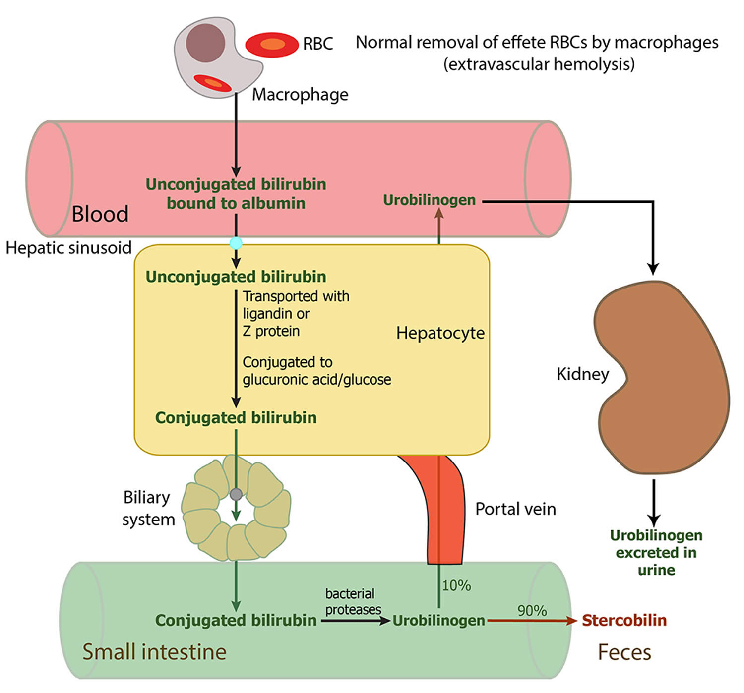
Red blood cells normally degrade after about 120 days in the blood circulation. As heme is released from hemoglobin, it is converted to bilirubin. This form of bilirubin is also called unconjugated bilirubin. Unconjugated bilirubin is carried by proteins to the liver; there, sugars are attached (conjugated) to bilirubin to form conjugated bilirubin. Conjugated bilirubin enters the bile and passes from the liver to the small intestines; there, it is further broken down by bacteria and eventually eliminated in the stool. Thus, the breakdown products of bilirubin give stool its characteristic brown color.
A small amount (approximately 250 to 350 milligrams) of bilirubin is produced daily in a normal, healthy adult. Most (85%) of bilirubin is derived from damaged or degraded red blood cells, with the remaining amount derived from the bone marrow or liver. Normally, small amounts of unconjugated bilirubin are released into the blood, but virtually no conjugated bilirubin is present. Both forms can be measured or estimated by laboratory tests, and a total bilirubin result (a sum of these) may also be reported.
Bilirubin concentrations tend to be slightly higher in males than females. African Americans routinely show lower bilirubin concentrations than non-African Americans. Strenuous exercise may increase bilirubin levels.
Drugs that can decrease total bilirubin include barbiturates, caffeine, penicillin, and high doses of salicylates. The drug atazanavir increases unconjugated (indirect) bilirubin.
If the bilirubin level increases in the blood, a person may appear jaundiced, with a yellowing of the skin and/or whites of the eyes. The pattern of bilirubin test results can give your doctor information regarding the condition that may be present. For example, unconjugated bilirubin may be increased when there is an unusual amount of red blood cell destruction (hemolysis) or when the liver is unable to process bilirubin (i.e., with liver diseases such as cirrhosis or inherited problems). Conversely, conjugated bilirubin can increase when the liver is able to process bilirubin but is not able to pass the conjugated bilirubin to the bile for removal; when this happens, the cause is often acute hepatitis or blockage of the bile ducts.
Increased total and unconjugated bilirubin levels are relatively common in newborns in the first few days after birth. This finding is called “physiologic jaundice of the newborn” and occurs because the newborn’s liver is not mature enough to process bilirubin yet. Usually, physiologic jaundice of the newborn resolves itself within a few days. However, in hemolytic disease of the newborn, red blood cells may be destroyed because of blood incompatibilities between the baby and the mother; in these cases, treatment may be required because high levels of unconjugated bilirubin can damage the newborn’s brain.
A rare (about 1 in 10,000 births) but life-threatening congenital condition called biliary atresia can cause increased total and conjugated bilirubin levels in newborns. This condition must be quickly detected and treated, usually with surgery, to prevent serious liver damage that may require liver transplantation within the first few years of life. Some children may require liver transplantation despite early surgical treatment.
Rare inherited disorders that cause abnormal bilirubin metabolism such as Rotor, Dubin-Johnson, and Crigler-Najjar syndromes, may also cause increased levels of bilirubin.
Crigler-Najjar syndromes type I and type II, are rare genetic disorders that are caused by a low or absent activity of bilirubin UDP-glucuronyl-transferase. In Crigler-Najjar syndromes type I, the enzyme activity is totally absent, the excretion rate of bilirubin is greatly reduced and the serum concentration of unconjugated bilirubin is greatly increased. Patients with Crigler-Najjar syndromes may die in infancy owing to the development of kernicterus.
Kernicterus isn’t common because babies usually are treated before jaundice becomes severe. If untreated, kernicterus can cause:
- Athetoid cerebral palsy. Babies with this condition have uncontrollable movements in the arms, legs, face and other body parts.
- Hearing loss
- Vision problems
- Dental problems
- Intellectual disabilities
Figure 3. Bilirubin metabolism
Unconjugated bilirubin
Though unconjugated bilirubin may be toxic to brain development in newborns (up to 2-4 weeks of age), it does not pose the same threat to older children and adults. In older children and adults, the “blood-brain barrier” is more developed and prevents bilirubin from gaining access to brain cells. Nevertheless, elevated bilirubin strongly suggests that a medical condition is present that must be evaluated and treated.
Conjugated bilirubin
Conjugated bilirubin is a water-soluble form of bilirubin formed in the liver by the chemical addition of sugar molecules to unconjugated bilirubin; when present in the blood, conjugated bilirubin can become chemically bound to albumin, forming delta-bilirubin also known as biliprotein.
Normal bilirubin levels
Direct (conjugated) Bilirubin
- > or =12 months: 0.0-0.3 mg/dL (less than 5.1 µmol/L)
Reference values have not been established for patients who are <12 months of age.
Total Bilirubin
- 0-6 days: Refer to www.bilitool.org for information on age-specific (postnatal hour of life) serum bilirubin values.
- 7-14 days: <15.0 mg/dL
- 15 days to 17 years: < or =1.0 mg/dL
- > or =18 years: 0.1 to 1.2 mg/dL (1.71 to 20.5 µmol/L)
Normal value ranges may vary slightly among different laboratories. Some labs use different measurements or may test different samples. Talk to your provider about the meaning of your specific test results.
Bilirubin in urine
Bilirubin is not normally present in the urine. However, conjugated bilirubin is water-soluble and may be eliminated from the body through the urine if it cannot pass into the bile. Measurable bilirubin in the urine usually indicates blockage of liver or bile ducts, hepatitis, or some other form of liver damage and may be detectable early in disease; for this reason, bilirubin testing is integrated into common dipstick testing used for routine urinalysis.
Jaundice causes
Jaundice is caused by the build-up in your body of a yellow substance called bilirubin. There are lots of possible reasons for this and some of them are serious.
Jaundice causes include:
- gallstones
- alcohol-related liver disease
- pancreatitis
- hepatitis (such as viral hepatitis A, B, C, D or E)
- sickle cell disease
- autoimmune disease, such as primary biliary cholangitis
- pancreatic cancer, liver cancer or gallbladder cancer
- hereditary conditions, such as Dubin–Johnson syndrome and Gilbert syndrome
- some medicines
- lymphoma
- surgery
- pregnancy
A retrospective study of more than 700 individuals found that most cases (55%) of acute jaundice in adults are caused by intrahepatic disorders, including viral hepatitis, alcoholic liver disease, and drug-induced liver injury. The remaining 45% of acute jaundice cases are extrahepatic and include gallstone disease, hemolysis, and cancer 3.
Unconjugated hyperbilirubinemia
Increased bilirubin production
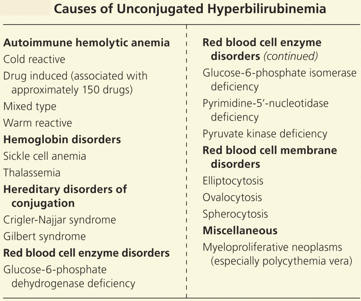
Unconjugated hyperbilirubinemia is usually a result of too much bilirubin presented to the conjugating machinery (from increased red blood cell destruction). Increased red blood cell breakdown may be caused by red blood cell membrane disorders, red blood cell enzyme disorders, hemoglobin disorders (Thalassemias), autoimmune red blood cell destruction (autoimmune hemolytic anemia), or some cancers 4, 5, 6, 7. The excess turnover of red blood cells results in increased heme metabolism, producing large amounts of bilirubin that overwhelm the conjugating machinery, leading to decreased excretion and clinical jaundice.
Impaired bilirubin conjugation
Deficiencies in the same conjugating machinery may also lead to jaundice in individuals with normal red blood cell turnover. Gilbert syndrome involves a deficiency in uridine diphosphate-glucuronosyltransferase, and it affects 10% of the white population 8. This is a benign condition that may be exacerbated by physical or emotional stress such as illness, strenuous exercise, or fasting. Crigler-Najjar syndrome is a more severe variant of the same uridine diphosphate-glucuronosyltransferase enzyme deficiency 8. Patients with impaired conjugation due to low levels of the bilirubin-UGT enzyme are particularly susceptible to jaundice from medications that inhibit this enzyme, such as protease inhibitors 9. Table 1 lists the causes of unconjugated hyperbilirubinemia.
Table 1. Causes of Unconjugated Hyperbilirubinemia
Conjugated hyperbilirubinemia
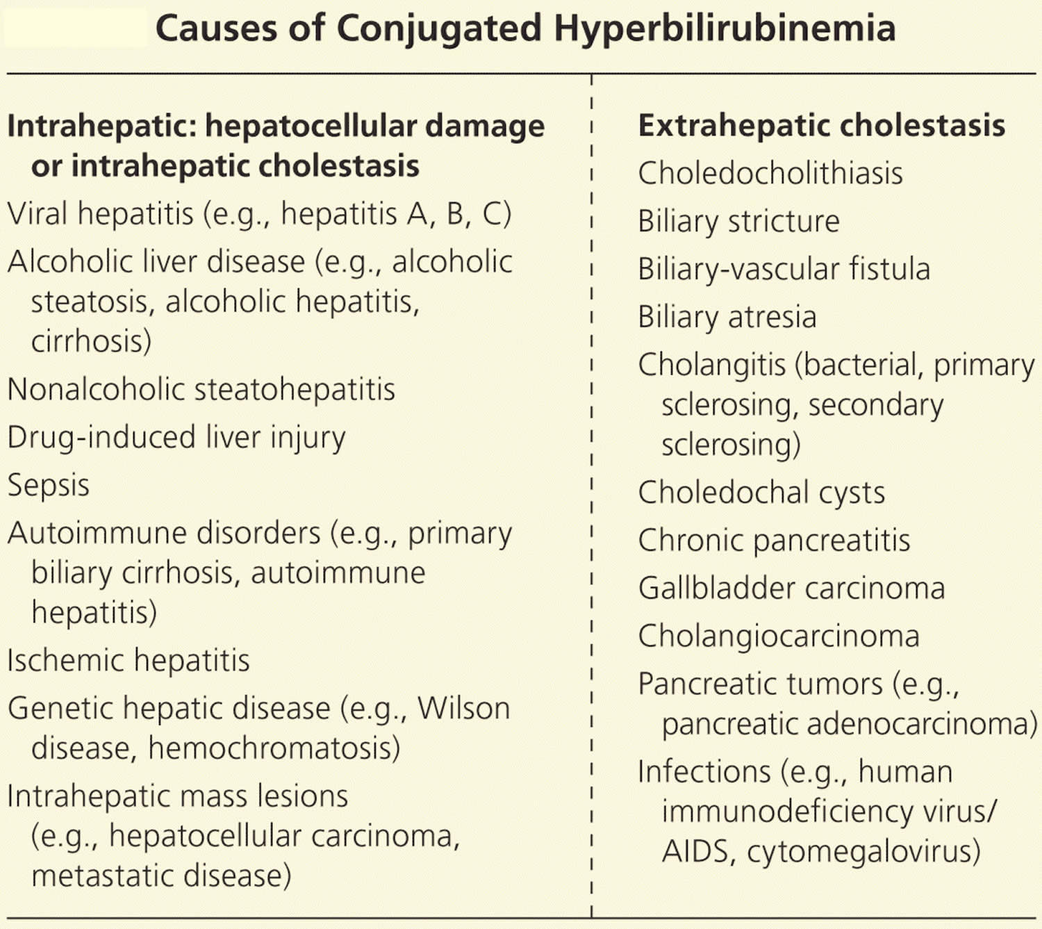
Intrahepatic disorders and intrahepatic cholestasis
The largest worldwide contributor to liver disease is viral hepatitis, mostly from hepatitis C 10. Viral hepatitis causes increased oxidative stress within liver cells (hepatocytes), leading to cell death, scarring, and diminished liver mass available for normal function 11, 12. Chronic alcohol consumption can cause various hepatic disorders, including fatty liver disease (steatosis) with minimal symptoms and often no jaundice; alcoholic hepatitis with acute onset jaundice and more severe symptoms; and cirrhosis, which is often associated with decompensation and liver failure in the setting of jaundice 13. Jaundice in persons with alcoholic liver disease can occur via multiple mechanisms, such as direct hepatocellular damage caused by ethanol metabolites or from alcohol’s effect on bile acid uptake and secretion contributing to cholestasis (reduced or stopped bile flow) 13, 14.
Approximately 30% to 40% of patients with nonalcoholic fatty liver disease (NAFLD) progress to nonalcoholic steatohepatitis (NASH), and approximately 40% to 50% of these patients develop fibrosis or cirrhosis that may lead to hyperbilirubinemia 15. Although the exact mechanism is poorly understood, liver lipid deposition may trigger inflammation and fibrosis, particularly when coupled with type 2 diabetes 15. Sepsis may also induce hyperbilirubinemia as circulating acute phase reactants and bacterial endotoxins disrupt bilirubin transport, leading to cholestasis and elevated bile salt levels 16, 17.
Extrahepatic disorders
Conjugated hyperbilirubinemia may also arise from extrahepatic obstruction. Patients with biliary obstruction may present with multiple signs and symptoms, including fever, itch (pruritus), abdominal pain, weight loss, muscle wasting, dark urine, and pale stools. Choledocholithiasis or the presence of gallstones within the common bile duct, is the most common non-neoplastic cause of biliary obstruction, accounting for 14% of all new cases of jaundice 3. An estimated 20 million Americans have gallstones, and risk factors for gallstones within the common bile duct (choledocholithiasis) include female sex, older age, increasing body mass index, and rapid weight loss 20.
Gallstones may cause jaundice by obstructing the biliary tree (typically the common bile duct) or by inducing a biliary stricture 21. Less commonly, stones in the gallbladder or cystic duct may mechanically compress the common hepatic duct causing jaundice, and, rarely, stones may cause the formation of a biliary-vascular fistula with accompanying jaundice 22. Biliary stricture causing postoperative jaundice is a rare complication of cholecystectomy (0.6% of cases) 21, 23.
Jaundice may be caused by surgeries such as liver transplantation and the Whipple and Billroth procedures, which both involve the creation of a choledochojejunostomy. Chronic pancreatitis may cause biliary strictures and jaundice, as may different forms of cholangitis 23, 24. In children, biliary atresia and choledochal cysts are the main causes of extrahepatic biliary obstruction 25.
Tumors are associated with 6.2% of new-onset cases of jaundice 3. Cholangiocarcinoma may affect the proximal or distal portions of the biliary tree by causing biliary strictures. Five-year survival for persons who have resection is 20% to 40%; survival in unresectable disease is less than one year 26, 27. Primary sclerosing cholangitis confers a 1,500-fold increased risk of cholangiocarcinoma, but more than 80% of cases have no risk factors for cholangiocarcinoma 26.
Gallbladder cancer, although rare, is the most common biliary tract malignancy; risk factors include gallstones, infection (Salmonella typhi), and female sex. Median survival is six to 12 months, depending on the stage at diagnosis 28. Ampullary cancers and bile duct compression from lymphadenopathy, or external tumors such as pancreatic cancer, may also cause bile duct obstruction.
Table 2. Causes of Conjugated Hyperbilirubinemia
Adults and children
Increased total bilirubin that is mainly unconjugated (indirect) bilirubin may be a result of:
- Hemolytic anemia or pernicious anemia
- Megaloblastic anemia
- A blood disorder called erythroblastosis fetalis
- Transfusion reaction in which red blood cells that were given in a transfusion are destroyed by the person’s immune system
- Cirrhosis (scarring of the liver)
- A relatively common inherited condition called Gilbert syndrome, due to low levels of the enzyme that produces conjugated bilirubin
If conjugated (direct) bilirubin is elevated more than unconjugated (indirect) bilirubin, there typically is a problem associated with decreased elimination of bilirubin by the liver cells. Some conditions that may cause this include:
- Viral hepatitis
- Drug reactions
- Alcoholic liver disease
Conjugated (direct) bilirubin is also elevated more than unconjugated (indirect) bilirubin when there is blockage of the bile ducts. This may occur, for example, with:
- Gallstones present in the bile ducts
- Tumors
- Scarring of the bile ducts or abnormal narrowing of the common bile duct (biliary stricture)
- Cancer of the pancreas or gallbladder cancer
In hepatobiliary diseases of various causes, bilirubin uptake, storage, and excretion are impaired to varying degrees. Thus, both conjugated and unconjugated bilirubin are retained and a wide range of abnormal serum concentrations of each form of bilirubin may be observed. Both conjugated and unconjugated bilirubins are increased in hepatitis and space-occupying lesions of the liver; and obstructive lesions such as carcinoma of the head of the pancreas, common bile duct, or ampulla of Vater.
Newborns
An elevated bilirubin level in a newborn may be temporary and resolve itself within a few days to two weeks. However, if the bilirubin level is above a critical threshold or increases rapidly, an investigation of the cause is needed so appropriate treatment can be initiated. Increased bilirubin concentrations may result from the accelerated breakdown of red blood cells due to:
- Blood type incompatibility between the mother and her newborn
- Certain congenital infections
- Lack of oxygen (hypoxia)
- Diseases that can affect the liver
In most of these conditions, only unconjugated (indirect) bilirubin is increased. An elevated conjugated (direct) bilirubin is seen in the rare conditions of biliary atresia and neonatal hepatitis. Biliary atresia requires surgical intervention to prevent liver damage.
Physiologic jaundice should resolve in 5 to 10 days in full-term infants and by 14 days in preterm infants.
Jaundice symptoms
Jaundice may appear suddenly or develop slowly over time. Symptoms of jaundice commonly include:
- Yellow skin and the white part of the eyes (sclera) — when jaundice is more severe, these areas may look brown
- Yellow color inside the mouth
- Dark or brown-colored urine
- Pale or clay-colored stools
- Itching (pruritis) usually occurs with jaundice
Other symptoms depend on the disorder causing the jaundice:
- Cancers may produce no symptoms, or there may be fatigue, weight loss, or other symptoms.
- Hepatitis may produce nausea, vomiting, fatigue, or other symptoms.
Jaundice complications
If the cause of your jaundice stays untreated, you may experience complications. These can vary depending on the underlying condition causing the jaundice.
Indirect (insoluble) bilirubin is harmful to cells and cellular structures. Due to the physiologic mechanisms that protect against elevated bilirubin, the toxic effects are limited to neonates due to the poorly developed blood-brain barrier. High levels of bilirubin are neurotoxic and can lead to permanent neurologic injury (kernicterus) in newborns 29.
Jaundice diagnosis
Your doctor will talk to you, ask about your symptoms, medical history, your lifestyle and perform a physical exam. The physical examination will show jaundice and possibly liver swelling. Specific tests vary but may include liver function and bilirubin blood tests to determine how well your liver is working.
Your doctor may order blood and urine tests. These allow your doctor to check your level of bilirubin and assess the health of your liver.
Your doctor may also order an ultrasound, MRI or CT scan to check for blockages. These can also be used to check for signs of liver and pancreatic disease. In some cases, your doctor may request a liver biopsy to confirm liver disease.
Other tests may include:
- Hepatitis virus panel to look for infection of the liver
- Liver function tests to determine how well the liver is working
- Complete blood count to check for low blood count or anemia
- Abdominal ultrasound
- Abdominal CT scan
- Magnetic resonance cholangiopancreatography (MRCP)
- Endoscopic retrograde cholangiopancreatography (ERCP)
- Percutaneous transhepatic cholangiogram (PTCA)
- Liver biopsy
- Cholesterol level
- Prothrombin time
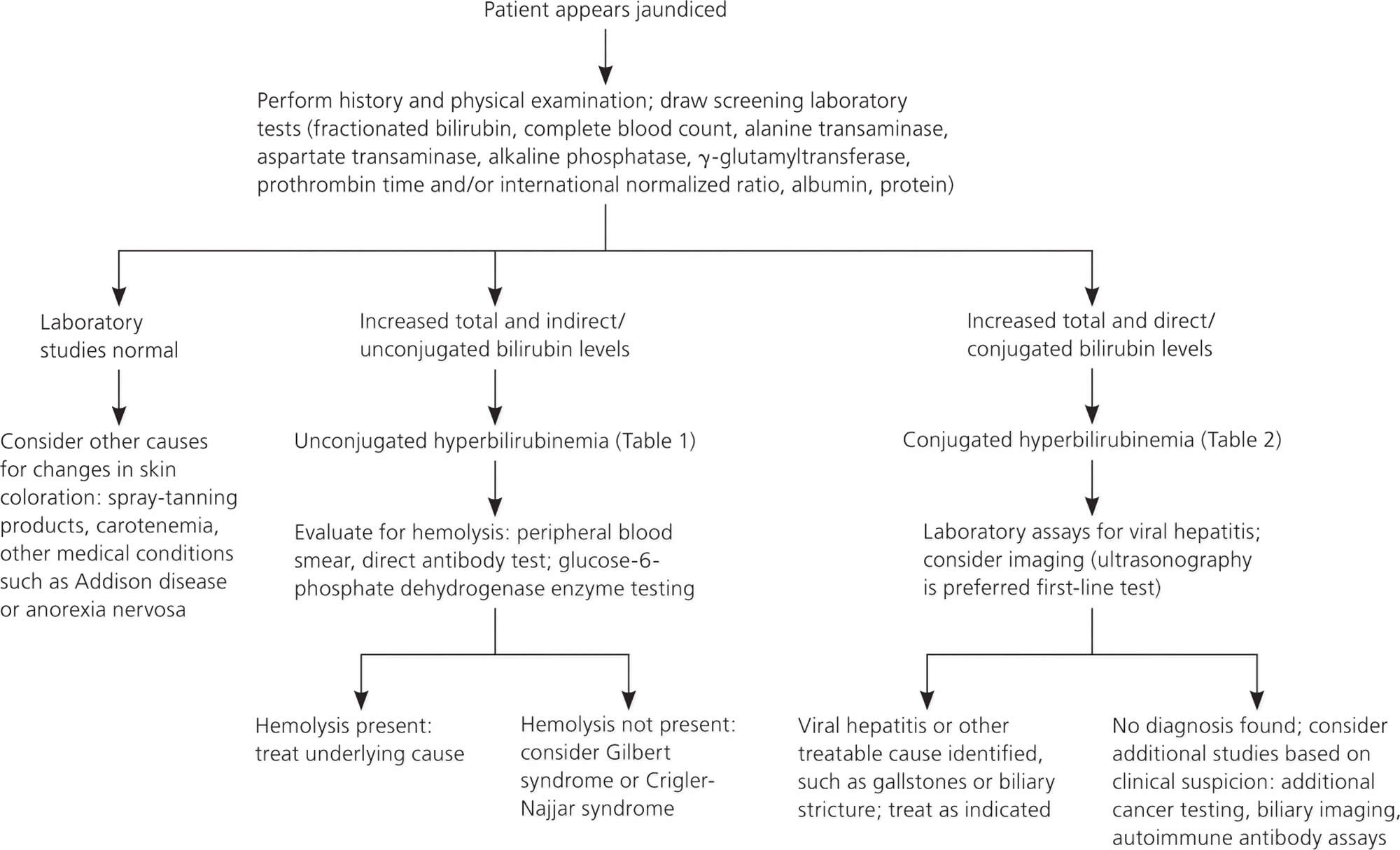
Figure 4. Jaundice in adults diagnostic algorithm
Bilirubin test
Bilirubin test measures the amount of bilirubin in your blood to evaluate your liver function or to help diagnose anemias caused by red blood cell destruction (hemolytic anemia).
A bilirubin test is used to detect an elevated bilirubin level in the blood. Bilirubin test may be used to help determine the cause of jaundice and/or help diagnose conditions such as liver disease, hemolytic anemia, and blockage of the bile ducts.
Bilirubin is an orange-yellow pigment, a waste product primarily produced by the normal breakdown of heme. Heme is a component of hemoglobin, which is found in red blood cells. Bilirubin is ultimately processed by the liver to allow its elimination from the body. Any condition that accelerates the breakdown of red blood cells or affects the processing and elimination of bilirubin may cause an elevated blood level.
Two forms of bilirubin can be measured or estimated by laboratory tests:
- Unconjugated bilirubin—when heme is released from hemoglobin, it is converted to unconjugated bilirubin. It is carried by proteins to the liver. Small amounts may be present in the blood.
- Conjugated bilirubin—formed in the liver when sugars are attached (conjugated) to bilirubin. It enters the bile and passes from the liver to the small intestines and is eventually eliminated in the stool. Normally, no conjugated bilirubin is present in the blood.
Usually, a chemical test is used to first measure the total bilirubin level (unconjugated plus conjugated bilirubin). If the total bilirubin level is increased, the laboratory can use a second chemical test to detect water-soluble forms of bilirubin, called “direct” bilirubin. The direct bilirubin test provides an estimate of the amount of conjugated bilirubin present. Subtracting direct bilirubin level from the total bilirubin level helps estimate the “indirect” level of unconjugated bilirubin. The pattern of bilirubin test results can give your healthcare provider information regarding the condition that may be present.
In adults and older children, bilirubin is measured to:
- Diagnose and/or monitor diseases of the liver and bile duct (e.g., cirrhosis, hepatitis, or gallstones)
- Evaluate people with sickle cell disease or other causes of hemolytic anemia; these people may have episodes called crises when excessive red blood cell destruction increases bilirubin levels.
In newborns with jaundice, bilirubin is used to distinguish the causes of jaundice.
- In both physiologic jaundice of the newborn and hemolytic disease of the newborn, only unconjugated (indirect) bilirubin is increased.
- In much less common cases, damage to the newborn’s liver from neonatal hepatitis and biliary atresia will increase conjugated (direct) bilirubin concentrations as well, often providing the first evidence that one of these less common conditions is present.
It is important that an elevated level of bilirubin in a newborn be identified and quickly treated because excessive unconjugated bilirubin damages developing brain cells. The consequences of this damage include mental retardation, learning and developmental disabilities, hearing loss, eye movement problems, and death.
When is bilirubin test ordered?
A healthcare practitioner usually orders a bilirubin test in conjunction with other liver function tests (alkaline phosphatase [ALP], aspartate aminotransferase [AST], alanine aminotransferase [ALT]) when someone shows signs of abnormal liver function.
A bilirubin level may be ordered when a person:
- Shows evidence of jaundice
- Has a history of drinking excessive amounts of alcohol
- Has suspected drug toxicity
- Has been exposed to hepatitis-causing viruses
Other symptoms that may be present include:
- Dark, amber-colored urine
- Nausea/vomiting
- Abdominal pain and/or swelling
- Fatigue and general malaise that often accompany chronic liver disease
Measuring and monitoring bilirubin in newborns with jaundice is considered standard medical care.
Tests for bilirubin may also be ordered when someone is suspected of having (or known to have) hemolytic anemia as a cause of anemia. In this case, it is often ordered along with other tests used to evaluate hemolysis, such as complete blood count, reticulocyte count, haptoglobin, and lactate dehydrogenase (LDH).
Imaging tests
Noninvasive imaging modalities in persons with jaundice include ultrasonography, computed tomography (CT), and magnetic resonance cholangiopancreatography. Ultrasonography or computed tomography is usually the first-line option to evaluate for obstruction, cirrhosis, and vessel patency, with ultrasonography being the least invasive and least expensive modality 13. Visualization of the intra- and extrahepatic biliary tree can be further evaluated using magnetic resonance cholangiopancreatography or endoscopic retrograde cholangiopancreatography, with the latter allowing for therapeutic options, such as biliary stent placement to relieve obstruction 23. Endoscopic ultrasonography can be used in addition to endoscopic retrograde cholangiopancreatography for evaluation of common bile duct obstructions and can help determine if the obstruction is from a mass or stone.
Liver biopsy
Liver biopsy is reserved for cases of jaundice in which the diagnosis is unclear after the initial history and physical examination, laboratory studies, and imaging 2. It should be performed only if biopsy results are required to determine treatment and prognosis. Biopsy may alter care in only about one-third of cases 23.
Jaundice treatment
The underlying cause of jaundice in adults needs to be treated, not the jaundice itself.
Your treatment options depend on the cause of your jaundice.
To relieve symptoms of jaundice caused by hepatitis, you should:
- get plenty of rest
- drink lots of fluids
- avoid alcohol
- avoid medicines that impact the liver
There are effective medicines for the treatment of hepatitis B and hepatitis C.
Your doctor might suggest surgery for other causes of jaundice such as:
- gallstones
- a blocked bile duct
- pancreatic cancer
Surgery may involve placement of a stent. This lets the bile (digestive fluid made by your liver) flow past the blockage.
Jaundice prognosis
Prognosis of jaundice depends on the underlying cause 1.
Causes of jaundice with excellent prognosis include jaundice from resorption of hematomas, physiologic jaundice of newborn, breastfeeding, breast milk jaundice, Gilbert syndrome, choledocholithiasis (gallstone within the common bile duct).
As a general rule, cancer causing biliary obstructions and cirrhosis with jaundice predict a poorer prognosis 30.
Neonatal jaundice
Jaundice is one of the most common conditions requiring medical attention in newborn babies. Approximately 60% of term and 80% of preterm babies develop jaundice in the first week of life, and about 10% of breastfed babies are still jaundiced at 1 month of age 31. In most babies with jaundice there is no underlying disease, and this early jaundice (termed ‘physiological jaundice’) is generally harmless. However, there are pathological causes of jaundice in the newborn, which, although rare, need to be detected. Such pathological jaundice may co-exist with physiological jaundice.
Neonatal jaundice refers to yellow coloration of the skin and the sclera (whites of the eyes) of newborn babies that results from accumulation of bilirubin in the skin and mucous membranes. This is associated with a raised level of bilirubin in the circulation, a condition known as hyperbilirubinemia. The term “kernicterus” refers to the ‘yellow staining of the basal nuclei of the brain’ caused by bilirubin. This is seen in parts of the brain on autopsy.
Factors significantly associated with hyperbilirubinemia are gestational age < 38 weeks, visible jaundice within 24 hours of birth, mother’s intention to breastfeed exclusively and family history of neonatal jaundice requiring treatment with phototherapy.
The level of bilirubinemia that results in kernicterus in a given infant is unknown. Poor-quality studies have shown a link between kernicterus (acute and chronic brain effects of severe hyperbilirubinemia) and both high serum bilirubin levels and free bilirubin levels in all babies.
Severe jaundice requiring exchange transfusion (bilirubin > 340 micromol/liter) and early onset of jaundice (within 24 hours) are statistically significant risk factors for hearing loss 31. Deafness is a clinical manifestation of kernicterus. Hemolytic disorders such as G6PD deficiency and ABO incompatibility (ABO incompatible blood) may cause a rapid increase in bilirubin level, and these disorders have been over-represented in international kernicterus registries and population studies of significant hyperbilirubinemia. A study of low-birthweight babies found a weak association between high serum bilirubin levels (> 340 micromol/liter) and neurodevelopmental impairment, hearing impairment and psychomotor impairment.
Bilirubin encephalopathy
Bilirubin encephalopathy is a rare neurological condition that occurs in some newborns with severe jaundice. Bilirubin encephalopathy is a serious condition. Many infants with late-stage nervous system complications die.
Bilirubin encephalopathy most often develops in the first week of life, but may be seen up until the third week. Some newborns with Rh hemolytic disease are at high risk for severe jaundice that can lead to this condition. Rarely, bilirubin encephalopathy can develop in seemingly healthy babies.
Bilirubin encephalopathy symptoms
The symptoms depend on the stage of bilirubin encephalopathy. Not all babies with kernicterus on autopsy have had definite symptoms.
Early stage:
- Extreme jaundice
- Absent startle reflex
- Poor feeding or sucking
- Extreme sleepiness (lethargy) and low muscle tone (hypotonia)
Middle stage:
- High-pitched cry
- Irritability
- May have arched back with neck hyperextended backwards, high muscle tone (hypertonia)
- Poor feeding
Late stage:
- Stupor or coma
- No feeding
- Shrill cry
- Muscle rigidity, markedly arched back with neck hyperextended backwards
- Seizures
Possible complications
Complications may include:
- Permanent brain damage
- Hearing loss
- Death
Prevention
Treating jaundice or conditions that may lead to it can help prevent this problem. Infants with the first signs of jaundice have bilirubin level measured within 24 hours. If the level is high, the infant should be screened for diseases that involve the destruction of red blood cells (hemolysis).
All newborns have a follow-up appointment within 2 to 3 days after leaving the hospital. This is very important for late preterm or early term babies (born more than 2 to 3 weeks before their due date).
Neonatal jaundice treatment
Treatment depends on how old the baby is (in hours) and whether the baby has any risk factors (such as prematurity). It may include:
- Light therapy (phototherapy)
- Exchange transfusions (removing the child’s blood and replacing it with fresh donor blood or plasma)
In preterm infants, the risk of a handicap increases by 30% for each 2.9 mg/dL increase of maximal total bilirubin concentration. While central nervous system damage is rare when total serum bilirubin is less than 20 mg/dL, premature infants may be affected at lower levels. The decision to institute therapy is based on a number of factors including total serum bilirubin, age, clinical history, physical examination, and coexisting conditions. Phototherapy typically is discontinued when total serum bilirubin level reaches 14 to 15 mg/dL.
How to manage hyperbilirubinemia in newborn babies
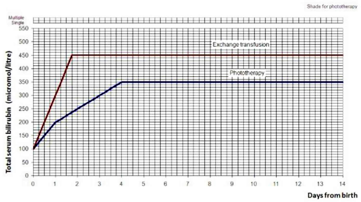
Use the bilirubin level to determine the management of hyperbilirubinemia in all babies (see threshold table (see Table 3) and treatment threshold graphs (Figures 5 and 6).
Table 3. Bilirubin threshold table for the management of babies of 38 weeks or more gestational age with hyperbilirubinemia
| Age (hours) | Bilirubin measurement (micromol/liter) | |||
|---|---|---|---|---|
| 0 | > 100 | > 100 | ||
| 6 | > 100 | > 112 | > 125 | > 150 |
| 12 | > 100 | > 125 | > 150 | > 200 |
| 18 | > 100 | > 137 | > 175 | > 250 |
| 24 | > 100 | > 150 | > 200 | > 300 |
| 30 | > 112 | > 162 | > 212 | > 350 |
| 36 | > 125 | > 175 | > 225 | > 400 |
| 42 | > 137 | > 187 | > 237 | > 450 |
| 48 | > 150 | > 200 | > 250 | > 450 |
| 54 | > 162 | > 212 | > 262 | > 450 |
| 60 | > 175 | > 225 | > 275 | > 450 |
| 66 | > 187 | > 237 | > 287 | > 450 |
| 72 | > 200 | > 250 | > 300 | > 450 |
| 78 | > 262 | > 312 | > 450 | |
| 84 | > 275 | > 325 | > 450 | |
| 90 | > 287 | > 337 | > 450 | |
| 96 | > 300 | > 350 | > 450 | |
| Action | ||||
| Repeat bilirubin measurement in 6–12 hours | Consider phototherapy and repeat bilirubin measurement in 6 hours | Start phototherapy | Perform an exchange transfusion unless the bilirubin level falls below threshold while the treatment is being prepared | |
Figure 5. Hyperbilirubinemia threshold graph for babies
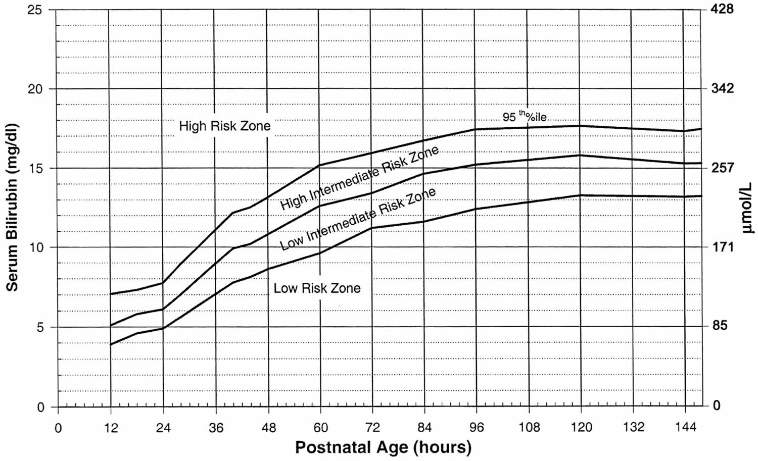
Figure 6. Hyperbilirubinemia treatment threshold graph for babies with neonatal jaundice >=38 weeks gestation
Phototherapy
Phototherapy is treatment with light. It is used in some cases of newborn jaundice to lower the bilirubin levels in your baby’s blood through a process called photo-oxidation.
Photo-oxidation adds oxygen to the bilirubin so it dissolves easily in water. This makes it easier for your baby’s liver to break down and remove the bilirubin from their blood.
There are two main types of phototherapy.
- Conventional phototherapy – where your baby is laid under a halogen or fluorescent lamp with their eyes covered
- Fibreoptic phototherapy – where your baby lies on a blanket that incorporates fibreoptic cables; light travels through the fibreoptic cables and shines on to your baby’s back
In both methods of phototherapy, the aim is to expose your baby’s skin to as much light as possible.
Conventional phototherapy is the treatment tried first in most cases, although fibreoptic phototherapy may be used first if your baby was born prematurely.
These types of phototherapy will usually be stopped for 30 minutes every three to four hours so you can feed your baby, change their nappy and give them a hug.
If your baby’s jaundice doesn’t improve after conventional or fibreoptic phototherapy, continuous multiple phototherapy may be offered. This involves using more than one light and often a fibreoptic blanket at the same time.
Treatment won’t be stopped during continuous multiple phototherapy. Instead, milk that has been squeezed out of your breasts in advance may be given through a tube into your baby’s stomach, or fluids may be given into one of their veins (intravenously).
During phototherapy, you baby’s temperature will be monitored to ensure they’re not getting too hot and they’ll be checked for signs of dehydration. Your baby may need intravenous fluids if they’re becoming dehydrated and aren’t able to drink a sufficient amount.
The bilirubin levels will be tested every four to six hours after phototherapy has started. Once levels start to fall, they’ll be checked every six to 12 hours.
Phototherapy will be stopped when the bilirubin level falls to a safe level, which usually takes a day or two.
Phototherapy is generally very effective for newborn jaundice and has very few side effects, although your baby may develop a temporary rash or tan as a result of the treatment.
Intravenous immunoglobulin
If jaundice is caused by an underlying health problem, such as an infection, this usually needs to be treated.
If the jaundice is caused by rhesus disease (when the mother has rhesus-negative blood and the baby has rhesus-positive blood), intravenous immunoglobulin (IVIG) may be used.
Intravenous immunoglobulin (IVIG) is usually only used if phototherapy alone hasn’t worked and the level of bilirubin in the blood is continuing to rise.
Exchange transfusion
A blood transfusion, known as an exchange transfusion, may be recommended if your baby has particularly high levels of bilirubin in their blood or if phototherapy hasn’t been effective.
During an exchange transfusion, small amounts of your baby’s blood are removed through a thin plastic tube placed into blood vessels in their umbilical cord, arms or legs. The blood is then replaced with blood from a suitable matching donor (someone with the same blood group).
As the new blood won’t contain bilirubin, the overall level of bilirubin in your baby’s blood will fall quickly.
Your baby will be monitored throughout the transfusion process, which can take several hours to complete. Any problems that may arise, such as bleeding, will be treated.
Your baby’s blood will be tested within two hours of treatment to check if it’s been successful. If the level of bilirubin in your baby’s blood remains high, the procedure may need to be repeated.
Only certain hospitals can perform exchange transfusions.
Breast milk jaundice
Jaundice also known as hyperbilirubinemia, is a condition that causes your baby’s skin to turn yellow in the first few days after birth. You may also notice that the sclera (white parts) of the baby’s eyes are yellow. The yellow color of the skin and sclera in newborns with jaundice comes from a build up of bilirubin. Small to medium increases in bilirubin are normal in newborns and will not hurt your baby. Very high levels of bilirubin can cause hearing loss, seizures and brain damage. There are two common problems that may occur in newborns receiving breast milk.
- Breast milk jaundice: Jaundice seen after the first week of life in a breastfed baby who is otherwise healthy.
- Breastfeeding failure jaundice also called “starvation jaundice”: Jaundice that occurs when your baby does not get enough breast milk, instead of from the breast milk itself.
Breast milk jaundice typically presents in the first or second week of life and usually spontaneously resolves even without discontinuation of breastfeeding 33. However, breast milk jaundice can persist for 8-12 weeks of life before resolution 34. Infants with breast milk jaundice often have higher peaks of serum bilirubin and an overall slower decline than infants without it, leading to the longer resolution time 35. Usually pathologic causes of persistent, unconjugated hyperbilirubinemia are ruled out before a diagnosis of breast milk jaundice can be made.
The frequency of breast milk jaundice within the United States is thought to be 20-30% for newborns from 3 to 4 weeks of age whose feeding is predominantly via breastfeeding 33. About 30-40% of breastfed infants are expected to have bilirubin levels greater than or equal to 5mg/dL with about 2-4% of exclusively breastfed infants having bilirubin levels above 10 mg/dL in week 3 of life 36. International studies in countries such as Turkey and Taiwan found that 20-28% of newborns had breast milk jaundice present at four weeks of age. Total serum bilirubin levels were also noted to be greater than or equal to 5mg/dL in these cases 37. The remaining international frequency of breast milk jaundice is not extensively reported but is thought to be similar to the frequency in the United States. No reports exist demonstrating a sex predilection.
In most cases of breastfeeding jaundice and breast-milk jaundice, doctors recommend that the mother continue breastfeeding. Early, frequent, unrestricted breastfeeding helps to eliminate bilirubin from baby’s body. Bilirubin exits the body in the infant’s stools, and because your milk has a laxative effect, breastfeeding frequently will result in lots of soiled diapers and thus, lower bilirubin levels. Your newborn should breastfeed a minimum of eight times per day.
Treatment is not necessary for breast milk jaundice unless the total serum bilirubin level of the infant is greater than 20mg/dL 33. If this occurs, the recommendation is for phototherapy treatment. Phototherapy is the use of light to convert bilirubin molecules into water-soluble isomers that can be excreted by the body. If the total serum bilirubin level remains below 12 mg/dL, the recommendation is to continue breastfeeding of the infant and to expect resolution of jaundice by 12 weeks of age. If the total serum bilirubin level is between 12-20 mg/dL and further investigation shows no evidence of hemolysis, the recommendations are the same 34. When the bilirubin is greater than 20 mg/dL, a brief 24-hour cessation of breastfeeding often leads to a sharp decline in the bilirubin levels.
What is breast milk jaundice levels?
About 30-40% of breastfed infants are expected to have bilirubin levels greater than or equal to 5mg/dL with about 2-4% of exclusively breastfed infants having bilirubin levels above 10 mg/dL in week 3 of life 36. International studies in countries such as Turkey and Taiwan found that 20-28% of newborns had breast milk jaundice present at four weeks of age. Total serum bilirubin levels were also noted to be greater than or equal to 5 mg/dL in these cases 37.
Breast milk jaundice cause
The exact cause of breast milk jaundice has not been determined 33. Most of the proposed causes involve factors present in the human breast milk itself. Another area of investigation has been the potential genetic mutations present in the affected newborns. Breast milk jaundice may run in families. It occurs just as often in males and females and affects about a third of all newborns who get only their mother’s milk.
Some factors in human breast milk that may be related to the cause of breast milk jaundice include pregnane-3a,20ß-diol, interleukin IL1ß e IL6, ß-glucuronidase, epidermal growth factor, and alpha-fetoprotein 38. The presence of pregnane-3a,20ß-diol is thought to inhibit the conjugation of bilirubin that allows for its excretion. ß-glucuronidase is an enzyme naturally present in the body that deconjugates bilirubin in the intestinal brush border, leading to increased unconjugated bilirubin levels 34. Studies have shown that the activity of this enzyme within formula milk is negligible, but it is considerable in human breast milk 39. Interleukin IL1ß e IL6 is thought to have a cholestatic effect that leads to hyperbilirubinemia 38. Epidermal growth factor is present in higher concentrations in human breast milk and in the serum of strictly breastfed infants. The thinking is that this substance enhances intestinal resorption of bilirubin and reduces intestinal motility in the neonatal period, leading to increased unconjugated bilirubin levels 34. The serum of babies with breast milk jaundice often has elevated levels of alpha-fetoprotein. The mechanism underlying this is not yet understood.
Several studies have shown that mutations in the coding region of the UGT1A1 gene increase the risk of developing breast milk jaundice. Mutations in this gene’s regulatory region are known to cause Crigler-Najjar and Gilbert syndrome, two syndromes known to cause persistent hyperbilirubinemia 37.
Bilirubin is a yellow pigment that is produced as the body recycles old red blood cells. The liver helps break down bilirubin so that it can be removed from the body in the stool. Newborns have markedly increased bilirubin production as compared to their adult counterparts secondary to a higher blood volume and hemoglobin concentration and a shorter red blood cell lifespan. This bilirubin production is in its unconjugated form, which is not water soluble and must, therefore, be conjugated into its water-soluble form before excretion from the body in the stool. This process takes place within the liver and specifically within the hepatocyte itself by the hepatic enzyme, bilirubin uridine diphosphate glucuronosyltransferase (bilirubin-UGT). The UGT1A1 gene codes production of this enzyme, and therefore those with genetic mutations in this gene are unable to conjugate bilirubin properly. This enzyme is also significantly less active in neonates than adults, leading to less efficient conjugation. After conjugation and excretion from hepatocytes within bile, the bilirubin travels to the small intestine where it converts to stercobilin by gut flora and gets excreted via the stool.
Infants have a decreased concentration of gut flora needed for this process compared to adults, leading to a higher concentration of bilirubin remaining within the intestinal tract. The enzyme ß-glucuronidase will deconjugate bilirubin remaining within the intestine. After this process, it is returned to the liver for re-conjugation via the portal circulation. This process is known as “enterohepatic circulation.” Newborns have significantly higher activity of the enzyme ß-glucuronidase as well as a more permeable gut wall, leading to overall higher concentrations of unconjugated bilirubin and increased enterohepatic circulation 34.
Identification of the substances within breast milk and the genetic mutations that maybe causes or risk factors for breast milk jaundice interfere with the healthy metabolism and excretion of bilirubin at the various stages are covered above.
Breast milk jaundice symptoms
True breast milk jaundice manifests after the first 4-7 days of life. A second peak in serum bilirubin level is noted by age 14 days. Your baby’s skin, and possibly the whites of the eyes (sclerae), will look yellow. Breast milk jaundice typically presents in the first or second week of life with an unconjugated hyperbilirubinemia in an otherwise healthy infant whose nutritional intake is predominantly via breastfeeding. These infants exhibit normal weight gain with normal production of urine and stools 34. A total serum bilirubin level above 1.5 mg/dL is considered elevated at this time, but most infants will not appear jaundiced unless the level is above 5 mg/dL. If the infant does appear jaundiced, this yellowish discoloration of their skin and/or sclera is typically first noted in the face and then proceeds to the infant’s trunk and extremities.
Breast milk jaundice complications
The most feared complication of all neonatal hyperbilirubinemia including breast milk jaundice is kernicterus or chronic bilirubin encephalopathy. This concern is due to its potential for permanent neurodevelopmental delay, and is a rare complication of breast milk jaundice and occurs in less than 2% of breastfed term infants who have no evidence of hemolytic anemia 40.
Breast milk jaundice diagnosis
Evaluation of an infant presenting with hyperbilirubinemia suspicious for breast milk jaundice must include methods to rule out other pathologic causes of hyperbilirubinemia. First, both unconjugated and conjugated bilirubin levels must be measured. Conjugated bilirubin levels less than 1 mg/dL or 20% of the total bilirubin level are considered normal. Conjugated bilirubin levels in excess of this indicate other disorders such as biliary atresia, neonatal hepatitis, and disorders of bilirubin excretion. Both breast milk jaundice and hemolytic anemias cause elevated unconjugated bilirubin levels. Hemolytic causes for hyperbilirubinemia, such as ABO incompatibility, G6PD deficiency, and hereditary spherocytosis must be ruled out.
Laboratory tests that may be done include:
- Bilirubin level (total and direct)
- Coombs’ test
- Blood smear to look at blood cell shapes and sizes
- Blood type
- Complete blood count (measurement of hemoglobin and hematocrit)
- Reticulocyte count (number of slightly immature red blood cells)
- Genetic testing
In some cases, a blood test to check for glucose-6-phosphate dehydrogenase (G6PD) may be done. G6PD is a protein that helps red blood cells work properly.
These tests are done to be sure that there are no other, more dangerous causes of the jaundice.
Another test that may be considered consists of stopping breastfeeding and giving formula for 12 to 24 hours. This is done to see if the bilirubin level goes down. This test is not always necessary.
Physiologic jaundice usually manifests after the first 24 hours of life. This can be accentuated by breastfeeding, which, in the first few days of life, may be associated with suboptimal milk and suboptimal caloric intake, especially if milk production is delayed. This is known as breastfeeding jaundice. Jaundice that manifests before the first 24 hours of life should always be considered pathologic until proven otherwise. In this situation, a full diagnostic workup with emphasis on infection and hemolysis should be undertaken.
In clinical practice, differentiating between physiologic jaundice from breast milk jaundice is important so that the duration of hyperbilirubinemia can be predicted. Identifying the infants who become dehydrated secondary to inadequate breastfeeding is also important. These babies need to be identified early and given breastfeeding support and formula supplementation as necessary. Depending on their serum bilirubin concentration, neonates with hyperbilirubinemia may become sleepy and feed poorly.
The clinician should consider other non-hemolytic causes of prolonged hyperbilirubinemia such as Crigler-Najjar or Gilbert syndrome but need not investigate for them unless jaundice does not resolve by 12 weeks of age. Galactosemia and hypothyroidism have also been identified as causes of unconjugated hyperbilirubinemia and should be ruled out via newborn screening tests 34.
Breast milk jaundice treatment
Treatment is not necessary for breast milk jaundice unless the total serum bilirubin level of the infant is greater than 20mg/dL 33. If this occurs, the recommendation is for phototherapy treatment. Phototherapy is the use of light to convert bilirubin molecules into water-soluble isomers that can be excreted by the body. If the total serum bilirubin level remains below 12 mg/dL, the recommendation is to continue breastfeeding of the infant and to expect resolution of jaundice by 12 weeks of age. If the total serum bilirubin level is between 12-20 mg/dL and further investigation shows no evidence of hemolysis, the recommendations are the same 34. When the bilirubin is greater than 20 mg/dL, a brief 24-hour cessation of breastfeeding often leads to a sharp decline in the bilirubin levels.
Treatments to lower the level of bilirubin in your baby’s blood may include:
- Light therapy (phototherapy). Your baby may be placed under a special lamp that emits light in the blue-green spectrum. The light changes the shape and structure of bilirubin molecules in such a way that they can be excreted in both the urine and stool. During treatment, your baby will wear only a diaper and protective eye patches. Light therapy may be supplemented with the use of a light-emitting pad or mattress.
- Exchange transfusion. Rarely, when severe jaundice doesn’t respond to other treatments, a baby may need an exchange transfusion of blood. This involves repeatedly withdrawing small amounts of blood and replacing it with donor blood, thereby diluting the bilirubin and maternal antibodies — a procedure that’s performed in a newborn intensive care unit.
Breast milk jaundice prognosis
Overall, the prognosis of infants with breast milk jaundice is excellent as it is a self-limited condition that usually resolves around 12 weeks of age 41.
Breast milk jaundice in otherwise healthy full-term infants rarely causes kernicterus (bilirubin encephalopathy). Case reports suggest that some breastfed infants who suffer from prolonged periods of inadequate breast milk intake and whose bilirubin levels exceeded 25 mg/dL may be at risk of kernicterus 42. Note that kernicterus is a preventable cause of cerebral palsy. Another group of breastfed infants who may be at risk of complications is late preterm infants who are nursing poorly.
Kernicterus may occur in exclusively breastfed infants in the absence of hemolysis or other specific pathologic conditions. Distinguishing between breastfeeding jaundice and breast milk jaundice is important, because bilirubin-induced encephalopathy occurs more commonly in breastfeeding jaundice. Near-term infants are more likely to manifest breastfeeding jaundice because of their difficulty in achieving adequate nursing, greater weight loss, and hepatic immaturity.
- Joseph A, Samant H. Jaundice. [Updated 2022 Aug 8]. In: StatPearls [Internet]. Treasure Island (FL): StatPearls Publishing; 2022 Jan-. Available from: https://www.ncbi.nlm.nih.gov/books/NBK544252[↩][↩][↩]
- Fargo MV, Grogan SP, Saguil A. Evaluation of Jaundice in Adults. Am Fam Physician. 2017 Feb 1;95(3):164-168. https://www.aafp.org/pubs/afp/issues/2017/0201/p164.html[↩][↩][↩][↩]
- Vuppalanchi R, Liangpunsakul S, Chalasani N. Etiology of new-onset jaundice: how often is it caused by idiosyncratic drug-induced liver injury in the United States? Am J Gastroenterol. 2007 Mar;102(3):558-62; quiz 693. doi: 10.1111/j.1572-0241.2006.01019.x[↩][↩][↩]
- Gallagher PG. Abnormalities of the erythrocyte membrane. Pediatr Clin North Am. 2013 Dec;60(6):1349-62. doi: 10.1016/j.pcl.2013.09.001[↩]
- Koralkova, P., van Solinge, W.W. and van Wijk, R. (2014), Rare hereditary red blood cell enzymopathies associated with hemolytic anemia – pathophysiology, clinical aspects, and laboratory diagnosis. Int. Jnl. Lab. Hem., 36: 388-397. https://doi.org/10.1111/ijlh.12223[↩]
- Martin A, Thompson AA. Thalassemias. Pediatr Clin North Am. 2013 Dec;60(6):1383-91. doi: 10.1016/j.pcl.2013.08.008[↩]
- Bass GF, Tuscano ET, Tuscano JM. Diagnosis and classification of autoimmune hemolytic anemia. Autoimmun Rev. 2014 Apr-May;13(4-5):560-4. doi: 10.1016/j.autrev.2013.11.010[↩]
- Strassburg CP. Hyperbilirubinemia syndromes (Gilbert-Meulengracht, Crigler-Najjar, Dubin-Johnson, and Rotor syndrome). Best Pract Res Clin Gastroenterol. 2010 Oct;24(5):555-71. doi: 10.1016/j.bpg.2010.07.007[↩][↩]
- Erlinger S, Arias IM, Dhumeaux D. Inherited disorders of bilirubin transport and conjugation: new insights into molecular mechanisms and consequences. Gastroenterology. 2014 Jun;146(7):1625-38. doi: 10.1053/j.gastro.2014.03.047[↩]
- Sun S, Song Z, Cotler SJ, Cho M. Biomechanics and functionality of hepatocytes in liver cirrhosis. J Biomech. 2014 Jun 27;47(9):2205-10. doi: 10.1016/j.jbiomech.2013.10.050[↩]
- Suhail M, Abdel-Hafiz H, Ali A, Fatima K, Damanhouri GA, Azhar E, Chaudhary AG, Qadri I. Potential mechanisms of hepatitis B virus induced liver injury. World J Gastroenterol. 2014 Sep 21;20(35):12462-72. doi: 10.3748/wjg.v20.i35.12462[↩]
- Levitt DG, Levitt MD. Quantitative assessment of the multiple processes responsible for bilirubin homeostasis in health and disease. Clin Exp Gastroenterol. 2014 Sep 2;7:307-28. doi: 10.2147/CEG.S64283[↩]
- Roche SP, Kobos R. Jaundice in the adult patient. Am Fam Physician. 2004;69(2):299-304. https://www.aafp.org/pubs/afp/issues/2004/0115/p299.html[↩][↩][↩][↩][↩]
- Rocco A, Compare D, Angrisani D, Sanduzzi Zamparelli M, Nardone G. Alcoholic disease: liver and beyond. World J Gastroenterol. 2014 Oct 28;20(40):14652-9. doi: 10.3748/wjg.v20.i40.14652[↩]
- Byrne CD, Targher G. NAFLD: a multisystem disease. J Hepatol. 2015 Apr;62(1 Suppl):S47-64. doi: 10.1016/j.jhep.2014.12.012[↩][↩]
- Bauer M, Press AT, Trauner M. The liver in sepsis: patterns of response and injury. Curr Opin Crit Care. 2013 Apr;19(2):123-7. doi: 10.1097/MCC.0b013e32835eba6d[↩]
- Kosters A, Karpen SJ. The role of inflammation in cholestasis: clinical and basic aspects. Semin Liver Dis. 2010 May;30(2):186-94. doi: 10.1055/s-0030-1253227[↩]
- Chen M, Suzuki A, Borlak J, Andrade RJ, Lucena MI. Drug-induced liver injury: Interactions between drug properties and host factors. J Hepatol. 2015 Aug;63(2):503-14. doi: 10.1016/j.jhep.2015.04.016[↩]
- Wooton-Kee CR, Jain AK, Wagner M, Grusak MA, Finegold MJ, Lutsenko S, Moore DD. Elevated copper impairs hepatic nuclear receptor function in Wilson’s disease. J Clin Invest. 2015 Sep;125(9):3449-60. doi: 10.1172/JCI78991[↩]
- Cafasso DE, Smith RR. Symptomatic cholelithiasis and functional disorders of the biliary tract. Surg Clin North Am. 2014 Apr;94(2):233-56. doi: 10.1016/j.suc.2013.12.001[↩]
- Fang Y, Gurusamy KS, Wang Q, Davidson BR, Lin H, Xie X, Wang C. Pre-operative biliary drainage for obstructive jaundice. Cochrane Database Syst Rev. 2012 Sep 12;9(9):CD005444. doi: 10.1002/14651858.CD005444.pub3[↩][↩]
- Luu MB, Deziel DJ. Unusual complications of gallstones. Surg Clin North Am. 2014 Apr;94(2):377-94. doi: 10.1016/j.suc.2014.01.002[↩]
- Winger J, Michelfelder A. Diagnostic approach to the patient with jaundice. Prim Care. 2011 Sep;38(3):469-82; viii. doi: 10.1016/j.pop.2011.05.004[↩][↩][↩][↩]
- Mortelé KJ, Wiesner W, Cantisani V, Silverman SG, Ros PR. Usual and unusual causes of extrahepatic cholestasis: assessment with magnetic resonance cholangiography and fast MRI. Abdom Imaging. 2004 Jan-Feb;29(1):87-99. doi: 10.1007/s00261-003-0062-6[↩]
- Krishna RP, Lal R, Sikora SS, Yachha SK, Pal L. Unusual causes of extrahepatic biliary obstruction in children: a case series with review of literature. Pediatr Surg Int. 2008 Feb;24(2):183-90. doi: 10.1007/s00383-007-2087-3[↩]
- Dickson PV, Behrman SW. Distal cholangiocarcinoma. Surg Clin North Am. 2014 Apr;94(2):325-42. doi: 10.1016/j.suc.2013.12.004[↩][↩]
- Brown KM, Geller DA. Proximal biliary tumors. Surg Clin North Am. 2014 Apr;94(2):311-23. doi: 10.1016/j.suc.2013.12.003[↩]
- Wernberg JA, Lucarelli DD. Gallbladder cancer. Surg Clin North Am. 2014 Apr;94(2):343-60. doi: 10.1016/j.suc.2014.01.009[↩]
- Le Pichon JB, Riordan SM, Watchko J, Shapiro SM. The Neurological Sequelae of Neonatal Hyperbilirubinemia: Definitions, Diagnosis and Treatment of the Kernicterus Spectrum Disorders (KSDs). Curr Pediatr Rev. 2017;13(3):199-209. doi: 10.2174/1573396313666170815100214[↩]
- Shin SJ, Park H, Sung YN, Yoo C, Hwang DW, Park JH, Kim KP, Lee SS, Ryoo BY, Seo DW, Kim SC, Hong SM. Prognosis of Pancreatic Cancer Patients with Synchronous or Metachronous Malignancies from Other Organs Is Better than Those with Pancreatic Cancer Only. Cancer Res Treat. 2018 Oct;50(4):1175-1185. doi: 10.4143/crt.2017.494[↩]
- National Collaborating Centre for Women’s and Children’s Health (UK). Neonatal Jaundice. London: RCOG Press; 2010 May. (NICE Clinical Guidelines, No. 98.) Available from: https://www.ncbi.nlm.nih.gov/books/NBK65119[↩][↩][↩][↩]
- https://bilitool.org/[↩]
- Bratton S, Stern M. Breast Milk Jaundice. [Updated 2019 Jan 1]. In: StatPearls [Internet]. Treasure Island (FL): StatPearls Publishing; 2019 Jan-. Available from: https://www.ncbi.nlm.nih.gov/books/NBK537334[↩][↩][↩][↩][↩]
- Preer GL, Philipp BL. Understanding and managing breast milk jaundice. Arch. Dis. Child. Fetal Neonatal Ed. 2011 Nov;96(6):F461-6.[↩][↩][↩][↩][↩][↩][↩][↩]
- Auerbach KG, Gartner LM. Breastfeeding and human milk: their association with jaundice in the neonate. Clin Perinatol. 1987 Mar;14(1):89-107.[↩]
- Ullah S, Rahman K, Hedayati M. Hyperbilirubinemia in Neonates: Types, Causes, Clinical Examinations, Preventive Measures and Treatments: A Narrative Review Article. Iran. J. Public Health. 2016 May;45(5):558-68.[↩][↩]
- Maruo Y, Morioka Y, Fujito H, Nakahara S, Yanagi T, Matsui K, Mori A, Sato H, Tukey RH, Takeuchi Y. Bilirubin uridine diphosphate-glucuronosyltransferase variation is a genetic basis of breast milk jaundice. J. Pediatr. 2014 Jul;165(1):36-41.e1.[↩][↩][↩]
- Soldi A, Tonetto P, Varalda A, Bertino E. Neonatal jaundice and human milk. J. Matern. Fetal. Neonatal. Med. 2011 Oct;24 Suppl 1:85-7.[↩][↩]
- el-Kholy MS, Halim HY, Marzouk AH. Beta-glucuronidase and hyperbilirubinemia in breast-fed versus formula-fed babies. J Egypt Public Health Assoc. 1992;67(3-4):237-48.[↩]
- Muchowski KE. Evaluation and treatment of neonatal hyperbilirubinemia. Am Fam Physician. 2014 Jun 01;89(11):873-8.[↩]
- Breast milk jaundice. https://emedicine.medscape.com/article/973629-overview[↩]
- Harris MC, Bernbaum JC, Polin JR, Zimmerman R, Polin RA. Developmental follow-up of breastfed term and near-term infants with marked hyperbilirubinemia. Pediatrics. 2001 May. 107(5):1075-80.[↩]


