Contents
Boerhaave syndrome
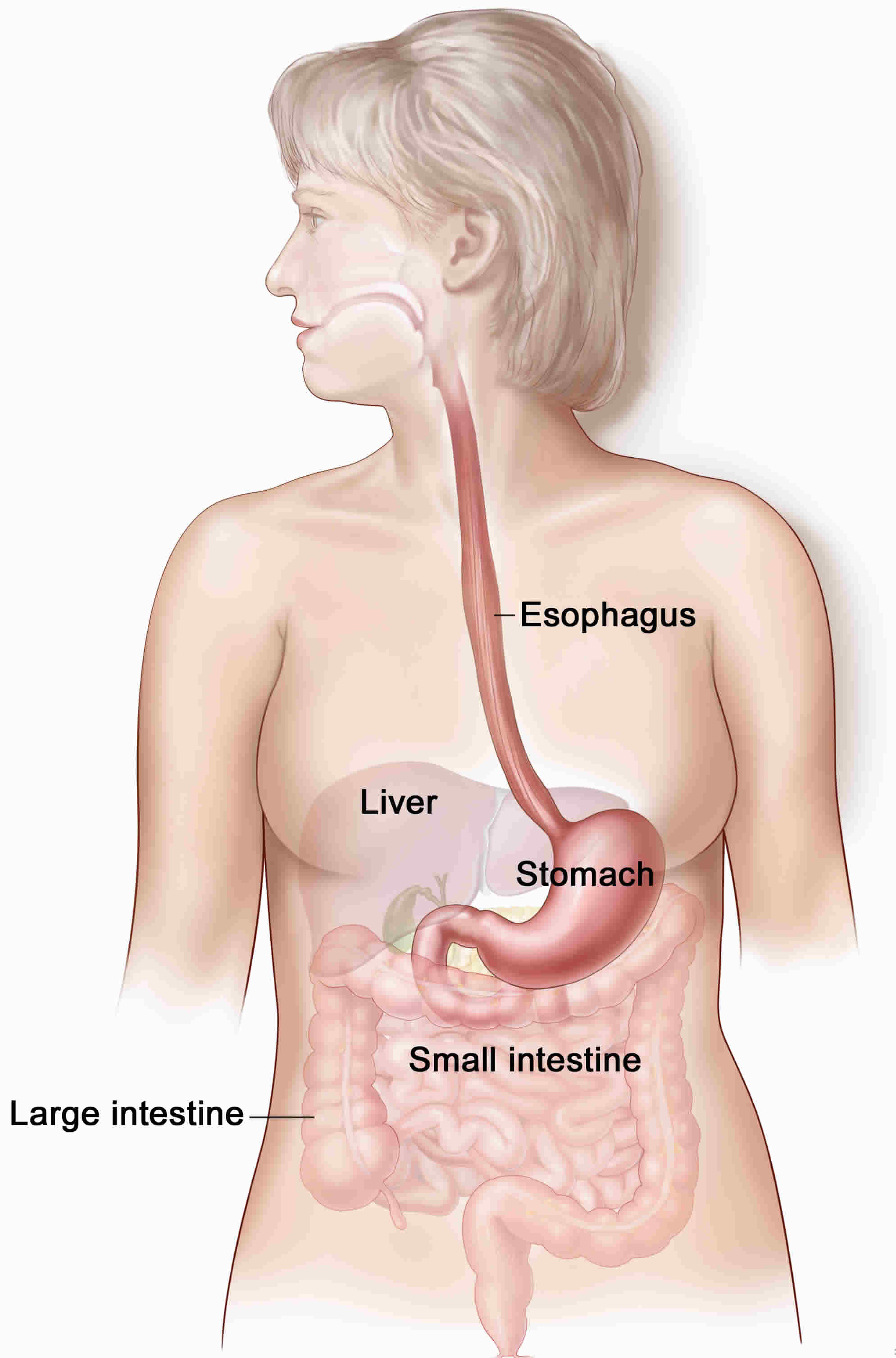
Boerhaave syndrome is also known as spontaneous rupture of the esophagus, which typically occurs after forceful vomiting 1. The esophagus is the tube food passes through as it goes from the mouth to the stomach. The contents of the esophagus can pass into the surrounding area in the chest (mediastinum), when there is a hole in the esophagus. This often results in infection of the mediastinum (mediastinitis). The classic presentation of Boerhaave syndrome is characterized by Mackler’s triad, consisting of chest pain, vomiting, and subcutaneous emphysema. However, Boerhaave syndrome rarely presents with all the features of Mackler’s triad; instead, the common presentation of Boerhaave syndrome includes chest or epigastric pain, severe retching and vomiting, dyspnea, and shock. These symptoms are typically misdiagnosed as cardiogenic in origin 1. Boerhaave syndrome itself is rare, with an annual incidence of 3.1 per 1,000,000 2. Due to Boerhaave syndrome atypical presentation, rarity, and mimicry of emergent conditions, diagnosis of Boerhaave syndrome is often delayed, resulting in a high mortality rate up to 40% at the time of diagnosis and with a subsequent exponential increase in mortality if treatment is delayed by greater than 48 hours 3. In addition, several factors, including difficulty assessing the esophagus and the unusual blood supply of the organ, contribute to the condition’s high morbidity. Without treatment, survival of Boerhaave’s syndrome is in days. Management relies on rapid recognition and intervention, as lack of therapeutic interventions can be fatal 4.
Boerhaave syndrome causes
Boerhaave’s syndrome is also known as spontaneous a transmural perforation of the esophagus or effort rupture of the esophagus. Although vomiting is thought to be the most common cause, other causes include weightlifting, defecation, epileptic seizures, abdominal trauma, compressed air injury, and childbirth, all of which can increase the pressure in the esophagus and cause a barogenic esophageal rupture. Most cases occur in patients with a normal underlying esophagus, although the presence of esophagitis or ulcers has also been found in a subset of individuals.
The esophagus may also become perforated as the result of:
- A tumor
- Gastric reflux with ulceration
- Previous surgery on the esophagus
- Swallowing a foreign object or caustic chemicals, such as household cleaners, disk batteries, and battery acid
- Trauma or injury to the chest and esophagus
- Violent vomiting (Boerhaave syndrome)
Less common causes include injuries to the esophagus area (blunt trauma) and injury to the esophagus during surgery of another organ near the esophagus.
Spontaneous esophageal rupture is caused by a sudden increase in intra-esophageal pressure, leading to a complete transmural tear through the esophagus. The complications will depend on the location of the rupture, as the esophagus abuts different areas of the body. The mid esophagus lies next to the right pleura while the lower esophagus abuts the left pleura. Rupture occurs most commonly in the left posterolateral wall of the distal third of the esophagus with extension into the left pleural cavity. Intrathoracic esophagus perforations can lead to mediastinal inflammation, emphysema or necrosis due to gastric contents entering the area. However, rupture may also occur in the cervical or upper thoracic area. Upper thoracic or mid-esophagus perforations tend to produce pleural effusion or hydropneumothorax on the right. The cervical ruptures are usually more localized and benign as the spread to the mediastinum through the retroesophageal space is slow and limited 5.
Boerhaave syndrome symptoms
Mackler’s triad, consisting of chest pain, vomiting, and subcutaneous emphysema represents a classic presentation of Boerhaave syndrome. However, Mackler’s triad is only present in about 5% of case, with chest pain being the most common feature 6. A healthcare provider should suspect Boerhaave’s syndrome when a patient presents with retrosternal chest pain with or without subcutaneous emphysema when associated with heavy alcohol intake and severe or repeated vomiting. Up to one-third of patients do not present with these symptoms.
The actual clinical presentation of Boerhaave syndrome will depend on the level of the perforation, the degree of leakage, and the time since onset of injury. Typically, the patient will present with pain at the site of perforation, usually in the neck, chest, epigastric region, or upper abdomen. Cervical perforations can present with neck pain, dysphagia, or dysphonia; intra-thoracic perforations with chest pain; and intra-abdominal perforations with epigastric pain radiating to the shoulder or back. History of increased intra-esophageal pressure for any reason followed by chest pain should prompt consideration of this condition. Physical exam findings may include abnormal vitals (tachycardia, tachypnea, fever), decreased breath sounds on the perforated side, mediastinal emphysema, and Hamman’s sign (mediastinal “crackling” accompanying every heart beat) in left lateral decubitus position.
A perforation in the middle or lower most part of the esophagus may cause:
- Swallowing problems
- Chest pain
- Breathing problems
Boerhaave syndrome complications
Esophageal rupture may lead to the development of septicemia, pneumomediastinum, mediastinitis, massive pleural effusion, empyema, pneumomediastinum, or subcutaneous emphysema.
If the esophageal rupture extends directly into the pleura, hydropneumothorax is expected. In adults, this occurs more commonly on the left side of the pleura. In neonates, esophageal rupture usually occurs on the right side.
After esophageal rupture, free air enters the mediastinum and also may spread to the adjacent structures, resulting in mediastinal abscess or superimposed secondary infection.
Other complications include acute respiratory distress syndrome, pneumomediastinum, pneumothorax, and hydrothorax.
Boerhaave syndrome diagnosis
Unfortunately, laboratory tests provide little help in Boerhaave syndrome diagnosis; however, they can exclude more common conditions in the differential including heart attack (myocardial infarction) and pancreatitis.
Your health care provider will look for:
- Fast breathing.
- Fever.
- Low blood pressure .
- Rapid heart rate.
- Neck pain or stiffness and air bubbles underneath the skin if the perforation is in the top part of the esophagus.
You may have a chest x-ray to look for:
- Air in the soft tissues of the chest.
- Fluid that has leaked from the esophagus into the space around the lungs.
- Collapsed lung. X-rays taken after you drink a non-harmful dye can help pinpoint the location of the perforation.
You may also have a chest CT scan to look for an abscess in the chest or esophageal cancer.
Imaging is of high importance in diagnosing Boerhaave’s Syndrome. While chest X-ray is readily available, it is normal for approximately 15% of cases and can not be used to exclude the diagnosis. Potential chest X-ray findings include subcutaneous or mediastinal emphysema, mediastinal widening, pleural effusion, and in 20% of cases, the “V sign” (radiolucent streak of air dissecting the retrocardiac fascial planes). The diagnostic tool of choice is the contrast esophagogram using a water-soluble contrast agent such as gastrograffin. Such a study will show extravasation of contrast material at the site of the perforation. Even though barium is superior in demonstrating small perforations, its use is not advised since extravasation of this material can lead to mediastinitis with subsequent fibrosis. CT scan is being used my many instead of contrast esophagogram due to its higher sensitivity and more detailed assessment of the involved organs. Endoscopy should be used with caution due to the risk of further esophageal perforation. If a chest tube is present, one can also use the methylene blue dye test. When sweetened methylene blue is taken orally, it gives a bluish discolouration to the chest tube effluent within 12 – 24 hours.
Boerhaave syndrome treatment
Treatment is typically tailored to the patient’s presentation, the type and extent of rupture, the time to diagnosis, and the viability of the esophageal wall. Early perforations, those diagnosed within 12-24 hours, have the best outcomes. Three common treatment options include conservative, endoscopic, or surgical.
Mainstays of Boerhaave syndrome treatment include the following:
- Fluids given through a vein (IV)
- IV antibiotics to prevent or treat infection
- Draining of fluid around the lungs with a chest tube
- Mediastinoscopy to remove fluid that has collected in the area behind the breastbone and between the lungs (mediastinum)
The decision to use a conservative (medical intervention only) or an aggressive (medical plus surgical intervention) approach depends on the following factors:
- Time delay in presentation and diagnosis
- Extent of perforation
- Overall medical condition of the patient
Conservative management consists of the following:
- Intravenous fluids should be administered.
- Antibiotics: Imipenem/cilastatin or ticarcillin/clavulanate offer good broad-spectrum coverage.
- Nasogastric suction should be applied.
- Keep the patient nil per os (NPO).
- Adequate drainage with tube thoracostomy or formal thoracotomy is vital.
- Early use of nutritional supplementation: Evidence suggests that for hastening recovery, a jejunostomy tube feeding may be favored over hyperalimentation.
A stent may be placed in the esophagus if only a small amount of fluid has leaked. This may help avoid surgery.
The mainstay of treatment includes volume replacement, broad-spectrum antibiotic coverage, and surgical evaluation. Surgical intervention includes primary esophageal repair through open thoracotomy versus video-assisted thoracic surgery (VATS) with fundic reinforcement, which is the gold standard within the first twenty-four hours. Endoscopic placement of stents has been used to prevent fistula formations or seal esophageal leaks in both patients with delayed diagnoses and those with the early diagnosis without widespread contamination. Conservative measurements are usually reserved for small or contained ruptures. Controversy occurs when a late perforation, those diagnosed after 24 hours, is diagnosed, as the wound edges are typically edematous, stiff, or friable rendering primary repair risky. Taking this into consideration, many manage late perforations through debridement of pleural cavity and mediastinum, esophagostomy, and feeding gastrostomy. Esophageal replacement is usually done after 6 weeks.
Surgery is often needed to repair a perforation in the middle or bottom portions of the esophagus. The leak may be treated by simple repair or by removing the esophagus, depending on the extent of the problem.
A perforation in the uppermost (neck region) part of the esophagus may heal by itself if you do not eat or drink for a period of time. In this case, you will need a stomach feeding tube or another way to get nutrients. Nutritional supplementation is required if prolonged “nothing through the mouth” status is necessary 7.
Boerhaave syndrome prognosis
The prognosis of Boerhaave syndrome is directly contingent on its early recognition and appropriate intervention 3. Boerhaave syndrome can progress to shock, even death, if untreated. Early diagnosis allows prompt surgical repair. Diagnosis and surgery within 24 hours carries a 75% survival rate but drops to approximately 50% after a 24-hour delay and approximately 10% after 48 hours.
Morbidity/mortality
Morbidity and mortality are high. Esophageal perforation is the most lethal perforation of the gastrointestinal tract. Early recognition and appropriate surgical intervention are essential for survival.
Overall, the mortality rate is approximately 30%. Mortality is usually due to subsequent infection, including mediastinitis, pneumonitis, pericarditis, or empyema.
As noted earlier, patients who undergo surgical repair within 24 hours of injury have a 70-75% chance of survival. This falls to 35-50% if surgery is delayed longer than 24 hours and to approximately 10% if delayed longer than 48 hours. Therefore, early diagnosis and intervention of esophageal perforation significantly reduces mortality.
Cases of patients surviving without surgery exist but are rare enough to warrant case reports in the medical literature.
- Lieu MT, Layoun ME, Dai D, Soo Hoo GW, Betancourt J. Tension hydropneumothorax as the initial presentation of Boerhaave syndrome. Respir Med Case Rep. 2018;25:100–103. Published 2018 Jul 31. doi:10.1016/j.rmcr.2018.07.007 https://www.ncbi.nlm.nih.gov/pmc/articles/PMC6083431[↩][↩]
- Vidarsdottir H.1, Blondal S., Alfredsson H., Geirsson A., Gudbjartsson T. Oesophageal perforations in Iceland: a whole population study on incidence, aetiology and surgical outcome. Thorac. Cardiovasc. Surg. 2010;58(8):476–480.[↩]
- Turner AR, Turner SD. Boerhaave Syndrome. [Updated 2019 Jun 3]. In: StatPearls [Internet]. Treasure Island (FL): StatPearls Publishing; 2019 Jan-. Available from: https://www.ncbi.nlm.nih.gov/books/NBK430808[↩][↩]
- Ciriano Hernández P, Grao Torrente I, Viejo Martínez E, Turégano Fuentes F. Boerhaave syndrome presenting as gastric emphysema. Cir Esp. 2019 Apr;97(4):231.[↩]
- Y K, F AB, A T, D H. Boerhaave syndrome in an elderly man successfully treated with 3-month indwelling esophageal stent. Radiol Case Rep. 2018 Oct;13(5):1084-1086.[↩]
- Vallabhajosyula S., Sundaragiri P.R., Berim I.G. Boerhaave syndrome presenting as tension pneumothorax: first reported north american case. J. Intensive Care Med. 2016;31(5):349–352.[↩]
- Still S, Mencio M, Ontiveros E, Burdick J, Leeds SG. Primary and Rescue Endoluminal Vacuum Therapy in the Management of Esophageal Perforations and Leaks. Ann Thorac Cardiovasc Surg. 2018 Aug 20;24(4):173-179.[↩]


