Contents
What is a hip pointer
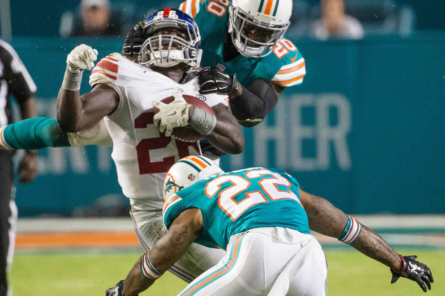
A hip pointer is a contusion of the iliac crest and/or the greater trochanteric region of the femur following a direct impact or collision that causes a contusion or subperiosteal hematoma 1. Hip pointer injuries are common in collision sports 2. A hip pointer is usually caused by a direct blow or from a fall striking the iliac crest or greater trochanter. The typical hip pointer injury is characterized by localized pain and severe palpable tenderness along a variable distance of the iliac crest 3. Athletes can often return to the field of play, and in professional sports their pain can be controlled with local anesthetic injection. These athletes usually recover in 1 to 3 weeks 3.
The assessment of routine hip pointers usually does not involve radiological investigation, and most of these injuries are managed clinically 2. The athlete’s symptoms and clinical signs are well localized to the iliac crest. It is common practice in professional athletes to treat pain with a guided or unguided local anesthetic injection to enable them to return to play on the same day or subsequent days to weeks. Although uncommon, iliac crest fractures can occur in collision sports, and an inability to return to play after a local anesthetic injection would warrant radiography to rule out a fracture.
The management of hip pointer injuries requires coordinated care between athletic trainers, clinical providers, and primary care and sports medicine specialists. The majority of hip pointer injuries improve with standard, first-line nonoperative management modalities.
Injuries to the hip and pelvis in sports are much less common compared to injuries about the knee and/or ankle 4. Hip injuries comprise 5 to 10% of athletic injuries, and hip pointers comprise an even smaller relative subset percentage of these hip injuries. A review of injuries in the National Football League from 1997 to 2006 reported an incidence of 0.3%, with an average of 5.6 days of training lost due to injury 5.
Figure 1. Hip pointer injury (bruising on the left anterolateral abdominal wall on day 3 post injury)
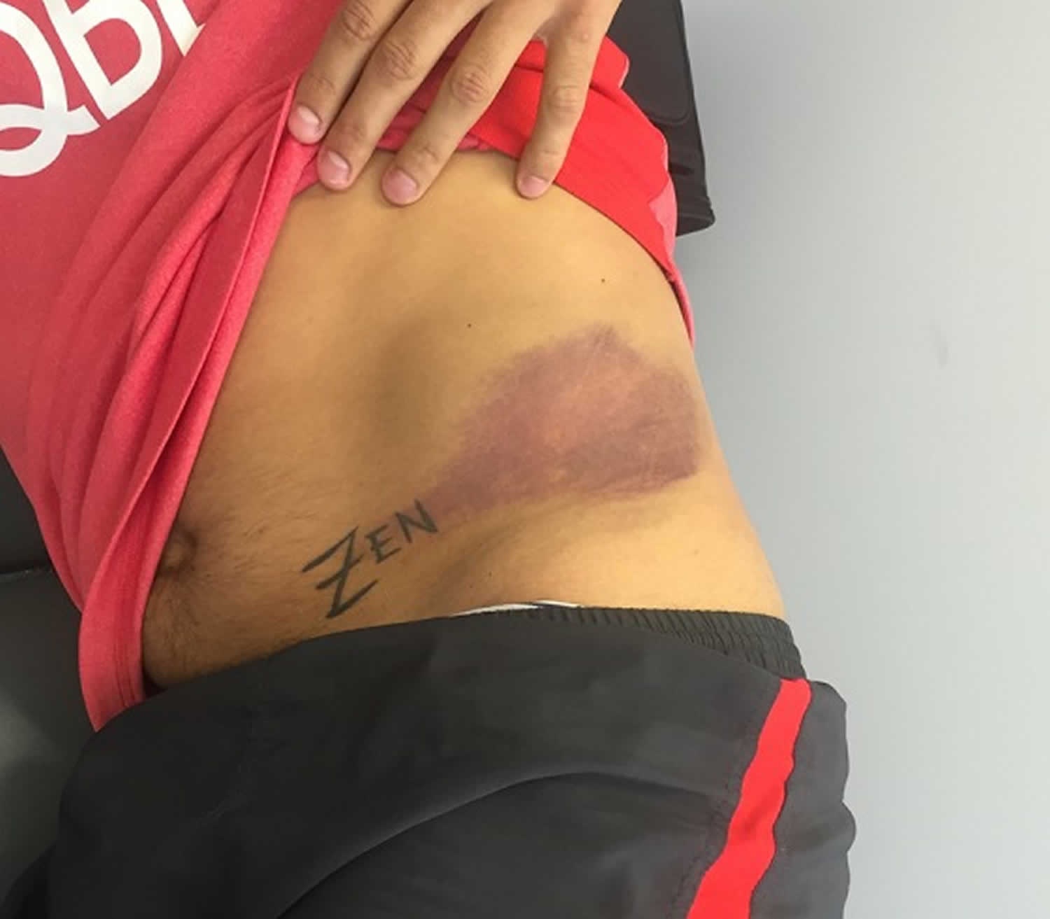
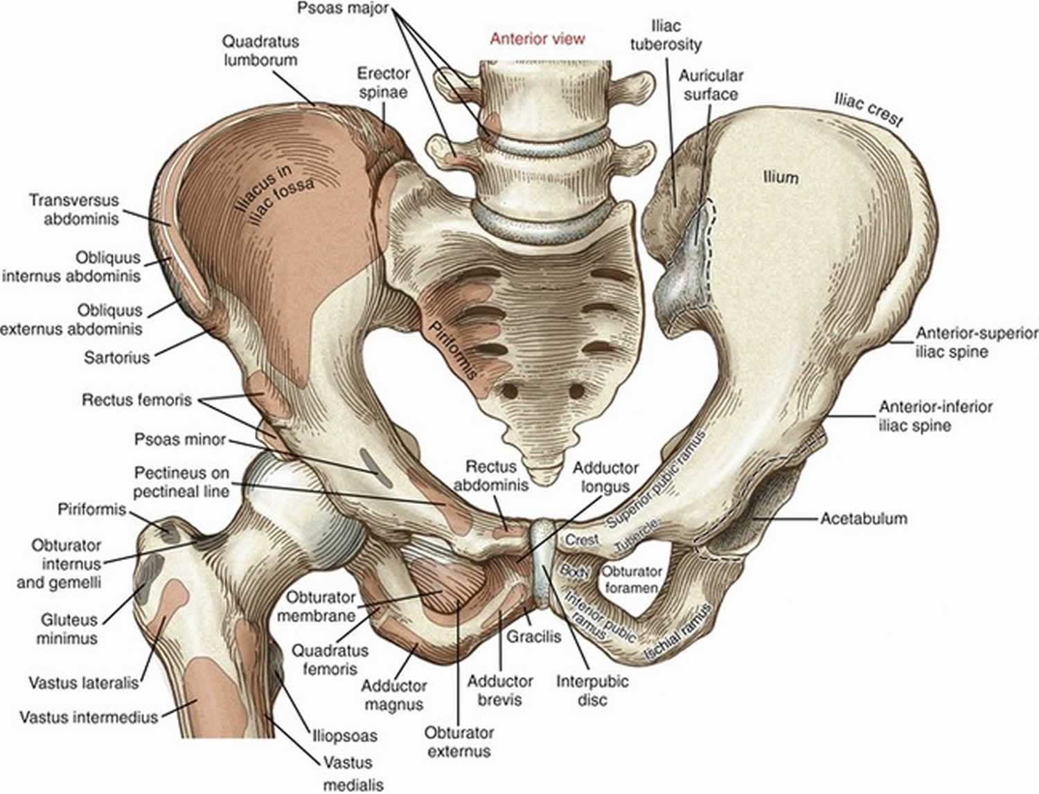
Figure 2. Hip anatomy
Hip pointer causes
The anterior iliac crest region and greater trochanter of the femur have, in most patients, minimal overlying fatty tissue and thus these areas are more susceptible to injury from direct trauma 6.
The primary cause of hip pointer is a direct blow to the iliac crest or greater trochanter during contact sports. American football and hockey are the most common sports that result in these injuries 6. The former implicates tackling and incidental collisions that regularly occur during competition. The same underlying mechanism can be said for rugby as well. In ice hockey, hip pointers can result from being checked into the boards or by contact with another player or player’s equipment 7. Other non-contact sports or high- versus low-energy traumatic mechanisms can also cause iliac crest contusions. In this scenario, a direct fall or a direct traumatic blow (i.e., secondary to a motor vehicle collision) is often the precipitating factor 8.
Following direct impact to the iliac crest, an ensuing hematoma develops around the area and often includes varying degrees of bleeding into the hip abductor musculature. The iliac crest is the origin of several muscles which can be affected depending on the degree and extent of the zone of injury 9:
- Sartorius
- Gluteus medius
- Tensor fascia lata
- Abdominal musculature – Transverse or oblique muscles
Hip pointer symptoms
Most commonly, patients present with varying degrees of bruising (ecchymoses) or contusion around the area of impact. The patient will be tender directly over these areas, and often range of motion (ROM) about the hip is limited secondary to pain. The hip abductors are typically weak, while the motor strength of the hip flexor and extensors should be intact. Hip abductors and external rotators may exhibit limited strength secondary to pain.
Hip pointer injury complications
- Chronic pain/dysfunction
- Inability to return to prior athletic performance
- Missed diagnosis leading to persistent pain/disability
Hip pointer injury diagnosis
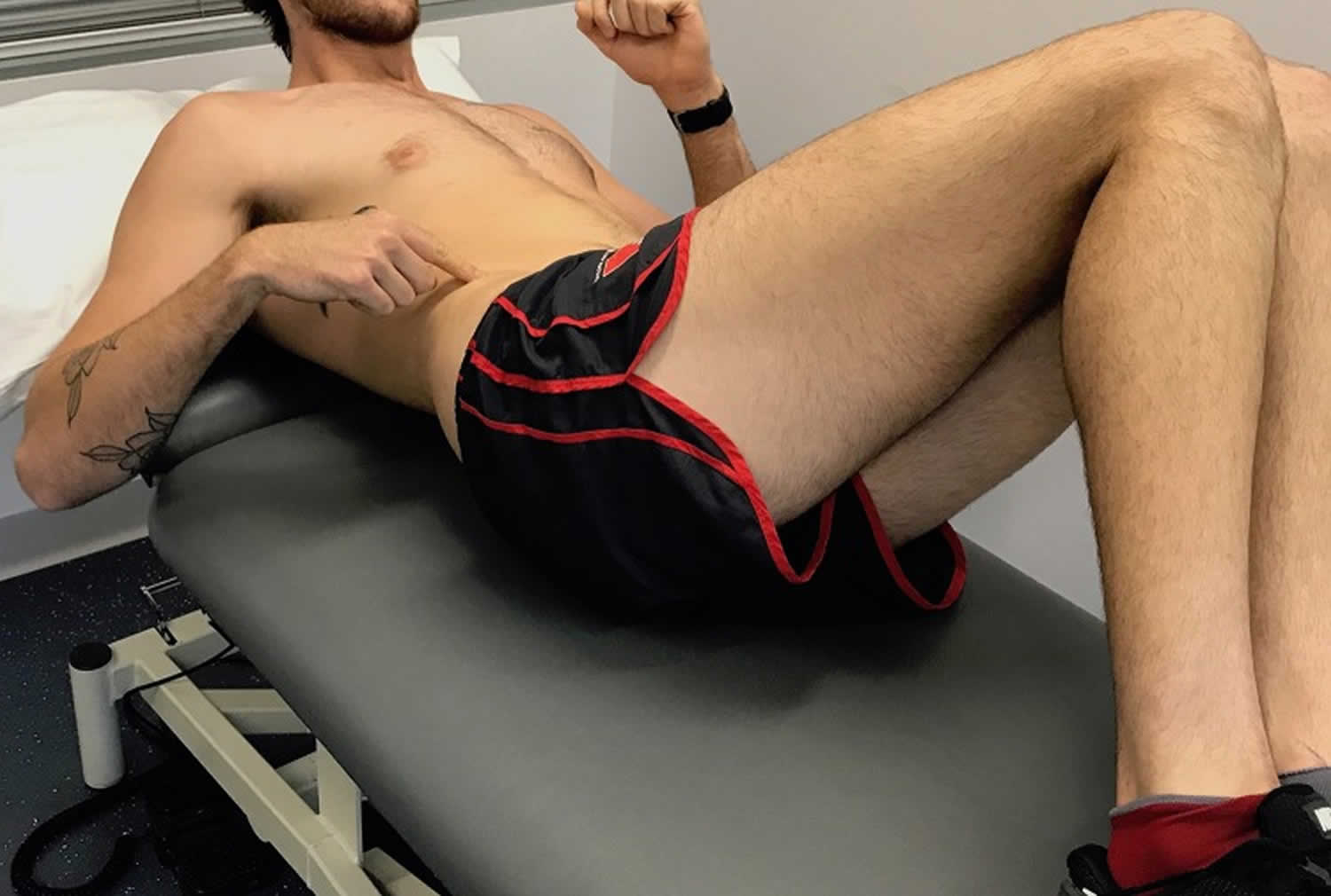
A useful clinical test, not only to initially diagnose the hip pointer injury but also to track recovery, is the “resisted oblique sit-up test”; this maneuver entails stressing the involved anterolateral abdominal and assessing pain and strength, while palpating for a palpable defect or tenderness (Figure 3).
Figure 3. Resisted oblique sit-up test
Radiographs are needed if a fracture or myositis ossificans is suspected. Hip pointer injuries are an important consideration for high school and college athletes because the ischial tuberosity and anterior superior iliac spine may fuse as late as the 3rd decade of life. Pediatric and adolescent athletes should be evaluated to rule out potential pelvic avulsion injuries including 7:
- Iliac crest avulsion injuries secondary to abdominal musculature avulsions
- Anterior superior iliac spine (ASIS) avulsion injuries sartorius and/or tensor fascia lata avulsion(s)
- Anterior inferior iliac spine (AIIS) avulsion injuries rectus femoris avulsion injury
- Ischial tuberosity proximal hamstring avulsion injuries
Otherwise, radiographs obtained in the setting of a true, isolated, iliac crest contusion (hip pointer injury), radiographs are unremarkable.
Consider CT scans if the patient has continued pain or the pain exceeds that expected from examination findings. A bone scan can exclude stress fractures if initial symptoms do not resolve. An MRI obtained in the setting of an isolated iliac crest contusion would likely reveal a large hematoma but is otherwise unremarkable.
A hip pointer must be distinguished from chronic exertional compartment syndrome, a femoral neck fracture, avascular necrosis, hip fracture or dislocation, tendonitis, iliotibial band syndrome, osteitis pubis, sacroiliac joint injury, snapping hip syndrome, and slipped capital femoral epiphysis.
Hip pointer treatment
The initial therapy of a hip pointer injury includes ice, anti-inflammatory pain medication, compression, rest, and avoidance of weight bearing as guided by the patient’s reported symptoms 6. As the pain decreases, the initiation of range of motion and active resistance exercises should begin. If a hematoma is present, aspiration can provide some pain relief and may potentially mitigate the risks of developing myositis ossificans. A local anesthetic such as 1% to 2% lidocaine or 0.5% bupivacaine (5mL to 9 mL), may provide short-term pain relief. It is considered safe, but little data support this practice. Even though there are no studies supporting corticosteroids, it is an accepted practice in the sports medicine community. If compartment syndrome is suspected, measure compartment pressures and consult an orthopedic surgeon or sports medicine specialist. An orthopedic surgeon should also be consulted if there is an avulsion fracture or for pain lasting longer than two weeks.
Physical therapy is often prescribed. Graded strengthening program for lumbopelvic region, initially focusing on isometric co-contractions and progressing to more challenging exercises involving planking and side planking to strengthen and remodel the damaged anterolateral abdominal wall 3. In the later stages of the rehabilitation, perform dynamic strengthening of the thoracolumbar region, using gym equipment (free weights, pulleys, and clinical Pilates) 3.
Hip pointer recovery time
Most patients can gradually return to normal levels of activity within two to three weeks 6.
Padding at the injury site may decrease the incidence of re-injury, limit pain, and allow a return to activity sooner. Patients with a hip pointer may return to activity once the pain and swelling resolve, and normal function returns. Patients with a hip pointer are at risk for recurrent injury and should be advised to avoid activities that may induce a repeat injury.
- Blazina ME. The “hip-pointer,” a term to describe a specific kind of athletic injury. Calif Med. 1967;106(6):450[↩]
- Hall M, Anderson J. Hip pointers. Clin Sports Med. 2013;32(2):325–330.[↩][↩]
- Gultekin S, Cross T. The Franklin-Naismith Lesion: A Severe Variant of Hip Pointer. Orthop J Sports Med. 2019;7(1):2325967118820507. Published 2019 Jan 9. doi:10.1177/2325967118820507 https://www.ncbi.nlm.nih.gov/pmc/articles/PMC6329028/[↩][↩][↩][↩][↩]
- Bordoni B, Varacallo M. StatPearls [Internet]. StatPearls Publishing; Treasure Island (FL): Dec 15, 2018. Anatomy, Bony Pelvis and Lower Limb, Thigh Quadriceps Muscle.[↩]
- Feeley BT, Powell JW, Muller MS, Barnes RP, Warren RF, Kelly BT. Hip injuries and labral tears in the national football league. Am J Sports Med. 2008 Nov;36(11):2187-95.[↩]
- Varacallo M, Bordoni B. Hip Pointer Injuries (Iliac Crest Contusions) [Updated 2019 Feb 9]. In: StatPearls [Internet]. Treasure Island (FL): StatPearls Publishing; 2019 Jan-. Available from: https://www.ncbi.nlm.nih.gov/books/NBK538258[↩][↩][↩][↩]
- Bordoni B, Varacallo M. StatPearls [Internet]. StatPearls Publishing; Treasure Island (FL): Dec 15, 2018. Anatomy, Bony Pelvis and Lower Limb, Thigh Quadriceps Muscle[↩][↩]
- Smith JM, Varacallo M. StatPearls [Internet]. StatPearls Publishing; Treasure Island (FL): Jan 19, 2019. Osgood Schlatter’s Disease [Tibial Tubercle Apophysitis][↩]
- Walters BB, Varacallo M. StatPearls [Internet]. StatPearls Publishing; Treasure Island (FL): Nov 14, 2018. Anatomy, Bony Pelvis and Lower Limb, Thigh Sartorius Muscle.[↩]


