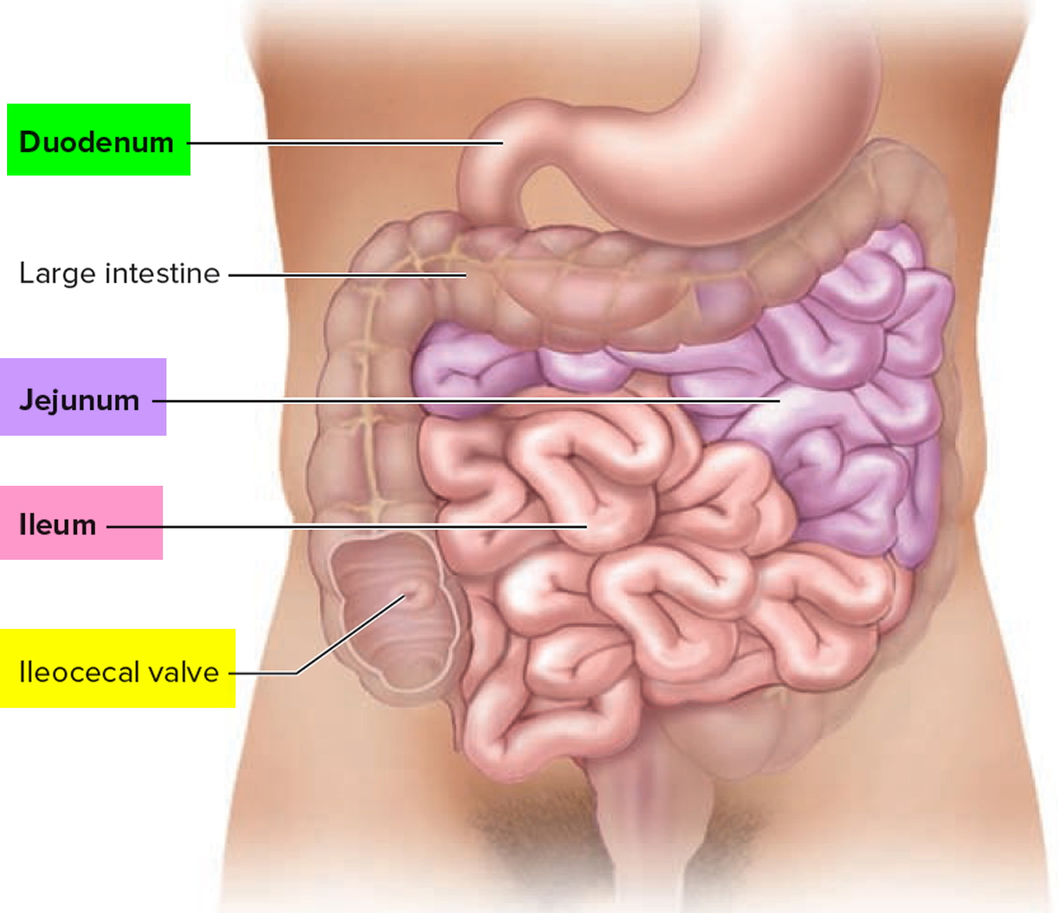
What is ileum
Ileum (“twisted intestine”) is the final and the longest portion of the small intestine, the ileum measures about 3 m (9.8 ft ) and contributes to almost 60% of the length of the small intestine. The ileum joins the large intestine at a smooth muscle sphincter called the ileocecal sphincter (valve). The ileum terminates at the ileocaecal junction in a valve at the superior aspect of the cecum before it goes on to form the ascending colon. The ileum absorbs any nutrients that got past the jejunum, with major absorptive products being vitamin B12 and bile acids 1. After participating in the emulsification and absorption of lipids, most of the bile salts are reabsorbed by active transport in the ileum and returned by the blood to the liver through the hepatic portal system for recycling. This cycle of bile salt secretion by hepatocytes into bile, reabsorption by the ileum, and resecretion into bile is called the enterohepatic circulation. Vitamin B12 combines with intrinsic factor produced by the stomach and the combination is absorbed in the ileum via an active transport mechanism.
The lamina propria of the small intestinal mucosa contains areolar connective tissue and has an abundance of mucosa-associated lymphoid tissue (MALT). Solitary lymphatic nodules are most numerous in the distal part of the ileum. Groups of lymphatic nodules referred to as aggregated lymphatic nodules or Peyer’s patches, are also present in the ileum. About 40 of these lymphatic nodules (Peyer’s patches) are present, averaging about a centimeter long and a centimeter wide. Besides destroying the microorganisms that invade them, the aggregated lymphoid nodules (Peyer’s patches) sample many different antigens from within the ileum and generate a wide variety of memory lymphocytes to protect the body.
The opening of the ileum of the small intestine into the cecum’s medial wall is surrounded internally by the ileocecal valve (Figure 3), which is formed by two raised edges of the mucosa. A sphincter in the distal ileum keeps the valve closed until there is food in the stomach, at which time the sphincter reflexively relaxes, opening the valve. As the cecum fills, its walls stretch, pulling the edges of the ileocecal valve together and closing the opening. This action prevents reflux of feces from the cecum back into the ileum.
The jejunum and ileum receive their blood supply from a rich network of arteries that travel through the mesentery and originate from the superior mesenteric artery. The multitude of arterial branches that split from the superior mesenteric artery is known as the arterial arcades, and they give rise to the vasa recta that deliver the blood to the jejunum and ileum.
Figure 1. Small intestine
Figure 2. Ileum anatomy
Figure 3. Terminal ileum
Ileum function
The ileum absorbs nutrients that did not get absorbed by the jejunum, with important nutrients being vitamin B12 and bile acids for reuse.
Solitary lymphatic nodules are most numerous in the distal part of the ileum. Besides destroying the microorganisms that invade them, the aggregated lymphoid nodules (Peyer’s patches) sample many different antigens from within the ileum and generate a wide variety of memory lymphocytes to protect the body.
The small bowel, in particular the ileum, has been identified as the primary site for active sulfate absorption by the intestine 2. Inorganic sulfate (SO2−4) is an important anion, vital for normal growth and development, and serves as an essential factor in numerous physiological processes 3. The majority of sulfate available to the body is sourced from the diet, either absorbed directly by the intestine in its inorganic form or released following the metabolism of sulfur-containing amino acids 4.
- Collins JT, Bhimji SS. Anatomy, Abdomen and Pelvis, Small Intestine. [Updated 2018 Sep 12]. In: StatPearls [Internet]. Treasure Island (FL): StatPearls Publishing; 2018 Jan-. Available from: https://www.ncbi.nlm.nih.gov/books/NBK459366[↩]
- Active sulfate absorption in rabbit ileum: dependence on sodium and chloride and effects of agents that alter chloride transport. Smith PL, Orellana SA, Field M. J Membr Biol. 1981; 63(3):199-206.[↩]
- Sulfate in fetal development. Dawson PA. Semin Cell Dev Biol. 2011 Aug; 22(6):653-9.[↩]
- Physiological roles and regulation of mammalian sulfate transporters. Markovich D. Physiol Rev. 2001 Oct; 81(4):1499-533.[↩]


