
Contents
- What is liver fluke
- Clonorchiasis
- Clonorchis sinensis liver fluke life cycle (Chinese or oriental liver fluke)
- How does one become infected with Clonorchis sinensis liver fluke?
- Can Clonorchis sinensis liver fluke be transmitted from person to person?
- Who is at risk of Clonorchis sinensis liver fluke infection?
- How can I prevent Clonorchis liver fluke infection?
- Clonorchis liver fluke signs and symptoms
- Clonorchis sinensis liver fluke diagnosis
- Clonorchis sinensis liver fluke treatment
- Opisthorchiasis
- How does one become infected with Opisthorchis liver fluke?
- Can Opisthorchis liver fluke be transmitted from person to person?
- Who is at risk of Opisthorchis liver fluke infection?
- How can I prevent Opisthorchis liver fluke infection?
- Opisthorchis liver fluke signs and symptoms
- Opisthorchis liver fluke diagnosis
- Opisthorchis liver fluke treatment
- Fascioliasis (Fasciola Infection)
- Fasciola liver fluke life cycle
- How do people get infected with Fasciola?
- Can Fasciola be spread directly from one person or animal to another?
- Can people get infected with Fasciola in the United States?
- How can Fasciola infection be prevented?
- Fasciola liver fluke infection signs and symptoms
- Fasciola liver fluke infection diagnosis
- Fasciola liver fluke infection treatment
- Clonorchiasis
What is liver fluke
Liver flukes are worms (trematodes) that can infect humans and cause liver, gallbladder and bile duct disease. There are two families of liver flukes that cause disease in humans: Opisthorchiidae which includes species of Clonorchis and Opisthorchis and Fasciolidae which includes species of Fasciola. These two families of liver flukes differ in their geographic distribution, life cycle, and long-term outcome after clinical infection.
Clonorchiasis
Clonorchiasis (Clonorchis infection) is a liver fluke parasite that humans can get by eating raw or undercooked fish, crabs, or crayfish from areas where the parasite is found. Found across parts of Asia, Clonorchis is also known as the Chinese or oriental liver fluke. Endemic areas are in Asia including Korea, China, Taiwan, and northern Vietnam. Clonorchiasis has been reported in non-endemic areas (including the United States). In such cases, the infection is found in Asian immigrants, or following ingestion of imported, undercooked or pickled freshwater fish containing metacercariae.
Liver flukes infect the liver, gallbladder, and bile duct in humans. While most infected persons do not show any symptoms, infections that last a long time can result in severe symptoms and serious illness. Untreated, infections may persist for up to 25–30 years, the lifespan of the parasite.
Most Clonorchis infections are asymptomatic. Most pathologic manifestations result from inflammation and intermittent obstruction of the biliary ducts. In mild cases, manifestations include dyspepsia, abdominal pain, diarrhea or constipation. With infections of longer duration, the symptoms can be more severe, and hepatomegaly and malnutrition may be present. In rare cases, cholangitis, cholecystitis, and chlolangiocarcinoma may develop.
Diagnosis of Clonorchis infection is based on microscopic identification of the parasite’s eggs in stool specimens. Safe and effective medication is available to treat Clonorchis infections. Adequately freezing or cooking fish will kill the parasite.
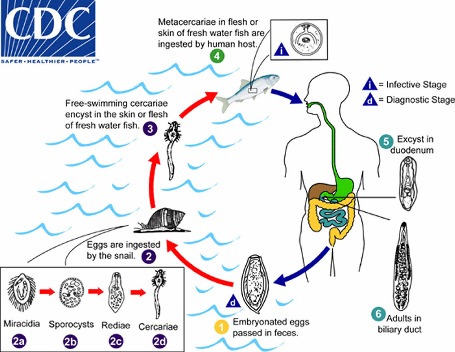
Clonorchis sinensis liver fluke life cycle (Chinese or oriental liver fluke)
Embryonated eggs are discharged in the biliary ducts and in the stool (number 1). Eggs are ingested by a suitable snail intermediate host (number 2). Each egg releases a miracidia (number 2a), which go through several developmental stages (sporocysts – number 2b, rediae – number 2c, and cercariae – number 2d). The cercariae are released from the snail and after a short period of free-swimming time in water, they come in contact and penetrate the flesh of freshwater fish, where they encyst as metacercariae(number 3). Infection of humans occurs by ingestion of undercooked, salted, pickled, or smoked freshwater fish (number 4). After ingestion, the metacercariae excyst in the duodenum (number 5) and ascend the biliary tract through the ampulla of Vater (number 6). Maturation takes approximately 1 month. The adult flukes (measuring 10 to 25 mm by 3 to 5 mm) reside in small and medium sized biliary ducts. In addition to humans, carnivorous animals can serve as reservoir hosts.
Figure 1. Clonorchis sinensis liver fluke life cycle
How does one become infected with Clonorchis sinensis liver fluke?
The eggs of Clonorchis are ingested by freshwater snails. After the eggs hatch, infected snails release microscopic larvae that then enter freshwater fish. People become infected by eating infected raw or undercooked fish containing the larvae. After being ingested by humans, the larvae grow into adult worms and live inside the human bile duct system. The life cycle takes three months to complete in humans. Infected people will then pass eggs in their stool(poop) or may cough them up.
Can Clonorchis sinensis liver fluke be transmitted from person to person?
No. Clonorchis cannot be directly transmitted from person to person.
Who is at risk of Clonorchis sinensis liver fluke infection?
Liver fluke infections occur mostly in people living in some areas where the parasites are found. Travelers to Asia who consume raw or poorly cooked fish are at risk for liver fluke infection. Chlonorchis is found in Asian countries including Korea, China, Taiwan, Northern Vietnam, Japan, and Asian Russia. Clonorchis infection has been reported in Asian immigrants in non-endemic areas. Some cases were found in people who had ingested imported freshwater fish (undercooked or pickled) containing parasitic cysts; the fish were imported to the US from areas where Clonorchis can be found.
Research has shown the liver fluke infections can persist for at most 25–30 years, the lifespan of the parasite.
How can I prevent Clonorchis liver fluke infection?
Clonorchis infection can be prevented by avoiding raw or undercooked freshwater fish. Lightly salted, smoked, or pickled fish can contain infectious parasites. Clonorchis infection does not result from drinking river water or other non-potable water.
The FDA recommends the following for fish preparation or storage to kill parasites.
Cooking
- Cook fish adequately (to an internal temperature of at least 145° F [~63° C]).
Freezing
- At -4°F (-20°C) or below for at least 7 days (total time); or
- At -31°F (-35°C) or below until solid, and storing at -31°F (-35°C) or below for a least 15 hours; or
- At -31°F (-35°C) or below until solid and storing at -4°F (-20°C) or below for at least 24 hours.
Clonorchis liver fluke signs and symptoms
Most infected persons have no symptoms. In mild cases, symptoms may include indigestion, abdominal pain, diarrhea, or constipation. Most signs and symptoms are related to inflammation and intermittent obstruction of the bile ducts. In severe cases, abdominal pain, nausea, and diarrhea can occur.
Untreated, infection may persist for up to 30 years, the lifespan of the parasite. In infections that last a long time, an enlarged liver and malnutrition may occur. In areas where liver flukes are endemic and a person may have multiple long-standing untreated liver fluke infections, the inflammation of the gallbladder and ducts caused by the parasite has been associated with liver and bile duct cancers, including cholangiocarcinoma. Liver flukes are one of many factors that have been associated with cholangiocarcinoma. Other known risk factors for cholangiocarcinoma include hepatitis B, hepatitis C, alcoholic liver disease and other causes of bile duct inflammation. These risk factors are thought to be more common causes of cholangiocarcinoma in the United States than liver fluke infection. Most patients in Western countries do not have an identifiable risk factor for cholangiocarcinoma.
Clonorchis sinensis liver fluke diagnosis
Diagnosis of Clonorchis infection is based on microscopic identification of eggs in stool specimens. More than one stool sample may be needed to detect the eggs. The eggs of Clonorchis are very similar to those of Opisthorchis, another liver fluke, but can be distinguished by microscopic features. Additionally, cysts containing the parasite can sometimes be detected by ultrasound, CT, or MRI. Testing the blood for Clonorchis is not useful for patient management. No blood test is available in the United States.
Clonorchis sinensis liver fluke treatment
Praziquantel or albendazole are the drugs of choice to treat Clonorchis infection.
Opisthorchiasis
Opisthorchis species are liver fluke worms that humans can get by eating raw or undercooked fish, crabs, or crayfish from areas in Asia and Europe where the parasite is found, including Thailand, Laos, Cambodia, Vietnam, Germany, Italy, Belarus, Russia, Kazakhstan, and Ukraine. Liver flukes infect the liver, gallbladder, and bile duct in humans. While most infected persons do not show any symptoms, infections that last a long time can result in severe symptoms and serious illness. Untreated, infections may persist for up to 25–30 years, the lifespan of the parasite. Typical symptoms include indigestion, abdominal pain, diarrhea, or constipation. In severe cases, abdominal pain, nausea, and diarrhea can occur.
Opisthorchis viverrini is known as the Southeast Asian liver fluke and Opisthorchis felineus as the cat liver fluke. Opisthorchis felineus, in addition to presenting with the typical symptoms also seen in Opisthorchis viverrini infections, can present with fever, facial swelling, swollen lymph glands, sore joints, and rash—similar to the signs and symptoms of schistosomiasis. Chronic Opisthorchis felineus infections may also involve the pancreatic ducts.
Diagnosis of Opisthorchis infection is based on microscopic identification of parasite eggs in stool specimens. Safe and effective medication is available to treat Opisthorchis infections. Adequately freezing or cooking fish will kill the parasite.
How does one become infected with Opisthorchis liver fluke?
The eggs of Opisthorchis are ingested by freshwater snails. After the eggs hatch, infected snails release microscopic larvae that then enter freshwater fish. People become infected when eating infected raw or undercooked fish containing the larvae. After being ingested by humans, the larvae grow into adult worms and live inside the human bile duct system. The life cycle takes three months to complete in humans. Infected people will then pass eggs in their stool or may cough them up.
Can Opisthorchis liver fluke be transmitted from person to person?
No. Opisthorchis cannot be directly transmitted from person to person.
Who is at risk of Opisthorchis liver fluke infection?
Liver fluke infections occur mostly in people living in areas where the parasites are found. Travelers to Asia who consume raw or undercooked fish are at risk for liver fluke infection.
Research has shown the liver fluke infections can persist for at most 25–30 years, the lifespan of the parasite.
How can I prevent Opisthorchis liver fluke infection?
Opisthorchis infection can be prevented by avoiding raw or undercooked freshwater fish. Lightly salted, smoked, or pickled fish can contain infectious parasites. Opisthorchis infection does not result from drinking river water or other non-potable water.
The FDA recommends the following for fish preparation or storage to kill parasites.
Cooking
- Cook fish adequately (to an internal temperature of at least 145° F [~63° C]).
Freezing
- At -4°F (-20°C) or below for at least 7 days (total time); or
- At -31°F (-35°C) or below until solid, and storing at -31°F (-35°C) or below for a least 15 hours; or
- At -31°F (-35°C) or below until solid and storing at -4°F (-20°C) or below for at least 24 hours.
Opisthorchis liver fluke signs and symptoms
Most infected persons have no symptoms. In mild cases, symptoms may include indigestion, abdominal pain, diarrhea, or constipation. Signs and symptoms are related to inflammation and blockage of the bile ducts that may come and go. In severe cases, abdominal pain, nausea, and diarrhea can occur.
Untreated, infection may persist for up to 25–30 years, the lifespan of the parasite. In infections that last a long time, an enlarged liver and malnutrition may occur. In areas where liver flukes are found and a person may have multiple long-standing untreated liver fluke infections, the inflammation of the gallbladder and ducts caused by the parasite has been associated with liver and bile duct cancers (bile duct cancer is also known as cholangiocarcinoma). Liver fluke infection is one of many factors that have been associated with bile duct cancer. Other known risk factors for bile duct cancer include hepatitis B, hepatitis C, alcoholic liver disease, and other causes of bile duct inflammation. These risk factors are thought to be more common causes of bile duct cancer in the United States than liver fluke infection. Most patients in Western countries do not have an identifiable risk factor for bile duct cancer.
In addition to the symptoms listed above, infections due to Opisthorchis felineus can also present with fever, facial swelling, swollen lymph glands, sore joints, and rash—similar to the signs and symptoms of schistosomiasis. Chronic Opisthorchis felineus infections may also involve the pancreatic ducts.
Opisthorchis liver fluke diagnosis
Diagnosis of Opisthorchis infection is based on microscopic identification of eggs in stool specimens. More than one stool sample may be needed to detect the eggs. The eggs of Opisthorchis are very similar to those of Clonorchis, another liver fluke, but can be distinguished by microscopic features. Additionally, cysts containing the parasite can sometimes be detected by ultrasound, CT, or MRI. Serologic testing (testing the blood) for Opisthorchis is not useful for patient management and is not available in the United States.
Opisthorchis liver fluke treatment
Praziquantel or albendazole are the drugs of choice to treat Opisthorchis infection.
Fascioliasis (Fasciola Infection)
Fascioliasis is a worm infection typically caused by Fasciola hepatica, which is also known as “the common liver fluke” or “the sheep liver fluke.” A related parasite, Fasciola gigantica, also can infect people. Fascioliasis is found in all continents except Antarctica, in over 70 countries, especially where there are sheep or cattle. Human infections with Fasciola hepatica are found in areas where sheep and cattle are raised, and where humans consume raw watercress or other water plants contaminated with immature parasite larvae, including Europe, the Middle East, and Asia. Infections with Fasciola gigantica have been reported, more rarely, in Asia, Africa, and Hawaii. The young worms move through the intestinal wall, the abdominal cavity, and the liver tissue, into the bile ducts, where they develop into mature adult flukes that produce eggs. The pathology typically is most pronounced in the bile ducts and liver. Fasciola infection is both treatable and preventable.
During the acute phase (caused by the migration of the immature fluke through the hepatic parenchyma), manifestations include abdominal pain, hepatomegaly, fever, vomiting, diarrhea, urticaria and eosinophilia, and can last for months. In the chronic phase (caused by the adult fluke within the bile ducts), the symptoms are more discrete and reflect intermittent biliary obstruction and inflammation. Occasionally, ectopic locations of infection (such as intestinal wall, lungs, subcutaneous tissue, and pharyngeal mucosa) can occur.
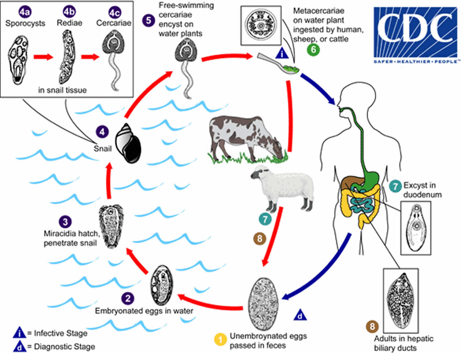
Fasciola liver fluke life cycle
Immature eggs are discharged in the biliary ducts and in the stool (number 1). Eggs become embryonated in water (number 2), eggs release miracidia (number 3), which invade a suitable snail intermediate host (number 4), including the genera Galba, Fossaria and Pseudosuccinea. In the snail the parasites undergo several developmental stages (sporocysts – number 4a, rediae – number 4b, and cercariae – number 4c). The cercariae are released from the snail (number 5) and encyst as metacercariae on aquatic vegetation or other surfaces. Mammals acquire the infection by eating vegetation containing metacercariae. Humans can become infected by ingesting metacercariae-containing freshwater plants, especially watercress (number 6). After ingestion, the metacercariae excyst in the duodenum (number 7) and migrate through the intestinal wall, the peritoneal cavity, and the liver parenchyma into the biliary ducts, where they develop into adults (number 8). In humans, maturation from metacercariae into adult flukes takes approximately 3 to 4 months. The adult flukes (Fasciola hepatica: up to 30 mm by 13 mm; Fasciola gigantica: up to 75 mm) reside in the large biliary ducts of the mammalian host. Fasciola hepatica infect various animal species, mostly herbivores.
Figure 2. Fasciola liver fluke life cycle
How do people get infected with Fasciola?
People get infected by accidentally ingesting (swallowing) the parasite. The main way this happens is by eating raw watercress or other contaminated freshwater plants. Another way people might get infected is by ingesting contaminated water, such as by drinking it or by eating vegetables that were washed or irrigated with contaminated water.
Can Fasciola be spread directly from one person or animal to another?
No. Fasciola cannot be passed directly from one person to another. The eggs passed in the stool of infected people (and animals) need to develop (mature) in certain types of freshwater snails, under favorable environmental conditions, to be able to infect someone else.
Under unusual circumstances, people might get infected by eating raw or undercooked sheep or goat liver that contains immature forms of the parasite.
Can people get infected with Fasciola in the United States?
Yes. It is possible, but few cases have been reported in published articles. Some cases have been documented in Hawaii, California, and Florida.
However, most reported cases in the United States have been in people, such as immigrants, who were infected in countries where fascioliasis is well known to occur.
How can Fasciola infection be prevented?
People can protect themselves by not eating raw watercress and other water plants, especially from Fasciola–endemic grazing areas. As always, travelers to areas with poor sanitation should avoid food and water that might be contaminated. No vaccine is available to protect people against Fasciola.
Fasciola liver fluke infection signs and symptoms
Some infected people don’t ever feel sick.
Some people feel sick early on in the infection, while immature flukes are passing (migrating) from the intestines through the abdominal cavity and liver. Symptoms from the acute (migratory) phase can start as soon as a few days after the exposure (typically, <1–2 weeks) and can last several weeks or months.
Some people feel sick during the chronic phase of the infection, when adult flukes are in the bile ducts (the duct system of the liver). The symptoms, if any, associated with this phase can start months to years after the exposure. For example, symptoms can result from inflammation and blockage of bile ducts.
During both phases of the infection, clinical features can include fever, malaise, abdominal pain, eosinophilia, hepatomegaly (an enlarged liver), and abnormal liver tests.
Fasciola liver fluke infection diagnosis
The infection typically is diagnosed by examining stool (fecal) specimens under a microscope. The diagnosis is confirmed if Fasciola eggs are seen. More than one specimen may need to be examined to find the parasite. Certain types of blood tests also may be helpful for diagnosing Fasciola infection.
Fasciola liver fluke infection treatment
Fascioliasis is a treatable disease. Triclabendazole is the drug of choice. It is given by mouth, usually in two doses. Most people respond well to the treatment.


