Contents
- What is actinomyces infection
- How you get actinomyces infection
- Actinomyces bacteria species
- Actinomyces infection clinical features
- Actinomyces bacteria diagnosis
- Actinomyces treatment
What is actinomyces infection
Actinomycosis is a rare chronic disease caused by Actinomyces spp., anaerobic filamentous Gram-positive bacteria that normally colonize the human mouth and digestive and genital tracts 1. Typical actinomycosis in humans is a chronic disease caused by Actinomyces israelii, Actinomyces gerencseriae and Propionibacterium propionicus (previously Arachnia propionica) 2. Actinomyces infection is characterized by persisting swelling, suppuration, and formation of abscesses with draining sinuses 2. Major types are cervicofacial, thoracic, and abdominal. In addition to the pathogens in actinomycosis some Actinomyces species have also been isolated from other mixed anaerobic infections, eye infections, blood and the urinary tract.
Actinomycosis typical clinical presentations (such as cervicofacial actinomycosis following dental focus of infection, pelvic actinomycosis in women with an intrauterine device, and pulmonary actinomycosis in smokers with poor dental hygiene), but also that actinomycosis may mimic the malignancy process in various anatomical sites 1. Bacterial cultures and pathology are the cornerstone of actinomyces diagnosis, but particular conditions are required in order to get the correct diagnosis. Prolonged bacterial cultures in anaerobic conditions are necessary to identify the Actinomyces bacterium and typical microscopic findings include necrosis with yellowish sulfur granules and filamentous Gram-positive fungal-like pathogens 1.
Actinomycosis is treated with antibiotics. Treatment starts off in hospital with antibiotics given directly into a vein (intravenously). When you’re well enough to go home, you’ll be given tablets to take for a few months. It’s important to keep taking antibiotics until they’re finished, even when you feel better.
You might also need surgery to drain areas of pus (abscesses) and cut out the surrounding area if it’s infected.
Patients with actinomyces infection require prolonged (6- to 12-month) high doses (to facilitate the drug penetration in abscess and in infected tissues) of penicillin G or amoxicillin, but the duration of antimicrobial therapy could probably be shortened to 3 months in patients in whom optimal surgical resection of infected tissues has been performed 1. Preventive measures, such as reduction of alcohol abuse and improvement of dental hygiene, may limit occurrence of pulmonary, cervicofacial, and central nervous system actinomycosis. In women, intrauterine devices must be changed every 5 years in order to limit the occurrence of pelvic actinomyces infection.
How you get actinomyces infection
The bacteria that cause actinomycosis normally live harmlessly in the body. Actinomyces bacteria only cause an infection if they get into the lining of areas such as the mouth or gut.
You can’t spread the Actinomyces infection to other people.
Any part of the body can be infected. Where it starts depends on what caused it.
| Possible causes | Symptoms |
|---|---|
| Jaw or mouth: tooth decay, an injury, dental surgery | dark lumps on your cheek or neck, difficulty chewing, pus leaking from small holes in your skin |
| Lungs: inhaling liquid or food contaminated with the bacteria | shortness of breath, chest pain, a cough, pus leaking from small holes in your skin |
| Tummy: burst appendix, surgery | diarrhoea or constipation, pain, a lump or swelling in your tummy, pus leaking from small holes in your skin |
| Pelvis: leaving an intrauterine device (IUD) contraceptive coil in for too long | pain low down in your tummy, vaginal bleeding or unusual discharge, a lump or swelling in your lower tummy |
You can’t always prevent actinomycosis
Actinomycosis is very rare, so the chances of getting it are extremely small.
You can help reduce your risk by:
- looking after your teeth and gums
- not leaving an intrauterine device (IUD) in for longer than recommended – they usually last 5 to 10 years, depending on the type you have
Actinomyces bacteria species
Bacteria of the genus Actinomyces belong to the Actinobacteria phylum and Actinomycetales order and are related to other genera such as Corynebacterium, Mycobacterium, Nocardia, and Propionibacterium. Besides Actinomyces, Propionibacterium propionicum (formerly Arachnia propionica), has often been reported as an agent of actinomycosis-like infections 3. More than 30 species of Actinomyces have been described. Actinomyces israelii is the most prevalent species isolated in human infections and is found in most clinical forms of actinomycosis 4. Actinomyces viscosus and Actinomyces meyeri are also often reported in typical actinomycosis, although they are less common 5 and Actinomyces meyeri is considered to have a great propensity for dissemination. Some species, including Actinomyces naeslundii, Actinomyces odontolyticus, Actinomyces gerencseriae (formerly Actinomyces israelii serotype 2), Actinomyces neuii, Actinomyces turicensis, and Actinomyces radingae, have been associated with particular clinical syndromes 6. Thus, Actinomyces israelii and Actinomyces gerencseriae are responsible for about 70% of orocervicofacial infections 7. Hematogenous dissemination of actinomycosis is extremely rare and has mainly been associated with Actinomyces meyeri, Actinomyces israelii, and Actinomyces odontolyticus 8. Of note, most of the Actinomyces spp. are present in polymicrobial flora. Therefore Actinomyces are often isolated with other normal commensals, such as Aggregatibacter actinomycetemcomitans, Eikenella corrodens, Capnocytophaga, fusobacteria, Bacteroides, staphylococci, streptococci, or Enterobacteriaceae, depending on the site of infection 9. As such, it is difficult to discriminate colonization of mucosa-contaminating samples and infection due to Actinomyces except when the culture is pure and associated with the presence of polynuclear neutrophils. On the other hand, Actinomyces infections could be polymicrobial and associated with other bacteria, named “companion microbes”, which contribute to initiation and development of infection by inhibiting host defenses or reducing oxygen tension 10. The multimicrobial nature of infection is well described in animal models and in human cervicofacial actinomycosis 11.
Actinomyces infection clinical features
Cervicofacial actinomycosis
Cervicofacial actinomycosis is the most frequent clinical form of actinomycosis, and “lumpy jaw syndrome”, which is associated with odontogenic infection, is the most common clinical manifestation, representing approximately 60% of all reported cases 12. Actinomyces spp. could also be responsible for maxillary osteomyelitis in patients with odontogenic maxillary sinusitis 13.
Actinomyces israelii and Actinomyces gerencseriae comprise almost 70% of cases, but many other species have been described, such as Actinomyces meyeri, Actinomyces odontolyticus, Actinomyces naeslundii, Actinomyces georgiae, Actinomyces pyogenes, or Actinomyces viscosus 7.
Actinomyces are commensals of the human oropharynx, and are particularly prevalent within gingival crevices, tonsillar crypts, periodontal pockets and dental plaques, as well as on carious teeth. Consequently, actinomycosis is mainly considered an endogenous infection that is triggered by a mucosal lesion 4. The pathophysiology of invasive disease following oral mucosal breach is unknown, but the invariably co-isolated commensals, such as E. corrodens, A. actinomycetemcomitans, or Haemophilus aphrophilus, may inhibit local host defenses, although their exact role is unclear 4. Cervicofacial actinomycosis could be associated with large abscesses and/or mandibular osteomyelitis with or without sinus tract (see Figure 1). Finally, cervicofacial actinomycosis can lead to distant organ dissemination, including brain, lungs, and digestive tract. Cervicofacial actinomycosis is a relatively rare condition worldwide, with no predilection for age, race, season, or occupation.
Physiopathological pathways of cervicofacial actinomycosis explain that predisposing conditions include poor oral hygiene (dental caries, gingivitis, infection in erupting secondary teeth) and oral mucosa trauma (dental extraction, gingival trauma, local tissue damage caused by neoplastic condition or irradiation, cervicofacial surgery). Other predisposing factors include male sex, diabetes mellitus, immunosuppression, alcoholism, and malnutrition 4.
Actinomyces spp. are considered to be involved in the pathogenesis of bisphosphonate severe osteonecrosis of the jaw, which, until recently, was considered a noninfectious disease. Most patients with osteoporosis receive bisphosphonate therapy. Occurrence of bisphosphonate osteonecrosis of the jaw is associated with duration of bisphosphonate therapy, concomitant use of corticosteroids, and mucosal disruption. The latter may facilitate Actinomyces colonization and invasion of the jaw, as Actinomyces spp. have been detected in biofilm in bone samples from patients with bisphosphonate osteonecrosis of the jaw 14.
Figure 1. Actinomyces lower jaw infection
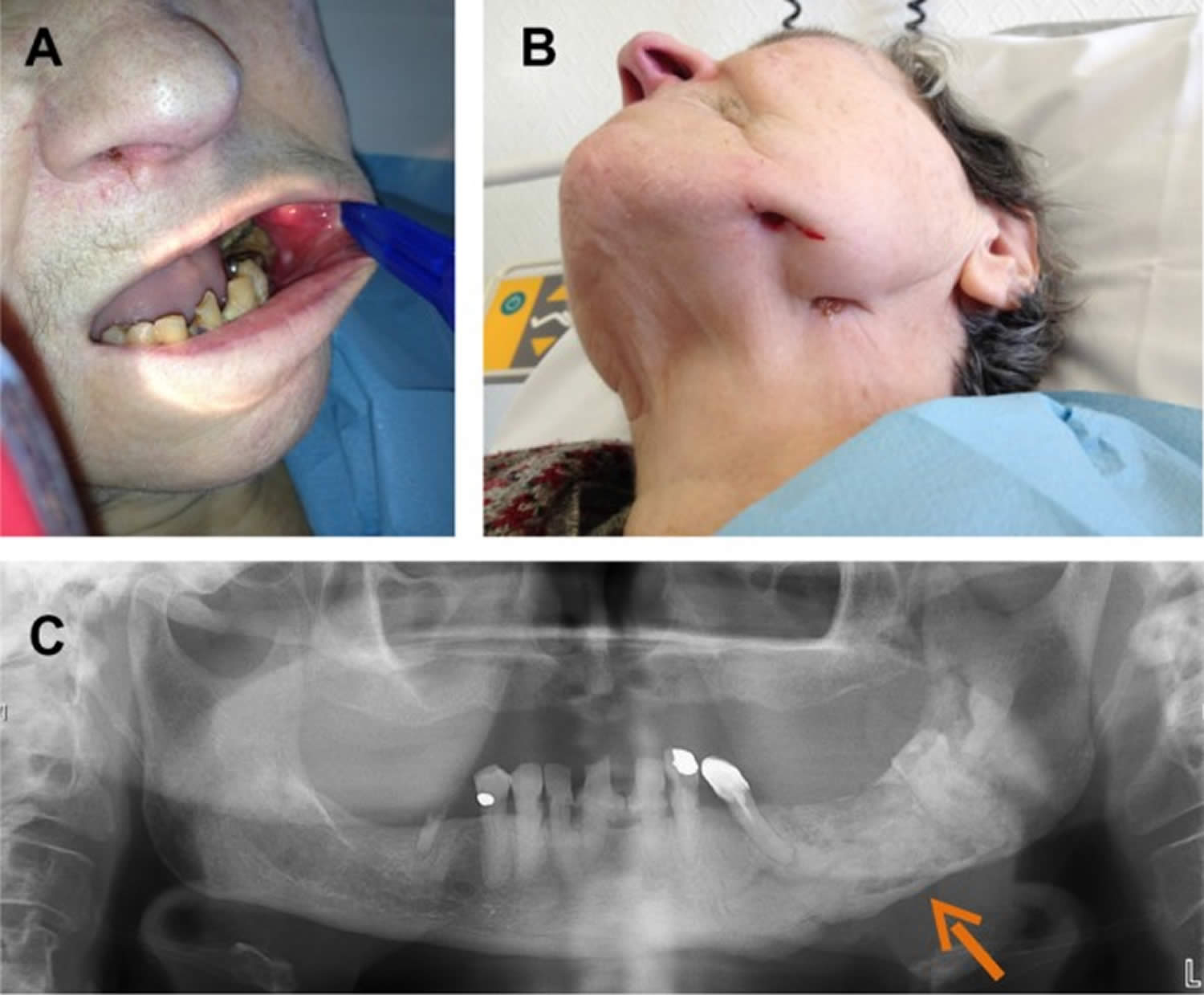
Footnote: A 77-year-old woman with past history of breast cancer was admitted 7 years after radiotherapy for left mandibular metastasis with left mandibular pain, buccal-sided bone exposure, and sinus tract. The patient had been receiving long-term trastuzumab therapy with bisphosphonate. As the patient was suspected to have chronic osteomyelitis with actinomycosis, left total hemimandibulectomy was performed. Pathology revealed suppurative osteomyelitis. No sample was sent for the microbiological diagnosis.
Left mandibular osteomyelitis with bone exposure (A) and sinus tract (B) following left mandibular radiotherapy in a patient receiving long-term bisphosphonate therapy. Panoramic dental X-ray shows mandibular lucencies (C). The arrow shows mandibular lucencies.
[Source 1 ]Cervicofacial actinomycosis signs and symptoms
Cervicofacial actinomycosis usually involves tissues surrounding the upper (maxillary expansion of the jaw) or lower mandible, including the mandible itself in approximately 50% of cases, cheek (15%), chin (15%), and submaxillary ramus and angle (10%). More rarely, the mandibular joint could be involved 15. Typically, the disease presents as a slowly progressive painless indurated mass, evolving into multiple abscesses with draining sinus tracts on the skin surface or oral mucosa, sometimes expressing a typical thick yellow exudate with characteristic sulfur granules 4. At advanced stages, pain and trismus can occur, linked with mastication muscles infiltration. Acute suppurative forms with rapid abscess formations are less common and are usually febrile and painful. Regional adenopathy is rare. Bone involvement is observed in approximately 10% of cases.
Although most cases are of odontogenic origin and concern the perimandibular regions, many other locations of primary infections have been described, including the tongue, sinuses, middle ear, larynx, lachrymal pathways, and thyroid gland 16.
Imaging findings are usually noncontributory to the positive diagnosis. Dental panoramic radiograph is mandatory to assess apical abscesses, which will require dental avulsions. CT scan and MRI may show a nonspecific involvement of skin and soft tissues, but are useful to assess bone involvement. In case of chronic osteomyelitis, osteolysis is common, with a possible periosteal reaction and intralesional gas 17.
Cervicofacial actinomycosis diagnosis
Diagnosis can be difficult, and especially making a distinction between neoplastic conditions, malignant hemopathy, and other cervicofacial infections such as nocardiosis or mycobacterial infections. A nonspecific and mild biological inflammatory syndrome can be found. The gold standard for diagnosing cervicofacial actinomycosis is histological examination and bacterial culture of an abscess or of a suspected bone, if osteomyelitis is suspected. Microbiological cultures of bone samples have to be incubated for 2 weeks, as bacteria frequently reduce their growth capacities in chronic osteomyelitis 18:1403–1409)). In patients with odontogenic cervicofacial actinomycosis, prescription of oral antimicrobials is common before surgery, leading frequently to false negative results of the cultures. Indeed, taking, for instance, the bacterial diagnosis for patients with prosthetic joint infection, the sensitivity decreased from 76.9% to 47.8% to 41.2% for bone sample culture as the antimicrobial-free interval before surgery decreased from greater than 14 days to 4–14 days, to 0–3 days, respectively 19. As a result, in patients with chronic mandibular osteomyelitis suspected to have cervicofacial actinomycosis, disruption of antimicrobials at least 14 days before surgery is mandatory to facilitate the growth of Actinomyces spp. in cultures. In typical cases, ie, especially in patients with lumpy jaw syndrome, Actinomyces spp. have always been targeted by antimicrobial therapy, regardless of the results of microbiological cultures or the result of pathology.
Cervicofacial actinomycosis treatment options
Surgical management can be required for drainage of voluminous abscesses, marsupialization of chronic sinus tracts, excision of recalcitrant fibrotic lesion, and/or debridement of necrotic bone tissue in case of osteomyelitis 4. Treatment of dental caries and/or apical abscesses is essential, often necessitating dental avulsions.
No randomized controlled trials have evaluated antibiotic regimens for cervicofacial actinomycosis. Most isolates are susceptible to beta-lactams, and the treatment of choice is a prolonged course of oral amoxicillin. As the penetration of beta-lactams in bone is low (10%–20% of the administered dose),16 intravenous high doses of amoxicillin (up to 200 mg/kg/day) or penicillin G (up to 24 MIU/day) has to be used initially in severe cases 4. Acceptable alternatives include clindamycin, macrolides (erythromycin, clarithromycin, or azithromycin), and doxycycline, which has a better bone penetration 20. The adjunction of a companion drug such as metronidazole or a beta-lactamase inhibitor is controversial, but may help in these frequent polymicrobial infections 21. The traditional prolonged course of up to 6–12 months of treatment can likely be shortened if an optimal surgical resection of infected tissues has been performed, in the absence of bone involvement, and if a satisfactory patient response to treatment is rapidly observed. Indeed, several observations have reported satisfactory cure rates with 4- to 6-week antimicrobial therapy 22.
Extrafacial bone and joint actinomycosis
Although cervicofacial actinomycosis is the most frequent form of actinomycosis with bone involvement, Actinomyces spp. could also be involved in extrafacial bone and joint infection. Various clinical forms of extrafacial bone and joint actinomycosis have been described: 1) hematogenous spread of localized actinomycosis; 2) contiguous spread of pulmonary actinomycosis to the spine; and 3) polymicrobial bone and joint infection following bone exposition, especially in patients with paraplegia and osteomyelitis of the ischial tuberosity 4.
No data are available on the epidemiology of extrafacial bone and joint actinomycosis. Few case reports have been described in the literature. Concerning hematogenous spread of localized actinomycosis, Brown et al 23 reported a case of hematogenous infection of total hip arthroplasty 9 months after a noninvasive dental procedure with Actinomyces spp. in intraoperative specimen cultures. Zaman et al 24 reported a case of chronic hematogenous infection due to Actinomyces spp. of prosthetic joint in an intravenous drug user.
Extrafacial bone and joint actinomycosis signs and symptoms
Most patients with extrafacial bone and joint actinomycosis have insidious onset of the disease, and signs and symptoms are usually similar to those of chronic bone and joint infection. Of note, patients suspected to have actinomycosis bone and joint infection of hematogenous origin usually experience clinical symptoms many months after the suspected bacteremia 24.
Extrafacial bone and joint actinomycosis treatment options
The treatment strategy for extrafacial bone and joint actinomycosis is similar to that of other chronic bone and joint infections. In patients with hematogenous spread of localized actinomycosis, surgery has to be performed if complications are noticed, and if the patient has implant-associated infection (the implant has to be removed) 25. In patients with contiguous spread of pulmonary actinomycosis to the spine, surgery is required if large abscesses or neurological complications are detected. Finally, in patients with polymicrobial bone and joint infection following bone exposition, surgery (debridement) is often required. In all patients with extrafacial bone and joint actinomycosis, antimicrobial therapy must be based on prolonged high-dose intravenous and then oral beta-lactam therapy, as described in the cervicofacial actinomycosis section.
Respiratory tract actinomycosis
Respiratory tract actinomycosis includes pulmonary, bronchial, and laryngeal actinomycosis. Pulmonary actinomycosis is the third most common type of actinomycosis, after that occurring in cervicofacial and abdominopelvic locations. In children, pulmonary involvement is uncommon 26. The peak incidence is reported to be in the fourth and fifth decades of life 27. Males are more often affected than women, with a 3:1 ratio 28. Pulmonary actinomycosis results mainly from aspiration of oropharyngeal or gastrointestinal secretions 29. Consequently, individuals with poor oral hygiene, preexisting dental disease, and alcoholism have an increased risk for developing pulmonary actinomycosis 30. Otherwise, patients with chronic lung disease such as emphysema, chronic bronchitis, and bronchiectasis, and patients with pulmonary complications following tuberculosis, are considered to also be at risk for pulmonary actinomycosis 31. The mechanism of immune response in actinomycosis remains unclear, but some factors, by altering this response, probably promote the disease. Human immunodeficiency virus infection, steroid use, infliximab treatment, lung and renal transplantation, and acute leukemia during chemotherapy have been described as risk factors, despite few data being available in such patients 32.
At early stages of the disease, a focal pulmonary consolidation occurs, which can be surrounded by pulmonary nodules, but there are often no associated physical symptoms at this stage. This primary pulmonary involvement could secondly lead to constitution of a peripheral mass, with or without cavitation, which could invade adjacent tissue 33. At this stage, pulmonary actinomycosis is usually characterized by fibrotic lesion with slow contiguous growth passing through the anatomical barriers 34. The mass is often confused with malignancy.
A direct or indirect extension from cervicofacial infection to the thorax may also lead to pulmonary actinomycosis. Conversely, pulmonary actinomycosis could be associated with extrapulmonary spread, from the lung to the pleura, mediastinum, and chest wall, with fistula and chronic suppuration 35. Finally, hematogenous dissemination with pulmonary location has been observed in patients with disseminated actinomycosis 36. Pulmonary actinomycosis can also be detected in children without any risk factor for the disease, and the most common presentation is a chest wall mass 33.
Bronchial actinomycosis is rare. It may occur after disruption of the mucosal barrier, especially in patients with endobronchial stent, or with a bronchial foreign body aspiration (for example, of a fish bone) 10.
Concerning laryngeal actinomycosis, various different forms have been described. Vocal cord actinomycosis may mimic primary carcinoma or papilloma, whereas in patients with past history of laryngeal carcinoma and radiotherapy, actinomycosis may mimic laryngeal cancer relapse, as it may present as an ulcerative lesion, most often without abscess or sinus tract 37.
Figure 2. Actinomyces chest infection
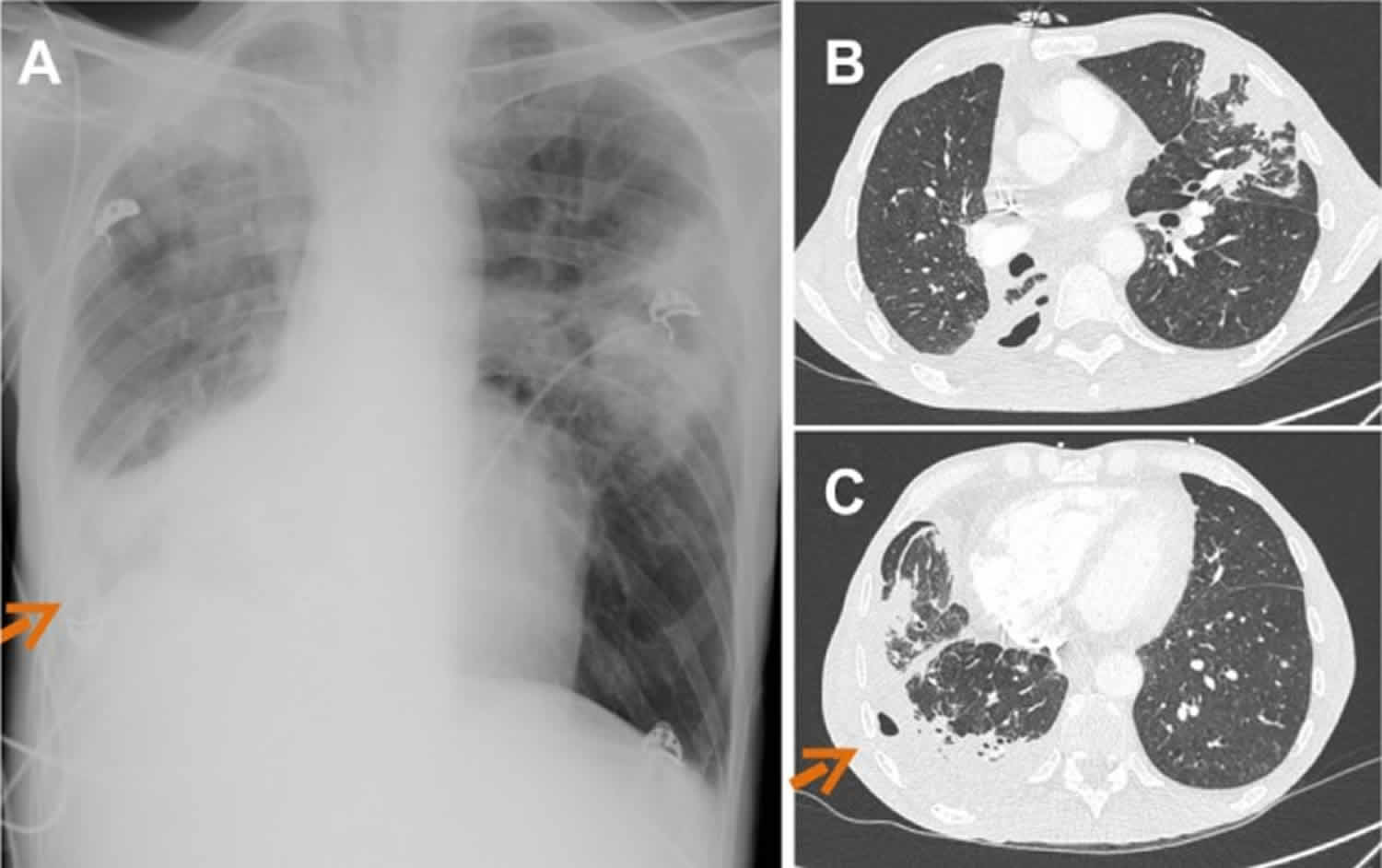
Footnote: A 53-year-old man with long-term tobacco and alcohol abuse was admitted for asthenia and loss of 20 kg in 6 months. The patient had had a cough for several months, but never experienced hemoptysis. Physical examination revealed a right pulmonary crackling sound. A sinus tract, which followed a 3-week history of subcutaneous abscess, was observed in the right face of the thorax. Chest X-ray and CT scan revealed multifocal pneumonia with right pleural cavitation, in the face of the sinus tract. The patient responded well to prolonged amoxicillin therapy (9 months).
Chest X-ray (A) and thorax computed tomography scan (B–C) revealing multifocal pneumonia with right pleural cavitation due to Actinomyces viscosus. Arrows indicate chest wall sinus tract.
[Source 1 ]Pulmonary actinomycosis signs and symptoms
Pulmonary actinomycosis could be acute or subacute, with lobar pulmonary involvement. However, the disease is mostly diagnosed at the chronic phase, in patients presenting mild fever and weight loss. The most common symptoms are nonspecific, similar to those of other chronic lung infections such as tuberculosis or thoracic cancer: productive cough, hemoptysis, dyspnea, and chest pain 38. General symptoms such as weight loss, fever, and night sweats may be present in the pulmonary location, but physicians should search for disseminated disease 26. Depending on the extension of the disease, the patient may have dyspnea and low oxygen saturation. Gradually, the pulmonary mass becomes soft and fluctuant, with a purulent center, which could be followed by cavitation. Cavitation may occur in patients with purulent discharge in bronchi, mimicking tuberculosis. Cavitation may also occur in patients with spontaneous drainage through the chest wall, forming a sinus tract. Patients with pulmonary cavitation associated with a chest-wall sinus tract should lead the physician to suspect actinomycosis 35. Patients with pulmonary actinomycosis may also have secondary cutaneous and/or muscular abscesses 39.
Pulmonary actinomycosis diagnosis
Imaging of pulmonary actinomycosis is not specific, and pulmonary actinomycosis is frequently confused with malignancy (mass) or tuberculosis (cavitation). The main CT findings are consolidation, lymph node enlargement, atelectasis, cavitation, ground glass opacity, and pleural effusion. There is no preferential localization in the lung. Pleural involvement, with thickening, effusion, or empyema is associated with about 15%–50% of cases of thoracic actinomycosis 40.
The gold standard for diagnosing pulmonary actinomycosis is histological examination and bacterial culture of a lung biopsy, obtained by percutaneous biopsy guided by CT scan or by open surgical resection 41.
Bronchoscopy should be performed to exclude malignancy. Simple culture of Actinomyces in bronchoalveolar lavage (BAL), as with sputum, is inappropriate for the diagnosis of pulmonary actinomycosis, except for patients with cavitation, as it may represent colonization 42. In patients with pulmonary actinomycosis associated with pleural effusion, it is of importance to note that Actinomyces almost never grow from pleural effusion samples. Actinomyces meyeri (nonbranching species with the greater propensity for dissemination) is the more frequent species described in pulmonary involvement cases 43.
Pulmonary actinomycosis treatment options
Patients with pulmonary actinomycosis require prolonged high doses of antimicrobial therapy with beta-lactam antibiotics, and penicillin G, cephalosporin, or amoxicillin are frequently used. For instance, it is recommended to intravenously administer a dose of 18–24 million units per day of penicillin G over 2–6 weeks, followed by oral therapy with penicillin V or amoxicillin for 6–12 months 4. Surgery could be required in patients with pulmonary actinomycosis, especially if the patient experienced hemoptysis 44. Importantly, in a recent report including 94 patients with pulmonary actinomycosis, half of the patients finally required surgery, especially those who had received cephalosporin (and not penicillin G), suggesting a lower microbiological activity of cephalosporin in comparison with penicillin G 40. Of note, a recent study suggested that patients who undergo surgical intervention have a better outcome, leading to recommendations of surgery for patients with complicated pulmonary actinomycosis (hemoptysis), in patients who do not respond well to high doses of penicillin therapy, or to definitively rule out the diagnosis of lung cancer 44. Finally, Kolditz et al, in 2009 45, published a report of a cohort of 49 patients with pulmonary actinomycosis who were exclusively medically treated. The authors suggest that the duration of antimicrobials has to be individualized in patients with pulmonary actinomycosis, but that treatment durations less than 3 months in medically treated patients should be avoided, as these patients are at risk for recurrence or local complications 45.
Actinomyces skin infection
Primary skin and soft-tissue actinomycosis is poorly described. Skin disruption may facilitate invasion of Actinomyces spp.
Figure 3. Actinomyces skin infection
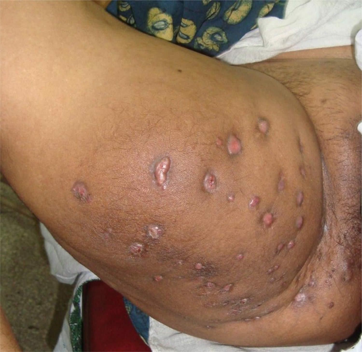
Footnote: Actinomycotic lesion over the thigh extending to the perineum. Multiple openings of sinuses can be seen.
A 42-year-old lady presented with complaints of a swelling with multiple discharging sinuses over her right thigh for the past two years. The swelling was insidious in onset and was gradually increasing in size. There was no history of any trauma or insect bite at the site of the lesion. The patient had no past history suggestive of tuberculosis or diabetes mellitus. She was postmenopausal for the last four years and had no past history of intra-uterine contraceptive device insertion. She had been treated by multiple doctors in various centres, over the last 6 months and had taken extended courses of oral anti-microbial therapy.
On examination there was a 15 cm × 20 cm, irregularly shaped, indurated swelling in the medial aspect of the right thigh extending from the perineum to mid-thigh (Figure 3). There were multiple sinuses draining yellow coloured pus along with sulphur granules. There were no other lesions elsewhere on the body.
[Source 46 ]Actinomyces skin infection signs and symptoms
Most patients have progressive skin and soft-tissue inflammation, which can become an abscess or cold mass, or nodular lesions with fistulas that need to be differentiated from chronic inflammatory skin disease, cutaneous mycobacterial infections, and sporotrichosis 46.
Actinomyces skin infection treatment options
In patients with primary skin and soft-tissue actinomycosis, abscesses have to be drained and prolonged antimicrobial therapy is required to achieve cure 46.
Genitourinary tract actinomycosis
Genitourinary tract actinomycosis is the second most frequent clinical form of actinomycosis. The main clinical feature of genitourinary tract actinomycosis is pelvic actinomycosis in women using an intrauterine device (IUD) 47. However, other clinical presentations have been described, such as primary bladder actinomycosis and testicular actinomycosis 48.
A. israelii is one of the most common species involved in pelvic actinomycosis. Colonization of the female genital tract by Actinomyces spp. is greatly promoted by the use of an IUD 49. Moreover, intrauterine devices (IUDs) have a traumatizing effect on endothelium by causing erosion, which may facilitate actinomycosis invasion. Actinomycosis intrauterine device (IUD)-associated infection is infrequent, but is clearly associated with the duration of the IUD use, hence it is recommended that an intrauterine device (IUD) be replaced every 5 years 50. There are no data comparing copper, hormonal, or inert IUDs in terms of the risk of actinomycosis. During intrauterine device (IUD)-associated actinomycosis, abscess formation is frequently observed in the genital tract, and creates dense adhesions with contiguous structures such as small bowel, promoting extensive fibrosis, fistulas, and peritonitis 47.
The pathogenesis of primary bladder actinomycosis is unclear, but could be due to cryptic locations, and usually mimics bladder carcinoma. The lesion may invade adjacent organs such as the uterus or the sigmoid colon. The diagnosis of primary bladder actinomycosis is of crucial importance, as it may avoid large surgical resection for suspected carcinoma 48.
Genitourinary tract actinomycosis signs and symptoms
Symptoms of patients with pelvic intrauterine device (IUD)-associated actinomycosis may mimic symptoms of gynecological malignant tumors, or uterine myoma or adenomyosis, by presenting as a genital mass without fever 51. Symptoms could be lower abdominal pain, constipation, and/or vaginal discharge. The duration of symptoms is usually 2 months at the time of diagnosis. Fever is usually not observed, except if a complication such as peritonitis occurs.
From blood test results, the white cell count is usually elevated with a high neutrophil count percentage and elevated C-reactive protein. Carbohydrate antigen 125, which can be associated with ovarian cancer, could also be elevated during pelvic actinomycosis 51.
CT scan usually reveals a pelvic mass with a mean size of 6–7 cm and with cystic lesions. A tubo-ovarian abscess strongly suggests pelvic actinomycosis, whereas some patients present with radiological findings suggesting malignant tumors. Lymphadenopathy is associated in 50% of cases 51.
Primary bladder actinomycosis can mimic bladder carcinoma, with macroscopic hematuria associated with thickening of the bladder wall 48.
Genitourinary tract actinomycosis diagnosis
Women carrying an intrauterine device (IUD) for over 5 years and presenting with a pelvic mass have a high index of suspicion for intrauterine device (IUD)-related actinomycosis. However, IUD-related actinomycosis has also been found within several months of intrauterine device (IUD) insertion 51. In patients suspected to have IUD-related actinomycosis, samples from surgical intrauterine device (IUD) removal, especially those containing pus, are required for bacterial cultures. Pathology must be done in patients with genitourinary tract masses showing sulfur granules and excluding gynecological malignant tumors.
IUD-related actinomycosis has to be distinguished from intrauterine device (IUD) colonization by Actinomyces spp. IUDs are frequently colonized by Actinomyces spp. and no antimicrobial treatment is required for asymptomatic women who systematically change their intrauterine device (IUD) in the 5 years following insertion 52.
In patients suspected to have primary bladder actinomycosis, guided biopsy should help toward diagnosis before performing surgical resection 53.
Genitourinary tract actinomycosis treatment options
Removal of the intrauterine device (IUD) is crucial in patients with intrauterine device (IUD)-associated actinomycosis 54. Open surgical resection, often required for the definite diagnosis of genitourinary tract actinomycosis, facilitates the cure, but may be mutilating, especially if hysterectomy or bladder resection is performed 55.
Antimicrobial therapy is the main treatment for genitourinary tract actinomycosis. Patients with genitourinary tract actinomycosis usually receive several weeks of intravenous high doses of a beta-lactam, followed by oral therapy for 2–6 months. There are no extensive data on the duration of antimicrobial therapy in such patients, but the duration of antimicrobials should probably be reduced in patients with extensive surgical resection of a small genital mass 55.
Digestive tract actinomycosis
Actinomyces spp. are saprophyte organisms of the mouth and digestive tract; actinomycosis of each part of the digestive tract has been previously described.
A. israelii is one of the most common species involved in abdominal actinomycosis. As with IUD-associated actinomycosis, a mucosal trauma causing erosion may facilitates actinomycosis invasion and infection. Digestive tract actinomycosis, as with Actinomyces spp. infections in other locations, may also mimic malignancy.
Esophageal actinomycosis is infrequent, with only around 20 cases described in the literature. Patients with esophageal actinomycosis are usually immunosuppressed by malignancy, HIV, or solid transplant. Most patients present with ulceration, and a few had perforation, an abscess, and sinus tract 56.
Appendix, cecum, and colon are the most common abdominal sites of actinomycosis, which can occur weeks to years after gastrointestinal mucosa disruption, and for which previous surgery such as for appendicitis or colonic diverticulitis with perforation are predisposing factors 4. Abdominal wall involvement with fistula may complicate abdominal actinomycosis.
Actinomycosis of the liver, the biliary tract, and the pancreas has also been described 57. Liver involvement mimicking malignancy or presenting as an abscess could be associated with digestive tract disease such as colonic diverticular disease. Pancreatic actinomycosis has been described in patients with pancreatic stents 58.
Digestive tract actinomycosis signs and symptoms
Signs and symptoms of patients with digestive tract actinomycosis depend on the anatomical location of the disease. Patients with ulcerative involvement of the esophagus mainly have dysphagia; patients with appendix, cecum, or colon actinomycosis frequently have abdominal pain with a palpable mass; patients with liver and biliary tract actinomycosis frequently have right upper quadrant pain and icterus 57.
Digestive tract actinomycosis diagnosis
As Actinomyces spp. are commensals of the digestive tract, pathology is crucial for the diagnosis of digestive tract actinomycosis, as Actinomyces spp. can be expected to contaminate digestive tract biopsies.
Digestive tract actinomycosis treatment options
As with other forms of actinomycosis, prolonged antimicrobial therapy is required for the treatment of digestive tract actinomycosis. Surgery is required in complicated cases, such as in patients with fistula and cell wall involvement 57.
Central nervous system actinomycosis
Actinomyces spp. are mainly involved in brain abscess, but meningitis, meningoencephalitis, epidural abscess, and subdural empyema have also been described. The central nervous system involvement occurs hematogenously from the lung or contiguously from a cervicofacial actinomycosis or following a penetrating head injury. Central nervous system actinomycosis is usually polymicrobial 59.
Central nervous system actinomycosis signs and symptoms
Symptoms are unspecific, and patients frequently experience focal weakness, sensory losses, and seizures. A contrast-enhanced, thick-walled ring lesion with secondary edema and vascular congestion is usually observed 59.
Central nervous system actinomycosis diagnosis
The diagnosis is mainly based on stereotaxic aspiration of pus, revealing Actinomyces spp. in cultures and sulfur granules in pathology 59.
Central nervous system actinomycosis treatment options
The treatment of actinomycosis brain abscess requires prolonged antimicrobial therapy after pus aspiration 59.
Actinomyces bacteria diagnosis
The bacteriological identification of Actinomyces from a sterile site confirms the diagnosis of actinomycosis. However, isolation and identification of these causative bacteria occur in only a minority of cases; the failure rate of culture is high because of previous antibiotic therapy, inhibition of Actinomyces growth by concomitant and/or contaminant microorganisms, inadequate culture conditions, or inadequate short-term incubation 60. Because of the microaerophilic or strict anaerobic character of Actinomyces, strict anaerobic processing (rapid transport to the laboratory and/or transport in an anaerobic transport medium) and anaerobic growth conditions should be used for primary isolation. The most appropriate clinical specimens are tissue from surgical biopsy or pus; swabs must be avoided. Finally, clinicians should indicate suspicion for actinomycosis to the microbiologist to ensure that prolonged culture on appropriate media and in an appropriate atmosphere is performed. Moreover, the identification of Actinomyces in mucosa, where these bacteria are normal inhabitants, is of little significance in the absence of sulfur granules or a typical clinical syndrome, highlighting the importance of microbiological investigations in combination with histologic analysis.
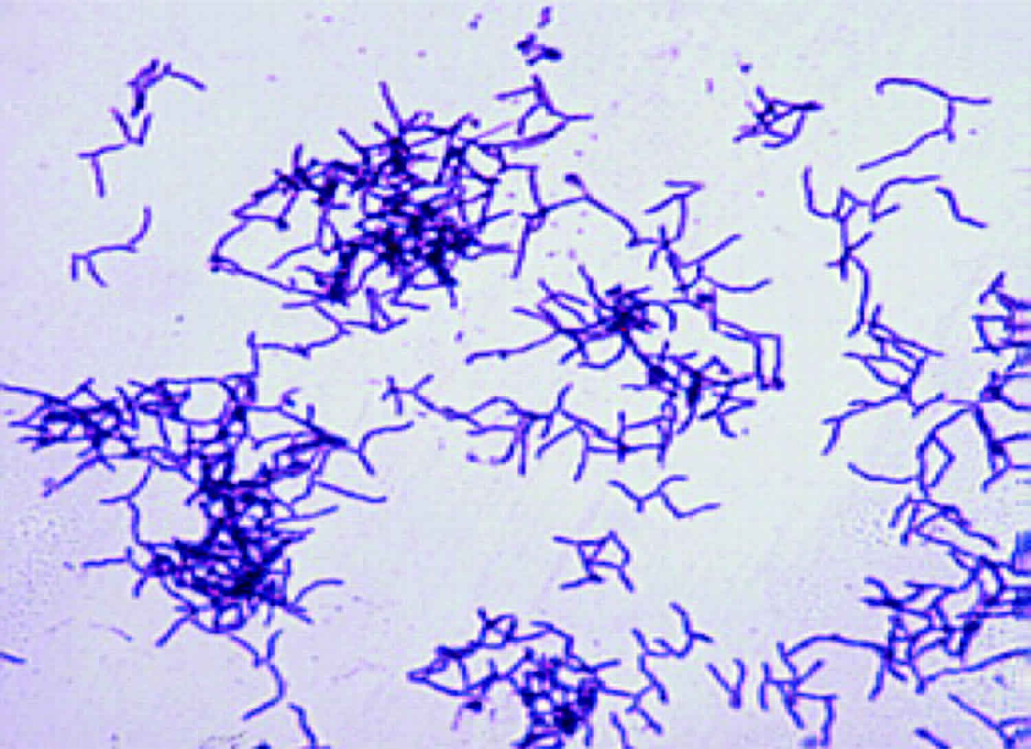
A Gram stain of the specimen is usually more sensitive than culture, especially if the patient had received antibiotics. Actinomyces are non-spore-forming Gram-positive rods. Except for Actinomyces meyeri, which is small and nonbranching, all the other species are branching filamentous rods.
Growth of Actinomyces is slow; it appears within at least 5 days and may take up to 15–20 days. Thus, incubation of at least 10 days is required before conclusion of a negative culture. Most Actinomyces spp. are facultative anaerobes, but some relevant species (such as Actinomyces meyeri), are strictly anaerobic, so cultures must be incubated in an anaerobic atmosphere. Actinomyces can be cultured on chocolate blood agar media at 37°C. Other enriched media can be used for Actinomyces isolation: brain heart infusion broth and Brucella Blood Agar with hemin and vitamin K1. The use of semi-selective media (such as phenylethyl alcohol or mupirocin-metronidazole blood agar) may increase isolation rates by inhibiting overgrowth of concomitant organisms 61. Actinomyces can be initially suspected by colony morphology and biochemical profiling. For example, Actinomyces israelii forms a “molar tooth” colony on agar and grows as clumps within broth, whereas Actinomyces odontolyticus forms rust-brown or red-colored colonies. Actinomyces are indole-negative bacteria. Identification was classically based on phenotypic tests (urease, catalase, fermentation of sugars, etc) or on commercial biochemical kits but, in fact, such tests can lead to misidentification of species and even of genus 62. Serological assays have been developed but need to be improved before they become clinically useful.
The classification of Actinomyces spp., initially based on phenotypic characters, has been recently revised, due to advances in microbiological taxonomy that use genotypic methods such as comparative 16S ribosomal RNA (rRNA) gene sequencing. Therefore, nowadays, molecular techniques such as 16S rRNA sequencing serve as the reference for identification 63. Besides 16S rRNA sequencing, a practical identification method consists of 16S ribosomal DNA restriction analysis 64. Polymerase chain reaction with specific primers can also be used for direct detection of Actinomyces in clinical material 65. Finally, matrix-assisted laser desorption ionization time-of-flight (MALDI-TOF) should be a quicker and accurate tool for Actinomyces identification in the future. Mass spectrometry uses an ionization source to charge and separate ionized bacterial proteins; then, a detector and mass analyzer generate a mass spectrum specific to bacterial species. Concerning Actinomyces, MALDI-TOF allows accurate identification at the genus level, but species identification remains uncertain and depends on the mass spectrometry system and the species studied 62. Improvement of this technique is needed for a definitive identification of Actinomyces spp., but it so far seems very promising.
Actinomyces treatment
Drug resistance is not considered a problem in actinomycosis. Indeed, Actinomyces spp. are usually extremely susceptible to beta-lactams, and especially penicillin G or amoxicillin. As a consequence, penicillin G or amoxicillin are considered drugs of choice for the treatment of actinomycosis 1. Third-generation cephalosporins are less frequently used even if they are considered to be active on Actinomyces israelii; however, it is important to note that some species are resistant to ceftriaxone (Actinomyces europaeus and Actinomyces graevenitzii) 4. Piperacillin–tazobactam, imipenem, and meropenem are considered to be active, but the use of these broad-spectrum antibiotics should be limited to avoid acquisition of resistant flora. Oxacillin, cloxacillin, and cephalexin, a first-generation cephalosporin, are not considered to be active. Metronidazole and aminoglycosides have no in vitro activity against Actinomyces. Fluoroquinolones (ciprofloxacin and moxifloxacin) are usually considered to be inactive, but data are limited and controversial. Doxycycline is considered to have a poor activity on Actinomyces spp., but clinical successes have been reported with this drug. Macrolides and clindamycin have been used successfully as alternatives. As Actinomyces spp. do not produce beta-lactamases, it is not useful to combine amoxicillin with beta-lactam inhibitors such as clavulanic acid, except if co-pathogens such as Enterobacteriaceae are involved in the disease 4.
- Valour F, Sénéchal A, Dupieux C, et al. Actinomycosis: etiology, clinical features, diagnosis, treatment, and management. Infect Drug Resist. 2014;7:183-97. Published 2014 Jul 5. doi:10.2147/IDR.S39601 https://www.ncbi.nlm.nih.gov/pmc/articles/PMC4094581/[↩][↩][↩][↩][↩][↩][↩]
- Bowden GHW. Actinomyces, Propionibacterium propionicus, and Streptomyces. In: Baron S, editor. Medical Microbiology. 4th edition. Galveston (TX): University of Texas Medical Branch at Galveston; 1996. Chapter 34. Available from: https://www.ncbi.nlm.nih.gov/books/NBK8385[↩][↩]
- Smego RA, Jr, Foglia G. Actinomycosis. Clin Infect Dis. 1998;26(6):1255–1261. https://www.ncbi.nlm.nih.gov/pubmed/9636842[↩]
- Wong VK, Turmezei TD, Weston VC. Actinomycosis. BMJ. 2011;343:d6099 https://www.bmj.com/content/343/bmj.d6099.long[↩][↩][↩][↩][↩][↩][↩][↩][↩][↩][↩][↩]
- Fazili T, Blair D, Riddell S, Kiska D, Nagra S. Actinomyces meyeri infection: case report and review of the literature. J Infect. 2012;65(4):357–361[↩]
- Cone LA, Leung MM, Hirschberg J. Actinomyces odontolyticus bacteremia. Emerg Infect Dis. 2003;9(12):1629–1632.[↩]
- Pulverer G, Schütt-Gerowitt H, Schaal KP. Human cervicofacial actinomycoses: microbiological data for 1997 cases. Clin Infect Dis. 2003;37(4):490–497.[↩][↩]
- Felz MW, Smith MR. Disseminated actinomycosis: multisystem mimicry in primary care. South Med J. 2003;96(3):294–299.[↩]
- Schaal KP, Lee HJ. Actinomycete infections in humans – a review. Gene. 1992;115(1–2):201–211.[↩]
- Mandell GL, Bennett JE, Dolin R, editors. Mandell, Douglas, and Bennett’s Principles and Practice of Infectious Diseases. 7th ed. Philadelphia, PA: Churchill Livingstone Elsevier; 2010.[↩][↩]
- Jordan HV, Kelly DM, Heeley JD. Enhancement of experimental actinomycosis in mice by Eikenella corrodens. Infect Immun. 1984;46(2):367–371.[↩]
- Mandell GL, Bennett JE, Dolin R, editors. Mandell, Douglas, and Bennett’s Principles and Practice of Infectious Diseases. 7th ed. Philadelphia, PA: Churchill Livingstone Elsevier; 2010[↩]
- Saibene AM, Di Pasquale D, Pipolo C, Felisati G. Actinomycosis mimicking sinonasal malignant disease. BMJ Case Rep. 2013;2013[↩]
- Gallay L, Bodard AG, Chidiac C, Ferry T. Bilateral bisphosphonate-related osteonecrosis of the jaw with left chronic infection in an 82-year-old woman. BMJ Case Rep. 2013;2013[↩]
- Schaal KP, Beaman BL. Clinical significance of actinomycetes. In: Goodfellow M, Mordarski M, Williams ST, editors. The Biology of the Actinomycetes. New York: Academic Press; 1983. p. 389.[↩]
- Vorasubin N, Wu AW, Day C, Suh JD. Invasive sinonasal actinomycosis: case report and literature review. Laryngoscope. 2013;123(2):334–338[↩]
- Sasaki Y, Kaneda T, Uyeda JW, et al. Actinomycosis in the mandible: CT and MR findings. AJNR Am J Neuroradiol. 2014;35(2):390–394.[↩]
- Schäfer P, Fink B, Sandow D, Margull A, Berger I, Frommelt L. Prolonged bacterial culture to identify late periprosthetic joint infection: a promising strategy. Clin Infect Dis. 2008;1(47(11[↩]
- Trampuz A, Piper KE, Jacobson MJ, et al. Sonication of removed hip and knee prostheses for diagnosis of infection. N Engl J Med. 2007;357(7):654–663[↩]
- Landersdorfer CB, Bulitta JB, Kinzig M, Holzgrabe U, Sörgel F. Penetration of antibacterials into bone: pharmacokinetic, pharmacodynamic and bioanalytical considerations. Clin Pharmacokinet. 2009;48(2):89–124[↩]
- Tanaka-Bandoh K, Watanabe K, Kato N, Ueno K. Susceptibilities of Actinomyces species and Propionibacterium propionicus to antimicrobial agents. Clin Infect Dis. 1997;25(Suppl 2):S262–S263[↩]
- Moghimi M, Salentijn E, Debets-Ossenkop Y, Karagozoglu K, Forouzanfar T. Treatment of cervicofacial actinomycosis: a report of 19 cases and review of literature. Med Oral Patol Oral Cir Bucal. 2013;18(4):e627–e632[↩]
- Brown ML, Drinkwater CJ. Hematogenous infection of total hip arthroplasty with Actinomyces following a noninvasive dental procedure. Orthopedics. 2012;35(7):e1086–e1089.[↩]
- Zaman R, Abbas M, Burd E. Late prosthetic hip joint infection with Actinomyces israelii in an intravenous drug user: case report and literature review. J Clin Microbiol. 2002;40(11):4391–4392.[↩][↩]
- Lew DP, Waldvogel FA. Osteomyelitis. Lancet. 2004;364(9431):369–379[↩]
- Bates M, Cruickshank G. Thoracic actinomycosis. Thorax. 1957;12(2):99–124[↩][↩]
- Chaudhry SI, Greenspan JS. Actinomycosis in HIV infection: a review of a rare complication. Int J STD AIDS. 2000;11(6):349–355.[↩]
- Bennhoff DF. Actinomycosis: diagnostic and therapeutic considerations and a review of 32 cases. Laryngoscope. 1984;94(9):1198–1217[↩]
- Apothéloz C, Regamey C. Disseminated infection due to Actinomyces meyeri: case report and review. Clin Infect Dis. 1996;22(4):621–625.[↩]
- Cohen RD, Bowie WR, Enns R, Flint J, Fitzgerald JM. Pulmonary actinomycosis complicating infliximab therapy for Crohn’s disease. Thorax. 2007;62(11):1013–1014.[↩]
- Bates M, Cruickshank G. Thoracic actinomycosis. Thorax. 1957;12(2):99–124.[↩]
- Han JY, Lee KN, Lee JK, et al. An overview of thoracic actinomycosis: CT features. Insights Imaging. 2013;4(2):245–252.[↩]
- Bartlett AH, Rivera AL, Krishnamurthy R, Baker CJ. Thoracic actinomycosis in children: case report and review of the literature. Pediatr Infect Dis J. 2008;27(2):165–169.[↩][↩]
- Brown JR. Human actinomycosis. A study of 181 subjects. Hum Pathol. 1973;4(3):319–330.[↩]
- Han JY, Lee KN, Lee JK, et al. An overview of thoracic actinomycosis: CT features. Insights Imaging. 2013;4(2):245–252[↩][↩]
- Smego RA, Jr, Foglia G. Actinomycosis. Clin Infect Dis. 1998;26(6):1255–1261 [↩]
- Yoshihama K, Kato Y, Baba Y. Vocal cord actinomycosis mimicking a laryngeal tumor. Case Rep Otolaryngol. 2013;2013:361986[↩]
- Kim SR, Jung LY, Oh IJ, et al. Pulmonary actinomycosis during the first decade of 21st century: cases of 94 patients. BMC Infect Dis. 2013;13(1):216.[↩]
- Liaudet L, Erard P, Kaeser P. Cutaneous and muscular abscesses secondary to Actinomyces meyeri pneumonia. Clin infect Dis. 1996;22:185–186[↩]
- Kim SR, Jung LY, Oh IJ, et al. Pulmonary actinomycosis during the first decade of 21st century: cases of 94 patients. BMC Infect Dis. 2013;13(1):216[↩][↩]
- Mabeza GF, Macfarlane J. Pulmonary actinomycosis. Eur Respir J. 2003;21(3):545–551.[↩]
- Nair PN, Brundin M, Sundqvist G, Sjögren U. Building biofilms in vital host tissues: a survival strategy of Actinomyces radicidentis. Oral Surg Oral Med Oral Pathol Oral Radiol Endod. 2008;106(4):595–603[↩]
- Colmegna I, Rodriguez-Barradas M, Rauch R, Clarridge J, Young EJ. Disseminated Actinomyces meyeri infection resembling lung cancer with brain metastases. Am J Med Sci. 2003;326(3):152–155.[↩]
- Song JU, Park HY, Jeon K, Um SW, Kwon OJ, Koh WJ. Treatment of thoracic actinomycosis: a retrospective analysis of 40 patients. Ann Thorac Med. 2010;5(2):80–85.[↩][↩]
- Kolditz M, Bickhardt J, Matthiessen W, Holotiuk O, Höffken G, Koschel D. Medical management of pulmonary actinomycosis: data from 49 consecutive cases. J Antimicrob Chemother. 2009;63(4):839–841[↩][↩]
- Khandelwal R, Jain I, Punia S, et al. Primary actinomycosis of the thigh – a rare soft tissue infection with review of literature. JRSM Short Rep. 2012;3(4):24. https://www.ncbi.nlm.nih.gov/pmc/articles/PMC3375841/[↩][↩][↩]
- Choi MH, Hong DG, Seong WJ, Lee YS, Park IS. Pelvic actinomycosis confirmed after surgery: single center experience. Arch Gynecol Obstet. 2010;281(4):651–656.[↩][↩]
- Bae JH, Song R, Lee A, Park JS, Kim MR. Computed tomography for the preoperative diagnosis of pelvic actinomycosis. J Obstet Gynaecol Res. 2011;37(4):300–304.[↩][↩][↩]
- Al-Kadhi S, Venkiteswaran KP, Al-Ansari A, Shamsudini A, Al-Bozom I, Kiliyanni AS. Primary vesical actinomycosis: a case report and literature review. Int J Urol. 2007;14(10):969–971.[↩]
- Westhoff C. IUDs and colonization or infection with Actinomyces. Contraception. 2007;75(Suppl 6):S48–S50.[↩]
- Sung HY, Lee IS, Kim SI, et al. Clinical features of abdominal actinomycosis: a 15-year experience of a single institute. J Korean Med Sci. 2011;26(7):932–937[↩][↩][↩][↩]
- Al-Kadhi S, Venkiteswaran KP, Al-Ansari A, Shamsudini A, Al-Bozom I, Kiliyanni AS. Primary vesical actinomycosis: a case report and literature review. Int J Urol. 2007;14(10):969–971[↩]
- Bae JH, Song R, Lee A, Park JS, Kim MR. Computed tomography for the preoperative diagnosis of pelvic actinomycosis. J Obstet Gynaecol Res. 2011;37(4):300–304[↩]
- Choi MH, Hong DG, Seong WJ, Lee YS, Park IS. Pelvic actinomycosis confirmed after surgery: single center experience. Arch Gynecol Obstet. 2010;281(4):651–656[↩]
- Sung HY, Lee IS, Kim SI, et al. Clinical features of abdominal actinomycosis: a 15-year experience of a single institute. J Korean Med Sci. 2011;26(7):932–937.[↩][↩]
- Abdalla J, Myers J, Moorman J. Actinomycotic infection of the oesophagus. J Infect. 2005;51(2):E39–E43.[↩]
- Joshi V, Koulaouzidis A, McGoldrick S, Tighe M, Tan C. Actinomycotic liver abscess: a rare complication of colonic diverticular disease. Ann Hepatol. 2010;9(1):96–98.[↩][↩][↩]
- Acevedo F, Baudrand R, Letelier LM, Gaete P. Actinomycosis: a great pretender. Case reports of unusual presentations and a review of the literature. Int J Infect Dis. 2008;12(4):358–362.[↩]
- Na KY, Jang JH, Sung JY, Kim YW, Park YK. Actinomycotic brain abscess developed 10 years after head trauma. Korean J Pathol. 2013;47(1):82–85[↩][↩][↩][↩]
- Bennhoff DF. Actinomycosis: diagnostic and therapeutic considerations and a review of 32 cases. Laryngoscope. 1984;94(9):1198–1217.[↩]
- Lewis R, McKenzie D, Bagg J, Dickie A. Experience with a novel selective medium for isolation of Actinomyces spp. from medical and dental specimens. J Clin Microbiol. 1995;33(6):1613–1616.[↩]
- Ng LS, Sim JH, Eng LC, Menon S, Tan TY. Comparison of phenotypic methods and matrix-assisted laser desorption ionisation time-of-flight mass spectrometry for the identification of aero-tolerant Actinomyces spp. isolated from soft-tissue infections. Eur J Clin Microbiol Infect Dis. 2012;31(8):1749–1752.[↩][↩]
- Garner O, Mochon A, Branda J, et al. Multi-centre evaluation of mass spectrometric identification of anaerobic bacteria using the VITEK® MS system. Clin Microbiol Infect. 2013 Jul 4; Epub[↩]
- Hall V, Talbot PR, Stubbs SL, Duerden BI. Identification of clinical isolates of Actinomyces species by amplified 16S ribosomal DNA restriction analysis. J Clin Microbiol. 2001;39(10):3555–3562.[↩]
- Hall V. Actinomyces – gathering evidence of human colonization and infection. Anaerobe. 2008;14:1–7.[↩]


