Contents
- What is an arthrogram
- What are some common uses of arthrogram?
- What are the benefits of an arthrogram?
- How should I prepare for an arthrogram?
- How is an arthrogram procedure performed?
- How long does an arthrogram take?
- What will I experience during and after an arthrogram?
- Are there any after effects of an arthrogram?
- Arthrogram results
- What are the limitations of arthrogram?
- Arthrogram vs MRI
- MRI arthrogram
- CT arthrogram
- Arthrogram risks
What is an arthrogram
An arthrogram or arthrography is an X-ray image or picture of the inside of a joint, such as the wrist, elbow, shoulder or knee, after a contrast medium (sometimes referred to as a contrast agent or “dye”) is injected into the joint. The type of contrast medium used depends on the exact nature of the arthrogram and the specialist doctor carrying out the arthrogram. If the arthrogram test uses X-rays (fluoroscopy or CT), this is generally iodinated contrast medium. Occasionally, a CT arthrogram will involve an injection of air either on its own or with a small amount of X-ray contrast medium. An arthrogram provides a clear image of the soft tissue in the joint (e.g. ligaments and cartilage) so that a more accurate diagnosis about an injury or cause of a symptom, such as joint pain or swelling, can be made. Arthrograms are usually taken with x-rays, using a special contrast dye that highlights ligaments, tendons and other connective tissues. Arthrogram is very effective at detecting disease within the ligaments, tendons and cartilage. Physicians use arthrograms to assess tears in the cartilage, ligaments or capsules within painful joints. It is often these connective structures, rather than muscles or bones, that are the cause of pain and dysfunction. X-ray arthrograms, in particular, help doctors make accurate diagnoses before resorting to more expensive imaging technologies such as MRI. Occasionally, an arthrogram is only used to confirm that an injection needle is correctly positioned in the joint. This is usually as part of a treatment of painful joints where medications or anaesthetic need to be injected.
A radiologist (specialist doctor) injects the contrast medium into the joint using fluoroscopy (a special type of X-ray), computed tomography (CT) or ultrasound to help guide the injection needle into the correct position.
Once the injection is finished, images of the joint are taken using magnetic resonance imaging (MRI) or CT. While a plain MRI or CT can provide some information of the soft tissue structures, an arthrogram can sometimes provide much more detailed information about what is wrong within the joint. Improvements in technology and X-ray imaging equipment mean that the use of arthrograms is becoming less common.
Arthrogram may be indirect, where contrast material is injected into the bloodstream, or direct, where contrast material is injected into the joint. Arthrogram may use computed tomography (CT) scanning, magnetic resonance imaging (MRI) or fluoroscopy – a form of real-time x-ray. Historically arthrograms were performed with fluoroscopy and plain radiographs only. Today all patients proceed to cross-sectional imaging after contrast injection. This is usually MRI but CT is sometimes performed if there are contraindications to MRI or if there is high suspicion of an associated bony abnormality.
With direct arthrogram, the contrast material is injected directly into the joint by a radiologist. Direct arthrogram is preferred over indirect arthrogram because it distends or enlarges the joint thus allowing for enhanced imaging of small internal structures. This leads to improved evaluation of diseases or conditions within the joint. Direct arthrogram is often performed only if a non-arthrographic exam is felt to be inadequate in assessing a joint abnormality.
There are several methods to perform direct arthrogram.
Conventional direct arthrogram of a joint often uses a special form of x-ray called fluoroscopy to guide and evaluate the injection of iodine contrast material directly into the joint. In some cases, ultrasound may be used to guide the procedure. Alternate methods of direct arthrogram examinations may also use magnetic resonance imaging (MRI) or computed tomography (CT) following contrast material injection into the joint.
An x-ray (radiograph) is a noninvasive medical test that helps physicians diagnose and treat medical conditions. Imaging with x-rays involves exposing a part of the body to a small dose of ionizing radiation to produce pictures of the inside of the body. X-rays are the oldest and most frequently used form of medical imaging.
Fluoroscopy makes it possible to see bones, joints and internal organs in real time. When iodine contrast is injected into the joint, it fills the entire joint which becomes clearly visible during x-ray evaluation, allowing the radiologist to assess the anatomy and function of the joint. Although the injection is typically monitored by fluoroscopy, the examination also commonly involves taking radiographs for documentation. These images are most often stored and viewed electronically.
Similarly, direct MRI arthrogram also involves the injection of a contrast material into the joint. The contrast material used for MRI evaluation is different from that used for x-ray; it contains gadolinium, which affects the local magnetic field within the joint and appears on the MR images. As in conventional direct arthrogram, the contrast material outlines the structures within the joint, such as cartilage, ligaments and bones, and allows them to be evaluated by the radiologist after the MR images are produced.
MRI uses a powerful magnetic field, radiofrequency pulses and a computer to produce detailed pictures of organs, soft tissues, bone and virtually all other internal body structures. The images can then be examined on a computer monitor connected to an image archive (PACS system) or printed or copied to CD. MRI does not use ionizing radiation (x-rays).
CT direct arthrogram uses the same type of contrast material as conventional direct arthrogram and may be supplemented by air to produce a double contrast CT arthrogram. CT makes cross sectional images processed by a computer using x-rays.
Your preparation may vary depending on which imaging method your exam will use. Tell your doctor if there’s a possibility that you are pregnant and discuss any recent illnesses, medical conditions, medications you’re taking, and allergies – especially to contrast materials. Leave jewelry at home and wear loose, comfortable clothing. You may be asked to wear a gown.
What are some common uses of arthrogram?
Arthrogram or arthrographic images help physicians evaluate alterations in structure and function of a joint and help to determine the possible need for treatment, including arthroscopy, open surgery or joint replacement.
Arthrogram procedure is most often used to identify abnormalities within the:
- shoulder
- elbow
- wrist
- hip
- knee
- ankle
Arthrogram is often used to help diagnose persistent, unexplained joint pain or discomfort. In some cases, local anesthetic medications or steroids may be injected into the joint along with the contrast material. These medications may temporarily decrease joint-related pain or inflammation and provide physicians additional information about possible sources of pain.
Your doctor will usually send you for an arthrogram as part of an MRI or CT to look at the soft tissue structures inside the joint. If the radiologist carrying out the test feels that using MRI or CT without contrast injection will provide sufficient information, then an arthrogram might not be done.
What are the benefits of an arthrogram?
The injection of contrast medium into the joint improves the quality of the MRI or CT to more accurately show damage to the internal structure of the joint.
Some common reasons for an arthrogram are:
- In the shoulder – where the joint is unstable or if an ultrasound or plain MRI has not shown a suspected tendon tear.
- In the hip – to show any tear of the cartilage labrum (or rim of the joint).
- In the wrist – to show any tear of the small ligaments of the wrist.
There are many other individual situations where your referring doctor may feel that the additional information obtained by an arthrogram could help to determine the best course of treatment.
How should I prepare for an arthrogram?
No special preparation is necessary before direct arthrogram. Food and fluid intake do not need to be restricted, unless a sedative will be given.
You should inform your physician of any medications you are taking and if you have any kidney problems or allergies, especially to iodinated or gadolinium-based contrast materials. Also, inform your doctor about recent illnesses or other medical conditions.
Some MRI arthrogram examinations may require you to receive an injection of contrast into the bloodstream. Some of this contrast material is absorbed into the joint resulting in an indirect arthrogram. The radiologist or technologist may ask if you have asthma, or allergies of any kind, such as an allergy to iodine or x-ray contrast material, drugs, food, or environmental agents. However, the contrast material used for an MRI exam, called gadolinium, does not contain iodine and is less likely to cause side effects or an allergic reaction.
Tell the radiologist if you have any serious health problems or if you have recently had surgery. Some conditions, such as severe kidney disease, may prevent you from being given MRI or CT arthrogram contrast material.
If you are scheduled to have MR or CT arthrogram and have claustrophobia (fear of enclosed spaces) or anxiety, you may want to ask your physician about being sedated prior to the scheduled examination.
Jewelry and other accessories should be left at home, if possible, or removed prior to the MRI arthrogram. Because they can interfere with the magnetic field of the MRI unit, metal and electronic items are not allowed in the exam room. In addition to affecting the MRI images, these objects can become projectiles within the MRI scanner room and may cause you and/or others nearby harm. These items include:
- jewelry, watches, credit cards and hearing aids, all of which can be damaged
- pins, hairpins, metal zippers and similar metallic items, which can distort MRI images
- removable dental work
- pens, pocket knives and eyeglasses
- body piercings
In most cases, an MRI exam is safe for patients with metal implants, except for a few types. People with the following implants cannot be scanned and should not enter the MRI scanning area:
- cochlear (ear) implant
- some types of clips used for brain aneurysms
- some types of metal coils placed within blood vessels
- nearly all cardiac defibrillators and pacemakers
You should tell the technologist if you have medical or electronic devices in your body. These objects may interfere with the exam or potentially pose a risk, depending on their nature and the strength of the MRI magnet. Many implanted devices will have a pamphlet explaining the MRI risks for that particular device. If you have the pamphlet, it is useful to bring that to the attention of the scheduler before the exam and bring it to your exam in case the radiologist or technologist has any questions. Some implanted devices require a short period of time after placement (usually six weeks) before being safe for MRI examinations. Examples include but are not limited to:
- artificial heart valves
- implanted drug infusion ports
- artificial limbs or metallic joint prostheses
- implanted nerve stimulators
- metal pins, screws, plates, stents or surgical staples
If there is any question of their presence, an x-ray may be taken to detect and identify any metal objects. In general, metal objects used in orthopedic surgery pose no risk during MRI. However, a recently placed artificial joint may require the use of another imaging procedure.
Patients who might have metal objects in certain parts of their bodies may also require an x-ray prior to an MRI arthrogram. You should notify the technologist or radiologist of any shrapnel, bullets, or other pieces of metal that may be present in your body due to prior accidents. Foreign bodies near and especially lodged in the eyes are particularly important because they may move during the scan, possibly causing blindness. Dyes used in tattoos may contain iron and could heat up during an MRI scan, but this is rare. Tooth fillings and braces usually are not affected by the magnetic field, but they may distort images of the facial area or brain, so you should let the radiologist know about them.
You will be asked to remove some of your clothes and to wear a gown during the exam. You may also be asked to remove jewelry, removable dental appliances, eye-glasses and any metal objects or clothing that might interfere with the x-ray images.
Women should always inform their physician and x-ray technologist if there is any possibility that they are pregnant. Many imaging tests are not performed during pregnancy so as not to expose the fetus to radiation. If an x-ray is necessary, precautions will be taken to minimize radiation exposure to the baby. See the Safety page for more information about pregnancy and x-rays.
Though MRI arthrogram does not use ionizing radiation, women should still inform their physician and technologist if they may be pregnant.
Children younger than 13 may need to be sedated in order to hold still for the procedure. Parents should ask about sedation before the procedure and realize that there are food and drink restrictions that may be required prior to sedation.
You should plan to have a relative or friend drive you home after your procedure.
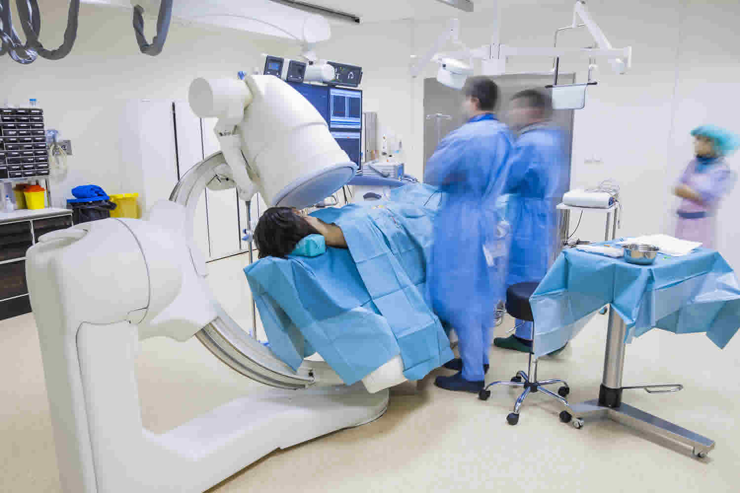
How is an arthrogram procedure performed?
This examination is usually done on an outpatient basis.
The patient is positioned on the examination table. X-rays of the joint may be taken prior to the procedure to help in guiding the injection and also to provide a baseline exam to be compared later with the arthrogram images. If recent x-rays are available, the physician may choose to use these for reference.
Next, the skin around the joint is cleansed with antiseptic and is often covered with a sterile surgical drape. Using a small needle, the physician injects local anesthetic into the area.
After the local anesthetic has taken effect, a longer needle is then inserted into the joint. The radiologist, a physician specially trained to supervise and interpret radiology examinations, will often use fluoroscopy or ultrasound to guide the needle into the correct position. The physician will sometimes use a syringe to drain (or aspirate) the joint fluid, which may be sent to a laboratory for analysis. Aspiration is typically performed when an infection is suspected.
The contrast material and sometimes air are injected into the joint space while the radiologist observes with fluoroscopy or ultrasound. In some cases, additional medications, such as anti-inflammatory steroids, may be injected into the joint along with the contrast material. After the needle is removed, the patient will be asked to move the affected joint to distribute the contrast material throughout the space. The radiologist may move the joint while evaluating the joint motion under fluoroscopy.
The type of contrast medium used depends on the exact nature of the arthrogram and the specialist doctor carrying out the arthrogram. If the arthrogram test uses X-rays (fluoroscopy or CT), this is generally iodinated contrast medium. Occasionally, a CT arthrogram will involve an injection of air either on its own or with a small amount of X-ray contrast medium.
If you are having an MRI arthrogram, the injection will be a very diluted mixture of MRI contrast medium (gadolinium chelates) together with sterile saline (mildly salty water).
After having the injection you will be taken to either an MRI suite (for an MRI arthrogram) or a CT suite (for a CT arthrogram), where detailed imaging of the joint will be carried out.
A conventional direct arthrogram exam is usually completed within 30 minutes. Exams involving MRI arthrogram may take more than one hour.
How long does an arthrogram take?
The injection of contrast medium usually takes about 15 minutes. You may then have to wait a short time before having the additional imaging of your joint. An MRI scan may take 30–45 minutes and a CT scan may take 15 minutes, depending on the joint and the number of scans that have to be done. You should allow approximately 2 hours from arrival at the radiology department.
What will I experience during and after an arthrogram?
You will experience a slight pinprick and may feel a momentary burning if a local anesthesia is used to numb the joint area. You may feel pressure or even pain when the needle is advanced into the joint. Inform the radiologist performing the procedure if you have pain so more local anesthetic can be injected into the area.
You may feel fullness in the joint as it is filled and possibly hear gurgling when the joint is moved.
If your arthrogram exam involves MR imaging:
It is normal for the area of your body being imaged to feel slightly warm, but if it bothers you, notify the radiologist or technologist. It is important that you remain perfectly still while the images are being obtained, which is typically only a few seconds to a few minutes at a time. You will know when images are being recorded because you will hear and feel loud tapping or thumping sounds when the coils that generate the radiofrequency pulses are activated. Some centers provide earplugs, while others use headphones to reduce the intensity of the sounds made by the MRI machine. You may be able to relax between imaging sequences, but will be asked to maintain your position without movement as much as possible.
You will usually be alone in the exam room during the MRI procedure. However, the technologist will be able to see, hear and speak with you at all times using a two-way intercom. Many MRI centers allow a friend or parent to stay in the room as long as they are also screened for safety in the magnetic environment.
Children will be given appropriately sized earplugs or headphones during the exam. MRI scanners are air-conditioned and well-lit. Music may be played through the headphones to help you pass the time.
In some cases, intravenous injection of contrast material may be administered before the images are obtained. The intravenous needle may cause you some discomfort when it is inserted and you may experience some bruising. There is also a very small chance of irritation of your skin at the site of the IV tube insertion. Some patients may sense a temporary metallic taste in their mouth after the contrast injection.
If you do not require sedation, no recovery period is necessary. You may resume your usual activities and normal diet immediately after the exam. On very rare occasions, a few patients experience side effects from the contrast material, including nausea, headache and pain at the site of injection. Similarly, patients are very rarely allergic to the contrast material and experience hives, itchy eyes or other reactions. If you experience allergic symptoms, notify the technologist. A radiologist or other physician will be available for immediate assistance.
If you experience allergic symptoms, a radiologist or other physician will be available for immediate assistance.
After the examination, you may experience swelling and discomfort. You may apply ice to the joint to reduce swelling if it is bothersome. A mild over-the-counter analgesic can be taken for pain. These symptoms usually disappear after 48 hours. Contact your doctor if they persist after two days.
Vigorous exercise is not recommended for at least 24 hours after the exam as there is a slight increased risk of dislocation after your procedure. Typically, if an arthrogram is performed on a joint, you will be asked to minimize activity using that joint for about 24 hours after the procedure to allow your body to eliminate the injected fluid from the joint.
If steroids or anesthetic medications are injected into the joint during the arthrogram, you may be asked to keep a log of your level of joint discomfort over the following days or weeks. This information may help your physician determine the cause of chronic joint pain and what therapies may be effective. It is also recommended that you refrain from vigorous exercise of the joint for about two weeks.
Are there any after effects of an arthrogram?
Many people referred for an arthrogram have symptoms of a sore joint. There can be some mild-to-moderate increase in soreness in the joint for 24–48 hours after the injection. The joint will then return to feeling the way it was before the test.
Arthrogram results
A radiologist, a physician specifically trained to supervise and interpret radiology examinations, will analyze the images and send a signed report to your primary care or referring physician, who will discuss the results with you.
The time that it takes your doctor to receive a written report on the test will vary, depending on:
- the urgency with which the result is needed;
- the complexity of the test;
- whether more information is needed from your doctor before the examination can be assessed by the radiologist;
- whether you have had previous X-rays or other medical imaging that needs to be compared with this new test;
- how the report is conveyed from the radiology facility or hospital to your doctor (i.e. phone, email, fax or mail).
Follow-up examinations may be necessary. Your doctor will explain the exact reason why another exam is requested. Sometimes a follow-up exam is done because a potential abnormality needs further evaluation with additional views or a special imaging technique. A follow-up examination may also be necessary so that any change in a known abnormality can be monitored over time. Follow-up examinations are sometimes the best way to see if treatment is working or if a finding is stable or changed over time.
What are the limitations of arthrogram?
The limitations of arthrogram include:
- Partial tears of the rotator cuff may not be detected with conventional direct arthrogram.
- Some joint injuries cannot be detected with conventional direct arthrogram, including defects of the cartilage, which can be found inside and along the edges of some joints, bruising of neighboring bones and injuries to ligaments outside the joint.
Arthrogram vs MRI
MRI or magnetic resonance imaging is a type of scan that uses strong magnetic fields and radio waves to produce detailed images of the inside of your body. Unlike conventional x-ray examinations and computed tomography (CT) scans, MRI does not utilize ionizing radiation. Instead, radiofrequency pulses re-align hydrogen atoms that naturally exist within your body. This does not cause any chemical changes in the tissues. As the hydrogen atoms return to their usual alignment, they emit different amounts of energy depending on the type of body tissue they are in. The MR scanner captures this energy and creates a picture of the tissues scanned based on this information. A computer then processes the signals and generates a series of images, each of which shows a thin slice of the body. The images can then be studied from different angles by the interpreting radiologist.
Frequently, the differentiation of abnormal (diseased) tissue from normal tissues is better with MRI than with other imaging modalities such as x-ray, CT and ultrasound.
An MRI scanner is a large tube that contains powerful magnets. You lie inside the tube during the scan.
An MRI scan can be used to examine almost any part of the body, including the:
- brain and spinal cord
- bones and joints
- breasts
- heart and blood vessels
- internal organs, such as the liver, womb or prostate gland
The results of an MRI scan can be used to help diagnose conditions, plan treatments and assess how effective previous treatment has been.
MRI benefits
- MRI is a noninvasive imaging technique that does not involve exposure to ionizing radiation.
- MRI enables the discovery of abnormalities that might be obscured by bone with other imaging methods.
- The contrast material used in MRI exams is less likely to produce an allergic reaction than the iodine-based contrast materials used for conventional x-rays and CT scanning.
What happens during an MRI scan?
During an MRI scan, you lie on a flat bed that’s moved into the scanner. Depending on the part of your body being scanned, you’ll be moved into the scanner either head first or feet first.
The MRI scanner is operated by a radiographer, who is trained in carrying out imaging investigations. They control the scanner using a computer, which is in a different room, to keep it away from the magnetic field generated by the scanner.
You’ll be able to talk to the radiographer through an intercom and they’ll be able to see you on a television monitor throughout the scan.
At certain times during the scan, the scanner will make loud tapping noises. This is the electric current in the scanner coils being turned on and off.
You’ll be given earplugs or headphones to wear.
It’s very important to keep as still as possible during your MRI scan.
The scan lasts 15 to 90 minutes, depending on the size of the area being scanned and how many images are taken.
How does an MRI scan work?
Most of the human body is made up of water molecules, which consist of hydrogen and oxygen atoms.
At the center of each hydrogen atom is an even smaller particle called a proton. Protons are like tiny magnets and are very sensitive to magnetic fields.
When you lie under the powerful scanner magnets, the protons in your body line up in the same direction, in the same way that a magnet can pull the needle of a compass.
Short bursts of radio waves are then sent to certain areas of the body, knocking the protons out of alignment.
When the radio waves are turned off, the protons realign. This sends out radio signals, which are picked up by receivers.
These signals provide information about the exact location of the protons in the body.
They also help to distinguish between the various types of tissue in the body, because the protons in different types of tissue realign at different speeds and produce distinct signals.
In the same way that millions of pixels on a computer screen can create complex pictures, the signals from the millions of protons in the body are combined to create a detailed image of the inside of the body.
MRI Safety
An MRI scan is a painless and safe procedure. You may find it uncomfortable if you have claustrophobia, but most people are able to manage it with support from the radiographer.
Going into the scanner feet first may be easier, although this isn’t always possible.
Extensive research has been carried out into whether the magnetic fields and radio waves used during MRI scans could pose a risk to the human body.
No evidence has been found to suggest there’s a risk, which means MRI scans are one of the safest medical procedures available.
But MRI scans may not be recommended in certain situations. For example, if you have a metal implant fitted, such as a pacemaker or artificial joint, you may not be able to have an MRI scan.
They’re also not usually recommended during pregnancy.
MRI arthrogram
An MRI arthrogram is a two-part diagnostic study that examines the inside of the joint (e.g., shoulder, knee, wrist, ankle) to assess an injury or a symptom you may be experiencing. The first part of the study is the arthrogram, in which contrast dye is injected into the joint with the help of an X-ray; the second part is the magnetic resonance imaging (MRI) scan. Injecting the contrast dye before the MRI helps provide a clearer image of the joint. The arthrogram is done using image guidance by an interventional radiologist.
MRI arthrogram involves the injection of a contrast material into the joint and the injection is normally done with the aid of local anesthesia. The MRI arthrogram solution used for MRI evaluation is different from that used for x-ray; it contains gadolinium, which affects the local magnetic field within the joint and appears on the MR images. The joint injection is performed under fluoroscopic guidance. As in conventional direct arthrogram, the contrast material outlines the structures within the joint, such as cartilage, ligaments and bones, and allows them to be evaluated by the radiologist after the MR images are produced.
MRI uses a powerful magnetic field, radiofrequency pulses and a computer to produce detailed pictures of organs, soft tissues, bone and virtually all other internal body structures. The images can then be examined on a computer monitor connected to an image archive (PACS system) or printed or copied to CD. MRI does not use ionizing radiation (x-rays).
The arthrogram takes 15 to 30 minutes.
The MRI scan may take 30 to 45 minutes, depending on the joint being scanned.
Once the MRI arthrogram is complete, you will return to the interventional radiology department where they will be assessed by a nurse and seen by an interventional radiologist before being discharged home.
CT arthrogram
CT arthrogram uses the same type of contrast material as conventional direct arthrogram and may be supplemented by air to produce a double contrast CT arthrogram. A computerized tomography (CT) scan uses X-rays and a computer to create detailed images of the inside of your body.
CT scans are sometimes referred to as CAT scans or computed tomography scans.
CT arthrogram is carried out by specially trained operators called radiographers, and can be done while you’re staying in hospital or during a short visit.
During the CT arthrogram, you’ll usually lie on your back on a flat bed that passes into the CT scanner.
The scanner consists of a ring that rotates around a small section of your body as you pass through it.
Unlike an MRI arthrogram, the scanner doesn’t surround your whole body at once, so you shouldn’t feel claustrophobic.
The radiographer will operate the scanner from the next room. While the CT scan is taking place, you’ll be able to hear and speak to them through an intercom.
While each scan is taken, you’ll need to lie very still and breathe normally. This ensures that the scan images aren’t blurred.
You may be asked to breathe in, breathe out, or hold your breath at certain points.
CT arthrogram will usually take around 10 to 20 minutes.
What happens afterwards?
You shouldn’t experience any after-effects from a CT arthrogram and can usually go home soon afterwards. You can eat and drink, go to work and drive as normal.
If a contrast was used, you may be advised to wait in the hospital for up to an hour to make sure you don’t have a reaction to it.
The contrast is normally completely harmless and will pass out of your body in your urine.
Your scan results won’t usually be available immediately. A computer will need to process the information from your scan, which will then be analyzed by a radiologist (a specialist in interpreting images of the body).
After analyzing the images, the radiologist will write a report and send it to the doctor who referred you for the scan so they can discuss the results with you. This normally takes a few days or weeks.
Is CT arthrogram safe?
CT scans are quick, painless and generally safe. But there’s a small risk you could have an allergic reaction to the contrast dye used and you’ll be exposed to X-ray radiation.
The amount of radiation you’re exposed to during a CT scan varies, depending on how much of your body is scanned.
CT scanners are designed to make sure you’re not exposed to unnecessarily high levels.
Generally, the amount of radiation you’re exposed to during each scan is the equivalent to between a few months and a few years of exposure to natural radiation from the environment.
It’s thought exposure to radiation during CT scans could slightly increase your chances of developing cancer many years later, although this risk is thought to be very small (less than 1 in 2,000).
Arthrogram risks
Arthrograms are considered low-risk procedures. However, any procedure where the skin is penetrated carries a risk of infection. The most serious complication of arthrogram is an infection of the joint. This is usually caused by organisms from the patient’s skin being transferred into the joint during the injection of contrast medium, and for this reason the procedure should not be carried out if there is broken or infected skin over the joint. The risk of infection is not precisely known, but the best available information suggests that it is in the order of 1 in 40,000 people having the test. If you develop increasing pain in your joint and feel unwell, particularly with a fever, you should see your doctor urgently or contact the radiology facility where you had the procedure done. There is always the possibility of injuring a vessel or a nerve adjacent to the joint. Injury to these structures, however, is minimal particularly when the procedure is performed under ultrasound guidance.
Occasionally, people may be allergic to the contrast medium that is injected, and this most commonly results in a rash, but may be more serious. The risk of a minor reaction (e.g. hives) has been reported in 1 in 2000 having the test. More serious reactions appear to be very rare.
Complications of gadolinium contrast medium used in an MRI have not been reported in the very small amounts used in arthrography.
The risk may increase depending on your condition, age, and health.
The risks of an arthrogram include:
- Allergic reaction
- Infection at the needle puncture site or in the joint that was injected
- Pain and discomfort in the joint
- Bleeding
Exams involving x-ray imaging:
- There is always a slight chance of cancer from excessive exposure to radiation. However, the benefit of an accurate diagnosis far outweighs the risk.
- Patients who have known allergies to iodine may have an adverse reaction to the contrast material. Because the contrast material is put in a joint and not a vein, allergic reactions are very rare, although in some cases, mild nausea to severe cardiovascular complications may result.
- Women should always inform their physician or x-ray technologist if there is any possibility that they are pregnant. See the Safety page for more information about pregnancy and x-rays.
- The effective radiation dose for this procedure varies. See the Safety page for more information about radiation dose.
Exams involving MR imaging:
- The MRI examination poses almost no risk to the average patient when appropriate safety guidelines are followed.
- If sedation is used, there are risks of excessive sedation. However, the technologist or nurse will monitor your vital signs to minimize this risk.
- Although the strong magnetic field is not harmful in itself, implanted medical devices that contain metal may malfunction or cause problems during an MRI exam.
- Nephrogenic systemic fibrosis is currently a recognized, but rare, complication of MRI believed to be caused by the injection of high doses of gadolinium-based contrast material in patients with very poor kidney function. Careful assessment of kidney function before considering a contrast injection minimizes the risk of this very rare complication.
- There is a very slight risk of an allergic reaction if contrast material is injected. Such reactions are usually mild and easily controlled by medication. If you experience allergic symptoms, a radiologist or other physician will be available for immediate assistance.


