Contents
- What is arthroplasty
- Hip arthroplasty
- Hip Joint Anatomy
- Partial hip replacement
- Total hip replacement
- Hip replacement alternatives
- Is Hip Replacement Surgery for You?
- Deciding to Have Hip Replacement Surgery
- Possible complications of hip replacement surgery
- Preparing for hip replacement surgery
- Hip replacement surgery
- Metal-on-Metal Implants Precautions
- Hip replacement procedure
- Traditional Hip Replacement
- Minimally Invasive Total Hip Replacement
- How long does hip replacement surgery take?
- Revision Total Hip Replacement
- Hip replacement surgery recovery
- Hip replacement recovery time
- Avoiding problems after hip replacement surgery
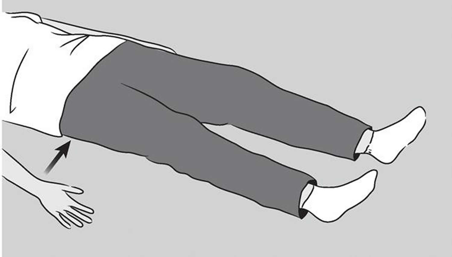
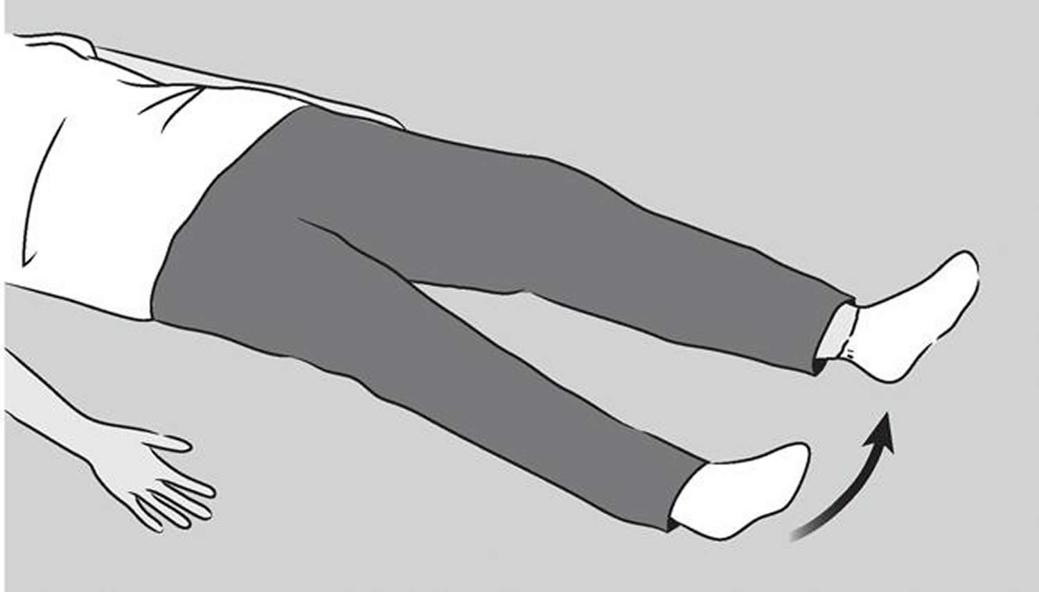
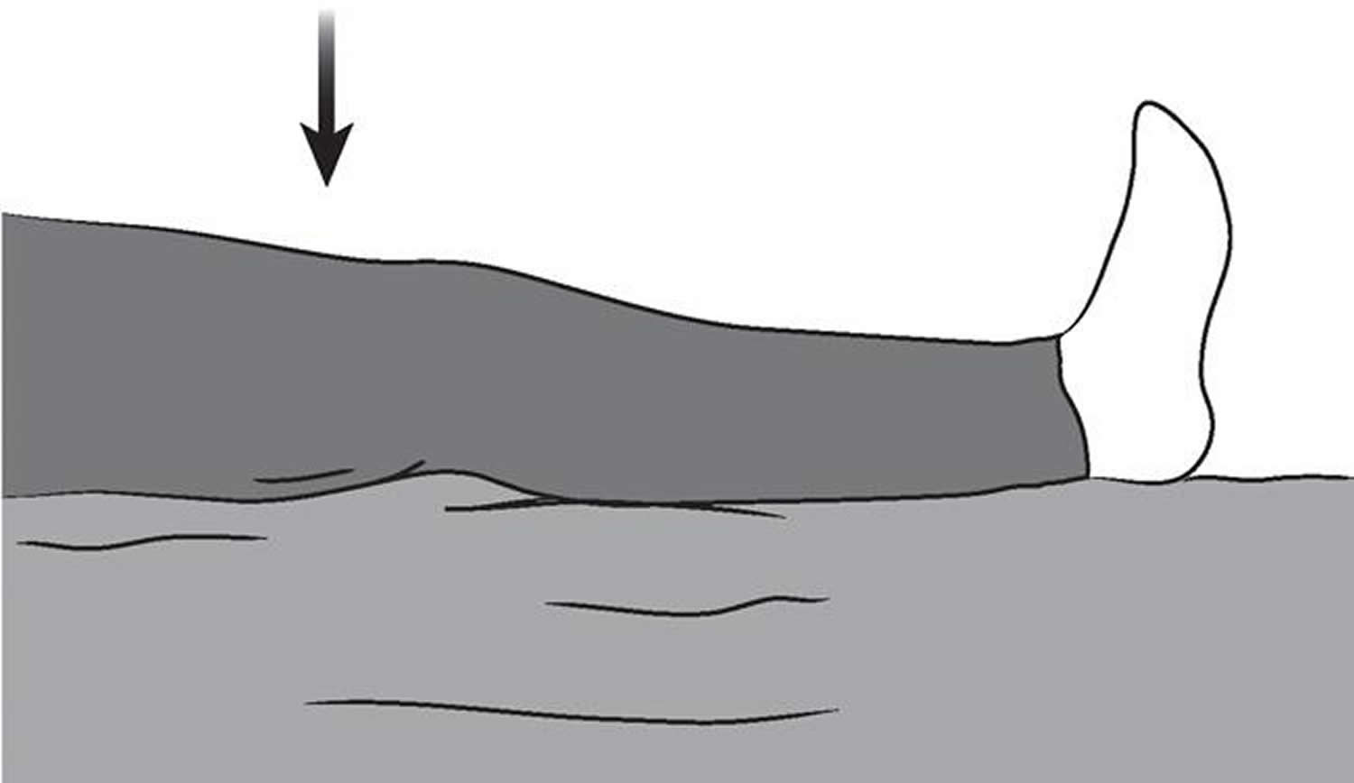
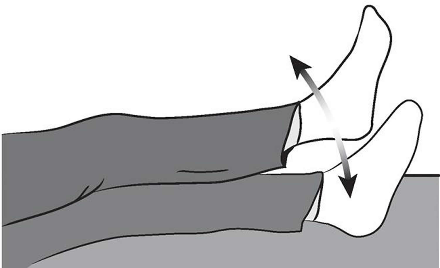
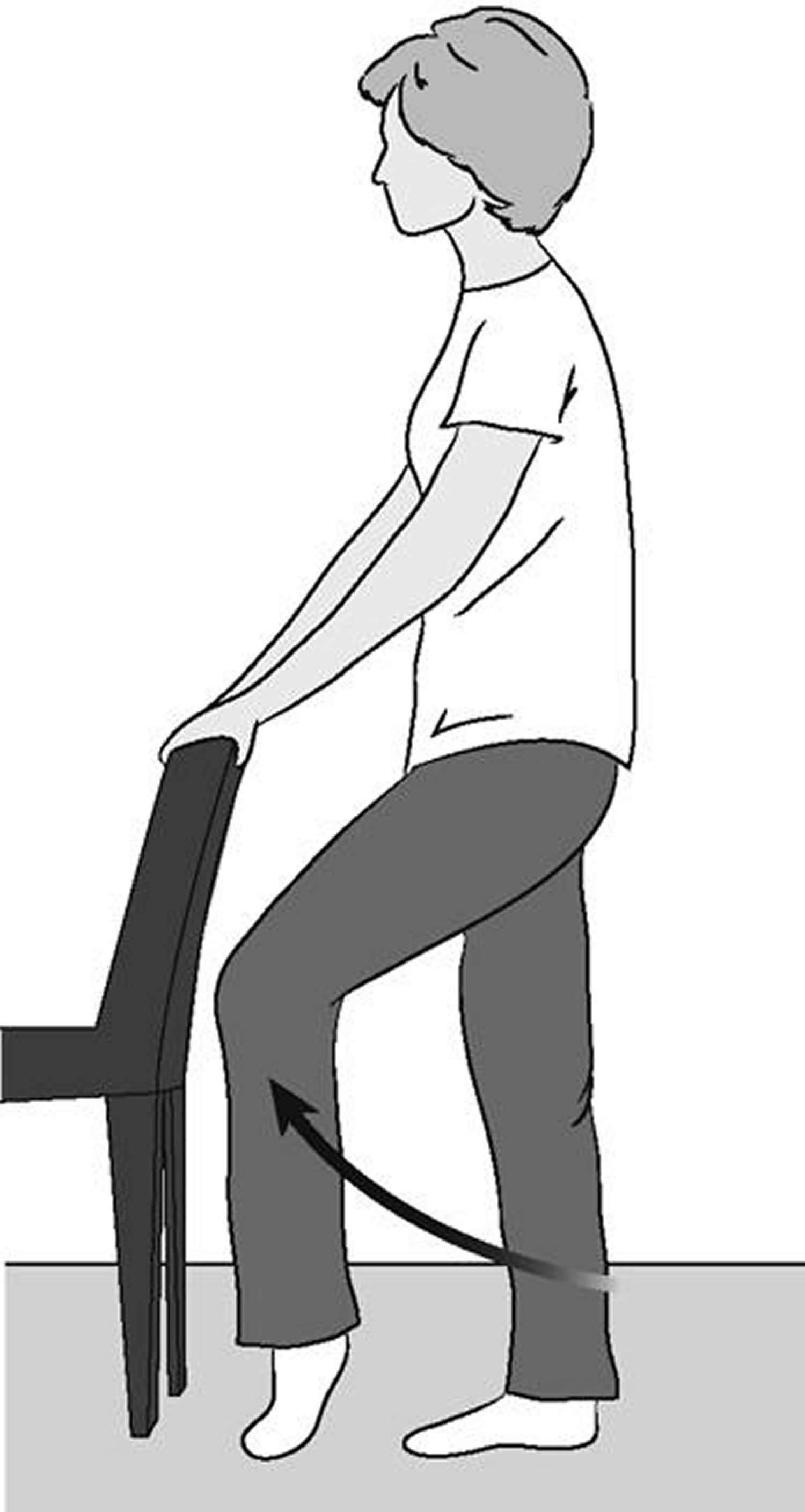
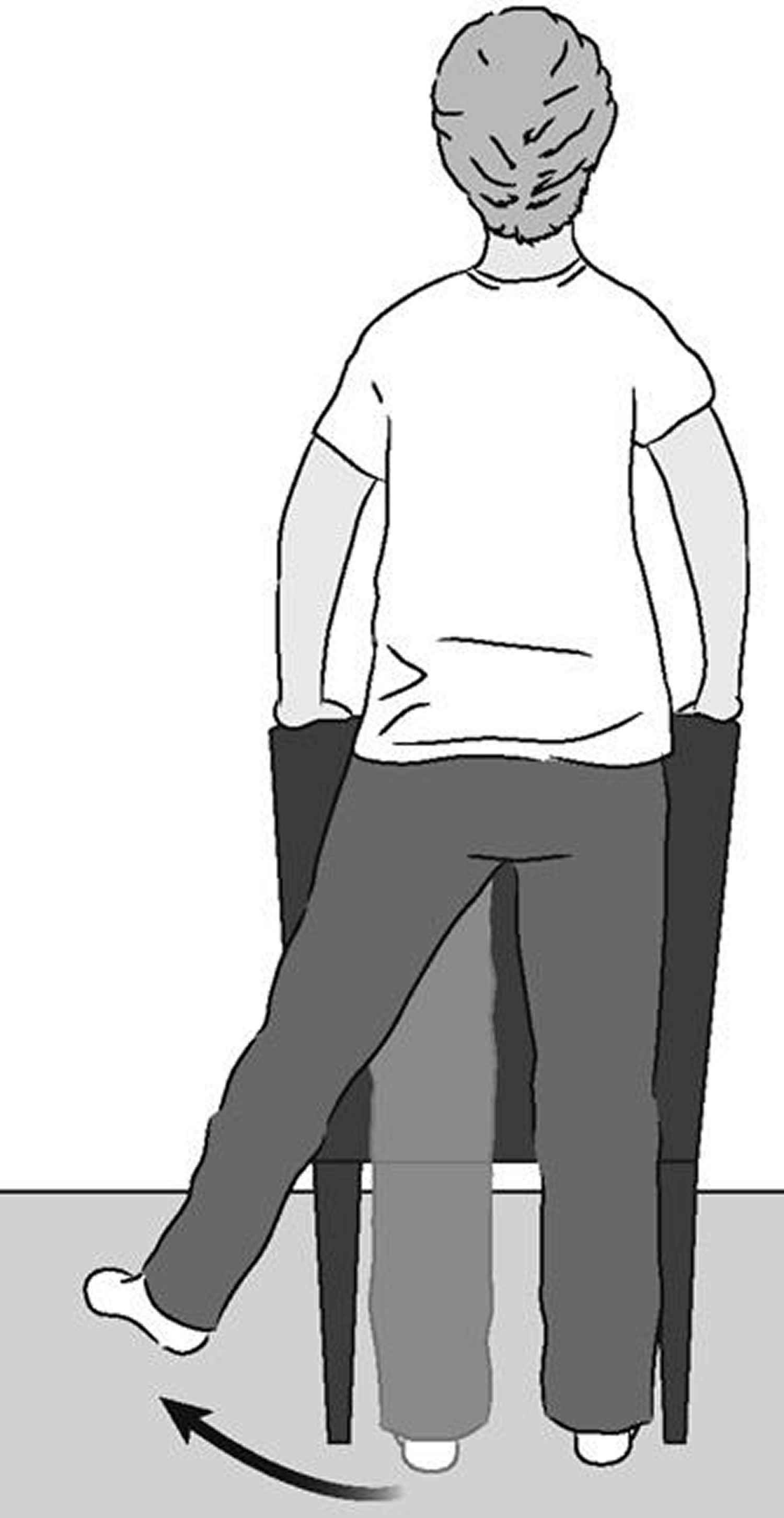
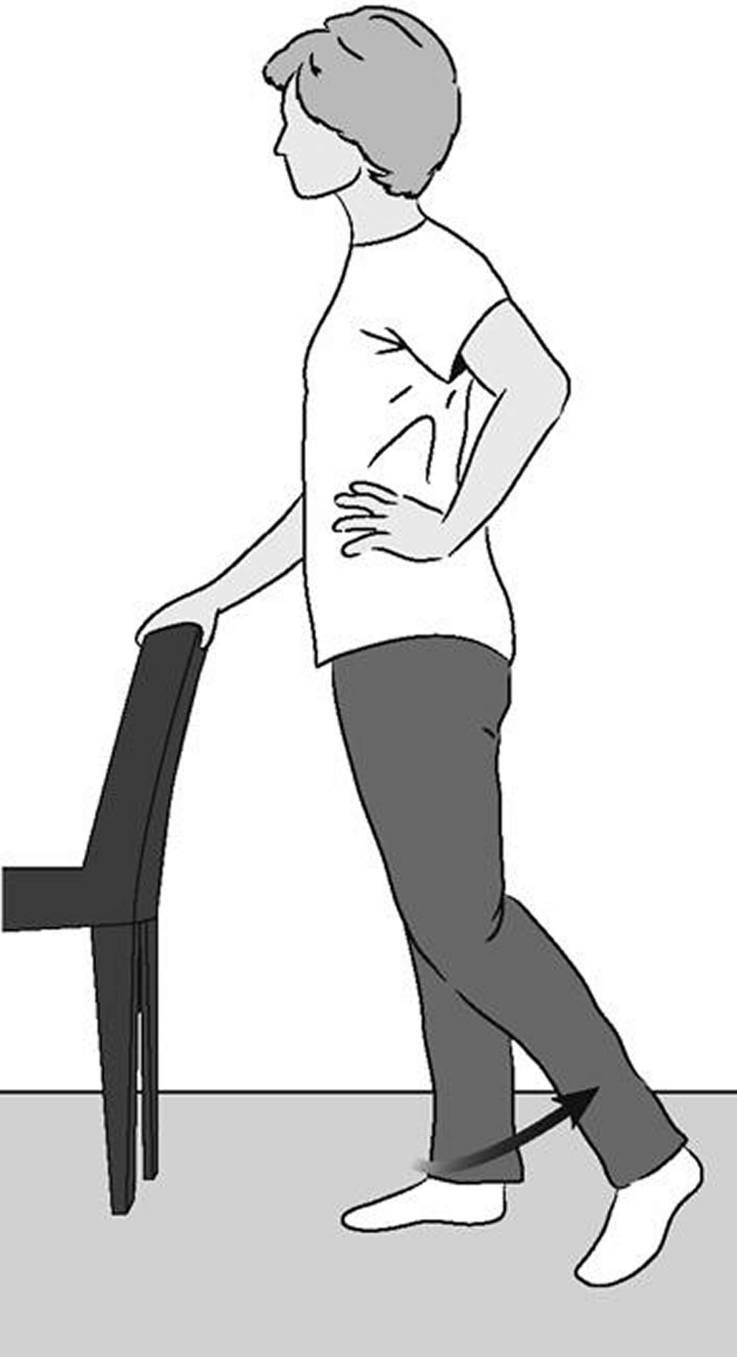
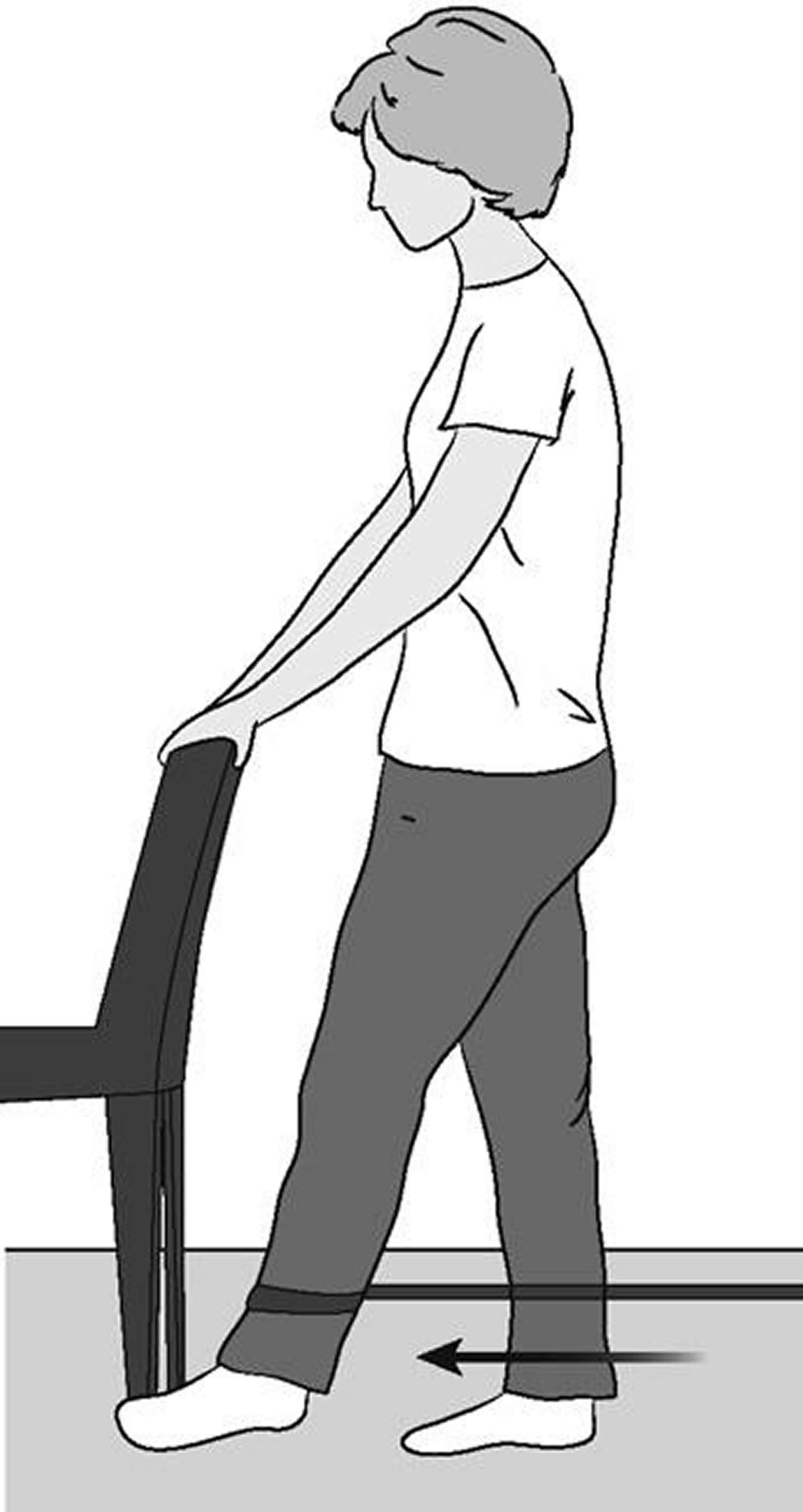
- Total Hip Replacement Exercise Guide
- Knee arthroplasty
- Knee replacement surgery risks
- How long will a replacement knee last?
- Deciding to Have Knee Replacement Surgery
- Knee Joint Anatomy
- Types of knee replacement surgery
- Knee replacement surgery alternatives
- Preparing for knee replacement surgery
- How knee replacement surgery is performed
- Total knee replacement
- Partial (half) knee replacement
- Other procedures
- After knee replacement surgery
- Knee replacement recovery
- Looking after your new knee
- Shoulder arthroplasty
- Shoulder joint anatomy
- Reasons for shoulder arthroplasty
- Is Shoulder Joint Replacement for You?
- Shoulder Replacement Options
- Total shoulder arthroplasty
- Reverse shoulder arthroplasty
- Candidates for reverse shoulder arthroplasty surgery
- Reverse shoulder arthroplasty surgical complications
- Preparing for reverse shoulder arthroplasty surgery
- Reverse shoulder arthroplasty surgical procedure
- Reverse shoulder arthroplasty recovery
- Do’s and Dont’s After Shoulder Replacement Surgery
- Long-Term Outcomes
- Thumb joint replacement
What is arthroplasty
Arthroplasty or joint replacement surgery is done as after other treatments (physical therapy and medications) have not helped. Joint replacement surgery is usually the last line of treatment, when all other treatments – including physical therapy and medications – have not helped the patient. Arthroplasty (joint replacement surgery) is a highly effective way of eliminating joint pain, correcting a deformity, and helping improve the patient’s mobility (movement). Joint replacement surgery is also performed to treat advanced arthritis.
People who are considered for joint replacement surgery often have severe joint pain, stiffness, limping, muscle weakness, limited motion, and swelling. Depending on the joint that is affected and the amount of damage, patients may have trouble with ordinary activities such as walking, putting on socks and shoes, getting into and out of cars, and climbing stairs.
What causes joint problems?
The most common causes of the joints not working properly are osteoarthritis and rheumatoid arthritis. While nobody is certain what causes arthritis, several things may contribute to joint weakening and lead to arthritis, including:
- Heredity (runs in the family)
- Problems with the development of the joint
- Genetic (inherited) tendency to problems with the cartilage
- Minor repetitive injures
- Severe trauma to the joint cartilage (the cushioning tissue at the end of the bones)
While being overweight does not necessarily cause arthritis, it can contribute to early joint problems that can get worse quickly.
What happens during joint replacement surgery?
Joint replacement surgery is designed to replace the damaged cartilage and any bone loss. During the procedure, the damaged joint is resurfaced, and the patient’s muscles and ligaments are used for support and function.
The prosthesis (replacement joint) is made of titanium, cobalt chrome, stainless steel, ceramic material, and polyethylene (plastic). It can be attached to the bone with acrylic cement or it can be press-fit, which allows bone to grow into the implant. Once the joint replacement is in place, the patient has physical therapy to be able to move and use the joint.
The 3 most common joint replacement surgeries are hip, knee, and shoulder.
Hip replacement
Total hip replacement is a surgery for replacing the hip socket and the “ball” or head of the thigh bone (femur). The surgeon resurfaces the socket and ball where cartilage and bone have been lost, and then inserts an artificial ball and socket into healthy bone.
Most people who have total hip replacements have serious changes in the hip joint caused by arthritis. A hip replacement is recommended if the person cannot bear the joint pain, and when the person can’t perform activities of daily living because the damaged hip is preventing it.
Knee replacement
Knee replacement surgery is performed to treat advanced or end-stage arthritis. When arthritis in the knee joint or joints has advanced to the point where it cannot be treated with medicine alone, or the deformity has become severe and keeps the patient from using the knee, replacement surgery may be recommended.
The need for knee replacement surgery is the damage to the coating or gliding surface called the articular cartilage. Depending on the amount of damage, ordinary activities such as walking and climbing stairs may become difficult. Damage to the knee joint cartilage and bone may also cause deformity. Knock-knee or bow-legged deformities and unusual knee sounds (crepitus) may become more noticeable as the deterioration gets worse.
Knee replacement surgery is designed to replace this damaged cartilage or gliding surface, as well as any loss of bone structure or ligament support. The material used for knee replacement is similar to that used for hip replacements.
Shoulder replacement
Total shoulder joint replacement is usually needed for people who have advanced forms of osteoarthritis or rheumatoid arthritis, and sometimes for those who have had severe injury from a shoulder fracture. The main goal of total shoulder replacement surgery is to relieve pain; other goals include improving motion, strength, and function.
Similar to the hip joint, the shoulder is a large ball-and-socket joint. The main reason for a total shoulder replacement is pain that is not being relieved with therapy or other treatment methods.
What is the post-operative (after surgery) management for a joint replacement surgery?
Hip replacement post-operative management
Most patients can stand at their bedside on the first day after surgery and can even begin exercising. By the second day after surgery, most patients begin walking with a walker or with crutches, and can apply 50 to 75% of their weight on the affected leg.
Most patients leave the hospital by the first or second day after surgery. Older individuals and patients who have major health problems are usually referred to a rehabilitation facility for 7 to 10 days for more therapy.
All patients will use either crutches or a walker for about 4 weeks after surgery. They are then allowed to place full weight on the affected leg while using a cane for balance. The cane also prevents the muscles from becoming tired.
Generally, by 6 to 12 weeks after surgery, the person can stop using the cane or walker (if the doctor or therapist agrees) and the hip can support the person’s full weight. Patients who have weaker muscles may need to use the cane or walker for a longer period.
Once you have completed therapy after the total hip replacement, you can take part in most activities, such as walking, bike riding, skiing, and golf. Activities in which there is repeated or frequent impact on the joint (such as tennis and racquetball) should be avoided or practiced only occasionally.
Knee replacement post-operative management
Most patients who have total knee surgery have a dramatic improvement within three months of the surgery. The pain caused by the damaged knee is relieved when a new gliding surface is built.
Patients who have knee replacement surgery are usually standing and moving the joint the day after surgery. After about 6 weeks, most patients are walking comfortably with very little support; however, it may take 6 months to a year before the most benefit is achieved. After muscle strength returns, patients who have knee replacement surgery can enjoy most activities (except running and jumping).
Approximately 85% of knee implants will last 20 years. Improvements in surgical techniques, prosthetic designs, bearing surfaces, and fixation methods may allow these implants to last even longer.
Shoulder replacement post-operative management
A successful result of your total shoulder joint replacement strongly depends on your performing the exercises that were prescribed for you. Through this structured exercise program, your muscles will be regularly and increasingly stretched and strengthened over one year’s time. The goal is to get your shoulder replacement working as well as it can.
In certain situations, patients may need extensive formal physical therapy after being discharged from the hospital. This can be done during outpatient therapy at home. Most patients, however, do not need any formal outpatient therapy.
Your rehabilitation will be continuing and always moving forward. It may take 6 months to 1 year to reach the most benefit. It is important to realize that progress is sometimes slow and not always steady. You must continue your therapy program without getting discouraged. Your doctor will check your progress during visits, which will be every 6 weeks for the first 4 to 5 months, and then less frequently for 1 year.
With improvements in materials, prosthetic designs, and surgical techniques, more than 95% of total joint replacement procedures should last 15 to 20 years or more. Follow-up after recovery from surgery should include X-rays after the first, third, fifth, and seventh years. After that, X-rays should be taken every 2 years to make sure that there is no wear on the replaced joint.
Hip arthroplasty
Hip replacement surgery is a procedure for people with severely damaged hip joint to replace it with an artificial one (known as a prosthesis). The most common reason for hip replacement is damaged hip caused by osteoarthritis. Osteoarthritis causes pain, swelling, and reduced motion in your joints. It can interfere with your daily activities. If your hip has been damaged by arthritis, a fracture, or other conditions, common activities such as walking or getting in and out of a chair may be painful and difficult. Your hip may be stiff, and it may be hard to put on your shoes and socks. You may even feel uncomfortable while resting.
If other treatments such as physical therapy, pain medicines, changes in your everyday activities, exercise and the use of walking supports do not adequately help your symptoms, you may consider hip replacement surgery.
Adults of any age can be considered for a hip replacement, although most are carried out on people between the ages of 60 and 80.
Hip replacement surgery is a safe and effective procedure that can relieve your pain, increase motion, and help you get back to enjoying normal, everyday activities.
Hip replacement surgery was first performed in 1960, hip replacement surgery is one of the most successful operations in all of medicine. Since 1960, improvements in joint replacement surgical techniques and technology have greatly increased the effectiveness of total hip replacement. According to the Agency for Healthcare Research and Quality, more than 300,000 total hip replacements are performed each year in the United States.
During a hip replacement operation, the surgeon removes damaged cartilage and bone from your hip joint and replaces them with new, man-made parts.
A modern artificial hip joint is designed to last for at least 15 years. Most people experience a significant reduction in pain and some improvement in their range of movement.
A hip replacement can:
- Relieve pain
- Help your hip joint work better
- Improve walking and other movements
The most common problem after a hip replacement surgery is hip dislocation. Because a man-made hip is smaller than the original joint, the ball can come out of its socket. The surgery can also cause blood clots and infections. With a hip replacement, you might need to avoid certain activities, such as jogging and high-impact sports.
Common causes of hip pain
The most common cause of chronic hip pain and disability is arthritis. Osteoarthritis, rheumatoid arthritis, and traumatic arthritis are the most common forms of this disease.
- Osteoarthritis. This is an age-related “wear and tear” type of arthritis. It usually occurs in people 50 years of age and older and often in individuals with a family history of arthritis. The cartilage cushioning the bones of the hip wears away. The bones then rub against each other, causing hip pain and stiffness. Osteoarthritis may also be caused or accelerated by subtle irregularities in how the hip developed in childhood.
- Rheumatoid arthritis. This is an autoimmune disease in which the synovial membrane becomes inflamed and thickened. This chronic inflammation can damage the cartilage, leading to pain and stiffness. Rheumatoid arthritis is the most common type of a group of disorders termed “inflammatory arthritis.”
- Post-traumatic arthritis. This can follow a serious hip injury or fracture. The cartilage may become damaged and lead to hip pain and stiffness over time.
- Avascular necrosis. An injury to the hip, such as a dislocation or fracture, may limit the blood supply to the femoral head. This is called avascular necrosis (also commonly referred to as “osteonecrosis”). The lack of blood may cause the surface of the bone to collapse, and arthritis will result. Some diseases can also cause avascular necrosis.
- Childhood hip disease. Some infants and children have hip problems. Even though the problems are successfully treated during childhood, they may still cause arthritis later on in life. This happens because the hip may not grow normally, and the joint surfaces are affected.
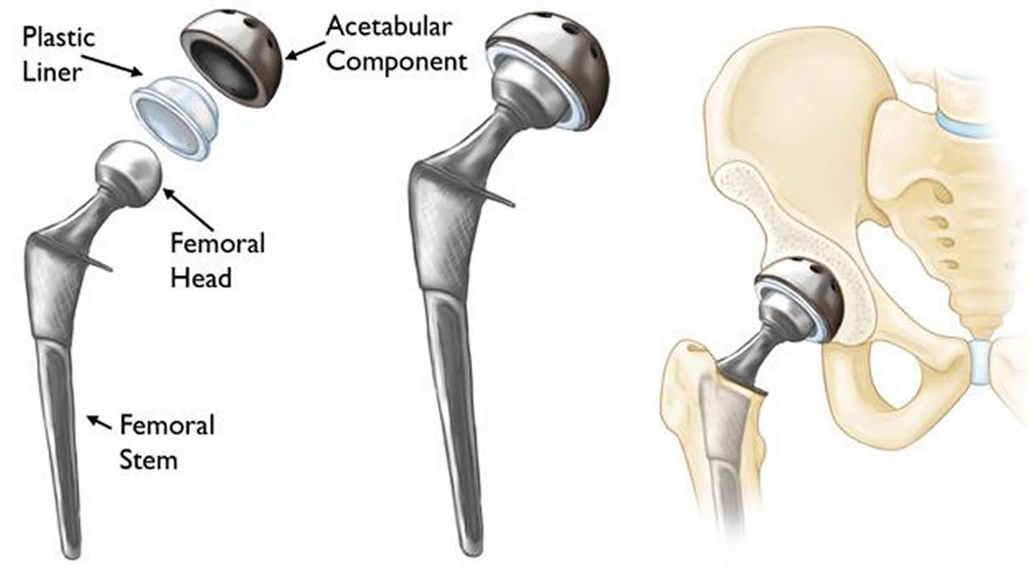
Figure 1. Hip replacement (total hip replacement) – Note: (Left) The individual components of a total hip replacement. (Center) The components merged into an implant. (Right) The implant as it fits into the hip.
Hip Joint Anatomy
The hip is one of the body’s largest joints. The hip joint (coxal joint) is a ball-and-socket joint formed by the head of the femur and the acetabulum of the hip bone. The socket is formed by the acetabulum, which is part of the large pelvis bone. The ball is the femoral head, which is the upper end of the femur (thighbone). The bone surfaces of the ball and socket are covered with articular cartilage, a smooth tissue that cushions the ends of the bones and enables them to move easily.
A thin tissue called synovial membrane surrounds the hip joint. In a healthy hip, this membrane makes a small amount of fluid that lubricates the cartilage and eliminates almost all friction during hip movement.
Bands of tissue called ligaments (the hip capsule) connect the ball to the socket and provide stability to the joint.
The hip joint allows flexion, extension, abduction, adduction, lateral rotation medial rotation, and circumduction of the thigh. The extreme stability of the hip joint is related to the very strong articular capsule and its accessory ligaments, the manner in which the femur fits into the acetabulum, and the muscles surrounding the joint.
Figure 2. Hip joint anatomy
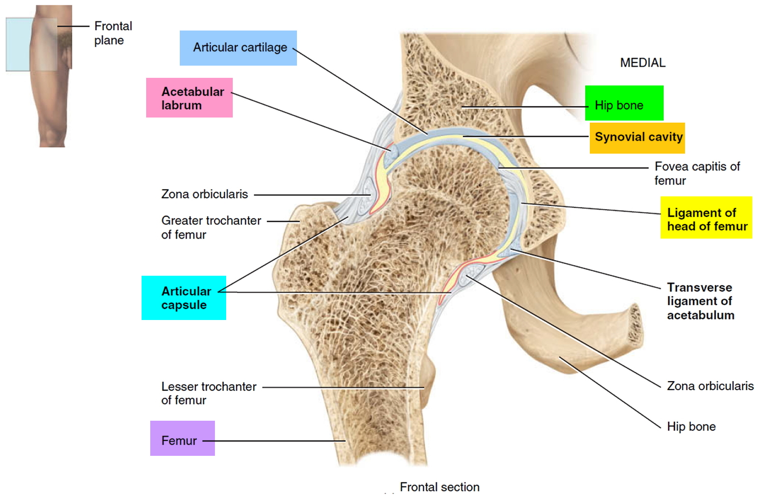
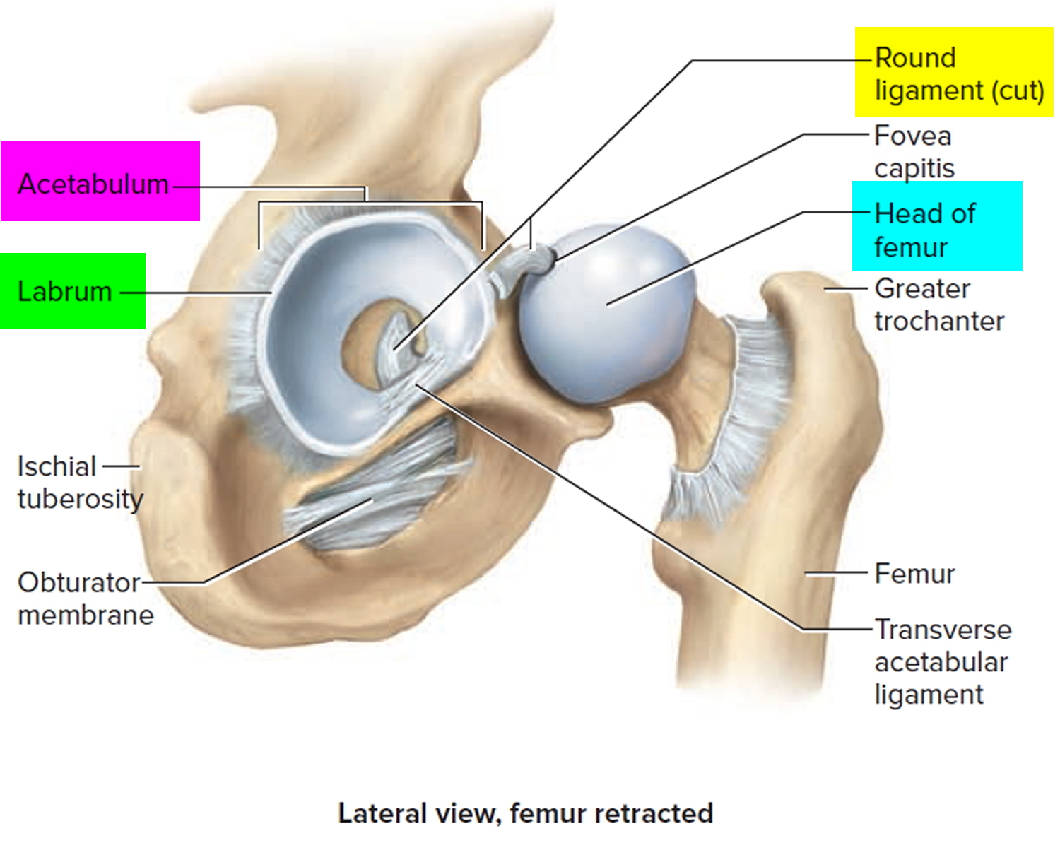 Hip joint anatomical components
1. Articular capsule
Very dense and strong capsule that extends from the rim of the acetabulum to the neck of the femur (Figure 2). With its accessory ligaments, this is one of the strongest structures of the body. The articular capsule consists of circular and longitudinal fibers. The circular fibers, called the zona orbicularis, form a collar around the neck of the femur. Accessory ligaments known as the iliofemoral ligament, pubofemoral ligament, and ischiofemoral ligament reinforce the longitudinal fibers of the articular capsule.
2. Iliofemoral ligament
Thickened portion of the articular capsule that extends from the anterior inferior iliac spine of the hip bone to the intertrochanteric line of the femur (Figure 2). This ligament is said to be the body’s strongest and prevents hyperextension of the femur at the hip joint during standing.
3. Pubofemoral ligament
Thickened portion of the articular capsule that extends from the pubic part of the rim of the acetabulum to the neck of the femur. This ligament prevents overabduction of the femur at the hip joint and strengthens the articular capsule.
4. Ischiofemoral ligament
Thickened portion of the articular capsule that extends from the ischial wall bordering the acetabulum to the neck of the femur. This ligament slackens during adduction, tenses during abduction, and strengthens the articular capsule.
5. Ligament of the head of the femur
Flat, triangular band (primarily a synovial fold) that extends from the fossa of the acetabulum to the fovea capitis of the head of the femur. The ligament usually contains a small artery that supplies the head of the femur.
6. Acetabular labrum
Fibrocartilage rim attached to the margin of the acetabulum that enhances the depth of the acetabulum. As a result, dislocation of the femur is rare.
7. Transverse ligament of the acetabulum
Strong ligament that crosses over the acetabular notch. It supports part of the acetabular labrum and is connected with the ligament of the head of the femur and the articular capsule.
Partial hip replacement
Partial hip replacement also called hemiarthroplasty replaces only the ball part (the head of femur or femoral head) of your hip joint. Partial hip replacement is generally considered to be the treatment of choice in the most elderly patients with a displaced fracture of the femoral neck.
In elderly patients suffering from a displaced femoral neck fracture, a cemented hip arthroplasty, compared to internal fixation, partial hip replacement (hemiarthroplasty) has been shown to reduce the reoperation rate and give better hip function and health-related quality of life 1. In the healthy, active elderly with a long life expectancy, a total hip replacement is probably the best treatment 2 while a partial hip replacement (Hemiarthroplasty) is generally considered to be sufficient for the most elderly patients with lower functional demands and a shorter life expectancy.
There are two different types of partial hip replacement (hemiarthroplasty): unipolar and bipolar. The theoretical advantage of the bipolar partial hip replacement (Hemiarthroplasty) is a reduction of acetabular wear due to the dual-bearing system 3. On the other hand, a potential disadvantage is the risk of polyethylene wear that may contribute to mechanical loosening over time and there is also a risk of inter-prosthetic dissociation in certain bipolar partial hip replacements necessitating open reduction 4. However, dissociation appears to be rare in modern bipolar surgical systems.
Based on the results of numerous studies 5, 6, 7, 8, 9, there does not appear to be any clinical disadvantage with the bipolar design. On the contrary, the results of this study 5 showed that the rate of acetabular erosion was significantly lower after the bipolar partial hip replacement, which in turn may indicate an advantage in the longer term. However, there is one frequently cited disadvantage associated with the bipolar partial hip replacement, i.e. the higher cost, the magnitude of which probably differs between countries. Since there were no differences in duration of surgery, need for blood transfusions, hospital stay and complications during the first year, this difference probably represents the total difference in primary costs for the two treatment modalities. This investment may well be justified in order to reduce the risk of future problems owing to acetabular erosion.
Figure 3. Partial hip replacement (Unipolar)
Figure 4. Partial hip replacement (Bipolar)
Total hip replacement
In a total hip replacement (also called total hip arthroplasty), the damaged bone and cartilage is removed and replaced with prosthetic components (see Figure 1 above).
- The damaged femoral head is removed and replaced with a metal stem that is placed into the hollow center of the femur. The femoral stem may be either cemented or “press fit” into the bone.
- A metal or ceramic ball is placed on the upper part of the stem. This ball replaces the damaged femoral head that was removed.
- The damaged cartilage surface of the socket (acetabulum) is removed and replaced with a metal socket. Screws or cement are sometimes used to hold the socket in place.
- A plastic, ceramic, or metal spacer is inserted between the new ball and the socket to allow for a smooth gliding surface.
Hip replacement alternatives
There is an alternative type of surgery to hip replacement, known as hip resurfacing. This involves removing the damaged surfaces of the bones inside the hip joint and replacing them with a metal surface.
An advantage to this approach is that it removes less bone. However, it may not be suitable for:
- adults over the age of 65 years – bones tend to weaken as a person becomes older
- women who have gone through the menopause – one of the side effects of the menopause is that the bones can become weakened and brittle (osteoporosis)
Resurfacing is much less popular now due to concerns about the metal surface causing damage to soft tissues around the hip.
Your surgeon should be able to tell you if you could be a suitable candidate for hip resurfacing.
Is Hip Replacement Surgery for You?
The decision to have hip replacement surgery should be a cooperative one made by you, your family, your primary care doctor, and your orthopaedic surgeon. The process of making this decision typically begins with a referral by your doctor to an orthopedic surgeon for an initial evaluation.
Hip replacement surgery is usually necessary when the hip joint is worn or damaged to the extent that your mobility is reduced and you experience pain even while resting.
The most common reason for hip replacement surgery is osteoarthritis. Other conditions that can cause hip joint damage include:
- rheumatoid arthritis
- a hip fracture
- septic arthritis
- ankylosing spondylitis
- disorders that cause unusual bone growth (bone dysplasias)
Candidates for hip replacement surgery
There are no absolute age or weight restrictions for total hip replacements. However, a hip replacement is major surgery, so is normally only recommended if other treatments, such as physiotherapy or steroid injections, haven’t helped reduce pain or improve mobility.
Recommendations for surgery are based on a patient’s pain and disability, not age. Most patients who undergo total hip replacement are age 50 to 80, but orthopedic surgeons evaluate patients individually. Total hip replacements have been performed successfully at all ages, from the young teenager with juvenile arthritis to the elderly patient with degenerative arthritis.
You may be offered hip replacement surgery if:
- you have severe pain, swelling and stiffness in your hip joint and your mobility is reduced
- your hip pain is so severe that it interferes with your quality of life and sleep
- everyday tasks, such as shopping or getting out of the bath, are difficult or impossible
- you’re feeling depressed because of the pain and lack of mobility
- you can’t work or have a normal social life
You’ll also need to be well enough to cope with both a major operation and the rehabilitation afterwards.
When hip replacement surgery is recommended
There are several reasons why your doctor may recommend hip replacement surgery. People who benefit from hip replacement surgery often have:
- Hip pain that limits everyday activities, such as walking or bending
- Hip pain that continues while resting, either day or night
- Stiffness in a hip that limits the ability to move or lift the leg
- Inadequate pain relief from anti-inflammatory drugs, physical therapy, or walking supports
The Orthopedic Evaluation
An evaluation with an orthopedic surgeon consists of several components.
- Medical history. Your orthopedic surgeon will gather information about your general health and ask questions about the extent of your hip pain and how it affects your ability to perform everyday activities.
- Physical examination. This will assess hip mobility, strength, and alignment.
- X-rays. These images help to determine the extent of damage or deformity in your hip.
- Other tests. Occasionally other tests, such as a magnetic resonance imaging (MRI) scan, may be needed to determine the condition of the bone and soft tissues of your hip.
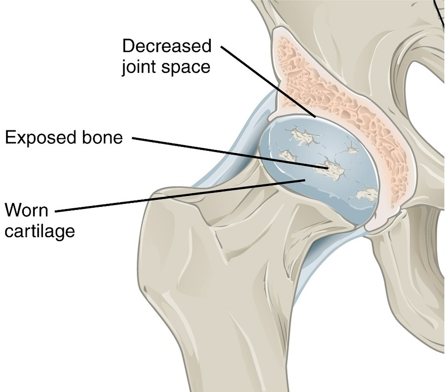
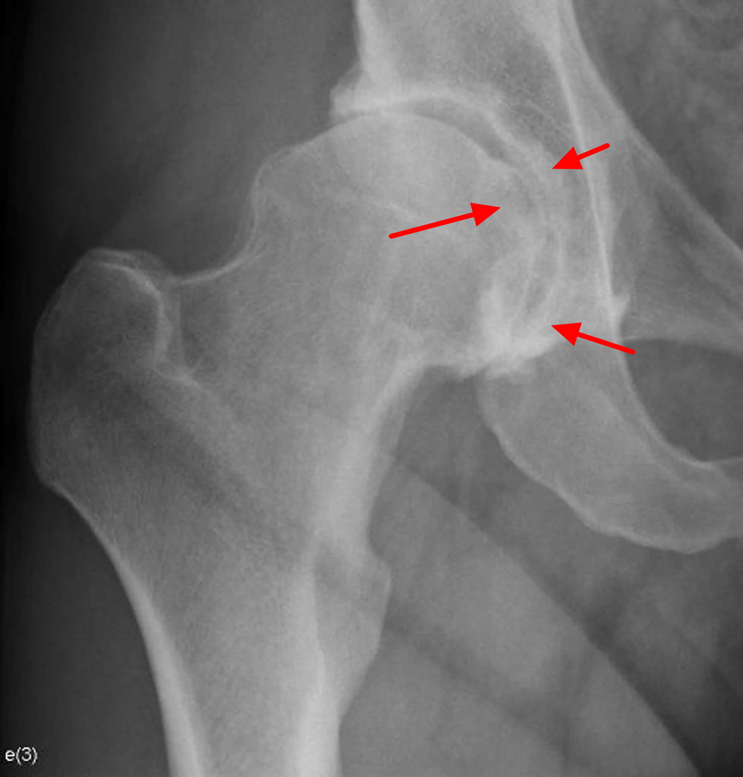
Figure 5. Hip osteoarthritis – note the smooth articular cartilage wears away and becomes frayed and rough
Figure 6. Hip osteoarthritis (X-ray)
Note: Dysplastic acetabulum with insufficient covering of the femoral head. Joint space narrowing, subchondral sclerosis and osteophytes at the femoroacetabular joint. Prominent bump at the antero-superior head-neck junction of the femur (cam deformity, also known as pistol grip deformity of the proximal femur).
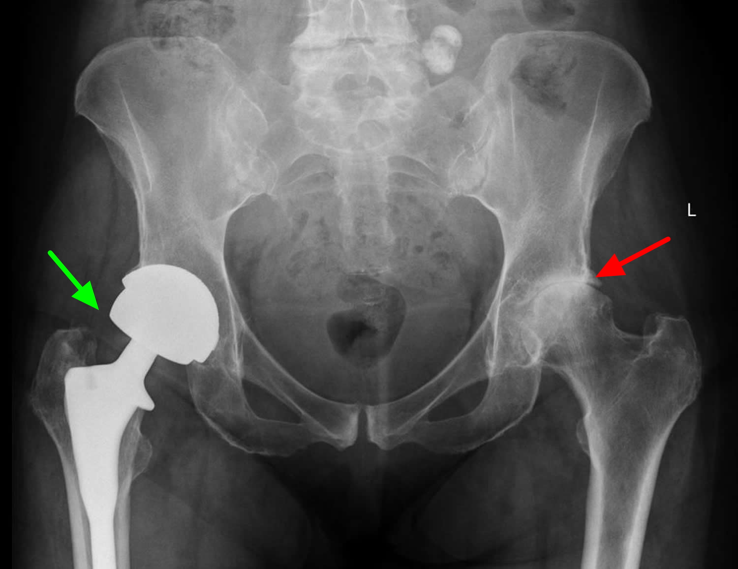
Figure 7. Total hip replacement (right hip – green arrow) and left hip osteoarthritis (red arrow) X-ray
Deciding to Have Hip Replacement Surgery
Talk with your doctor
Your orthopedic surgeon will review the results of your evaluation with you and discuss whether hip replacement surgery is the best method to relieve your pain and improve your mobility. Other treatment options — such as medications, physical therapy, or other types of surgery — also may be considered.
In addition, your orthopedic surgeon will explain the potential risks and complications of hip replacement surgery, including those related to the surgery itself and those that can occur over time after your surgery.
Never hesitate to ask your doctor questions when you do not understand. The more you know, the better you will be able to manage the changes that hip replacement surgery will make in your life.
Realistic expectations
An important factor in deciding whether to have hip replacement surgery is understanding what the procedure can and cannot do. Most people who undergo hip replacement surgery experience a dramatic reduction of hip pain and a significant improvement in their ability to perform the common activities of daily living.
With normal use and activity, the material between the head and the socket of every hip replacement implant begins to wear. Excessive activity or being overweight may speed up this normal wear and cause the hip replacement to loosen and become painful. Therefore, most surgeons advise against high-impact activities such as running, jogging, jumping, or other high-impact sports.
Realistic activities following total hip replacement include unlimited walking, swimming, golf, driving, hiking, biking, dancing, and other low-impact sports.
With appropriate activity modification, hip replacements can last for many years.
Possible complications of hip replacement surgery
The complication rate following hip replacement surgery is low. Serious complications, such as joint infection, occur in less than 2% of patients. Major medical complications, such as heart attack or stroke, occur even less frequently. However, chronic illnesses may increase the potential for complications. Although uncommon, when these complications occur they can prolong or limit full recovery.
Infection
A small percentage of patients undergoing hip replacement (roughly about 1 in 100) may develop an infection after the operation.
Infection may occur superficially in the wound or deep around the artificial implants. An infection may develop while in the hospital or after you go home. It may even occur years later.
Minor infections of the wound are generally treated with antibiotics. Major or deep infections may require more surgery and removal of the artificial implants. Any infection in your body can spread to your joint replacement.
Infections are caused by bacteria. Although bacteria are abundant in your gastrointestinal tract and on your skin, they are usually kept in check by your immune system. For example, if bacteria make it into your bloodstream, your immune system rapidly responds and kills the invading bacteria.
However, because joint replacements are made of metal and plastic, it is difficult for the immune system to attack bacteria that make it to these implants. If bacteria gain access to the implants, they may multiply and cause an infection.
Despite antibiotics and preventive treatments, patients with infected joint replacements often require surgery to cure the infection.
The most common ways bacteria enter the body include:
- Through breaks or cuts in the skin
- During major dental procedures (such as a tooth extraction or root canal)
- Through wounds from other surgical procedures
Some people are at a higher risk for developing infections after a joint replacement procedure. Factors that increase the risk for infection include:
- Immune deficiencies (such as HIV or lymphoma)
- Diabetes mellitus
- Peripheral vascular disease (poor circulation to the hands and feet)
- Immunosuppressive treatments (such as chemotherapy or corticosteroids)
- Obesity
Prevention
At the time of original joint replacement surgery, there are several measures taken to minimize the risk of infection. Some of the steps have been proven to lower the risk of infection, and some are thought to help but have not been scientifically proven. The most important known measures to lower the risk of infection after total joint replacement include:
- Antibiotics before and after surgery. Antibiotics are given within one hour of the start of surgery (usually once in the operating room) and continued at intervals for 24 hours following the procedure.
- Short operating time and minimal operating room traffic. Efficiency in the operation by your surgeon helps to lower the risk of infection by limiting the time the joint is exposed. Limiting the number of operating room personnel entering and leaving the room is thought to the decrease risk of infection.
- Use of strict sterile technique and sterilization instruments. Care is taken to ensure the operating site is sterile, the instruments have been autoclaved (sterilized) and not exposed to any contamination, and the implants are packaged to ensure their sterility.
- Preoperative nasal screening for bacterial colonization. There is some evidence that testing for the presence of bacteria (particularly the Staphylococcus species) in the nasal passages several weeks prior to surgery may help prevent joint infection. In institutions where this is performed, those patients that are found to have Staphylococcus in their nasal passages are given an intranasal antibacterial ointment prior to surgery. The type of bacteria that is found in the nasal passages may help your doctors determine which antibiotic you are given at the time of your surgery.
- Preoperative chlorhexidine wash. There is also evidence that home washing with a chlorhexidine solution (often in the form of soaked cloths) in the days leading up to surgery may help prevent infection. This may be particularly important if patients are known to have certain types of antibiotic-resistant bacteria on their skin or in their nasal passages (see above). Your surgeon will talk with you about this option.
- Long-term prophylaxis. Surgeons sometimes prescribe antibiotics for patients who have had joint replacements before they undergo dental work. This is done to protect the implants from bacteria that might enter the bloodstream during the dental procedure and cause infection. The American Academy of Orthopaedic Surgeons has developed recommendations for when antibiotics should be given before dental work and for which patients would benefit. In general, most people do not require antibiotics before dental procedures. There is little evidence that taking antibiotics before dental procedures is effective at preventing infection.
Antibiotics may also be considered before major surgical procedures; however, most patients do not require this.
Your orthopedic surgeon will talk with you about the risks and benefits of prophylactic antibiotics in your specific situation.
Symptoms of infection
Signs and symptoms of an infected joint replacement include:
- A high temperature (fever) of 38 °C (100.4 °F) or above
- Hip pain that can persist even when resting
- Increased pain or stiffness in a previously well-functioning joint
- Swelling
- Warmth and redness around the wound
- A discharge of liquid from the site of the surgery
- Wound drainage
- Fevers, chills and night sweats
- Fatigue
Blood Clots
There’s a small risk of developing a blood clot in the first few weeks after surgery – either deep vein thrombosis (DVT) in the leg or pulmonary embolism in the lung. Blood clots in the leg veins or pelvis are one of the most common complications of hip replacement surgery. These clots can be life-threatening if they break free and travel to your lungs. Your orthopedic surgeon will outline a prevention program which may include blood thinning medications, support hose, inflatable leg coverings, ankle pump exercises, and early mobilization.
Symptoms of DVT include:
- pain, swelling and tenderness in one of your legs (usually your calf)
- a heavy ache in the affected area
- warm skin in the area of the clot
Symptoms of pulmonary embolism include:
- breathlessness, which may come on suddenly or gradually
- chest pain, which may be worse when you breathe in
- coughing
If you suspect either of these types of blood clots you should seek immediate medical advice from your doctor or the doctor in charge of your care.
To reduce your risk of blood clots you may be given blood thinning medication such as warfarin, or asked to wear compression stockings.
Leg-length Inequality
Sometimes after a hip replacement, one leg may feel longer or shorter than the other. Your orthopedic surgeon will make every effort to make your leg lengths even, but may lengthen or shorten your leg slightly in order to maximize the stability and biomechanics of the hip. Some patients may feel more comfortable with a shoe lift after surgery.
Hip replacement dislocation
This occurs when the ball comes out of the socket. The risk for hip replacement dislocation is greatest in the first few months after surgery while the tissues are healing. Hip replacement dislocation is uncommon. If the ball does come out of the socket, a closed reduction usually can put it back into place without the need for more surgery. In situations in which the hip continues to dislocate, further surgery may be necessary.
Loosening and Implant Wear
Over years, the hip prosthesis may wear out or loosen. This is most often due to everyday activity. It can also result from a biologic thinning of the bone called osteolysis. If loosening is painful, a second surgery called a revision may be necessary.
Joint stiffening
The soft tissues can harden around the implant, causing reduced mobility.
This isn’t usually painful and can be prevented using medication or radiation therapy (a quick and painless procedure during which controlled doses of radiation are directed at your hip joint).
Other complications
Nerve and blood vessel injury, bleeding, fracture, and stiffness can occur. In a small number of patients, some pain can continue or new pain can occur after surgery.
Preparing for hip replacement surgery
Medical Evaluation
If you decide to have hip replacement surgery, your orthopedic surgeon may ask you to have a complete physical examination by your primary care doctor before your surgical procedure. This is needed to make sure you are healthy enough to have the surgery and complete the recovery process. Many patients with chronic medical conditions, like heart disease, may also be evaluated by a specialist, such a cardiologist, before the surgery.
Tests
Several tests, such as blood and urine samples, an electrocardiogram (EKG), and chest x-rays, may be needed to help plan your surgery.
Preparing your skin
Your skin should not have any infections or irritations before surgery. If either is present, contact your orthopedic surgeon for treatment to improve your skin before surgery.
Blood donations
You may be advised to donate your own blood prior to surgery. It will be stored in the event you need blood after surgery.
Medications
Tell your orthopedic surgeon about the medications you are taking. He or she or your primary care doctor will advise you which medications you should stop taking and which you can continue to take before surgery.
Weight Loss
If you are overweight, your doctor may ask you to lose some weight before surgery to minimize the stress on your new hip and possibly decrease the risks of surgery.
Dental evaluation
Although infections after hip replacement are not common, an infection can occur if bacteria enter your bloodstream. Because bacteria can enter the bloodstream during dental procedures, major dental procedures (such as tooth extractions and periodontal work) should be completed before your hip replacement surgery. Routine cleaning of your teeth should be delayed for several weeks after surgery.
Urinary evaluation
Individuals with a history of recent or frequent urinary infections should have a urological evaluation before surgery. Older men with prostate disease should consider completing required treatment before having surgery.
Social planning
Although you will be able to walk with crutches or a walker soon after surgery, you will need some help for several weeks with such tasks as cooking, shopping, bathing, and laundry.
If you live alone, your orthopedic surgeon’s office, a social worker, or a discharge planner at the hospital can help you make advance arrangements to have someone assist you at your home. A short stay in an extended care facility during your recovery after surgery also may be arranged.
Home planning
Several modifications can make your home easier to navigate during your recovery. The following items may help with daily activities:
- Securely fastened safety bars or handrails in your shower or bath
- Secure handrails along all stairways
- A stable chair for your early recovery with a firm seat cushion (that allows your knees to remain lower than your hips), a firm back, and two arms
- A raised toilet seat
- A stable shower bench or chair for bathing
- A long-handled sponge and shower hose
- A dressing stick, a sock aid, and a long-handled shoe horn for putting on and taking off shoes and socks without excessively bending your new hip
- A reacher that will allow you to grab objects without excessive bending of your hips
- Firm pillows for your chairs, sofas, and car that enable you to sit with your knees lower than your hips
- Removal of all loose carpets and electrical cords from the areas where you walk in your home
Hip replacement surgery
You will most likely be admitted to the hospital on the day of your surgery.
Anesthesia
After admission, you will be evaluated by a member of the anesthesia team. The most common types of anesthesia are general anesthesia (you are put to sleep) or spinal, epidural, or regional nerve block anesthesia (you are awake but your body is numb from the waist down). The anesthesia team, with your input, will determine which type of anesthesia will be best for you.
Implant Components
After you and your orthopedic surgeon have determined you are a candidate for hip replacement surgery, your surgeon will select a hip replacement device for you based on your body structure, medical history, and lifestyle.
There are more than 60 different types of implant or prosthesis. However, the options are usually limited to around four or five. Your surgeon can advise you on the type they think would suit you best.
Many different types of designs and materials are currently used in artificial hip joints. All of them consist of two basic components: the ball component (made of a highly polished strong metal or ceramic material) and the socket component (a durable cup of plastic/polyethylene, ceramic, or metal, which may have an outer metal shell). Sometimes, the socket is made of a different material than the ball, or is lined with a different material, and sometimes the ball and socket are made of the same material. Your orthopedic surgeon will recommend the best combination for you.
The prosthetic components may be “press fit” into the bone to allow your bone to grow onto the components or they may be cemented into place. The decision to press fit or to cement the components is based on a number of factors, such as the quality and strength of your bone. A combination of a cemented stem and a non-cemented socket may also be used.
Your orthopedic surgeon will choose the type of prosthesis that best meets your needs.
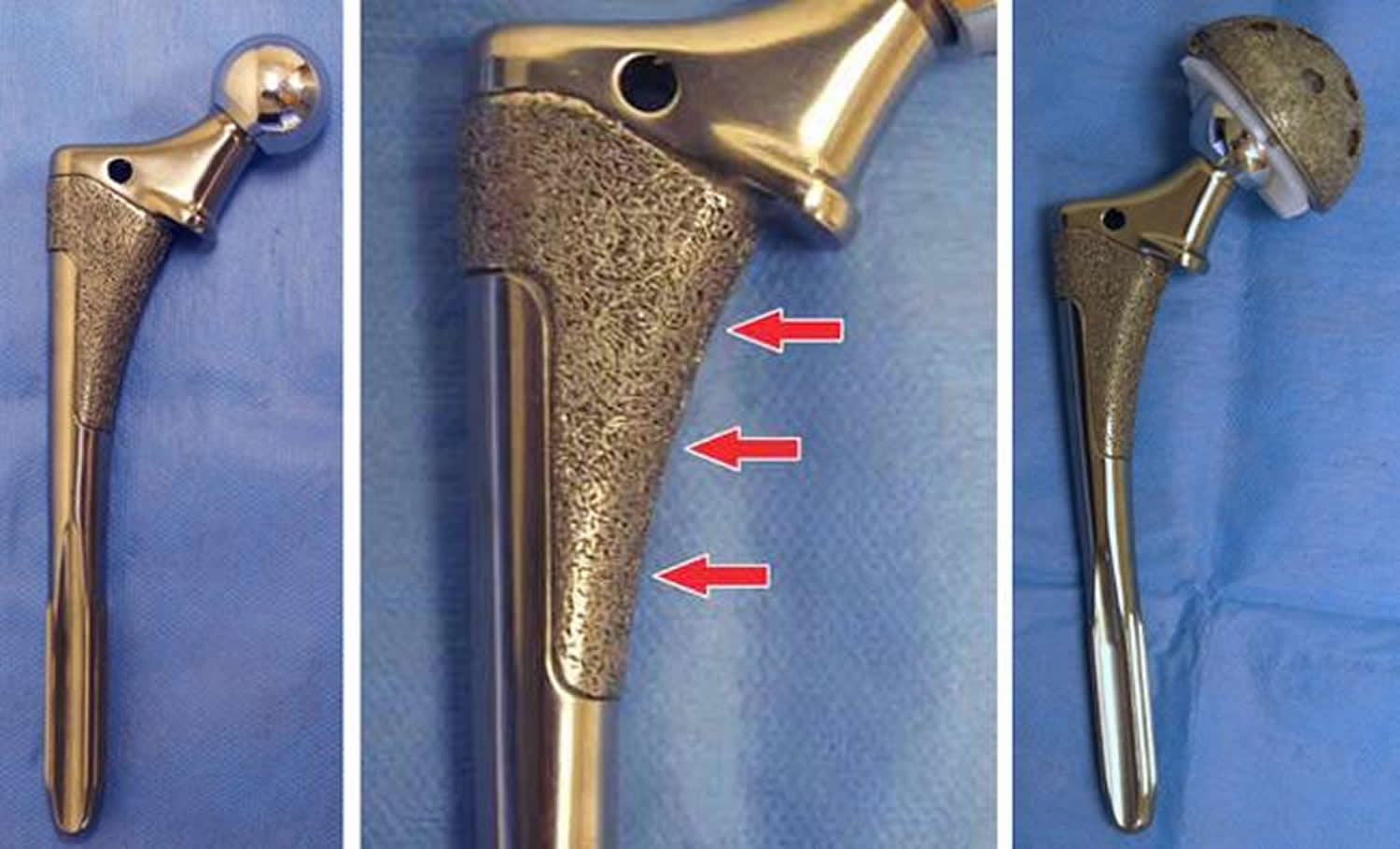
Figure 8. Femoral component of hip replacement – Note: (Left) A standard non-cemented femoral component. (Center) A close-up of this component showing the porous surface for bone ingrowth. (Right) The femoral component and the acetabular component working together.
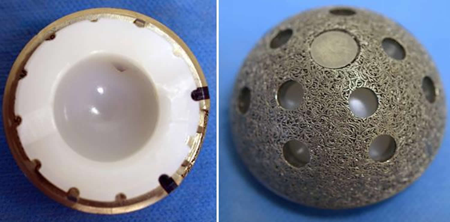
Figure 9. Acetabular component of hip replacement – Note: (Left) The acetabular component shows the plastic (polyethylene) liner inside the metal shell. (Right) The porous surface of this acetabular component allows for bone ingrowth. The holes around the cup are used if screws are needed to hold the cup in place.
Metal-on-Metal Implants Precautions
In metal-on-metal devices both the ball and socket components are made of metal. These metal implants have been used in total hip replacement surgeries and hip resurfacing procedures.
Metal-on-metal implants feature a joint made of two metal surfaces:
- a metal “ball” that replaces the ball found at the top of the thigh bone (femur)
- a metal “cup” that acts like the socket found in the pelvis
Because of metal’s durability, metal-on-metal devices were expected to last longer than other hip implants. In addition, the ball in a metal-on-metal device is larger, making the hip joint more stable and less likely to dislocate.
Metal-on-metal implants have also been used because they avoid the complication of debris wear from implants made of plastic/polyethylene. However, recent information about the wear of certain metal-on-metal devices has raised concerns about their use. Like polyethylene, metal surfaces give off small particles of debris. In addition, metal surfaces can corrode, giving off metal ions. Metal debris (ions and particles) can enter the space around the implant, as well as enter the bloodstream. This can cause a reaction in some patients, such as pain or swelling around the hip, osteolysis, and very rarely symptoms in other parts of the body.
Although the vast majority of patients have not had any problems with metal-on-metal implants, orthopedic surgeons and the U.S. Food and Drug Administration 10 are concerned because cases have been reported in the peer-reviewed literature and through a British database in which patients have local hip symptoms (pain and swelling). In addition, there have been a very small number of cases that have had other medical concerns seemingly unrelated to the hip.
- Patients who have metal-on-metal implants should be monitored regularly for the life of the implant, and have tests to measure levels of metal particles (ions) in their blood.
Patients with these implants who have symptoms may be investigated with MRI or ultrasound scans, and patients without symptoms should have a scan if the level of metal ions in their blood is rising.
If after a joint replacement surgery you experience pain or have other, new medical concerns or issues, please talk to your doctor or orthopedic surgeon.
You should also be aware of the warning signs that could signal a problem.
What are the warning signs?
You should contact your doctor if you have:
- pain in the groin, hip or leg
- swelling at or near the hip joint
- a limp or problems walking
- grinding or clunking from the joint
These symptoms don’t necessarily mean your device is failing, but they do need investigating.
Any changes in general health should also be reported, including:
- chest pain or shortness of breath
- numbness or weakness
- changes in vision or hearing
- fatigue
- feeling cold
- weight gain
What exactly is the problem with metal-on-metal implants?
Wear and tear
All hip implants wear down over time as the ball and cup slide against each other during movements, including walking and running.
Although many people live the rest of their lives without needing a replacement implant, some people may eventually need surgery to remove or replace its components.
Data suggests that certain types of metal-on-metal implant wear down at a faster rate than other types.
As friction acts upon their surfaces, it can cause tiny metal particles to break off and enter the space around the implant.
People are thought to react differently to the presence of these metal particles, but they can trigger inflammation and discomfort in the area around the implant in some people.
If not caught early, this can cause damage and deterioration in the bone and tissue surrounding the implant and joint over time. This in turn may cause the implant to become loose and cause painful symptoms, meaning further surgery is required.
Metal ions in the bloodstream
Some news coverage has focused on the Medicines and Healthcare products Regulatory Agency’s recommendation to check for the presence of metal ions in the bloodstream.
Ions are electrically charged molecules. Levels of ions in the bloodstream, particularly of the cobalt and chromium used in the surface of the implants, may therefore indicate how much wear there is to the artificial hip.
These ions in the blood are not blood poisoning and don’t lead to sepsis, which is an entirely different type of illness. Talk of this in some of the news reports is very misleading and completely wrong.
There has been no definitive link between ions from metal-on-metal implants and illness, although there has been a small number of cases in which high levels of metal ions in the bloodstream have been associated with symptoms or illnesses elsewhere in the body, including effects on the heart, nervous system and thyroid gland.
Is there a way to determine ahead of time if I might have a reaction to the metal in the metal-on-metal hip implant system?
Currently there is no widely accepted test to predict if you will develop a reaction to the metal from a hip system, and there is insufficient evidence to support using a skin patch test to determine your sensitivity to a metal-on-metal hip implant. If, however, you have a known sensitivity to metal, it is important to share that information with your surgeon.
Are there any ways to prevent the metal from reaching the joint and bloodstream if I get a metal-on-metal hip implant?
No. All artificial hips require one component to slide against another component and it is inevitable that material at the surfaces will wear as they interact. In metal-on-metal hip implants, some tiny metal particles and metal ions are released into the joint space and metal ions can potentially enter the bloodstream. Certain characteristics may place patients at risk for increased wear and metal ion production, and these patients will need closer follow-up after implantation. However, how a patient reacts to the metal is variable.
Which patients should not have a metal-on-metal hip implant system implanted?
Each type of hip implant system has its own set of benefits and risks. Metal-on-metal hip implant systems are not for everyone. You should discuss your situation with your orthopedic surgeon to determine whether you are a candidate or not. In general, metal-on-metal hip systems are not meant to be implanted in patients:
- Who have kidney problems
- Who have a known allergy or sensitivity to metals
- Who have a suppressed immune system
- Who are currently receiving high doses of corticosteroids such as prednisone
- Who are women of childbearing age
In addition, people with smaller body frames may be at increased risk for adverse events and device failure.
Why are women of child-bearing age not good candidates for metal-on-metal hip implants?
As discussed above, recent information shows that metal ions can leave the artificial joint and enter the bloodstream. It is not known how long they remain in blood or other organs of the body.
Some metallic ions may cross the barrier from mother to fetus through the placenta. It is not known if the amount of ions is great enough to have any effect on the growing fetus or if the presence of metal ions in the mother’s bloodstream will have any effect on future pregnancies.
For this reason, it is recommended that younger women who need hip replacement surgery consider implant options other than metal-on-metal.
With the risk of adverse reactions to metal-on-metal hip implant systems, why are these devices still being offered to patients?
It is known that every different type of hip implant system has its own set of risks as well as its own set of benefits. FDA’s assessment of medical devices such as metal-on-metal hip implants is based on a risk-benefit ratio with the data available. Metal-on-metal hip implants overall have been shown to provide significant benefits (e.g., high survivorship) in certain patient populations. Although the exact prevalence of adverse reactions to metal debris is not known, current experience leads us to consider the adverse outcomes to be relatively low or equal (with some designs) to other types of hip implants. Thus, for many patients, currently available information supports a favorable risk-benefit ratio.
The orthopedic surgeon should assess the patient’s individual needs and should avoid using metal-on-metal hip implants in patients where the risks outweigh the benefits.
What symptoms might a metal-on-metal hip implant cause?
Symptoms may include hip/groin pain, local swelling, numbness or changes in your ability to walk. There are many reasons a patient with a metal-on-metal hip implant may experience such symptoms and it is important that you contact your surgeon to help determine why you are having them.
Are there other medical effects that can occur with my metal-on-metal hip implant system?
Metal-on-metal hip implants, like other types of hip implants, are known to have adverse events, including infection and joint dislocation. There are some case reports of the metal particles causing a reaction around the joint, leading to deterioration of the tissue around the joint, loosening of the implant, and failure of the device, as well as some of the symptoms described above. In addition, some metal ions from the implant may enter into the bloodstream. There have been a few recent case reports of patients with metal-on-metal hip implants developing a reaction to these ions and experiencing medical problems that might have been related to their implants, including effects on the nervous system, heart, and thyroid gland.
What are my chances of developing a reaction to my metal-on-metal hip implant and having these types of medical problems?
Although current data suggests that these events are rare, it is currently unknown how often they occur in patients with metal-on-metal hip implants.
Part of the difficulty in answering this question is that individuals vary in how they react to metal ions in their bodies. For example, a reaction may develop in Patient A in response to a very small amount of metal, whereas Patient B may be able to tolerate a much larger amount before a reaction develops.
What should I do if I am experiencing adverse events associated with my metal-on-metal hip implant?
If you are experiencing hip/groin pain, difficulty walking or a worsening of your previous symptoms, you should make an appointment to see your orthopedic surgeon for further evaluation of your implant. Your orthopedic surgeon may wish to perform a physical exam and an evaluation based on your symptoms.
If you experience any new symptoms or medical conditions in your body other than at your hip, you should report these to your primary physician and remind them that you have a metal-on-metal hip implant system during their evaluation.
What should I do if I am not experiencing adverse events associated with my metal-on-metal hip implant?
If you are not having any symptoms and your orthopedic surgeon believes the metal-on-metal hip implant is functioning appropriately, there are no data to support the need for additional tests. You should continue to follow-up with your orthopedic surgeon for periodic examinations.
What should I discuss with my other healthcare providers including my general internist or family practice doctor?
There are rare case reports of patients with metal-on-metal hip implants who experienced medical problems in areas of the body away from their hip implant. This may have resulted from the metal ions released by the metal-on-metal hip implant.
If you see a healthcare provider for the evaluation of any new or worsening symptoms outside the hip/groin area, including symptoms related to your heart, nervous system, or thyroid gland, it is important that you tell that clinician of your metal-on-metal hip implant. This information may affect the types of tests that are ordered to further evaluate the cause of your symptoms.
When would a hip revision surgery be needed?
There are multiple reasons why a surgeon may recommend a device revision (a surgical procedure where your implant is removed and another put in its place). Many of these reasons, including infection, dislocation, and device fracture, apply to any type of hip implant. Your surgeon might also consider revision if you develop evidence of local or systemic reactions to the metal from your hip implant. In that case, the surgeon will take several factors into account in considering if and when a revision surgery is advisable.
What are the risks of revision surgery?
Any surgical procedure, including revision surgery, has risks associated with it, including reaction to the anesthesia, infection, bleeding, and blood clots. The revision surgery may be more difficult if you had a local reaction to the implant that may have affected your soft tissue and/or bone quality.
What does it mean when I see that a hip implant system has been “recalled?”
A hip system may be recalled by the manufacturer for a number of reasons. If your device is recalled, this does not necessarily mean that the device needs to be removed and replaced. In some cases, the recall just calls for different or more frequent monitoring. It is important to discuss the reason for the recall with your surgeon to determine the most appropriate course of action. If you are unsure if your hip implant was recalled, consult with your orthopedic surgeon. Additional information on the recall can be obtained from the manufacturer.
Hip replacement procedure
The traditional surgical approach to total hip replacement uses a single, long incision to view and access the hip joint. A variation of this approach is a minimally invasive procedure in which one or two shorter incisions are used. In minimally invasive surgery, a smaller surgical incision is used and fewer muscles around the hip are cut or detached. The goal of using shorter incisions is to reduce pain and speed recovery. Unlike traditional total hip replacement, the minimally invasive technique is not suitable for all patients. Despite this difference, however, both traditional hip replacement surgery and minimally invasive surgery are technically demanding and have better outcomes if the surgeon and operating team have considerable experience.
Your orthopedic surgeon will discuss different surgical options with you.
The surgical procedure takes a few hours. During any hip replacement surgery, the damaged bone is cut and removed, along with some soft tissues.
Your orthopedic surgeon will remove the damaged cartilage and bone and then position new metal, plastic, or ceramic implants to restore the alignment and function of your hip.
After surgery, you will be moved to the recovery room where you will remain for several hours while your recovery from anesthesia is monitored. After you wake up, you will be taken to your hospital room.
How the operation is carried out
Once you’ve been anaesthetised, the surgeon removes the existing hip joint completely. The upper part of the thigh bone (femur) is removed and the natural socket for the head of the femur is hollowed out.
A socket is fitted into the hollow in the pelvis. A short, angled metal shaft (the stem) with a smooth ball on its upper end (to fit into the socket) is placed into the hollow of the thigh bone. The cup and the stem may be pressed into place or fixed with acrylic cement.
Metal-on-metal hip resurfacing is carried out in a similar way. The main difference is that less of the bone is removed from the femur as only the joint surfaces are replaced with metal inserts.
Materials
The prosthetic parts can be cemented or uncemented:
- cemented parts are secured to healthy bone using acrylic cement
- uncemented parts are made from material that has a rough surface; this allows the bone to grow on to it, holding it in place
Most prosthetic parts are produced using high-density polythene for the socket, titanium alloys for the shaft and sometimes a separate ball made of an alloy of cobalt, chromium and molybdenum.
Some surgeons use a metal ball and socket and in some cases ceramic parts are used, which don’t wear as quickly as plastic.
Traditional Hip Replacement
To perform a traditional hip replacement:
- A 10- to 12-inch incision is made on the side of the hip. The muscles are split or detached from the hip, allowing the hip to be dislocated and fully viewed by the surgical team.
- The damaged femoral head is removed and replaced with a metal stem that is placed into the hollow center of the femur, then a metal or ceramic ball is placed on the upper part of the stem. This ball replaces the damaged femoral head that was removed.
- The damaged cartilage surface of the socket (acetabulum) is removed and replaced with a metal socket. Screws or cement are sometimes used to hold the socket in place.
- A plastic, ceramic or metal spacer is inserted between the new ball and the socket to allow for a smooth gliding surface.
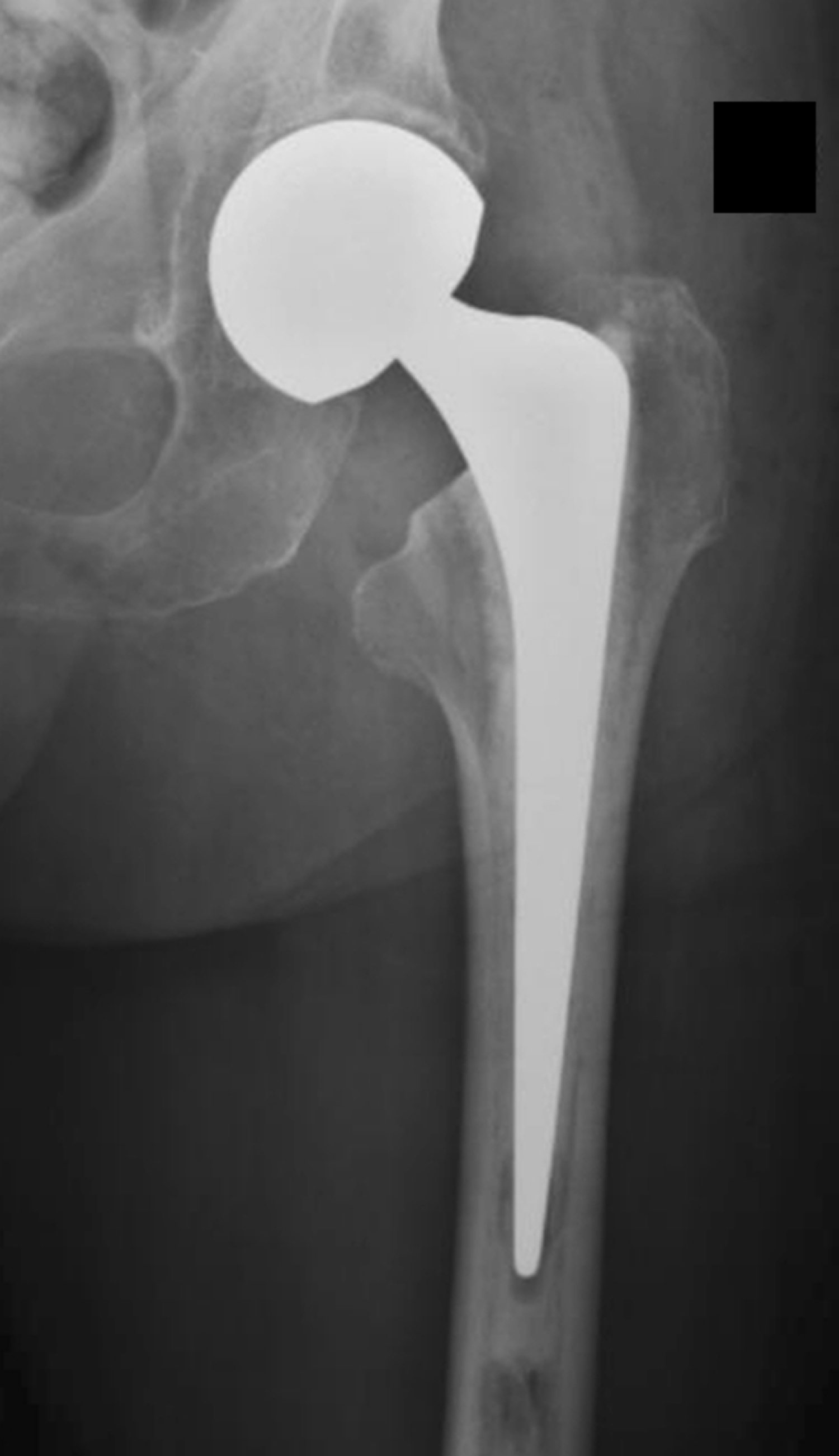
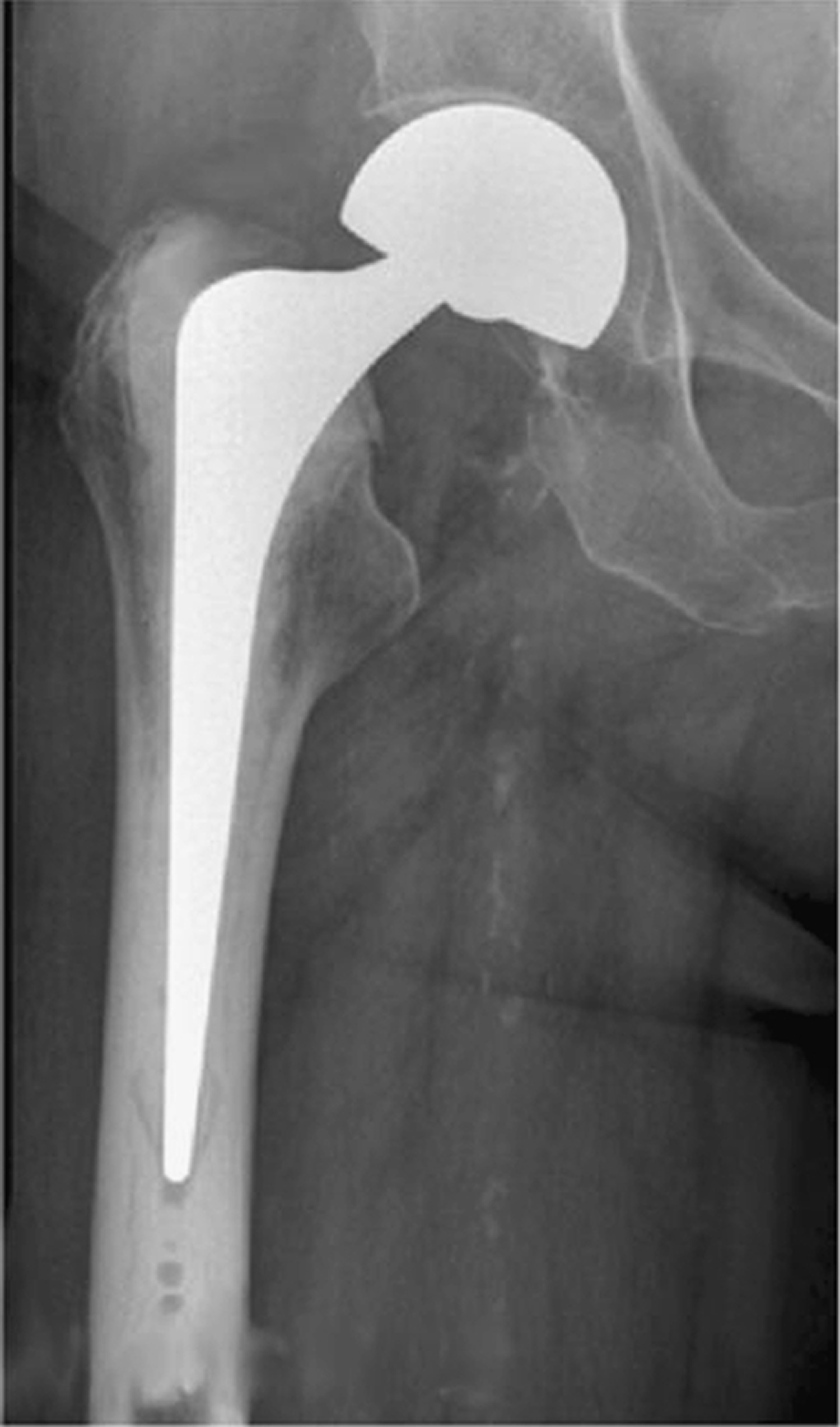
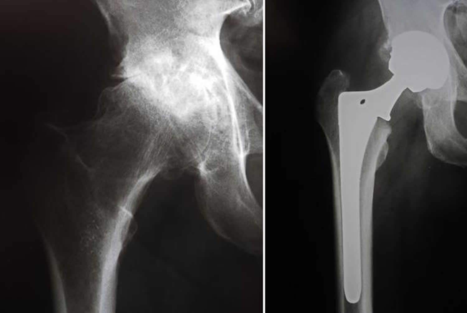
Figure 10. X-rays before and after total hip replacement – Note: In this case, non-cemented components were used.
Minimally Invasive Total Hip Replacement
In minimally invasive total hip replacement, the surgical procedure is similar, but there is less cutting of the tissue surrounding the hip. The artificial implants used are the same as those used for traditional hip replacement. However, specially designed surgical instruments are needed to prepare the socket and femur and to place the implants properly.
Minimally invasive total hip replacement can be performed with either one or two small incisions. Smaller incisions allow for less tissue disturbance.
- Single-incision surgery. In this type of minimally invasive hip replacement, the surgeon makes a single incision that usually measures from 3 to 6 inches. The length of the incision depends on the size of the patient and the difficulty of the procedure.
- The incision is usually placed over the outside of the hip. The muscles and tendons are split or detached from the hip, but to a lesser extent than in traditional hip replacement surgery. They are routinely repaired after the surgeon places the implants. This encourages healing and helps prevent dislocation of the hip.
- Two-incision surgery. In this type of minimally invasive hip replacement, the surgeon makes two small incisions:
- A 2- to 3-inch incision over the groin for placement of the socket, and
- A 1- to 2-inch incision over the buttock for placement of the femoral stem.
- To perform the two-incision procedure, the surgeon may need guidance from x-rays. It may take longer to perform the two-incision surgery than it does to perform traditional hip replacement surgery.
The hospital stay after minimally invasive surgery is similar in length to the stay after traditional hip replacement surgery–ranging from 1 to 4 days. Physical rehabilitation is a critical component of recovery. Your surgeon or a physical therapist will provide you with specific exercises to help increase your range of motion and restore your strength.
Candidates for Minimally Invasive Total Hip Replacement
Minimally invasive total hip replacement is not suitable for all patients. Your doctor will conduct a comprehensive evaluation and consider several factors before determining if the procedure is an option for you.
In general, candidates for minimal incision procedures are thinner, younger, healthier, and more motivated to participate in the rehabilitation process, compared with patients who undergo the traditional surgery.
Minimally invasive techniques are less suitable for patients who are overweight or who have already undergone other hip surgeries. In addition, patients who have a significant deformity of the hip joint, those who are very muscular, and those with health problems that may slow wound healing may be at a higher risk for problems from minimally invasive total hip replacement.
Summary of Minimally invasive total hip replacement
Minimally invasive and small incision total hip replacement surgery is an evolving area and more research is needed on the long-term function and durability of the implants.
The benefits of minimally invasive hip replacement have been reported to include less damage to soft tissues, leading to a quicker, less painful recovery and more rapid return to normal activities. Current evidence suggests that the long-term benefits of minimally invasive surgery do not differ from those of hip replacement performed with the traditional approach.
Like all surgery, minimally invasive surgery has a risk of complications. These complications include nerve and artery injuries, wound healing problems, infection, fracture of the femur, and errors in positioning the prosthetic hip implants.
Like traditional hip replacement surgery, minimally invasive surgery should be performed by a well-trained, highly experienced orthopedic surgeon. Your orthopaedic surgeon can talk to you about his or her experience with minimally invasive hip replacement surgery, and the possible risks and benefits of the techniques for your individual treatment.
How long does hip replacement surgery take?
The duration of a hip replacement varies a lot between patients and the orthopedic surgeon skills and experience. In general, the surgery usually takes around 60-90 minutes to complete.
It’s vitally important you choose a specialist who performs hip replacement regularly and can discuss their results with you.
This is even more important if you’re having a second or subsequent hip replacement (revision surgery), which is more difficult to perform.
Revision Total Hip Replacement
Total hip replacement is one of the most successful procedures in all of medicine. In the vast majority of cases, total hip replacement enables people to live more active lives without debilitating hip pain. Over time, however, a hip replacement can fail for a variety of reasons.
When this occurs, your doctor may recommend that you have a second operation to remove some or all of the parts of the original prosthesis and replace them with new ones. This procedure is called revision total hip replacement.
Although both procedures have the same goals—to relieve pain and improve function and quality of life—revision surgery is different than primary total hip replacement. Revision hip replacement is a longer, more complex procedure. It requires extensive planning, as well as the use of specialized implants and tools, in order to achieve a good result.
There are different types of revision surgery. In some cases, only some components of the prosthesis need to be revised. In other cases, the whole prosthesis needs to be removed or replaced and the bone around the hip needs to be rebuilt with augments (metal pieces that substitute for missing bone) or bone graft.
Damage to bone and soft tissue around the hip may make it difficult for the doctor to use standard primary hip implants for revision hip replacement. In most revisions, the doctor will use specialized implants that are designed to compensate for the damaged bone and soft tissue.
When revision total hip replacement is recommended
Hip replacement Loosening
The most common problem that can arise as a result of a hip replacement is loosening of the joint, which causes pain and feeling that the joint is unstable. This happens in around 10% of cases. Loosening of the joint can occur at any time, but it normally occurs 10-15 years after the original surgery was performed.
In order for a total hip replacement to function properly, an implant must remain firmly attached to the bone. During the initial surgery, the hip replacement components were either cemented into place or were “press fit” into the bone to allow bone to grow onto them. Sometimes, however, bone may fail to grow onto press-fit components. In addition, cemented or press-fit components that were once firmly fixed to the bone can eventually loosen, resulting in a painful hip.
The cause of loosening is not always clear, but repetitive high-impact activities, excessive body weight, and wear of the plastic liner between the ball and the metal cup are all factors that may contribute.
Implant Wear and Tear
Another common complication of hip replacement surgery is wear and tear of the artificial sockets. Particles that have worn off the artificial joint surfaces can be absorbed by surrounding tissue, causing loosening of the joint.
If wear or loosening is noticed on X-ray, your surgeon may request regular X-rays. Depending on the severity of the problem, you may be advised to have further surgery.
There have been reports about metal-on-metal implants wearing sooner than expected and causing complications. See Metal-on-Metal Implants Precautions above.
In addition, patients who are younger when they undergo the initial hip replacement may “outlive” the life expectancy of their artificial hip. For these patients, there is a higher long-term risk that revision surgery will be needed due to loosening or wear.
In some cases, tiny particles that wear off the cup’s plastic liner accumulate around the hip joint and are attacked by the body’s immune system. This immune response also attacks the healthy bone around the implant, leading to a condition called osteolysis. In osteolysis, the bone around the implant deteriorates, making the implant loose or unstable.
Fortunately, plastics have improved greatly over the years, so plastic wear and osteolysis occur less frequently today than they did with earlier generations of implants.
Infection
Infection is a potential complication of any surgical procedure, including total hip replacement. Infection occurs when bacteria attach in and around the surface of the prosthesis. Infection may occur while you are in the hospital or after you go home. It may even occur years later.
If a total hip replacement becomes infected, it can be painful and the implant may begin to lose its attachment to the bone. Even if the implant remains properly fixed to the bone, there may still be pain, instability, and drainage from the infection. Because bacteria cannot be easily eliminated from a joint replacement with antibiotics alone, revision surgery is usually necessary.
Revision surgery for infection can be done in different ways. To determine which procedure is best for you, your doctor will consider a number of factors, including:
- The type of bacteria
- The duration and severity of the infection
- Your preference for a specific treatment
Debridement. In this procedure, your doctor will open up your hip, wash out the bacteria, and exchange the ball and plastic liner. The metal implants that are firmly attached to the bone are left in place. After debridement, you will receive intravenous antibiotics for several weeks to help cure the infection.
Staged surgery. In some cases, the implants must be completely removed. If the implants are removed to treat the infection, your doctor will usually perform the revision in two separate surgeries.
In the first surgery, your doctor will remove the implants and place a temporary cement spacer in your hip. This spacer is treated with antibiotics to help fight the infection and will remain in your hip for several weeks. During this time, you will also receive intravenous antibiotics.
When the infection has been cleared, your doctor will perform a second surgery to remove the antibiotic spacer and insert a new prosthesis. In general, removing the implant leads to a higher chance of curing the infection, but is associated with a longer recovery.
In some cases, your doctor may be able to remove the implants, wash out the hip, and place a new prosthesis all in the same operation. This procedure, which is called a one-stage exchange, may be appropriate in limited situations.
Recurrent Dislocation
In around 3% of cases the hip joint can come out of its socket. This is most likely to occur in the first few months after surgery when the hip is still healing.
A hip replacement has a ball-and-socket structure like that of your natural hip. For a hip replacement to work well, the ball must remain inside the socket. Trauma or certain hip positions can sometimes cause the ball to become dislodged from the socket. This is called a “hip dislocation.” If you experience recurrent hip dislocations, you may need revision surgery to better align your hip joint or to insert a special implant designed to prevent dislocations.
Fracture
A periprosthetic fracture is a broken bone that occurs around the components of an implant. These fractures are most often the result of a fall, and often require revision surgery. To determine whether a revision is needed, your doctor will consider several factors, including the amount of remaining bone, whether your implant is loose, and the location of the fracture.
In rare circumstances, an implant itself can break. This also requires revision surgery.
Reaction to Metal Ions and Allergy to Metal
Over time, the metals used in implants can break down or wear, causing tiny particles to fall off the device into the space around the implant. This is more common with “metal-on-metal” devices, in which both the ball and socket components are made of metal. In some patients, sensitivity to the metal ions in these particles can result in damage to the bone and soft tissues around the hip and lead to the need for revision surgery.
In very rare cases, a patient allergy to the metal used in implants may cause pain around the site of the implant. There is no definitive agreement among doctors regarding metal allergy in this setting, however, and more studies are needed.
Other Factors
The likelihood of needing revision hip replacement is also impacted by individual differences among patients. For example, patients who are younger and more active are more likely to experience issues related to implant wear. Patients who have certain medical conditions, such as obesity or diabetes, and those who have had certain surgical procedures in the past, are more likely to experience infection or implant failure.
Long-Term Outcomes
The vast majority of patients who undergo revision surgery experience favorable long-term outcomes. This includes relief from pain, increased stability, and better function. Complete pain relief and restoration of function are not always achievable, however, and some patients may still experience some pain or dysfunction following revision surgery.
Hip replacement surgery recovery
The success of your hip replacement surgery will depend in large measure on how well you follow your orthopedic surgeon’s instructions regarding home care during the first few weeks after surgery.
Your hospital stay will typically last from 1 to 4 days, depending on the speed of your recovery. Before you are discharged from the hospital, you will need to accomplish several goals, such as:
- Getting in and out of bed by yourself.
- Having acceptable pain control.
- Being able to eat, drink, and use the bathroom.
- Walking with an assistive device (a cane, walker, or crutches) on a level surface and being able to climb up and down two or three stairs.
- Being able to perform the prescribed home exercises.
- Understanding any hip precautions you may have been given to prevent injury and ensure proper healing.
If you are not yet able to accomplish these goals, it may be unsafe for you to go directly home after discharge. If this is the case, you may be temporarily transferred to a rehabilitation or skilled nursing center.
When you are discharged, your healthcare team will provide you with information to support your recovery at home. Although the complication rate after total hip replacement is low, when complications occur they can prolong or limit full recovery. Hospital staff will discuss possible complications, and review with you the warning signs of an infection or a blood clot.
Wound Care
You may have stitches or staples running along your wound or a suture beneath your skin. The stitches or staples will be removed approximately 2 weeks after surgery.
Avoid getting the wound wet until it has thoroughly sealed and dried. You may continue to bandage the wound to prevent irritation from clothing or support stockings.
Diet
Some loss of appetite is common for several weeks after surgery. A balanced diet, often with an iron supplement, is important to promote proper tissue healing and restore muscle strength. Be sure to drink plenty of fluids.
Activity
Exercise is a critical component of home care, particularly during the first few weeks after surgery. You should be able to resume most normal light activities of daily living within 3 to 6 weeks following surgery. Some discomfort with activity and at night is common for several weeks.
Your activity program should include:
- A graduated walking program to slowly increase your mobility, initially in your home and later outside
- Resuming other normal household activities, such as sitting, standing, and climbing stairs
- Specific exercises several times a day to restore movement and strengthen your hip. You probably will be able to perform the exercises without help, but you may have a physical therapist help you at home or in a therapy center the first few weeks after surgery.
How your new hip is different
You may feel some numbness in the skin around your incision. You also may feel some stiffness, particularly with excessive bending. These differences often diminish with time, and most patients find these are minor compared with the pain and limited function they experienced prior to surgery.
Your new hip may activate metal detectors required for security in airports and some buildings. Tell the security agent about your hip replacement if the alarm is activated. You may ask your orthopedic surgeon for a card confirming that you have an artificial hip.
Protecting your hip replacement
There are many things you can do to protect your hip replacement and extend the life of your hip implant.
- Participate in a regular light exercise program to maintain proper strength and mobility of your new hip.
- Take special precautions to avoid falls and injuries. If you break a bone in your leg, you may require more surgery.
- Make sure your dentist knows that you have a hip replacement. Talk with your orthopedic surgeon about whether you need to take antibiotics prior to dental procedures.
- See your orthopedic surgeon periodically for routine follow-up examinations and x-rays, even if your hip replacement seems to be doing fine.
Hip replacement recovery time
Recovery times can vary depending on the individual and type of surgery carried out. It’s important to follow the advice your doctor gives you on looking after your hip. In general, your doctor will encourage you to use your “new” joint shortly after your operation. Although it may be challenging at times, following your doctor’s instructions will speed your recovery.
The rehabilitation process after surgery can be a demanding time and requires commitment. You may also be enrolled on an exercise programme that’s designed to help you regain and then improve the use of your new hip joint.
Most patients will experience some temporary pain in the replaced joint because the surrounding muscles are weak from inactivity, the body is adjusting to the new joint, and the tissues are healing. This pain should resolve in a few months.
Exercise is an important part of the recovery process. Your doctor or physical therapist will provide you with specific exercises to help restore movement and strengthen the joint.
The majority of patients are able to perform daily activities more easily after joint replacement surgery. Most people can expect their joint replacement to last for many years, providing them with an improved quality of life that includes less pain, along with improved motion and strength that would not have been possible otherwise.
For the first four to six weeks after the operation you’ll need a walking aid, such as crutches, to help support you.
Most people are able to resume normal activities within two to three months but it can take up to a year before you experience the full benefits of your new hip.
If you have any questions about limitations on your activities after total joint replacement, please consult your doctor.
After surgery
After the operation, you’ll be lying flat on your back and may have a pillow between your legs to keep your hip in the correct position. You may feel hazy or groggy as you come round from the general anaesthetic. A nurse may give you oxygen (through tubes in your nose or a mask) to help you feel better.
It’s common to feel sick or vomit after you’ve been given general anesthesia. Your nurse may offer you medicine to help with sickness. You may also have a sore throat and dry mouth.
The nursing staff will monitor your condition and you’ll have a large dressing on your leg to protect the wound.
You may be allowed to have a drink about an hour after you return to the ward and, depending on your condition, you may be allowed to have something to eat.
How soon will I be up and about?
The staff will help you to get up and walk about as quickly as possible after surgery. If you’ve had minimally invasive surgery or are on an enhanced recovery programme, you may be able to walk on the same day as your operation.
Initially, you’ll feel discomfort while walking and exercising, and your legs and feet may be swollen. You may be given an injection into your abdomen to help prevent blood clots forming in your legs, and possibly a short course of antibiotics to help prevent infection.
A physiotherapist may teach you exercises to help strengthen the hip and explain what should and shouldn’t be done after the operation. They’ll teach you how to bend and sit to avoid damaging your new hip.
Going home
You’ll usually be in hospital for around three to five days, depending on the progress you make and what type of surgery you have.
If you’re generally fit and well, the surgeon may suggest an enhanced recovery programme, where you start walking on the day of the operation and are discharged within one to three days.
Recovering at home
Don’t be surprised if you feel very tired at first. You’ve had a major operation and muscles and tissues surrounding your new hip will take time to heal. Follow the advice of the surgical team and call your doctor if you have any particular worries or queries.
You may be eligible for home help and there may be aids that can help you. You may want to arrange to have someone to help you for a week or so.
The exercises your physiotherapist gives you are an important part of your recovery. It’s essential you continue with them once you’re at home. Your rehabilitation will be monitored by a physiotherapist.
How soon will the pain go away?
The pain you may have experienced before the operation should go immediately. You can expect to feel some pain as a result of the operation itself, but this won’t last for long.
How long will it be before I feel back to normal?
Generally, you should be able to stop using your crutches within four to six weeks and feel more or less normal after three months, by which time you should be able to perform all your normal activities.
It’s best to avoid extreme movements or sports where there’s a risk of falling, such as skiing or riding. Your doctor or a physiotherapist can advise you about this.
When can I drive again?
You can usually drive a car after about six weeks, subject to advice from your surgeon. It can be tricky getting in and out of your car at first. It’s best to ease yourself in backwards and swing both legs round together.
When can I go back to work?
This depends on your job, but you can usually return to work 6-12 weeks after your operation.
How will it affect my sex life?
If you were finding sex difficult before because of pain, you may find that having the operation gives your sex life a boost. Your surgeon can advise when it’s OK to have sex again.
As long as you’re careful, you should be able to have sex after six to eight weeks. Avoid vigorous sex and more extreme positions.
Will I need another new hip?
Nowadays, most hip implants last for 15 years or more. If you’re older, your new hip may last your lifetime. If you’re younger, you may need another new hip at some point.
Revision surgery is more complicated and time-consuming for the surgeon to perform than a first hip replacement and complication rates are usually higher.
It can’t be performed in every patient, but most people who can have it report success for 10 years or more.
Avoiding problems after hip replacement surgery
Recognizing the Signs of a Blood Clot
Follow your orthopedic surgeon’s instructions carefully to reduce the risk of blood clots developing during the first several weeks of your recovery. He or she may recommend that you continue taking the blood thinning medication you started in the hospital. Notify your doctor immediately if you develop any of the following warning signs.
Warning signs of blood clots. The warning signs of possible blood clot in your leg include:
- Pain in your calf and leg that is unrelated to your incision
- Tenderness or redness of your calf
- New or increasing swelling of your thigh, calf, ankle, or foot
Warning signs of pulmonary embolism. The warning signs that a blood clot has traveled to your lung include:
- Sudden shortness of breath
- Sudden onset of chest pain
- Localized chest pain with coughing
Preventing Infection
A common cause of infection following hip replacement surgery is from bacteria that enter the bloodstream during dental procedures, urinary tract infections, or skin infections.
Following surgery, patients with certain risk factors may need to take antibiotics prior to dental work, including dental cleanings, or before any surgical procedure that could allow bacteria to enter your bloodstream. Your orthopedic surgeon will discuss with you whether taking preventive antibiotics before dental procedures is needed in your situation.
Warning signs of infection. Notify your doctor immediately if you develop any of the following signs of a possible hip replacement infection:
- Persistent fever (higher than 100°F or 37.8 °C orally)
- Shaking chills
- Increasing redness, tenderness, or swelling of the hip wound
- Drainage from the hip wound
- Increasing hip pain with both activity and rest
Looking after your new hip
With care, your new hip should last well. The following advice may be given by the hospital to help you care for your new hip. However, the advice may vary based on your doctors recommendations:
- avoid bending your hip more than 90° (a right angle) during any activity
- avoid twisting your hip
- don’t swivel on the ball of your foot
- when you turn around, take small steps
- don’t apply pressure to the wound in the early stages (so try to avoid lying on your side)
- don’t cross your legs over each other
- don’t force the hip or do anything that makes your hip feel uncomfortable
- avoid low chairs and toilet seats (raised toilet seats are available)
Avoiding Falls
A fall during the first few weeks after surgery can damage your new hip and may result in a need for more surgery. Stairs are a particular hazard until your hip is strong and mobile. You should use a cane, crutches, a walker, or handrails or have someone help you until you improve your balance, flexibility, and strength.
Your orthopedic surgeon and physical therapist will help you decide which assistive aides will be required following surgery, and when those aides can safely be discontinued.
Other precautions
To assure proper recovery and prevent dislocation of the prosthesis, you may be asked to take special precautions when sitting, bending, or sleeping — usually for the first 6 weeks after surgery. These precautions will vary from patient to patient, depending on the surgical approach your surgeon used to perform your hip replacement.
Prior to discharge from the hospital, your surgeon and physical therapist will provide you with any specific precautions you should follow.
Total Hip Replacement Exercise Guide
Regular exercise to restore strength and mobility to your hip and a gradual return to everyday activities are important for your full recovery after hip replacement. Your orthopedic surgeon and physical therapist may recommend that you exercise for 20 to 30 minutes, 2 or 3 times a day during your early recovery. They may suggest some of the exercises shown below.
This guide can help you better understand your exercise and activity program, supervised by your physical therapist and orthopedic surgeon. To ensure your safe recovery, be sure to check with your therapist or surgeon before performing any of the exercises shown.
Early Hip Replacement Postoperative Exercises
The following exercises will help increase circulation to your legs and feet, which is important for preventing blood clots. They will also help strengthen your muscles and improve hip movement.
Start the exercises as soon as you are able. You can begin them in the recovery room shortly after surgery. You may feel uncomfortable at first, but these exercises will help speed your recovery and actually diminish your postoperative pain.
Ankle Pumps
Slowly push your foot up and down. Repeat this exercise several times, as often as every 5 or 10 minutes.
Begin this exercise immediately after surgery and continue it until you are fully recovered.
Figure 12. Ankle pumps exercise after a hip replacement
Ankle Rotations
Move your ankle inward toward your other foot and then outward away from your other foot.
Repeat 5 times in each direction.
Do 3 or 4 sessions a day.
Figure 13. Ankle Rotations after a hip replacement
Bed-Supported Knee Bends
Slide your foot toward your buttocks, bending your knee and keeping your heel on the bed. Do not let your knee roll inward. Hold your knee in a maximally bent position for 5 to 10 seconds and then straighten.
Repeat 10 times.
Do 3 or 4 sessions a day.
Figure 14. Bed-Supported Knee Bends exercise after a hip replacement
Buttock Contractions
Tighten your buttock muscles and hold to a count of 5.
Repeat 10 times.
Do 3 or 4 sessions a day.
Figure 15. Buttock contractions after a hip replacement
Abduction Exercise
Slide your leg out to the side as far as you can and then back.
Repeat 10 times.
Do 3 or 4 sessions a day.
Figure 16. Abduction exercise after a hip replacement
Quadriceps Set
Tighten your thigh muscle. Try to straighten your knee. Hold for 5 to 10 seconds.
Repeat this exercise 10 times during a 10-minute period, rest one minute and repeat.
Continue until your thigh feels fatigued.
Figure 17. Quadriceps exercise after a hip replacement
Straight Leg Raises
Tighten your thigh muscle with your knee fully straightened on the bed. Lift your leg several inches. Hold for 5 to 10 seconds. Slowly lower.
Repeat until your thigh feels fatigued.
Figure 18. Straight Leg Raises exercise after hip replacement
Standing Exercises
Soon after your hip replacement surgery, you will be out of bed and able to stand. You will require help at first but, as you regain your strength, you will be able to stand independently. While doing these standing exercises, make sure you are holding on to a firm surface such as a bar attached to your bed or a wall.
Standing Knee Raises
Lift your operated leg toward your chest. Do not lift your knee higher than your waist. Hold for 2 or 3 counts and put your leg down.
Repeat 10 times.
Do 3 or 4 sessions a day.
Figure 19. Standing Knee Raises exercise after hip replacement
Standing Hip Abduction
Be sure your hip, knee and foot are pointing straight forward. Keep your body straight. With your knee straight, lift your leg out to the side. Slowly lower your leg so your foot is back on the floor.
Repeat 10 times.
Do 3 or 4 sessions a day.
Figure 20. Standing Hip Abduction exercise after hip replacement
Standing Hip Extensions
Lift your operated leg backward slowly. Try to keep your back straight. Hold for 2 or 3 counts. Return your foot to the floor.
Repeat 10 times.
Do 3 or 4 sessions a day.
Figure 21. Standing Hip Extensions exercise after hip replacement
Early Activity
Soon after surgery, you will begin to walk short distances in your hospital room and perform light everyday activities. This early activity aids your recovery and helps your hip regain its strength and movement.
Walking
Early on, walking will help you regain movement in your hip.
Proper walking is the best way to help your hip recover. At first, you will walk with a walker or crutches. Your surgeon or therapist will tell you how much weight to put on your leg.
Stand comfortably and erect with your weight evenly balanced on your walker or crutches. Advance your walker or crutches a short distance; then reach forward with your operated leg with your knee straightened so the heel of your foot touches the floor first. As you move forward, your knee and ankle will bend and your entire foot will rest evenly on the floor. As you complete the step, your toe will lift off the floor and your and knee and hip will bend so that you can reach forward for your next step. Remember, touch your heel first, then flatten your foot, then lift your toes off the floor.
Walk as rhythmically and smoothly as you can. Don’t hurry. Adjust the length of your step and speed as necessary to walk with an even pattern. As your muscle strength and endurance improve, you may spend more time walking. You will gradually put more weight on your leg. You may use a cane in the hand opposite your surgery and, eventually, walk without an aid.
Walk with a cane until you have regained your balance skills. In the beginning, walk for 5 or 10 minutes, 3 or 4 times a day. As your strength and endurance improve, you can walk for 20 to 30 minutes, 2 or 3 times a day. Once you have fully recovered, regular walks of 20 to 30 minutes, 3 or 4 times a week, will help maintain your strength.
When you can walk and stand for more than 10 minutes and your leg is strong enough so that you are not carrying any weight on your walker or crutches, you can begin using a single crutch or cane. Hold the aid in the hand opposite the side of your surgery.
Stair Climbing and Descending
The ability to go up and down stairs requires both strength and flexibility. At first, you will need a handrail for support and will be able to go only one step at a time. Always lead up the stairs with your good leg and down the stairs with your operated leg. Remember, “up with the good” and “down with the bad.” You may want to have someone help you until you have regained most of your strength and mobility.
Stair climbing is an excellent strengthening and endurance activity. Do not try to climb steps higher than the standard height (7 inches) and always use a handrail for balance. As you become stronger and more mobile, you can begin to climb stairs foot over foot.
Advanced Exercises and Activities
The pain from your hip problems before your surgery and the pain and swelling after surgery have weakened your hip muscles. A full recovery will take many months. The following exercises and activities will help your hip muscles recover fully.
These exercises should be done in 10 repetitions, 4 times a day. Place one end of the tubing around the ankle of your operated leg and attach the opposite end of the tubing to a stationary object such as a locked door or heavy furniture. Hold on to a chair or bar for balance.
Elastic Tube Exercises
Resistive Hip Flexion
Stand with your feet slightly apart. Bring your operated leg forward keeping the knee straight. Allow your leg to return to its previous position.
Figure 22. Resistive Hip Flexion exercise after a hip replacement
Resistive Hip Abduction
Stand sideways from the door to which the tubing is attached and extend your operated leg out to the side. Allow your leg to return to its previous position.
Figure 23. Resistive Hip Abduction exercise after hip replacement
Resistive Hip Extensions
Face the door to which the tubing is attached and pull your leg straight back. Allow your leg to return to its previous position.
Figure 24. Resistive Hip Extensions exercise after hip replacement
Exercycling
Exercycling is an excellent activity to help you regain muscle strength and hip mobility.
At first, adjust the seat height so that the bottom of your foot just touches the pedal with your knee almost straight. Pedal backwards at first. Ride forward only after a comfortable cycling motion is possible backwards.
As you become stronger (at about 4 to 6 weeks) slowly increase the tension on the exercycle. Exercycle for 10 to 15 minutes twice a day, gradually building up to 20 to 30 minutes, 3 or 4 times a week.
Knee arthroplasty
Knee replacement surgery is also called knee arthroplasty, is a routine operation that involves replacing a damaged, worn or diseased knee with an artificial joint made of metal and plastic parts. Adults of any age can be considered for a knee replacement, although most are carried out on people between the ages of 60 and 80. More people are now receiving this operation at a younger age.
Most people leave hospital 1-4 days after a knee replacement surgery. How quickly you get back to normal depends on many factors, including your age and general health and fitness.
You will probably need a cane, crutches or walking frame in the first few weeks and an exercise and physiotherapy program to help you recover.
Knee replacement surgery was first performed in 1968. Since then, improvements in surgical materials and techniques have greatly increased its effectiveness. Total knee replacements are one of the most successful procedures in all of medicine. According to the Agency for Healthcare Research and Quality, more than 600,000 knee replacements are performed each year in the United States.
There are four basic steps to a knee replacement procedure:
- Prepare the bone. The damaged cartilage surfaces at the ends of the femur and tibia are removed along with a small amount of underlying bone.
- Position the metal implants. The removed cartilage and bone is replaced with metal components that recreate the surface of the joint. These metal parts may be cemented or “press-fit” into the bone.
- Resurface the patella. The undersurface of the patella (kneecap) is cut and resurfaced with a plastic button. Some surgeons do not resurface the patella, depending upon the case.
- Insert a spacer. A medical-grade plastic spacer is inserted between the metal components to create a smooth gliding surface.
The decision to have total knee replacement surgery should be a cooperative one between you, your family, your family physician, and your orthopedic surgeon. Your physician may refer you to an orthopedic surgeon for a thorough evaluation to determine if you might benefit from this surgery.
When a knee replacement is needed
Knee replacement surgery is usually necessary when the knee joint is worn or damaged to the extent that your mobility is reduced and you experience pain even while resting.
If your knee is severely damaged by arthritis or injury, it may be hard for you to perform simple activities, such as walking or climbing stairs. You may even begin to feel pain while you are sitting or lying down.
If nonsurgical treatments like medications and using walking supports are no longer helpful, you may want to consider total knee replacement surgery. Knee joint replacement surgery is a safe and effective procedure to relieve pain, correct leg deformity, and help you resume normal activities.
The most common cause of chronic knee pain and disability is arthritis. Although there are many types of arthritis, most knee pain is caused by just three types: osteoarthritis, rheumatoid arthritis, and post-traumatic arthritis.
- Osteoarthritis. This is an age-related “wear and tear” type of arthritis. It usually occurs in people 50 years of age and older, but may occur in younger people, too. The cartilage that cushions the bones of the knee softens and wears away. The bones then rub against one another, causing knee pain and stiffness.
- Rheumatoid arthritis. This is a disease in which the synovial membrane that surrounds the joint becomes inflamed and thickened. This chronic inflammation can damage the cartilage and eventually cause cartilage loss, pain, and stiffness. Rheumatoid arthritis is the most common form of a group of disorders termed “inflammatory arthritis.”
- Post-traumatic arthritis. This can follow a serious knee injury. Fractures of the bones surrounding the knee or tears of the knee ligaments may damage the articular cartilage over time, causing knee pain and limiting knee function.
The most common reason for knee replacement surgery is osteoarthritis. Other conditions that cause knee damage include:
- Rheumatoid arthritis
- Hemophilia
- Gout
- Disorders that cause unusual bone growth (bone dysplasias)
- Death of bone in the knee joint following blood supply problems (avascular necrosis)
- Knee injury
- Knee deformity with pain and loss of cartilage
Who is offered knee replacement surgery
A knee replacement is major surgery, so is normally only recommended if other treatments, such as physiotherapy or steroid injections, haven’t helped reduce pain or improve mobility.
You may be offered knee replacement surgery if:
- You have severe knee pain, swelling and stiffness in your knee joint that limits your everyday activities, including walking, climbing stairs, and getting in and out of chairs. You may find it hard to walk more than a few blocks without significant pain and you may need to use a cane or walker
- Your knee pain is so severe that it interferes with your quality of life and sleep
- Moderate or severe knee pain while resting, either day or night
- Everyday tasks, such as shopping or getting out of the bath, are difficult or impossible
- You’re feeling depressed because of the pain and lack of mobility
- You can’t work or have a normal social life
- Chronic knee inflammation and swelling that does not improve with rest or medications
- Knee deformity — a bowing in or out of your knee
- Failure to substantially improve with other treatments such as anti-inflammatory medications, cortisone injections, lubricating injections, physical therapy, or other surgeries
You’ll also need to be well enough to cope with both a major operation and the rehabilitation afterwards.
Candidates for knee replacement surgery
There are no absolute age or weight restrictions for total knee replacement surgery.
Recommendations for knee replacement surgery are based on a patient’s pain and disability, not age. Most patients who undergo total knee replacement are age 50 to 80, but orthopedic surgeons evaluate patients individually. Total knee replacements have been performed successfully at all ages, from the young teenager with juvenile arthritis to the elderly patient with degenerative arthritis.
How your new knee is different
Improvement of knee motion is a goal of total knee replacement, but restoration of full motion is uncommon. The motion of your knee replacement after surgery can be predicted by the range of motion you have in your knee before surgery. Most patients can expect to be able to almost fully straighten the replaced knee and to bend the knee sufficiently to climb stairs and get in and out of a car. Kneeling is sometimes uncomfortable, but it is not harmful.
Most people feel some numbness in the skin around your incision. You also may feel some stiffness, particularly with excessive bending activities.
Most people also feel or hear some clicking of the metal and plastic with knee bending or walking. This is a normal. These differences often diminish with time and most patients find them to be tolerable when compared with the pain and limited function they experienced prior to surgery.
Your new knee may activate metal detectors required for security in airports and some buildings. Tell the security agent about your knee replacement if the alarm is activated.
Knee replacement surgery risks
Knee replacement surgery is a common operation and most people don’t experience complications. However, as with any operation, knee replacement surgery has risks as well as benefits.
Possible complications of knee replacement surgery are rare but can include:
Complications occur in about one in 20 cases, but most are minor and can be successfully treated. Possible complications include:
- infection of the wound – this is usually treated with antibiotics, but occasionally the wound can become deeply infected and require further surgery; in rare cases it may require replacement of the artificial knee joint.
- stiffness of the knee
- deep infection of the joint replacement, needing further surgery
- unexpected bleeding into the knee joint
- ligament, artery or nerve damage in the area around the knee joint
- blood clots or deep vein thrombosis (DVT) – clots may form in the leg veins as a result of reduced movement in the leg during the first few weeks after surgery. They can be prevented by using special support stockings, starting to walk or exercise soon after surgery, and by using anticoagulant medicines
- fracture in the bone around the artificial joint during or after surgery – treatment will depend on the location and extent of the fracture
- excess bone forming around the artificial knee joint and restricting movement of the knee – further surgery may be able to remove this and restore movement
- excess scar tissue forming and restricting movement of the knee – further surgery may be able to remove this and restore movement
- the kneecap becoming dislocated – surgery can usually repair this
- numbness in the area around the wound scar
- allergic reaction – you may have an allergic reaction to the bone cement if this is used in your procedure
- persistent pain the in the knee
- fracture – a break in the bone around the knee replacement during or after the operation
In some cases, the new knee joint may not be completely stable and further surgery may be needed to correct it.
The complication rate following total knee replacement is low. Serious complications, such as a knee joint infection, occur in fewer than 2% of patients. Major medical complications such as heart attack or stroke occur even less frequently. Chronic illnesses may increase the potential for complications. Although uncommon, when these complications occur, they can prolong or limit full recovery.
Discuss your concerns thoroughly with your orthopedic surgeon prior to surgery.
Infection. Infection may occur in the wound or deep around the prosthesis. It may happen while in the hospital or after you go home. It may even occur years later. Minor infections in the wound area are generally treated with antibiotics. Major or deep infections may require more surgery and removal of the prosthesis. Any infection in your body can spread to your joint replacement.
Blood clots. Blood clots in the leg veins are one of the most common complications of knee replacement surgery. These clots can be life-threatening if they break free and travel to your lungs. Your orthopedic surgeon will outline a prevention program, which may include periodic elevation of your legs, lower leg exercises to increase circulation, support stockings, and medication to thin your blood.
Implant problems. Although implant designs and materials, as well as surgical techniques, continue to advance, implant surfaces may wear down and the components may loosen. Additionally, although an average of 115° of motion is generally anticipated after surgery, scarring of the knee can occasionally occur, and motion may be more limited, particularly in patients with limited motion before surgery.
Continued pain. A small number of patients continue to have pain after a knee replacement. This complication is rare, however, and the vast majority of patients experience excellent pain relief following knee replacement.
Neurovascular injury. While rare, injury to the nerves or blood vessels around the knee can occur during surgery.
In some cases, the new knee joint may not be completely stable and further surgery may be needed to correct it.
Anesthesia
Anesthetics are extremely safe, but carry a risk of minor side effects such as sickness and confusion (usually temporary). There is also a slight risk of serious complications.
The risk of death in a healthy person having routine surgery is very small. Death occurs in around one in every 100,000 general anesthetics given.
The risk is higher if you’re older or have other health conditions, such as heart or lung disease.
Your anesthetist and surgeon can answer questions you may have about your personal risks from anesthetic or the surgery itself.
How long will a replacement knee last?
Wear and tear through everyday use means your replacement knee won’t always last forever. Some people will need further surgery.
According to the National Joint Registry, around 5% of patients who have a knee replacement (1 in 20) will need further surgery after 12 years. However, this varies depending on the type of replacement. Total knee replacements tend to last longer than partial knee replacements.
Extending the life of your knee implant
Currently, more than 90% of modern total knee replacements are still functioning well 15 years after the surgery. Following your orthopaedic surgeon’s instructions after surgery and taking care to protect your knee replacement and your general health are important ways you can contribute to the final success of your surgery.
Protecting your knee replacement
After knee replacement surgery, make sure you also do the following:
- Participate in regular light exercise programs to maintain proper strength and mobility of your new knee.
- Take special precautions to avoid falls and injuries. If you break a bone in your leg, you may require more surgery.
- Make sure your dentist knows that you have a knee replacement. Talk with your orthopedic surgeon about whether you need to take antibiotics prior to dental procedures.
- See your orthopedic surgeon periodically for a routine follow-up examination and x-rays, usually once a year.
Deciding to Have Knee Replacement Surgery
An important factor in deciding whether to have total knee replacement surgery is understanding what the procedure can and cannot do.
More than 90% of people who have total knee replacement surgery experience a dramatic reduction of knee pain and a significant improvement in the ability to perform common activities of daily living. But total knee replacement will not allow you to do more than you could before you developed arthritis.
With normal use and activity, every knee replacement implant begins to wear in its plastic spacer. Excessive activity or weight may speed up this normal wear and may cause the knee replacement to loosen and become painful. Therefore, most surgeons advise against high-impact activities such as running, jogging, jumping, or other high-impact sports for the rest of your life after surgery.
Realistic activities following total knee replacement include unlimited walking, swimming, golf, driving, light hiking, biking, ballroom dancing, and other low-impact sports.
With appropriate activity modification, knee replacements can last for many years.
Knee Joint Anatomy
The knee joint (tibiofemoral joint) is the largest and most complex joint of the body (Figures 1) and having healthy knees is required to perform most everyday activities. The knee joint (tibiofemoral joint) is a modified hinge joint (because its primary movement is a uniaxial hinge movement) that consists of three joints within a single synovial cavity:
- Laterally is a tibiofemoral joint, between the lateral condyle of the femur, lateral meniscus, and lateral condyle of the tibia, which is the weight-bearing bone of the leg.
- Medially is another tibiofemoral joint, between the medial condyle of the femur, medial meniscus, and medial condyle of the tibia.
- An intermediate patellofemoral joint is between the patella and the patellar surface of the femur.
The knee is made up of the lower end of the thighbone (femur), the upper end of the shinbone (tibia), and the kneecap (patella). The ends of these three bones where they touch are covered with articular cartilage, a smooth substance that protects the bones and enables them to move easily.
The menisci are located between the femur and tibia. These C-shaped wedges act as “shock absorbers” that cushion the joint.
Large ligaments hold the femur and tibia together and provide stability. The long thigh muscles give the knee strength.
All remaining surfaces of the knee are covered by a thin lining called the synovial membrane. This membrane releases a fluid that lubricates the cartilage, reducing friction to nearly zero in a healthy knee.
Normally, all of these components work in harmony. But disease or injury can disrupt this harmony, resulting in pain, muscle weakness, and reduced function.
Figure 25. Knee joint
Figure 26. Knee joint anatomy sagittal view
Figure 27. Different types of knee implants that are used in knee replacement to meet each patient’s individual needs.
Figure 28. Knee replacement
Figure 29. Knee replacement surgery before and after (X-rays) – (Left) An x-ray of a severely arthritic knee. (Right) The x-ray appearance of a total knee replacement. Note that the plastic spacer inserted between the components does not show up in an x-ray.
Types of knee replacement surgery
There are two main types of surgery, depending on the condition of the knee:
- Total knee replacement (TKR) – both sides of your knee joint are replaced
- Partial (half) knee replacement (PKR) – only one side of your joint is replaced in a smaller operation with a shorter hospital stay and recovery period
Knee replacement surgery alternatives
There are alternative surgeries to knee replacement, but results are often not as good in the long term. Your doctor will discuss the best treatment option with you. Alternatives may include:
- Arthroscopic washout and debridement – an arthroscope (tiny telescope) is inserted into the knee, which is then washed out with saline to clear out any bits of bone or cartilage
- Osteotomy – during an open operation, the surgeon cuts the shin bone and realigns it so that weight is no longer focused on the damaged part of the knee
- Mosaicplasty – a keyhole operation that involves transferring plugs of hard cartilage, together with some underlying bone from another part of your knee, to repair the damaged surface
Preparing for knee replacement surgery
Before you go into hospital, find out as much as you can about what’s involved in your operation. Your surgeon and hospital should provide written information or videos.
Stay as active as you can. Strengthening the muscles around your knee will aid your recovery. If you can, continue to take gentle exercise, such as walking and swimming, in the weeks and months before your operation. You can be referred to a physiotherapist, who will give you helpful exercises.
Orthopedic Evaluation
An evaluation with an orthopedic surgeon consists of several components:
- A medical history. Your orthopedic surgeon will gather information about your general health and ask you about the extent of your knee pain and your ability to function.
- A physical examination. This will assess knee motion, stability, strength, and overall leg alignment.
- X-rays. These images help to determine the extent of damage and deformity in your knee.
- Other tests. Occasionally blood tests, or advanced imaging such as a magnetic resonance imaging (MRI) scan, may be needed to determine the condition of the bone and soft tissues of your knee.
Medical Evaluation
If you decide to have total knee replacement surgery, your orthopaedic surgeon may ask you to schedule a complete physical examination with your family physician several weeks before the operation. This is needed to make sure you are healthy enough to have the surgery and complete the recovery process. Many patients with chronic medical conditions, like heart disease, may also be evaluated by a specialist, such as a cardiologist, before the surgery.
Tests
Several tests, such as blood and urine samples, and an electrocardiogram, may be needed to help your orthopaedic surgeon plan your surgery.
Medications
Tell your orthopaedic surgeon about the medications you are taking. He or she will tell you which medications you should stop taking and which you should continue to take before surgery.
Dental Evaluation
Although the incidence of infection after knee replacement is very low, an infection can occur if bacteria enter your bloodstream. To reduce the risk of infection, major dental procedures (such as tooth extractions and periodontal work) should be completed before your total knee replacement surgery.
Urinary Evaluations
People with a history of recent or frequent urinary infections should have a urological evaluation before surgery. Older men with prostate disease should consider completing required treatment before undertaking knee replacement surgery.
Your orthopedic surgeon will review the results of your evaluation with you and discuss whether total knee replacement is the best method to relieve your pain and improve your function. Other treatment options — including medications, injections, physical therapy, or other types of surgery — will also be considered and discussed.
In addition, your orthopedic surgeon will explain the potential risks and complications of total knee replacement, including those related to the surgery itself and those that can occur over time after your surgery.
Social Planning
Although you will be able to walk on crutches or a walker soon after surgery, you will need help for several weeks with such tasks as cooking, shopping, bathing, and doing laundry.
If you live alone, your orthopaedic surgeon’s office, a social worker, or a discharge planner at the hospital can help you make advance arrangements to have someone assist you at home. They also can help you arrange for a short stay in an extended care facility during your recovery, if this option works best for you.
Home Planning
Several modifications can make your home easier to navigate during your recovery. The following items may help with daily activities:
- Safety bars or a secure handrail in your shower or bath
- Secure handrails along your stairways
- A stable chair for your early recovery with a firm seat cushion (and a height of 18 to 20 inches), a firm back, two arms, and a footstool for intermittent leg elevation
- A toilet seat riser with arms, if you have a low toilet
- A stable shower bench or chair for bathing
- Removing all loose carpets and cords
- A temporary living space on the same floor because walking up or down stairs will be more difficult during your early recovery
How knee replacement surgery is performed
You’ll usually be admitted to hospital on the day of your knee replacement surgery. The surgeon and anesthetist will usually come and see you to discuss what will happen and answer any questions you have.
Most people would have seen their surgeon at a pre-assessment clinic and had the chance to discuss any relevant details of the knee replacement surgery.
Anesthesia
The most common types of anesthesia are general anesthesia (you are put to sleep) or spinal, epidural, or regional nerve block anesthesia (you are awake but your body is numb from the waist down). The anesthesia team, with your input, will determine which type of anesthesia will be best for you. There’s no need to be anxious about having a general anesthetic: the anesthetist will be by your side the whole time you’re asleep, carefully monitoring you, and will be there when you wake up.
Knee replacement surgery is usually performed either under general anesthetic (you’re asleep throughout the procedure) or under spinal or epidural anesthetic (you’re awake but have no feeling from the waist down).
Knee replacement procedure
The knee replacement procedure itself takes approximately 1 to 3 hours. Your orthopedic surgeon will remove the damaged cartilage and bone in your knee joint, and then position the new metal and plastic implants (a prosthesis), which have been measured to fit, to restore the alignment and function of your knee.
You may have either a total or a half-knee replacement. This will depend on how damaged your knee is. Total knee replacements are the most common.
Total knee replacement
In a total knee replacement (TKR), both sides of your knee joint are replaced. The total knee replacement procedure takes one to three hours:
- Your surgeon makes a cut down the front of your knee to expose your kneecap. This is then moved to the side so the surgeon can get to the knee joint behind it.
- The damaged ends of your thigh bone (femur) and shin bone (tibia) are carefully cut away. The ends are precisely measured and shaped to fit the appropriately sized prosthetic replacement. A dummy joint is positioned to test that the joint is working properly. Adjustments are made, the bone ends are cleaned, and the final prosthesis is fitted.
- The end of your femur is replaced by a curved piece of metal, and the end of your tibia is replaced by a flat metal plate. These are fixed using special bone ‘cement’, or are treated to encourage your bone to fuse with the replacement parts. A plastic spacer is placed between the pieces of metal. This acts like cartilage, reducing friction as your joint moves.
- The back of the knee cap may also be replaced, depending on the reasons for replacement.
- The wound is closed with either stitches or clips and a dressing is applied to the wound. In rare cases a splint is used to keep your leg immobile, but you’re usually encouraged to move your knee as early as possible.
Total knee replacement is a common procedure but you’re still likely to have some difficulty moving, especially bending your knee, and kneeling may be difficult because of the scar.
Partial (half) knee replacement
If only one side of your knee is damaged, you may be able to have a partial (half) knee replacement (PKR). Partial (half) knee replacement is a smaller operation, which uses a smaller incision, and involves less bone being removed. It’s suitable for around one in four people with osteoarthritis.
There are advantages to partial (half) knee replacement including a shorter hospital stay and recovery period. Blood transfusions are also rarely needed. Partial (half) knee replacement often results in more natural movement in the knee and you may be able to be more active than after a total knee replacement.
Talk to your surgeon about the type of surgery they intend to use and why they think it’s the best choice for you.
Other procedures
In some cases, there may be other types of procedure used, including:
Kneecap replacement
If only your kneecap is damaged, an operation called a patellofemoral replacement or patellofemoral joint arthroplasty can be performed. This involves a smaller surgery with a faster recovery time. However, the long-term results are still unclear and it’s not suitable for most people with osteoarthritis.
Mini-incision total knee replacement surgery
This new technique has been recommended by the National Institute for Health and Care Excellence. It can be used for either total or half knee replacements, but is currently more commonly used for partial knee replacements.
The surgeon makes a smaller cut (an incision 10 to 12 cm long over the knee) than in standard total knee replacement surgery which requires an incision 20 to 30 cm long. Specialized instruments are then used to maneuver around the tissue, rather than cutting through it. The same prostheses (knee replacement implants) are inserted using specially designed instruments. This should lead to a quicker recovery.
Image-guided knee replacement surgery
The surgeon performs this operation using computerized images, which are generated by attaching infrared beacons to parts of your leg and to the operating tools. These are tracked on infrared cameras in the operating theater. Results so far suggest that this may enable the new knee joint to be positioned more accurately.
Most hospitals don’t yet have the equipment to do this and only around 1% of knee replacements are performed in this way.
Patient-specific knee replacement
This is a more recent advance in knee replacement surgery. A guide is created using magnetic resonance imaging (MRI) scans. This helps to create the best fitting implant for each patient.
The potential advantage of this procedure is that the implant may last longer, as the most accurate fitting is used. However, as this is a new technique, the results and long-term effects aren’t fully known yet.
After knee replacement surgery
After surgery, you’ll be moved back to the ward (after local anesthetic) or a recovery room (after general anesthetic or epidural), where you’ll be told how the operation went.
You may feel hazy or groggy as you come round from the general anesthetic. A nurse may give you oxygen (through tubes in your nose or a mask) to help you feel better.
In the surgical ward, you may be given a switch that enables you to self-administer painkillers at a safe rate. You may also be given oxygen through a mask or tubes. If necessary, you’ll be given a blood transfusion.
It’s common to feel sick or vomit after you’ve been given general anesthesia. Your nurse may offer you medicine to help with sickness. You may also have a sore throat and dry mouth.
Your blood pressure will be taken regularly. This will either be done by a nurse or by using an automatic cuff that squeezes tightly at regular times. Your temperature will also be taken.
You’ll have a large dressing on your knee to protect your wound. Various drains will syphon off blood from the operation site to prevent it collecting inside the wound.
Your wound dressing will be changed regularly until it’s healed over.
Research shows the earlier you get out of bed and start walking, eating and drinking after your operation, the better.
Your hospital may offer an enhanced recovery programme if you have had major surgery. This rehabilitation programme aims to get you back to full health quickly.
Pain Management
After surgery, you will always feel some pain. This is a natural part of the healing process. Your doctor and nurses will work to reduce your pain, which can help you recover from surgery faster. Tell your nurse as soon as you start to feel any pain so they can give you painkilling medication as soon as possible.
Medications are often prescribed for short-term pain relief after surgery. Many types of medicines are available to help manage pain, including opioids, non-steroidal anti-inflammatory drugs (NSAIDs), and local anesthetics. Your doctor may use a combination of these medications to improve pain relief, as well as minimize the need for opioids.
Be aware that although opioids help relieve pain after surgery, they are a narcotic and can be addictive. Opioid dependency and overdose has become a critical public health issue in the U.S. It is important to use opioids only as directed by your doctor. As soon as your pain begins to improve, stop taking opioids. Talk to your doctor if your pain has not begun to improve within a few days of your surgery.
Blood Clot Prevention
Your orthopedic surgeon may prescribe one or more measures to prevent blood clots and decrease leg swelling. You may be given special support stockings to wear after surgery to help your blood circulation. Your nurse or doctor will explain how you should use these.
Some people are given an injection to thin the blood slightly to help reduce the risk of clots.
Foot and ankle movement also is encouraged immediately following surgery to increase blood flow in your leg muscles to help prevent leg swelling and blood clots. These may be as simple as flexing your knees or ankles and rotating your feet.
The sooner you start to move around, the better. Lying in bed for too long can cause some of your blood to pool in your legs. This puts you at risk of a blood clot.
Physical Therapy
Most patients begin exercising their knee the day after surgery. In some cases, patients begin moving their knee on the actual day of surgery. A physical therapist will teach you specific exercises to strengthen your leg and restore knee movement to allow walking and other normal daily activities soon after your surgery.
To restore movement in your knee and leg, your surgeon may use a knee support that slowly moves your knee while you are in bed. The device is called a continuous passive motion (CPM) exercise machine. Some surgeons believe that a continuous passive motion machine decreases leg swelling by elevating your leg and improves your blood circulation by moving the muscles of your leg.
Figure 30. Continuous passive motion exercise machine
Preventing Pneumonia
It is common for patients to have shallow breathing in the early postoperative period. This is usually due to the effects of anesthesia, pain medications, and increased time spent in bed. This shallow breathing can lead to a partial collapse of the lungs (termed “atelectasis”) which can make patients susceptible to pneumonia. To help prevent this, it is important to take frequent deep breaths. Your nurse may provide a simple breathing apparatus called a spirometer to encourage you to take deep breaths.
How soon will you be up and about?
The nursing staff will help you to get up and walk about as quickly as possible. If you’ve had minimally invasive knee replacement surgery or are on an enhanced recovery programme, you may be able to walk on the same day as your operation. Generally, you’ll be helped to stand within 12-24 hours after your knee replacement operation.
Walking with a frame or crutches is encouraged. Most people are able to walk independently with sticks after about a week but this can vary depending on the individual.
During your stay in hospital, a physiotherapist will teach you exercises to help strengthen your knee. You can usually begin these the day after your operation. It’s important to follow the physiotherapist’s advice to avoid complications or dislocation of your new joint.
It’s normal to experience initial discomfort while walking and exercising, and your legs and feet may be swollen.
You may be put on a passive motion machine to restore movement in your knee and leg. This support will slowly move your knee while you are in bed. It helps to decrease swelling by keeping your leg raised and helps improve your circulation.
Going home
You’ll usually be in hospital for three to five days, depending on what progress you make and what type of knee replacement you have. Patients who have a half knee replacement usually have a shorter hospital stay.
If you’re generally fit and well, the surgeon may suggest an enhanced recovery programme where you start walking on the day of the operation and are discharged within one to three days.
Before you leave hospital, you may (depending on the type of operation you had) have an appointment with a physiotherapist. They’ll be able to advise you about any exercises you need to carry out.
You’ll also be given advice about how to care for your wound, any equipment you may require, such as dressings, bandages, crutches and splints, and maybe a dose of painkillers.
Each hospital will have its own policy and arrangements for discharging patients.
Your discharge will be affected by:
- how quickly your health improves while you’re in hospital
- what support you’ll need after you return home
You may want to ask some questions before you leave hospital, such as:
- Who should I call if I have any concerns once I’m home?
- What should I be trying to do on my own – for example, going to the bathroom and getting out of bed?
- Is there anything I should avoid doing?
- When can I go back to work?
- How much pain, bruising or swelling should I expect when I get home?
- When and where will any stitches be removed?
- Do I need to return to hospital or my doctor for follow-up? If so, when will this be?
The outcome of your operation
It’s important to find out how your operation went.
Here are some questions you may want to ask:
- Was the operation as successful as expected?
- What effect has the operation had on my condition?
- How will I expect to feel when I get home?
- How long will it be before I’m back to normal?
Transport home
You won’t usually be able to drive yourself home after surgery. Instead, you could ask someone to pick you up or take you home in a taxi.
It’s a good idea to have an adult available to help you for at least 24 hours after leaving the hospital.
Knee replacement recovery
Recovery times can vary depending on the individual and type of surgery carried out. It’s important to follow the advice the hospital gives you on looking after your knee.
You’ll usually be in hospital for three to five days, but recovery times can vary depending on the individual and type of surgery being carried out.
Once you’re able to be discharged, your hospital will give you advice about looking after your knee at home. You’ll need to use a frame or crutches at first and a physiotherapist will teach you exercises to help strengthen your knee.
Most people can stop using walking aids around six weeks after surgery, and start driving after about eight to 12 weeks.
Three to six weeks after surgery, you generally can resume most daily activities, such as shopping and light housekeeping. Driving is also possible at around three weeks if you can bend your knee far enough to sit in a car, if you have enough muscle control to operate the brakes and accelerator, and if you’re not still taking narcotic pain medications.
After recovery, you can engage in various low-impact activities, such as walking, swimming, golfing or biking. But you should avoid higher impact activities — such as jogging, skiing, tennis and sports that involve contact or jumping. Talk to your doctor about your limitations.
Full recovery can take up to two years as scar tissue heals and your muscles are restored by exercise. A very small amount of people will continue to experience some pain after two years.
After having a knee replacement surgery, contact your doctor if:
- you develop hot, reddened, hard or painful areas in your legs in the first few weeks after your operation – although this may just be bruising from the surgery, it could mean a blood clot has developed
- you experience chest pains or breathlessness – although it’s very rare, you could have a clot on your lung (pulmonary embolism) which needs urgent treatment
Recovery at Home
The success of your knee replacement surgery will depend largely on how well you follow your orthopedic surgeon’s instructions at home during the first few weeks after surgery.
Don’t be surprised if you feel extremely tired at first. You’ve had a major operation and muscles and tissues surrounding your new knee will take time to heal. Follow the advice of the surgical team and call your doctor if you have any particular worries or queries.
You may be eligible for home help and there may be aids that can help you. You may also want to arrange for someone to help you out for a week or so.
The exercises your physiotherapist gives you are an important part of your recovery. It’s essential you continue with them once you’re at home. Your rehabilitation will be monitored by a physiotherapist.
Wound Care
You will have stitches or staples running along your wound or a suture beneath your skin on the front of your knee. The stitches or staples will be removed several weeks after surgery. A suture beneath your skin will not require removal.
Avoid soaking the wound in water until it has thoroughly sealed and dried. You may continue to bandage the wound to prevent irritation from clothing or support stockings.
Blood Clot Prevention
Follow your orthopedic surgeon’s instructions carefully to reduce the risk of blood clots developing during the first several weeks of your recovery. He or she may recommend that you continue taking the blood thinning medication you started in the hospital. Notify your doctor immediately if you develop any of the following warning signs.
Warning signs of blood clots. The warning signs of possible blood clots in your leg include:
- Increasing pain in your calf
- Tenderness or redness above or below your knee
- New or increasing swelling in your calf, ankle, and foot
Warning signs of pulmonary embolism. The warning signs that a blood clot has traveled to your lung include:
- Sudden shortness of breath
- Sudden onset of chest pain
- Localized chest pain with coughing
Preventing Infection
A common cause of infection following total knee replacement surgery is from bacteria that enter the bloodstream during dental procedures, urinary tract infections, or skin infections. These bacteria can lodge around your knee replacement and cause an infection.
After knee replacement, patients with certain risk factors may need to take antibiotics prior to dental work, including dental cleanings, or before any surgical procedure that could allow bacteria to enter the bloodstream. Your orthopedic surgeon will discuss with you whether taking preventive antibiotics before dental procedures is needed in your situation.
Warning signs of infection. Notify your doctor immediately if you develop any of the following signs of a possible knee replacement infection:
- Persistent fever (higher than 100 °F or 37.8 °C orally)
- Shaking chills
- Increasing redness, tenderness, or swelling of the knee wound
- Drainage from the knee wound
- Increasing knee pain with both activity and rest
An infected knee replacement usually requires surgery to remove the artificial parts and antibiotics to kill the bacteria. After the infection is cleared, another surgery is performed to install a new knee.
Avoiding Falls
A fall during the first few weeks after surgery can damage your new knee and may result in a need for further surgery. Stairs are a particular hazard until your knee is strong and mobile. You should use a cane, crutches, a walker, hand rails, or have someone to help you until you have improved your balance, flexibility, and strength.
Your surgeon and physical therapist will help you decide what assistive aides will be required following surgery and when those aides can safely be discontinued.
Diet
Some loss of appetite is common for several weeks after surgery. A balanced diet, often with an iron supplement, is important to help your wound heal and to restore muscle strength.
Activity
Exercise is a critical component of home care, particularly during the first few weeks after surgery. You should be able to resume most normal activities of daily living within 3 to 6 weeks following surgery. Some pain with activity and at night is common for several weeks after surgery.
Your activity program should include:
- A graduated walking program to slowly increase your mobility, initially in your home and later outside
- Resuming other normal household activities, such as sitting, standing, and climbing stairs
- Specific exercises several times a day to restore movement and strengthen your knee. You probably will be able to perform the exercises without help, but you may have a physical therapist help you at home or in a therapy center the first few weeks after surgery.
You will most likely be able to resume driving when your knee bends enough that you can enter and sit comfortably in your car, and when your muscle control provides adequate reaction time for braking and acceleration. Most people resume driving approximately 4 to 6 weeks after surgery.
Looking after your new knee
- Continue to take any prescribed painkillers or anti-inflammatories to help manage any pain and swelling
- Use your walking aids but aim to gradually decrease the amount you rely on them as your leg feels stronger
- Keep up your exercises to help prevent stiffness and do not force your knee
- Do not sit with your legs crossed for six weeks after your operation
- Do not put a pillow underneath your knee when sleeping as this can result in a permanently bent knee
- Avoid twisting at your knee
- Wear supportive outdoor shoes
- Do not kneel on your operated knee until your surgeon says you can
- Raise your leg when sitting and use ice packs to help with any swelling
How long will it be before I feel normal?
You should be able to stop using your crutches or walking frame and resume normal leisure activities six weeks after surgery. However, it may take up to three months for pain and swelling to settle down. It can take up to a year for any leg swelling to disappear.
Your new knee will continue to recover up to two years after your operation. During this time, scar tissue will heal and muscles will be restored by exercise.
Even after you have recovered, it’s best to avoid extreme movements or sports where there’s a risk of falling, such as skiing or mountain biking. Your doctor or a physiotherapist can advise you.
When can I drive again?
You can resume driving when you can bend your knee enough to get in and out of a car and control the car properly.
This is usually around four to six weeks after your surgery, but check with your physiotherapist or doctor whether it’s safe for you to drive.
When can I go back to work?
This depends on your job, but you can usually return to work six to 12 weeks after your operation.
When can I do housework?
For the first three months, you should be able to manage light chores, such as dusting and washing up.
Avoid heavy household tasks such as vacuuming and changing the beds. Do not stand for long periods as this may cause ankle swelling and avoid stretching up or bending down for the first six weeks.
How will knee replacement affect my sex life?
You may find that having the operation gives your sex life a boost. Your surgeon can advise when you can have sex again. As long as you’re careful, it should be fine after six to eight weeks. Avoid vigorous sex and kneeling positions.
Will I have to go back to the hospital?
You’ll be given an outpatient appointment to check on your progress, usually six to 12 weeks after your knee replacement.
Will I need another new knee?
The knee can be replaced as often as necessary, although results tend to be slightly less effective each time. Recovery may take longer, but once you’ve recovered, results are usually good.
Shoulder arthroplasty
Shoulder joint replacement or shoulder arthroplasty is less common than knee or hip replacement, it is just as successful in relieving joint pain.
Shoulder joint replacement surgery was first performed in the United States in the 1950s to treat severe shoulder fractures. Over the years, shoulder arthroplasty has come to be used for many other painful conditions of the shoulder, such as different forms of arthritis.
Today, about 53,000 people in the U.S. have shoulder arthroplasty surgery each year, according to the Agency for Healthcare Research and Quality. This compares to more than 900,000 Americans a year who have knee and hip replacement surgery.
If nonsurgical treatments like medications and activity changes are no longer helpful for relieving pain, you may want to consider shoulder joint replacement surgery. Joint replacement surgery is a safe and effective procedure to relieve pain and help you resume everyday activities.
Shoulder joint anatomy
The shoulder joint is a ball-and-socket joint, much like the hip joint. Your shoulder is made up of three bones: your upper arm bone (humerus), your shoulder blade (scapula), and your collarbone (clavicle). The ball, or head, of your upper arm bone (humerus) fits into a shallow socket in your shoulder blade (scapula). This socket is called the glenoid. This joint allows people an enormous range of motion at the shoulder.
The surfaces of the bones where they touch are covered with articular cartilage, a smooth substance that protects the bones and enables them to move easily. A thin, smooth tissue called synovial membrane covers all remaining surfaces inside the shoulder joint. In a healthy shoulder, this membrane makes a small amount of fluid that lubricates the cartilage and eliminates almost any friction in your shoulder.
The muscles and tendons that surround the shoulder provide stability and support.
All of these structures allow the shoulder to rotate through a greater range of motion than any other joint in the body.
The shoulder joint allows flexion, extension, hyperextension, abduction, adduction, medial rotation, lateral rotation, and circumduction of the arm. It has more freedom of movement than any other joint of the body. This freedom results from the looseness of the articular capsule and the shallowness of the glenoid cavity in relation to the large size of the head of the humerus.
Although the ligaments of the shoulder joint strengthen it to some extent, most of the strength results from the muscles that surround the joint, especially the rotator cuff muscles. These muscles (supraspinatus, infraspinatus, teres minor, and subscapularis) anchor the humerus to the scapula. The tendons of the rotator
cuff muscles encircle the joint (except for the inferior portion) and intimately surround the articular capsule. The rotator cuff muscles work as a group to hold the head of the humerus in the glenoid cavity.
The Pectoral Girdle (shoulder girdle)
The pectoral girdle (shoulder girdle) supports the arm and links it to the axial skeleton. It consists of two bones on each side of the body: the clavicle (collarbone) and scapula (shoulder blade). The medial end of the clavicle articulates with the sternum at the sternoclavicular joint, and its lateral end articulates with the scapula at the acromioclavicular joint. The scapula also articulates with the humerus at the glenohumeral joint. These are loose attachments that result in a shoulder far more flexible, but they also make the shoulder joint easy to dislocate.
Figure 31. Shoulder girdle (pectoral girdle)
 Figure 32. Shoulder joint
 Figure 33. Shoulder joint (frontal view)
Figure 35. Shoulder joint rotator cuff muscles
Reasons for shoulder arthroplasty
Several conditions can cause shoulder pain and disability, and lead patients to consider shoulder joint replacement surgery.
Osteoarthritis (Degenerative Joint Disease)
This is an age-related “wear and tear” type of arthritis. It usually occurs in people 50 years of age and older, but may occur in younger people, too. The cartilage that cushions the bones of the shoulder softens and wears away. The bones then rub against one another. Over time, the shoulder joint slowly becomes stiff and painful.
Unfortunately, there is no way to prevent the development of osteoarthritis. It is a common reason people have shoulder replacement surgery.
Rheumatoid Arthritis
This is a disease in which the synovial membrane that surrounds the joint becomes inflamed and thickened. This chronic inflammation can damage the cartilage and eventually cause cartilage loss, pain, and stiffness. Rheumatoid arthritis is the most common form of a group of disorders termed “inflammatory arthritis.”
Post-traumatic Arthritis
This can follow a serious shoulder injury. Fractures of the bones that make up the shoulder or tears of the shoulder tendons or ligaments may damage the articular cartilage over time. This causes shoulder pain and limits shoulder function.
Rotator Cuff Tear Arthropathy
A patient with a very large, long-standing rotator cuff tear may develop cuff tear arthropathy. In this condition, the changes in the shoulder joint due to the rotator cuff tear may lead to arthritis and destruction of the joint cartilage.
Avascular Necrosis (Osteonecrosis)
Avascular necrosis is a painful condition that occurs when the blood supply to the bone is disrupted. Because bone cells die without a blood supply, osteonecrosis can ultimately cause destruction of the shoulder joint and lead to arthritis. Chronic steroid use, deep sea diving, severe fracture of the shoulder, sickle cell disease, and heavy alcohol use are risk factors for avascular necrosis.
Severe Fractures
A severe fracture of the shoulder is another common reason people have shoulder replacements. When the head of the upper arm bone is shattered, it may be very difficult for a doctor to put the pieces of bone back in place. In addition, the blood supply to the bone pieces can be interrupted. In this case, a surgeon may recommend a shoulder replacement. Older patients with osteoporosis are most at risk for severe shoulder fractures.
Failed Previous Shoulder Replacement Surgery
Although uncommon, some shoulder replacements fail, most often because of implant loosening, wear, infection, and dislocation. When this occurs, a second joint replacement surgery — called a revision surgery — may be necessary.
Is Shoulder Joint Replacement for You?
The decision to have shoulder replacement surgery should be a cooperative one between you, your family, your family physician, and your orthopaedic surgeon.
There are several reasons why your doctor may recommend shoulder replacement surgery.
People who benefit from surgery often have:
- Severe shoulder pain that interferes with everyday activities, such as reaching into a cabinet, dressing, toileting, and washing.
- Moderate to severe pain while resting. This pain may be severe enough to prevent a good night’s sleep.
- Loss of motion and/or weakness in the shoulder.
- Failure to substantially improve with other treatments such as anti-inflammatory medications, cortisone injections, or physical therapy.
Orthopaedic Evaluation
Your family physician may refer you to an orthopaedic surgeon for a thorough evaluation to determine if you can benefit from this surgery.
An evaluation with an orthopaedic surgeon consists of several components:
- A medical history. Your orthopaedic surgeon will gather information about your general health and ask you about the extent of your shoulder pain and your ability to function.
- A physical examination. This will assess shoulder motion, stability, and strength.
- X-rays. These images help to determine the extent of damage in your shoulder. They can show loss of the normal joint space between bones, flattening or irregularity in the shape of the bone, bone spurs, and loose pieces of cartilage or bone that may be floating inside the joint.
- Other tests. Occasionally blood tests, a magnetic resonance imaging (MRI) scan, or a bone scan may be needed to determine the condition of the bone and soft tissues of your shoulder.
Your orthopaedic surgeon will review the results of your evaluation with you and discuss whether shoulder joint replacement is the best method to relieve your pain and improve your function. Other treatment options — including medications, injections, physical therapy, or other types of surgery — will also be discussed and considered.
Possible Complications of Shoulder Replacement Surgery
Your orthopaedic surgeon will explain the potential risks and complications of shoulder joint replacement, including those related to the surgery itself and those that can occur over time after your surgery.
The risks of the shoulder replacement surgery include but are not limited to:
- Infection
- Instability of the joint replacement
- Fracture of either the humerus or glenoid bone
- Nerve injury
- Loosening of the joint replacement
- Anesthesia problems
- Hematoma or blood clots
When complications occur, most are successfully treatable. Possible complications include the following.
- Infection
Infection is a complication of any surgery. In shoulder joint replacement, infection may occur in the wound or deep around the prosthesis. It may happen while in the hospital or after you go home. It may even occur years later. Minor infections in the wound area are generally treated with antibiotics. Major or deep infections may require more surgery and removal of the prosthesis.
Any infection in your body can spread to your joint replacement.
- Prosthesis Problems
Although prosthesis designs and materials, as well as surgical techniques, continue to advance, the prosthesis may wear down and the components may loosen. The components of a shoulder replacement may also dislocate. Excessive wear, loosening, or dislocation may require additional surgery (revision procedure).
- Nerve Injury
Nerves in the vicinity of the joint replacement may be damaged during surgery, although this type of injury is infrequent. Over time, these nerve injuries often improve and may completely recover.
Shoulder Replacement Options
Shoulder replacement surgery is highly technical. It should be performed by a surgical team with experience in this procedure.
There are different types of shoulder replacements. Your surgeon will evaluate your situation carefully before making any decisions. He or she will discuss with you which type of replacement would best meet your health needs. Do not hesitate to ask what type of implant will be used in your situation, and why that choice is right for you.
When shoulder replacement surgery is performed for severe arthritis, the ball is removed from the top of the humerus and replaced with a half-moon shaped metal implant. The implant is attached to a stem inserted down the center of the arm bone. The socket portion of the joint is shaved clean and replaced with a plastic socket that is cemented into the scapula.
A conventional total shoulder replacement device mimics the normal anatomy of the shoulder: a plastic “cup” is fitted into the shoulder socket (glenoid), and a metal “ball” is attached to the top of the upper arm bone (humerus). In a reverse total shoulder replacement, the socket and metal ball are switched. The metal ball is fixed to the socket and the plastic cup is fixed to the upper end of the humerus.
Figure 36. Shoulder arthroplasty options – total shoulder arthroplasty and reverse total shoulder arthroplasty
Stemmed Hemiarthroplasty
Depending on the condition of your shoulder, your surgeon may replace only the ball. This procedure is called a hemiarthroplasty. In a traditional hemiarthroplasty, the head of the humerus is replaced with a metal ball and stem, similar to the component used in a total shoulder replacement. This is called a stemmed hemiarthroplasty.
Some surgeons recommend hemiarthroplasty when the humeral head is severely fractured but the socket is normal. Other indications for a hemiarthroplasty include:
- Arthritis that only involves the head of the humerus with a glenoid that has a healthy and intact cartilage surface
- Shoulders with severely weakened bone in the glenoid
- Some shoulders with severely torn rotator cuff tendons and arthritis
Sometimes, surgeons make the decision between a total shoulder replacement and a hemiarthroplasty in the operating room at the time of the surgery.
Studies show that patients with osteoarthritis get better pain relief from total shoulder arthroplasty than from hemiarthroplasty.
Figure 37. Stemmed Hemiarthroplasty
Resurfacing arthroplasty
Resurfacing hemiarthroplasty involves replacing just the joint surface of the humeral head with a cap-like prosthesis without a stem. With its bone preserving advantage, it offers those with arthritis of the shoulder an alternative to the standard stemmed shoulder replacement.
Cases in which a total replacement of the shoulder is not necessary in circumstances such as trauma, where the shoulder has dislocated and part of the shoulder is broken, or avascular necrosis, where the humeral head loses blood and the cartilage effectively dies, it may only be necessary to replace part of the humeral head.
In resurfacing hemiarthroplasty either all or part of the humeral head bone is replaced with a smooth metal ball. Unlike total shoulder replacemtent or hemiarthroplasty, however, the glenoid and the humeral canal are not violated.
The advantage of resurfacing hemiarthroplasty surgery is that the native glenoid bone is preserved as is the majority of the humeral head.
The disadvantage is that the longevity of the implant may be less than a total shoulder replacement. Often in cases where the glenoid cartilage is of good quality and the problem is limited to trauma, arthritis or avascular necrosis of the humeral head resurfacing arthroplasty may be an option.
Resurfacing arthroplasty may be an option for you if:
- The glenoid still has an intact cartilage surface
- There has been no fresh fracture of the humeral neck or head
- There is a desire to preserve humeral bone
For patients who are young or very active, resurfacing hemiarthroplasty avoids the risks of component wear and loosening that may occur with conventional total shoulder replacements in this patient population. Due to its more conservative nature, resurfacing hemiarthroplasty may be easier to convert to total shoulder replacement, if necessary at a later time.
Figure 38. Shoulder resurfacing arthroplasty – this x-ray shows the cap-like prosthesis used in resurfacing hemiarthroplasty
Total shoulder arthroplasty
The typical total shoulder arthroplasty involves replacing the arthritic joint surfaces with a highly polished metal ball attached to a stem, and a plastic socket.
These components come in various sizes. They may be either cemented or “press fit” into the bone. If the bone is of good quality, your surgeon may choose to use a non-cemented (press-fit) humeral component. If the bone is soft, the humeral component may be implanted with bone cement. In most cases, an all-plastic glenoid (socket) component is implanted with bone cement.
Implantation of a glenoid component is not advised if:
- The glenoid has good cartilage
- The glenoid bone is severely deficient
- The rotator cuff tendons are irreparably torn
Patients with bone-on-bone osteoarthritis and intact rotator cuff tendons are generally good candidates for conventional total shoulder replacement.
Every year, thousands of conventional total shoulder replacements are successfully done in the United States for patients with shoulder arthritis. This type of surgery, however, is not as beneficial for patients with large rotator cuff tears who have developed a complex type of shoulder arthritis called “cuff tear arthropathy.” For these patients, conventional total shoulder replacement may result in pain and limited motion, and reverse total shoulder replacement is a better option.
Preparing for Surgery
Medical Evaluation
If you decide to have shoulder replacement surgery, your orthopaedic surgeon may ask you to schedule a complete physical examination with your family physician several weeks before surgery. This is needed to make sure you are healthy enough to have the surgery and complete the recovery process. Many patients with chronic medical conditions, like heart disease, must also be evaluated by a specialist, such a cardiologist, before the surgery.
Medications
Be sure to talk to your orthopaedic surgeon about the medications you take. Some medications may need to be stopped before surgery. For example, the following over-the-counter medicines may cause excessive bleeding and should be stopped 2 weeks before surgery:
- Non-steroidal anti-inflammatory medications, such as aspirin, ibuprofen, and naproxen sodium
- Most arthritis medications
If you take blood thinners, either your primary care doctor or cardiologist will advise you about stopping these medications before surgery.
Home Planning
Making simple changes in your home before surgery can make your recovery period easier.
For the first several weeks after your surgery, it will be hard to reach high shelves and cupboards. Before your surgery, be sure to go through your home and place any items you may need afterwards on low shelves.
When you come home from the hospital, you will need help for a few weeks with some daily tasks like dressing, bathing, cooking, and laundry. If you will not have any support at home immediately after surgery, you may need a short stay in a rehabilitation facility until you become more independent.
Before Your Operation
Wear loose-fitting clothes and a button-front shirt when you go to the hospital for your surgery. After surgery, you will be wearing a sling and will have limited use of your arm.
You will most likely be admitted to the hospital on the day of your surgery. After admission, you will be taken to the preoperative preparation area and will meet a doctor from the anesthesia department.
You, your anesthesiologist, and your surgeon will discuss the type of anesthesia to be used. You may be provided a general anesthetic (you are asleep for the entire operation), a regional anesthetic (you may be awake but have no feeling around the surgical area), or a combination of both types.
Total shoulder arthroplasty surgical procedure
The total shoulder arthroplasty procedure to replace your shoulder joint with an artificial device usually takes about 2 hours.
After surgery, you will be moved to the recovery room, where you will remain for several hours while your recovery from anesthesia is monitored. After you wake up, you will be taken to your hospital room.
Total shoulder arthroplasty recovery
Your medical team will give you several doses of antibiotics to prevent infection. Most patients are able to eat solid food and get out of bed the day after surgery. You will most likely be able to go home on the first, second or third day after surgery.
Pain Management
After surgery, you will feel some pain. This is a natural part of the healing process. Your doctor and nurses will work to reduce your pain, which can help you recover from surgery faster.
Medications are often prescribed for short-term pain relief after surgery. Many types of medicines are available to help manage pain, including opioids, non-steroidal anti-inflammatory drugs (NSAIDs), and local anesthetics. Your doctor may use a combination of these medications to improve pain relief, as well as minimize the need for opioids.
Be aware that although opioids help relieve pain after surgery, they are a narcotic and can be addictive. Opioid dependency and overdose has become a critical public health issue in the U.S. It is important to use opioids only as directed by your doctor. As soon as your pain begins to improve, stop taking opioids. Talk to your doctor if your pain has not begun to improve within a few days of your surgery.
Pain management is an important part of your recovery. Physical therapy will begin soon after surgery, and when you feel less pain, you can start moving sooner and get your strength back more quickly. Talk with your doctor if postoperative pain becomes a problem.
Total shoulder arthroplasty rehabilitation
A careful, well-planned rehabilitation program is critical to the success of a shoulder replacement. You usually start gentle physical therapy soon after the operation. Your surgeon or physical therapist will provide you with a home exercise program to strengthen your shoulder and improve flexibility.
Your Recovery At Home
When you leave the hospital, your arm will be in a sling. You will need the sling to support and protect your shoulder for the first 2 to 4 weeks after surgery.
Wound care. You will have staples running along your wound or a suture beneath your skin. The staples will be removed several weeks after surgery. A suture beneath your skin will not require removal.
Avoid soaking the wound in water until it has thoroughly sealed and dried. You may continue to bandage the wound to prevent irritation from clothing.
Activity. Exercise is a critical component of home care, particularly during the first few weeks after surgery. Follow your surgeon’s home exercise plan to help you regain strength. Most patients are able to perform simple activities such as eating, dressing and grooming within 2 weeks after surgery. Some pain with activity and at night is common for several weeks after surgery.
Driving a car is not allowed for 2 to 4 weeks after surgery.
Do’s and Don’ts after total shoulder replacement surgery
The success of your surgery will depend largely on how well you follow your orthopaedic surgeon’s instructions at home during the first few weeks after surgery. Here are some common do’s and don’ts for when you return home:
- Don’t use the arm to push yourself up in bed or from a chair because this requires forceful contraction of muscles.
- Do follow the program of home exercises prescribed for you. You may need to do the exercises 2 to 3 times a day for a month or more.
- Don’t overdo it! If your shoulder pain was severe before the surgery, the experience of pain-free motion may lull you into thinking that you can do more than is prescribed. Early overuse of the shoulder may result in severe limitations in motion.
- Don’t lift anything heavier than a glass of water for the first 2 to 4 weeks after surgery.
- Do ask for assistance. Your physician may be able to recommend an agency or facility if you do not have home support.
- Don’t participate in contact sports or do any repetitive heavy lifting after your shoulder replacement.
- Do avoid placing your arm in any extreme position, such as straight out to the side or behind your body for the first 6 weeks after surgery.
Many thousands of patients have experienced an improved quality of life after total shoulder joint replacement surgery. They experience less pain, improved motion and strength, and better function.
Reverse shoulder arthroplasty
Another type of shoulder replacement is called reverse total shoulder replacement. Reverse total shoulder replacement is used for people who have:
- Completely torn rotator cuffs with severe arm weakness
- The effects of severe arthritis and rotator cuff tearing (cuff tear arthropathy)
- Had a previous shoulder replacement that failed
For these individuals, a conventional total shoulder replacement can still leave them with pain. They may also be unable to lift their arm up past a 90-degree angle. Not being able to lift one’s arm away from the side can be severely debilitating.
In reverse total shoulder replacement, the socket and metal ball are switched. That means a metal ball is attached to the shoulder bone and a plastic socket is attached to the upper arm bone. This allows the patient to use the deltoid muscle instead of the torn rotator cuff to lift the arm.
The reverse total shoulder replacement surgery was originally designed in the 1980s in Europe. The Food and Drug Administration (FDA) approved its use in the United States in 2003.
A reverse total shoulder replacement works better for people with shoulder rotator cuff tear arthropathy because it relies on different muscles to move the arm. In a healthy shoulder, the rotator cuff muscles help position and power the arm during range of motion. A conventional replacement device also uses the rotator cuff muscles to function properly. In a patient with a large rotator cuff tear and cuff tear arthropathy, these muscles no longer function. The reverse total shoulder replacement relies on the deltoid muscle, instead of the rotator cuff, to power and position the arm.
Candidates for reverse shoulder arthroplasty surgery
Reverse total shoulder replacement may be recommended if you have:
- A completely torn rotator cuff that cannot be repaired
- Cuff tear arthropathy
- A previous shoulder replacement that was unsuccessful
- Severe shoulder pain and difficulty lifting your arm away from your side or over your head
- A complex fracture of the shoulder joint
- A chronic shoulder dislocation
- A tumor of the shoulder joint
- Tried other treatments, such as rest, medications, cortisone injections, and physical therapy, that have not relieved shoulder pain
Reverse shoulder arthroplasty surgical complications
Reverse total shoulder replacement is a highly technical procedure. Your surgeon will evaluate your particular situation carefully and discuss the risks of surgery with you.
Risks for any surgery include bleeding, nerve damage, and infection. Complications specific to a total joint replacement include wear, loosening, or dislocation of the components. If any of these occur, the new shoulder joint may need to be revised, or re-operated on.
Preparing for reverse shoulder arthroplasty surgery
Your orthopaedic surgeon will help you plan and prepare for your reverse shoulder arthroplasty surgery.
Medical Evaluation
Most patients must have a complete physical by their primary care doctor before surgery. This is needed to make sure you are healthy enough to have the surgery and complete the recovery. Many patients with chronic medical conditions, like heart disease, must also be evaluated by a specialist, such a cardiologist, before the surgery.
Medications
Be sure to talk to your orthopaedic surgeon about the medications you take. Some medications may need to be stopped before surgery. For example, the following over-the-counter medicines may cause excessive bleeding and should be stopped 2 weeks before surgery:
- Non-steroidal anti-inflammatory medications, such as aspirin, ibuprofen, and naproxen sodium
- Some arthritis medications
If you take blood thinners, either your primary care doctor or cardiologist will advise you about stopping these medications before surgery.
Home Planning
Making simple changes in your home before surgery can make your recovery period easier.
For the first several weeks after your surgery, it will be hard to reach high shelves and cupboards. Before your surgery, be sure to go through your home and place any items you may need afterwards on low shelves.
When you come home from the hospital, you will need help for a few weeks with some daily tasks like dressing, bathing, cooking, and laundry. If you will not have any support at home immediately after surgery, you may need a short stay in a rehabilitation facility until you become more independent.
Before Your Operation
Wear loose-fitting clothes and a button-front shirt when you go to the hospital for your surgery. After surgery, you will be wearing a sling and will have limited use of your arm.
You will most likely be admitted to the hospital on the day of your surgery. After admission, you will be taken to the preoperative preparation area and will meet a doctor from the anesthesia department.
You, your anesthesiologist, and your surgeon will discuss the type of anesthesia to be used. You may be provided a general anesthetic (you are asleep for the entire operation), a regional anesthetic (you may be awake but have no feeling around the surgical area), or a combination of both types.
Reverse shoulder arthroplasty surgical procedure
The reverse shoulder arthroplasty procedure to replace your shoulder joint with an artificial device usually takes about 2 hours.
Your surgeon will make an incision either on the front or the top of your shoulder. He or she will remove the damaged bone and then position the new components to restore function to your shoulder.
Reverse shoulder arthroplasty recovery
After a reverse shoulder arthroplasty surgery, your medical team will give you several doses of antibiotics to reduce your risk for infection, and pain medication to keep you comfortable. Most patients are able to eat solid food and get out of bed the day after surgery. You will most likely be able to go home on the second or third day after surgery.
Pain Management
After surgery, you will feel some pain. This is a natural part of the healing process. Your doctor and nurses will work to reduce your pain, which can help you recover from surgery faster.
Medications are often prescribed for short-term pain relief after surgery. Many types of medicines are available to help manage pain, including opioids, nonsteroidal anti-inflammatory drugs (NSAIDs), and local anesthetics. Your doctor may use a combination of these medications to improve pain relief, as well as minimize the need for opioids.
Be aware that although opioids help relieve pain after surgery, they are a narcotic and can be addictive. Opioid dependency and overdose has become a critical public health issue. It is important to use opioids only as directed by your doctor. As soon as your pain begins to improve, stop taking opioids. Talk to your doctor if your pain has not begun to improve within a few weeks of your surgery.
Rehabilitation
When you leave the hospital, your arm will be in a sling. Your surgeon may instruct you to do gentle range of motion exercises to increase your mobility and endurance. A formal physical therapy program may also be recommended to strengthen your shoulder and improve flexibility.
You should be able to eat, dress, and groom yourself within a few weeks after surgery.
Your surgeon may ask you to return for office visits and x-rays in order to monitor your shoulder.
Do’s and Dont’s After Shoulder Replacement Surgery
- Do follow the home exercise program prescribed by your doctor.
- Do avoid extreme arm positions, such as behind your body or your arm straight out to the side for the first 6 weeks.
- Don’t overdo it.
- Don’t lift anything heavier than 5 lbs. for the first 6 weeks after surgery.
- Don’t push yourself up out of a chair or bed, as this requires forceful muscle contractions.
- Don’t participate in repetitive heavy lifting after shoulder replacement.
Long-Term Outcomes
After rehabilitation, you will most likely be able to lift your arm to just above shoulder height and bend your elbow to reach the top of your head or into a cupboard. Reverse total shoulder replacement provides outstanding pain relief and patient satisfaction is typically very high.
Early and mid-term studies of the results of this surgery have been very promising. Currently, very few long-term studies exist. This is an area for future research.
Thumb joint replacement
Arthritis of the thumb (trapeziometacarpal or first carpometacarpal) joint represents a common manifestation of osteoarthritis with some estimates reporting an incidence as high as 15% in the general population 11. Osteoarthritis of the basal joint of the thumb is a common disorder affecting 1 in 4 women and 1 in 12 men 12. Traditionally, treatment has been stage-based 13; stage I with ligament reconstruction, stage II and III with 1 of hemitrapeziectomy, joint fusion or implant arthroplasty, and stage IV with complete trapeziectomy with or without ligament reconstruction 14. Effective treatment of advanced disease (stages II–IV) is contingent on the removal or replacement of the entire joint surface. This can be achieved through several means including: (1) simple joint fusion, which will alleviate pain, but results in a significant mobility deficit 15; (2) implant arthroplasty, which has been shown to have implant failure and joint instability 15; and (3) trapeziectomy with ligament reconstruction and tendon interposition, the most common intervention to treat osteoarthritis in the thumb carpometacarpal joint 16, 17.
There are numerous surgical techniques for the treatment of thumb joint (first carpometacarpal joint) osteoarthritis, however, controversy exists as to whether outcomes differ between techniques. Recent studies 18, 19, 20 suggest equivalent outcomes with regard to long-term pain, mobility, and strength, in patients undergoing either trapeziectomy alone or trapeziectomy with ligament reconstruction and tendon interposition. However, a more recent study 21 suggests that there is an association between reported trapeziectomy having fewer complications than trapeziectomy with ligament reconstruction and tendon interposition (T + LRTI). These results are consistent with the results of others as summarized in a Cochrane review of treatment for thumb arthritis in 2009 22. Another study 23 suggests that simple trapeziectomy is as effective and may be safer than trapeziectomy and ligament reconstruction (LR) with or without tendon interposition (TI).
- Keating JF, Grant A, Masson M, Scott NW, Forbes JF. Randomized comparison of reduction and fixation, bipolar hemiarthroplasty, and total hip arthroplasty Treatment of displaced intracapsular hip fractures in healthy older patients. J Bone Joint Surg Am. 2006;88:249–260. doi: 10.2106/JBJS.E.00215[↩]
- Baker RP, Squires B, Gargan MF, Bannister GC. Total hip arthroplasty and hemiarthroplasty in mobile, independent patients with a displaced intracapsular fracture of the femoral neck. A randomized, controlled trial. J Bone Joint Surg Am. 2006;88:2583–2589. doi: 10.2106/JBJS.E.01373[↩]
- Hedbeck CJ, Blomfeldt R, Lapidus G, Törnkvist H, Ponzer S, Tidermark J. Unipolar hemiarthroplasty versus bipolar hemiarthroplasty in the most elderly patients with displaced femoral neck fractures: a randomised, controlled trial. International Orthopaedics. 2011;35(11):1703-1711. doi:10.1007/s00264-011-1213-y. https://www.ncbi.nlm.nih.gov/pmc/articles/PMC3193971/[↩]
- Stability of hip hemiarthroplasties. Varley J, Parker MJ. https://www.ncbi.nlm.nih.gov/pmc/articles/PMC3456984/pdf/264_2004_Article_572.pdfInt Orthop. 2004 Oct; 28(5):274-7.[↩]
- Hedbeck CJ, Blomfeldt R, Lapidus G, Törnkvist H, Ponzer S, Tidermark J. Unipolar hemiarthroplasty versus bipolar hemiarthroplasty in the most elderly patients with displaced femoral neck fractures: a randomised, controlled trial. International Orthopaedics. 2011;35(11):1703-1711. doi:10.1007/s00264-011-1213-y https://www.ncbi.nlm.nih.gov/pmc/articles/PMC3193971/[↩][↩]
- Cornell CN, Levine D, O’Doherty J, Lyden J (1998) Unipolar versus bipolar hemiarthroplasty for the treatment of femoral neck fractures in the elderly. Clin Orthop Relat Res 348:67–71[↩]
- Davison JN, Calder SJ, Anderson GH, Ward G, Jagger C, Harper WM, Gregg PJ. Treatment for displaced intracapsular fracture of the proximal femur. A prospective, randomised trial in patients aged 65 to 79 years. J Bone Joint Surg Br. 2001;83:206–212. doi: 10.1302/0301-620X.83B2.11128[↩]
- Raia FJ, Chapman CB, Herrera MF, Schweppe MW, Michelsen CB, Rosenwasser MP (2003) Unipolar or bipolar hemiarthroplasty for femoral neck fractures in the elderly? Clin Orthop Relat Res 259–265[↩]
- Calder SJ, Anderson GH, Jagger C, Harper WM, Gregg PJ. Unipolar or bipolar prosthesis for displaced intracapsular hip fracture in octogenarians: a randomised prospective study [see comments] J Bone Joint Surg Br. 1996;78:391–394[↩]
- Concerns about Metal-on-Metal Hip Implants. https://www.fda.gov/MedicalDevices/ProductsandMedicalProcedures/ImplantsandProsthetics/MetalonMetalHipImplants/ucm241604.htm[↩]
- Osteoarthritis in the carpometacarpal joint of the thumb. Prevalence and associations with disability and mortality. Haara MM, Heliövaara M, Kröger H, Arokoski JP, Manninen P, Kärkkäinen A, Knekt P, Impivaara O, Aromaa A. J Bone Joint Surg Am. 2004 Jul; 86-A(7):1452-7. https://www.ncbi.nlm.nih.gov/pubmed/15252092/[↩]
- Comparison of ligament reconstruction tendon interposition and trapeziometacarpal interposition arthroplasty for basal joint arthritis. Catalano L, Horne LT, Fischer E, Barron OA, Glickel SZ. Orthopedics. 2008 Mar; 31(3):228. https://www.ncbi.nlm.nih.gov/pubmed/19292246/[↩]
- Eaton RG, Glickel SZ. Trapeziometacarpal osteoarthritis. Staging as a rationale for treatment. Hand Clin. 1987;3:455–471. https://www.ncbi.nlm.nih.gov/pubmed/3693416[↩]
- Wolfe SW, Hotchkiss RN, Pederson WC, et al. Green’s Operative Hand Surgery. 2011 6th ed Philadelphia, Pa.: Elsevier.[↩]
- Vermeulen GM, Slijper H, Feitz R, et al. Surgical management of primary thumb carpometacarpal osteoarthritis: a systematic review. J Hand Surg Am. 2011;36:157–169 https://www.ncbi.nlm.nih.gov/pubmed/21193136[↩][↩]
- Aliu O, Davis MM, DeMonner S, et al. The influence of evidence in the surgical treatment of thumb basilar joint arthritis. Plast Reconstr Surg. 2013;131:816–828. https://www.ncbi.nlm.nih.gov/pmc/articles/PMC4797989/[↩]
- Barron OA, Eaton RG. Save the trapezium: double interposition arthroplasty for the treatment of stage IV disease of the basal joint. J Hand Surg Am. 1998;23:196–204. https://www.ncbi.nlm.nih.gov/pubmed/9556256[↩]
- Belcher HJ, Nicholl JE. A comparison of trapeziectomy with and without ligament reconstruction and tendon interposition. J Hand Surg Br. 2000;25(4):350-356. https://www.ncbi.nlm.nih.gov/pubmed/11058002[↩]
- Downing ND, Davis TR. Trapezial space height after trapeziectomy: mechanism of formation and benefits. J Hand Surg Am. 2001;26(5):862-868. https://www.ncbi.nlm.nih.gov/pubmed/11561239[↩]
- Gangopadhyay S, McKenna H, Burke FD, Davis TR. Five- to 18-year follow-up for treatment of trapeziometacarpal osteoarthritis: a prospective comparison of excision, tendon interposition, and ligament reconstruction and tendon interposition. J Hand Surg Am. 2012;37(3):411-417. doi:10.1016/j.jhsa.2011.11.027. https://www.ncbi.nlm.nih.gov/pubmed/22305824[↩]
- Naram A, Lyons K, Rothkopf DM, et al. Increased Complications in Trapeziectomy With Ligament Reconstruction and Tendon Interposition Compared With Trapeziectomy Alone. Hand (New York, NY). 2016;11(1):78-82. doi:10.1177/1558944715617215. https://www.ncbi.nlm.nih.gov/pmc/articles/PMC4920518/[↩]
- Wajon A, Carr E, Edmunds I, Ada L. Surgery for thumb (trapeziometacarpal joint) osteoarthritis. Cochrane Database Syst Rev. 2009(4):CD004631. doi:10.1002/14651858.CD004631.pub3.[↩]
- Aliu O, Davis MM, DeMonner S, Chung KC. The Influence of Evidence in the Surgical Treatment of Thumb Basilar Joint Arthritis. Plastic and reconstructive surgery. 2013;131(4):816-828. doi:10.1097/PRS.0b013e3182818d08. https://www.ncbi.nlm.nih.gov/pmc/articles/PMC4797989/[↩]

