Contents
What is a birthmark
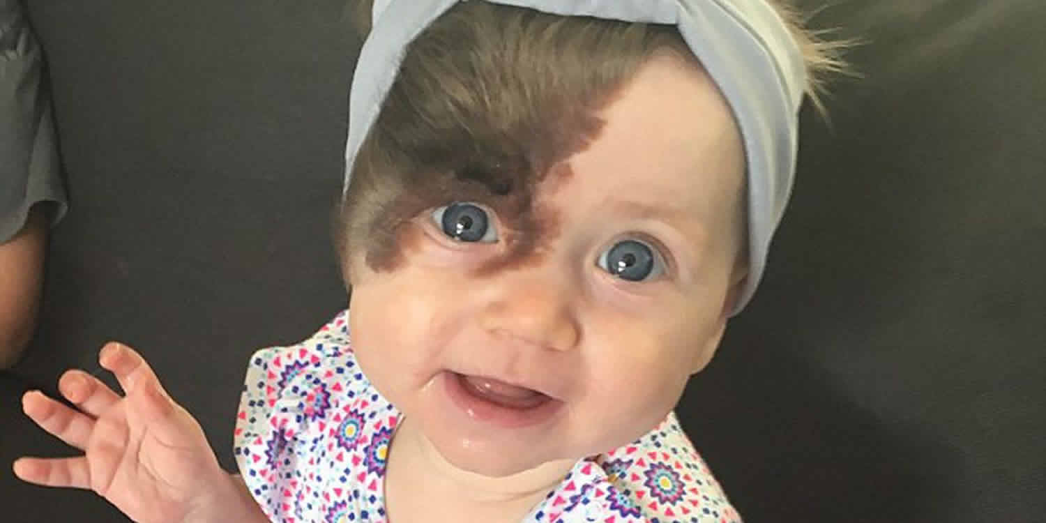
Birthmarks are marks that are present on the skin of a lot of newborn babies. A baby can develop birthmarks on his/her skin at birth either before being born or soon after birth. Birthmarks range from hardly noticeable to disfiguring, but no matter how large or small they are, they can be upsetting. One of the most common types of birthmarks is called a hemangioma. Hemangiomas are a bunch of tiny blood vessels that grow in a specific area on the skin — that’s why they usually look red or purple. Blood vessels are tiny tubes that carry blood through the body.
No one knows what causes blood vessels to group together, but it’s good to know that most birthmarks aren’t a sign of any kind of illness and usually don’t hurt at all. Lots of newborns have these birthmarks on their bodies, perhaps between the eyebrows (where they’re called angel kisses). These birthmarks are from wider blood vessels and usually disappear early in life.
For most birthmarks you don’t have to do anything. Babies who have vascular birthmarks usually need to see a skin doctor (dermatologist) to find out if it needs to be treated. This is important when a birthmark is in a certain spot like near the eye or near the lip.
A skin doctor also can tell you if there are ways to make a birthmark fade or become lighter. Here are some things the doctor might use or suggest:
- Medicine taken by mouth or given in a shot.
- Special types of lasers that can make birthmarks lighter or smaller.
- Surgery might be used to get rid of moles.
- Special makeup sometimes can hide birthmarks that don’t go away on their own.
Figure 1. Infantile hemangioma
Why do we have birthmarks?
Nobody really knows why babies get birthmarks or why some kids have small birthmarks and others have bigger ones. Birthmarks just seem to happen. Some types of birthmarks run in families, but not always.
Birthmarks can’t be prevented and they’re not caused by anything done or not done during pregnancy. There’s no truth to old wives’ tales about “stains” being caused by something the mother did or ate. The cause of most birthmarks is unknown. They can be inherited, but usually are not. They are typically not related to trauma to the skin during childbirth.
Does everyone have a birthmark?
Almost everyone’s have a birthmark. Birthmarks come in different shapes, sizes and colors and can be anywhere on the skin. Some are so little and pale that you might not even notice them. Most people have at least a few of these.
Other birthmarks are bigger and are purple, red, or black. You might notice these more, especially if they are on someone’s face. These large birthmarks might make some kids embarrassed. Other kids aren’t bothered at all. Usually a big birthmark doesn’t mean there’s anything wrong, though. Some kids just happen to have them. Birthmarks usually don’t hurt. No one really knows what causes them. Some go away on their own, and others might stick around your whole life. If you have a birthmark that bothers you, talk to your mom or dad about it. You can ask them to take you to a skin doctor (dermatologist). The skin doctor can talk to you about your birthmark and decide if it needs to be treated or if you should just leave it alone.
Types of birthmarks
There are two main types of birthmarks: pigmented birthmarks and vascular birthmarks.
Pigmented birthmarks
Pigment is a fancy word for color. These types of birthmarks happen when you have more pigment in one part of your skin. It’s like a spot on your skin. The types of pigmented birthmarks are:
Moles
Congenital moles also known as congenital melanocytic naevi. They’re relatively large brown or black moles that are present from birth. People often call these birthmarks “beauty marks.” Congenital moles are fairly common and are caused by an overgrowth of pigment cells in the skin. Most congenital moles (congenital melanocytic naevi) become proportionally smaller and less obvious with time, although they may darken during puberty or become bumpy or hairy. But not all moles are birthmarks. Moles usually are small, round brown spots (no bigger than about the size of a pencil eraser), but they sometimes can be larger and can be different colors. They can be pink, skin-colored or black. Some are flat and smooth; others are raised above the skin like a slight bump. Some moles go away, but you also might get more moles on your body as you get older.
Congenital moles can range in size from less than 1.5cm (about 0.6 inches) to more than 20cm (about 7.9 inches) in diameter. The risk of a mole developing into skin cancer is low, but the risk increases the larger it is. If you notice a mole that itches or bleeds, or if it looks a lot different than your other moles, ask your parents to take you to the doctor. It is important to have it checked out and make sure it’s OK.
Figure 2. Congenital mole
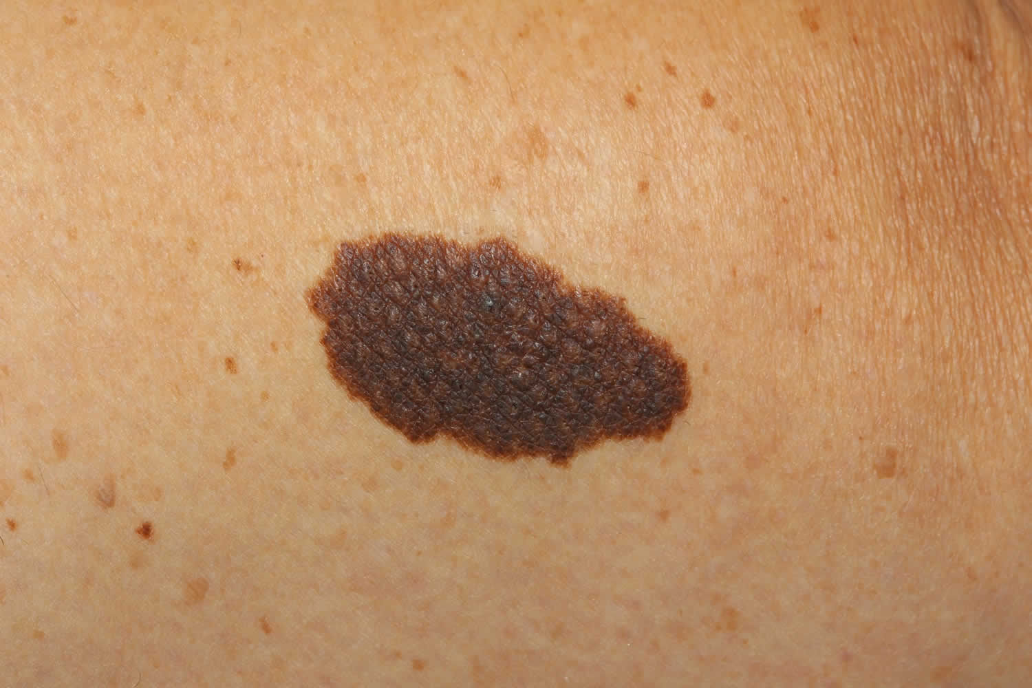
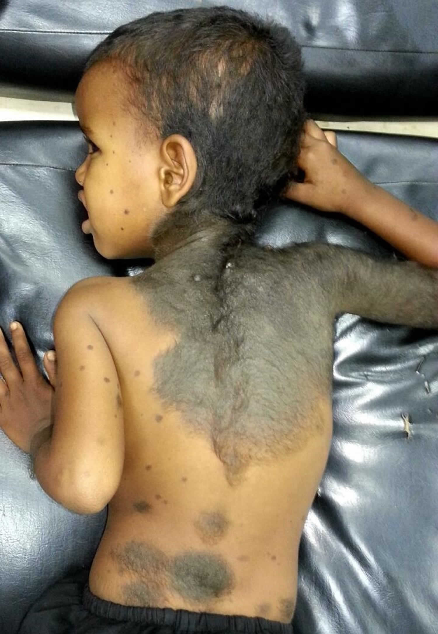
Figure 3. Giant hairy congenital nevus – difficult to manage due to its size and potential to develop melanoma
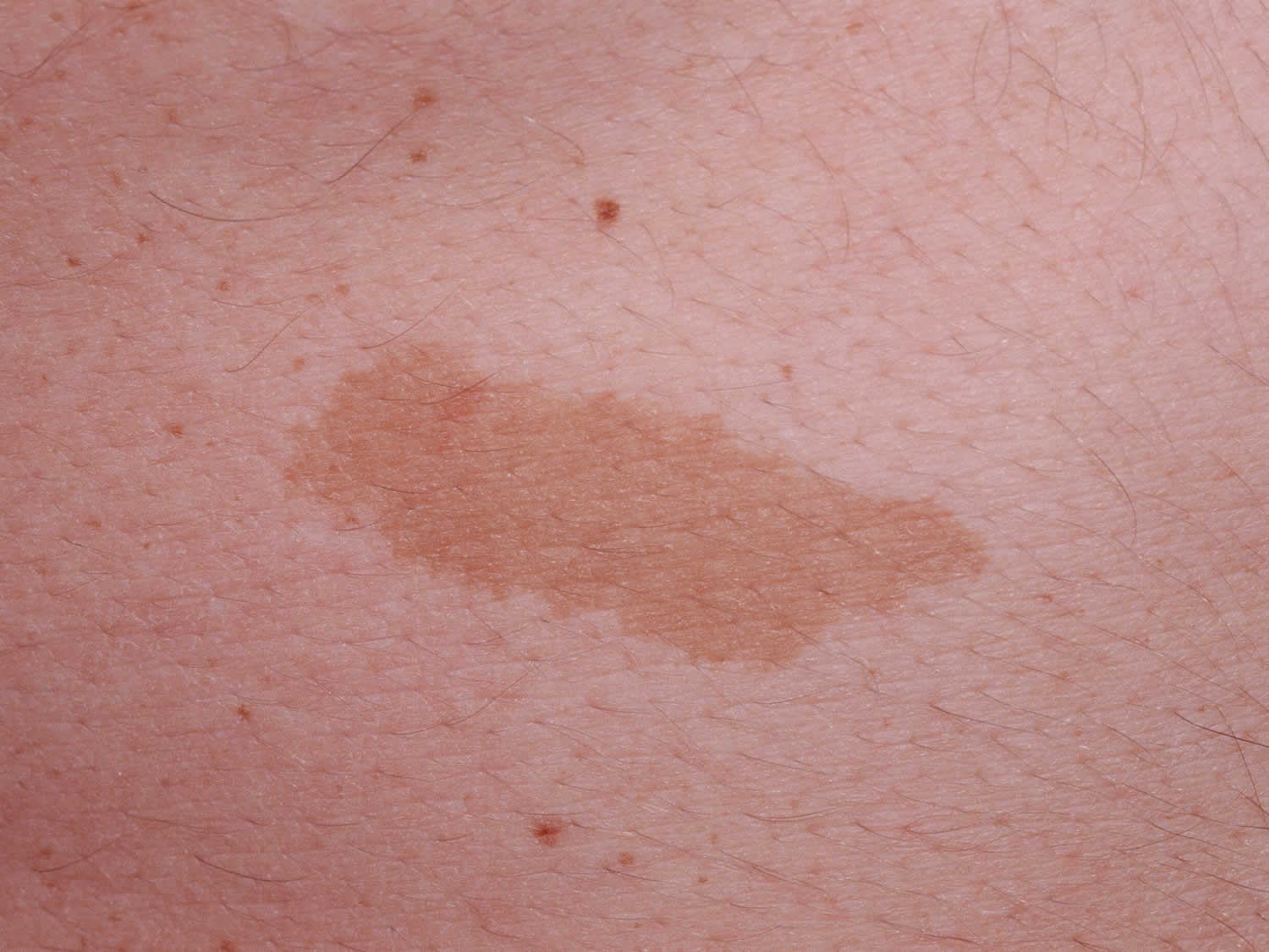
Cafe-au-lait spot
Cafe-au-lait is French for “coffee with milk,” which is the color of these spots, kind of light brown, when they’re on light skin. On dark skin they can be the color of black coffee. They can be small or big and often are oval-shaped. The spots might fade as you get older, but they probably won’t go away totally. Many children have one or two, but if more than six have developed by the time the child is five, you should see your doctor. Cafe-au-lait spot could be a sign of neurofibromatosis (a number of genetic conditions that cause tumors to grow along the nerves).
Figure 4. Cafe-au-lait spot
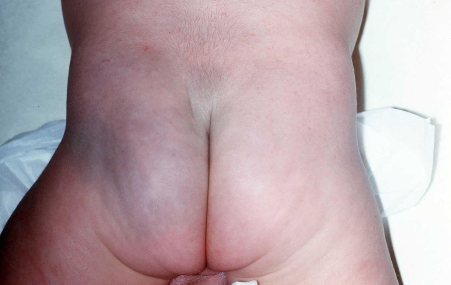
Mongolian spots
Mongolian spots are blue-grey or bruised-looking birthmarks that are present from birth.
Mongolian spots are more commonly seen in darker-skinned people and usually occur over the lower back or buttocks. However, they can also appear elsewhere on the body or limbs.
Mongolian spots may last for months or years, but they usually disappear by the time a child reaches four years of age. Mongolian spots are completely harmless and don’t need treatment. They may sometimes be mistaken for a bruise.
Figure 5. Mongolian spot
Vascular birthmarks
Your heart and blood vessels — the little highways that move blood through your body — are your vascular system. Sometimes a bunch of extra blood vessels will clump together, and you can see this clump in your skin. This is called a vascular birthmark. More than one in 10 babies has this type of birthmark. The different kinds are:
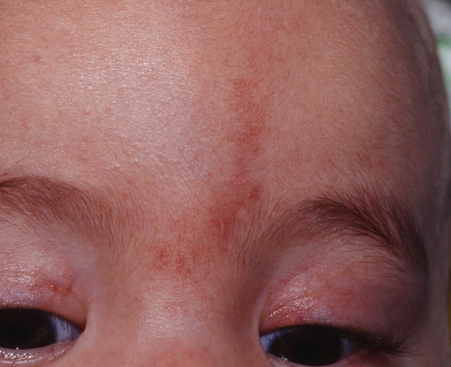
Salmon patches
These marks are flat and kind of pink or red (like salmon). If you get them on your face, people call them “angel’s kisses.” If you get them on the back of your neck, they’re called “stork bites” (red spots that look like bite marks — they’re not, of course). Sometimes they fade away, but sometimes they don’t.
Figure 6. Salmon patch
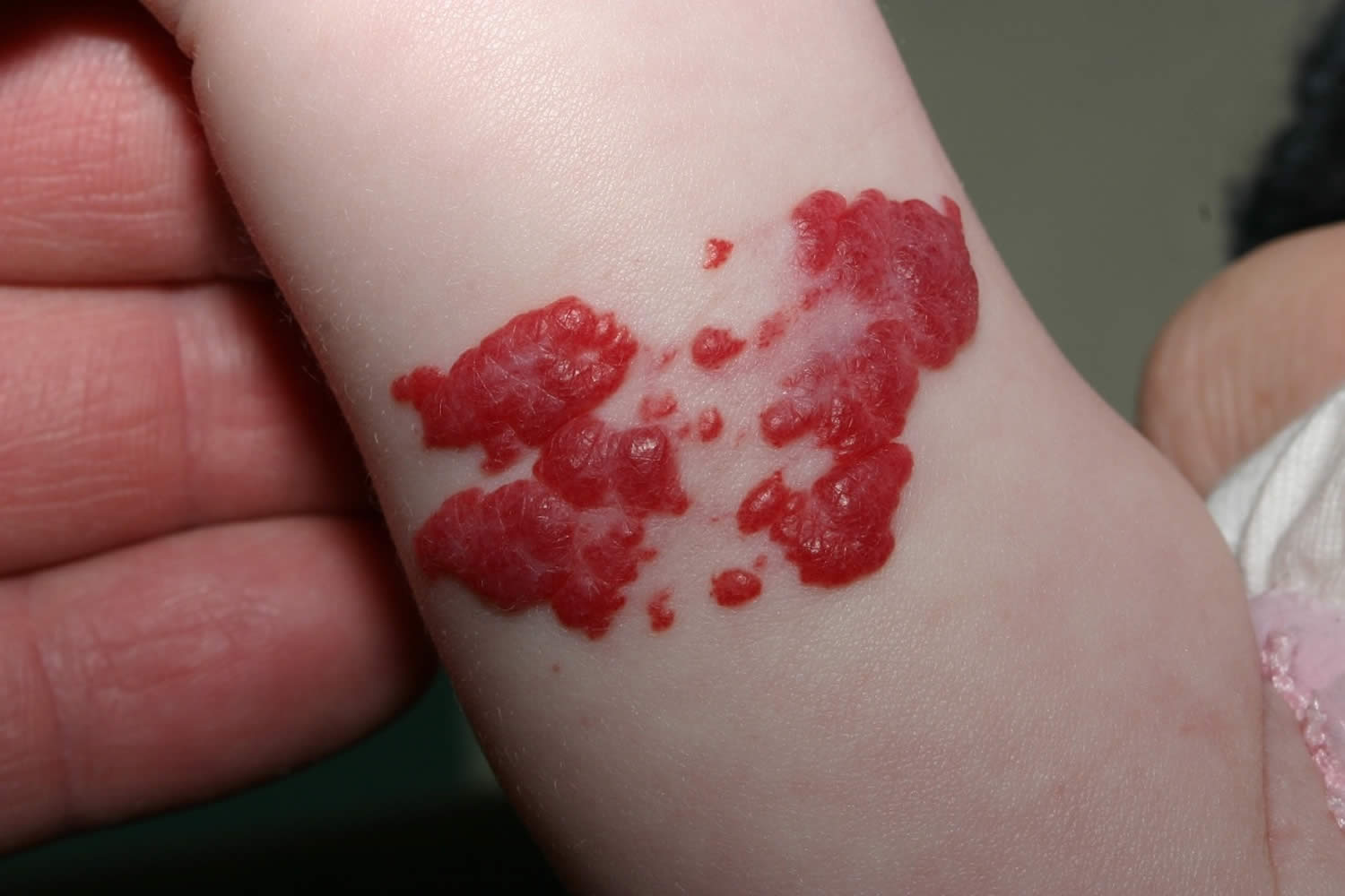
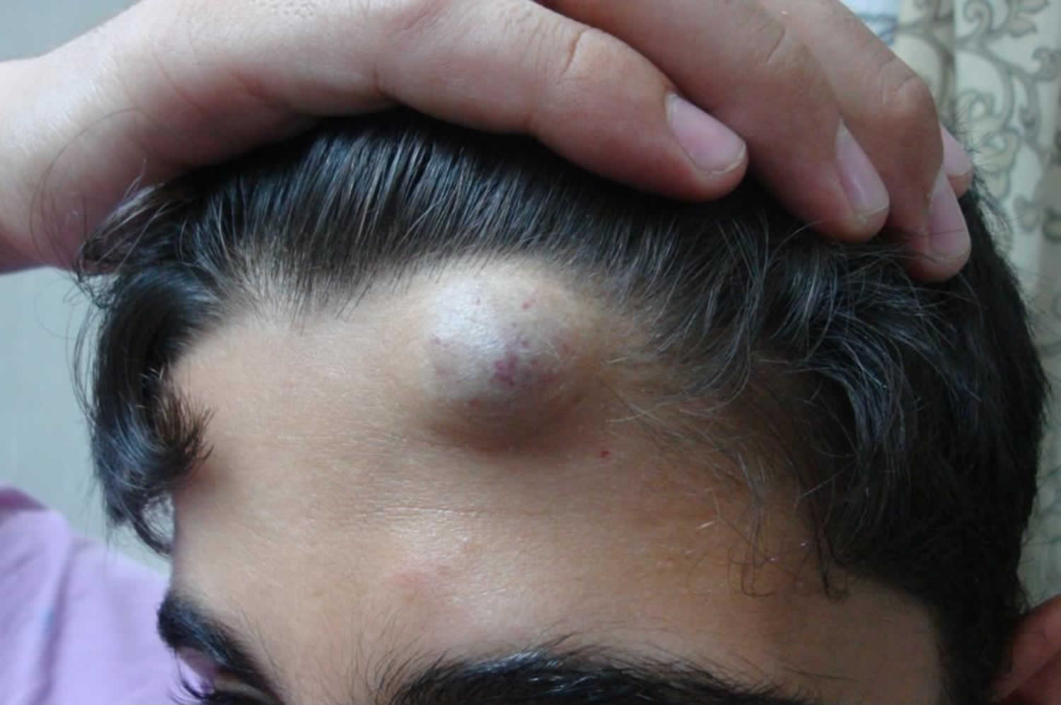
Hemangiomas
Hemangiomas birthmarks are usually harmless. There are two types: the kind that shows up on top of your skin and the kind that is deep in your skin. The ones on top are called strawberry hemangiomas (see Figure 1 above) because they’re bright red and look like the fruit. Deep hemangiomas are bluish-purple and make the skin swell and bulge. This kind shows up after a baby is born. For the first year, both types can get bigger and bigger, which can look a little scary to parents. The good news is they usually start shrinking. Most hemangiomas become flat by age 5 to 10, and many become flat even earlier. They can leave a light mark behind.
Some, particularly larger ones, may leave a scar as they regress that can be corrected by minor plastic surgery. Most hemangiomas are on the head or neck, although they can be anywhere on the body, and can cause problems if their location interferes with sight, feeding, breathing, or other body functions. Open sores sometimes form with hemangiomas and can get infected.
Figure 7. Deep hemangioma
Port wine stains
These marks often show up on the face, and they’re the color of wine or grape juice: pink, red or purple. They don’t go away on their own and can get bigger as kids grow.
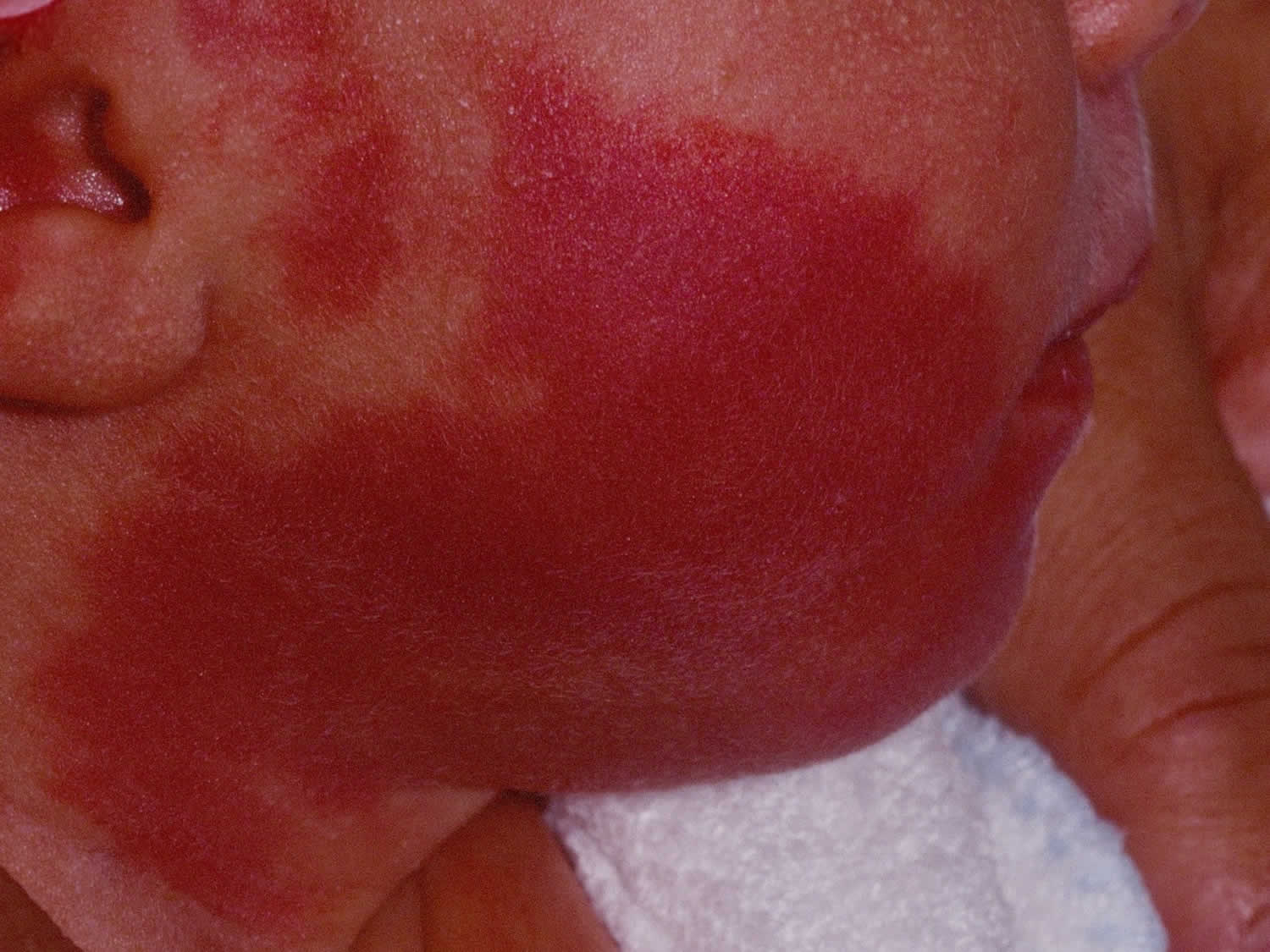
Figure 8. Port wine stain on the cheek of an infant
What causes birthmarks
It’s not fully understood why birthmarks occur, but they’re not usually inherited. Vascular birthmarks are caused by abnormal blood vessels in or under the skin, and pigmented birthmarks are caused by clusters of pigment cells.
Port wine stains are thought to occur because the nerves that control the widening or narrowing of the capillaries (tiny blood vessels) don’t function properly, or there aren’t enough of them. This means that blood is constantly supplied to the skin in that area, which makes it permanently red or purple in colour.
Port wine stains are sometimes related to other conditions, such as Sturge-Weber syndrome and Klippel-Trenaunay syndrome.
Birthmarks complications
Most birthmarks are harmless. But in rare cases, complications can occur that need to be treated.
Hemangiomas
Although it’s rare, some hemangiomas can cause severe problems and can even be life-threatening. They need to be treated if they interfere with eating, breathing or eyesight.
If your child has a hemangioma near their eye, nose, mouth or nappy area, they may need to be referred to a specialist. Hemangiomas in these areas are more likely to become infected. If the birthmark bleeds, apply pressure to it until the bleeding stops.
See your doctor if your child’s hemangioma forms an ulcer. It may become infected and be very painful. Keep the wound clean and covered with a dressing. It should heal within two weeks.
Infected hemangiomas need urgent treatment with antibiotics. An infected ulcer may leave an unsightly scar.
If your child has more than five haemangiomas, they may also have internal hemangiomas. An ultrasound scan or magnetic resonance imaging (MRI) scan may be used to find out whether any internal hemangiomas are present.
It’s very unusual for internal hemangiomas to cause problems. But in rare cases they can cause coughing and difficulty breathing, which may indicate airway hemangiomas. Another possible symptom is blood in the stools, which may indicate a hemangioma in the bowel.
Port wine stains (capillary malformation)
Port wine stains (capillary malformation) can lead to the following complications:
- glaucoma (raised pressure within the eye that affects vision) – this may develop if the birthmark affects both the upper and lower eyelids on the same side
- Sturge-Weber syndrome – a rare disorder affecting the eyes and brain that’s usually associated with a large port wine stain that extends across the forehead or scalp
- soft tissue hypertrophy (the tissue beneath the birthmark enlarging) – this may occur on the lip, for example Klippel-Trenaunay syndrome – a rare disorder that’s present at birth where the blood vessels fail to form properly; if your child has an enlarged port wine stain on their limb, they may have Klippel-Trenaunay syndrome.
All of the above conditions will need to be treated by a specialist.
Congenital mole (congenital melanocytic naevi)
If a congenital mole (congenital melanocytic naevi) increases in size or changes shape or colour, your doctor may recommend that you have a biopsy (where a tissue sample is taken so that it can be examined under a microscope).
You should see your doctor if you notice any of the following changes in your birthmark:
- bleeding
- inflammation (swelling)
- itching
- open sores
- pain
- changes in color, size or texture
Although it’s very rare, some congenital mole (congenital melanocytic naevi) can develop into skin cancer. This risk increases with the size of the birthmark – the larger it is, the greater the risk.
How to get rid of birthmarks
Most birthmarks are harmless and don’t need to be treated. Some types of birthmarks will fade over time, whereas other types such as port wine stains will be permanent if they’re not treated.
In some cases, a birthmark will need to be treated for medical reasons – for example, if a hemangioma blocks the airways, affects vision or becomes ulcerated. Some people may also decide to seek treatment for cosmetic reasons.
Hemangiomas
Hemangiomas sometimes disappear without treatment, but they often don’t change until your child is two years old.
Hemangiomas will sometimes disappear by the time a child reaches five years of age. In other cases, they may last until a child is 12.
Plastic surgery may be an option if a hemangioma has left the skin deformed or stretched. The aim of surgery will be to improve the appearance of the distorted skin.
If the hemangioma has formed an ulcer, extra measures may need to be taken to prevent infection, and surgery or laser treatment may be offered.
Large or complicated hemangiomas
Some hemangiomas may cause complications that will need treatment.
A hemangioma near your child’s eye, nose or mouth may cause vision, breathing or feeding problems.
Hemangiomas on the lip or around the nappy area are more likely to form ulcers that can sometimes bleed and be painful.
The exact treatment will depend on where and how severe the hemangioma is. Most hemangiomas can be effectively treated with medicine, such as propranolol which is given by mouth as a liquid. This will shrink the birthmark.
If propranolol doesn’t work, other medicines can be used, such as steroids or vincristine. Surgery is rarely necessary.
If your child has breathing difficulties caused by a hemangioma in their airway, they may need laser treatment.
This will usually be carried out during an examination of their airway using a small telescope called an endoscope. The procedure is known as microlaryngoscopy and bronchoscopy. They may also be given propranolol.
Occasionally, a child with a hemangioma in their airway may need to have a temporary tracheostomy (an artificial opening in the windpipe) to improve their breathing.
Propranolol
Medication may be recommended if the hemangioma is complicated or large. This is usually a beta-blocker called propranolol. The full side effects of using propranolol to treat a hemangioma are still being monitored.
Beta-blockers work by blocking the release of noradrenaline in certain parts of the body. Noradrenaline is a chemical released by nerves when they’re stimulated. The noradrenaline passes messages to other parts of the body, such as the muscles, blood vessels and heart.
It’s thought that propranolol helps narrow the blood vessels, reducing the amount of blood flowing through them. This makes the hemangioma lighter in colour and softer. The cells that cause the hemangioma to grow are also affected so that it gets smaller.
Hemangiomas often leave a patch of stretched and thinned skin, with a small amount of swelling of the tissues under the skin. This is barely noticeable in most areas.
However, in areas such as the nose, lips, ears and cheek, these small changes can be unsightly. In these areas, treatment with propranalol may be considered during the first few weeks of life, immediately after the hemangioma has been diagnosed, so that the long-term appearance can be improved.
Monitoring internal hemangiomas
If your child has a hemangioma in an internal organ, they may need an ultrasound scan or magnetic resonance imaging (MRI) scan to confirm its location and size. MRI scans use a magnetic field and radio waves to produce detailed pictures of the inside of the body.
Port wine stains (capillary malformation)
Port wine stains (capillary malformation) are permanent, but treatment helps fade the mark, making it less noticeable. You can also disguise it using cosmetics.
Laser treatment
Laser treatment is the only treatment for a port wine stain. It lightens the affected area of skin. Laser treatment often works better in younger children because in adults a port wine stain may become bumpy and raised after a number of years.
The most common type of laser treatment is pulsed dye laser treatment. The laser passes through a fiberoptic cable. On the end of the cable is a device that looks like a pen. It’s gently held against the surface of your child’s skin and a button is pressed, which sends a beam of light to the skin.
The light goes less than 1mm into the skin. It’s absorbed by the blood vessel just beneath the surface, causing it to heat up. The heat damages the blood vessel, which creates a bruise that will fade within a week or two.
During or after treatment, your child’s skin will be cooled to reduce discomfort. A jet of cold air may be blown onto the skin during treatment.
Some of the possible side effects of laser treatment include:
- bruising – after some types of laser treatment, it’s normal for the mark on the skin to look worse because of bruising, but this will fade after one to two weeks
- pain – the laser stings, so most children have laser treatment under general anaesthetic (where they’re put to sleep), but some children can tolerate treatment with the help of a local anaesthetic cream that numbs the skin; a cold gel pad may also be put on the area before treatment to minimise any discomfort
- increased sensitivity to sunlight – your child’s skin will be very prone to sunburn for up to six months after laser treatment
Between three and 30 treatment sessions may be needed at intervals of six to eight weeks.
How effective the treatment is will depend on how prominent and dark the affected area is. The best results are often seen in marks that are already smaller and lighter.
Camouflage make-up
You can get a prescription for a special type of camouflage make-up that covers up the birthmark.
Congenital mole (congenital melanocytic naevi)
As congenital mole (congenital melanocytic naevi) can affect a person’s appearance, surgery may be considered. However, surgery will leave scarring and may not be possible if the affected area is very large.
Surgery involves removing the birthmark and stitching together the edges of skin. If the area is large, a skin graft may be needed. This involves taking skin from another part of the body and using it to cover the wound.


