Contents
What is caput succedaneum
Caput succedaneum is also called scalp edema or a collection of serosanguinous fluid in the subcutaneous layer of the scalp, causing a significant swelling of the soft tissues of the baby’s scalp that develops as the baby travels through the birth canal 1. Some babies have some bruising of the area. Caput succedaneum is most often brought on by pressure from the uterus or vaginal wall during a head-first (vertex) delivery. Babies delivered by vacuum extraction are more likely to have this condition.
Your health care provider will look at the swelling to confirm that it is a caput succedaneum. No other testing is needed.
No treatment is needed. A caput succedaneum most often goes away on its own within a few days. Complete recovery can be expected. The scalp will go back to a normal shape. Complications may include a yellow color to the skin (jaundice) if bruising is involved.
Figure 1. Caput succedaneum
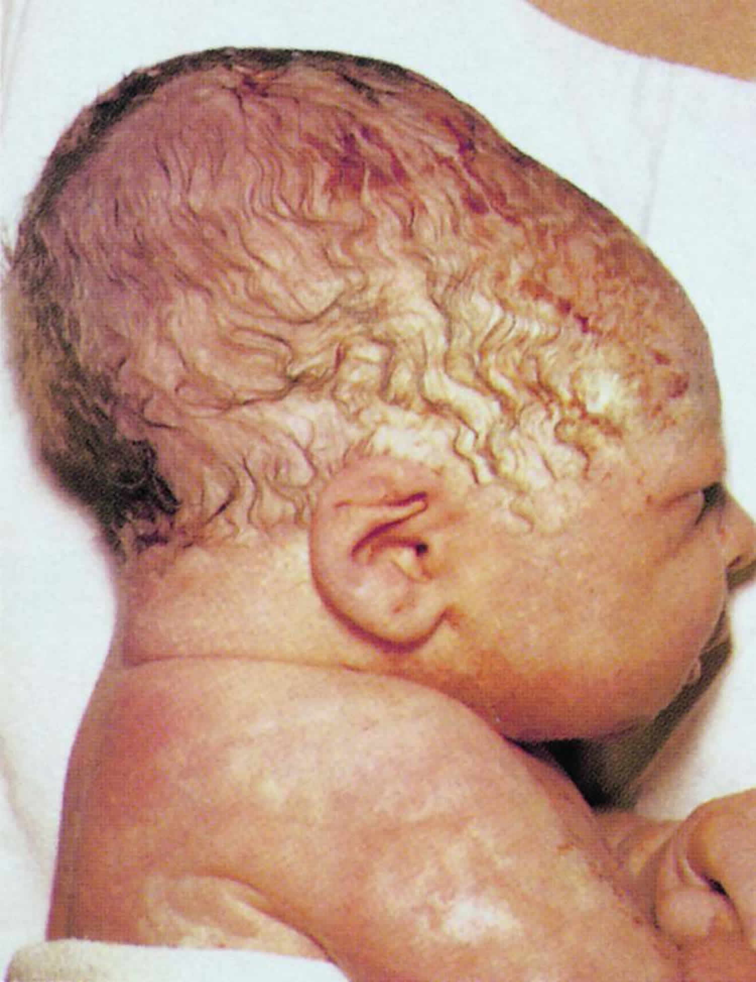
Figure 2. Scalp and skull anatomy
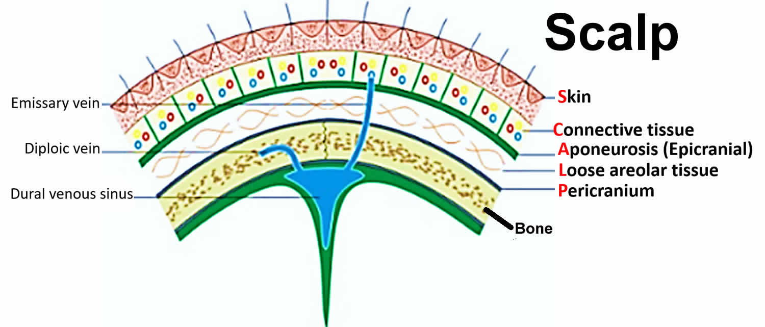
Figure 3. Hemorrhages by location within the different layers of the scalp (right of image) and meninges (left of image)
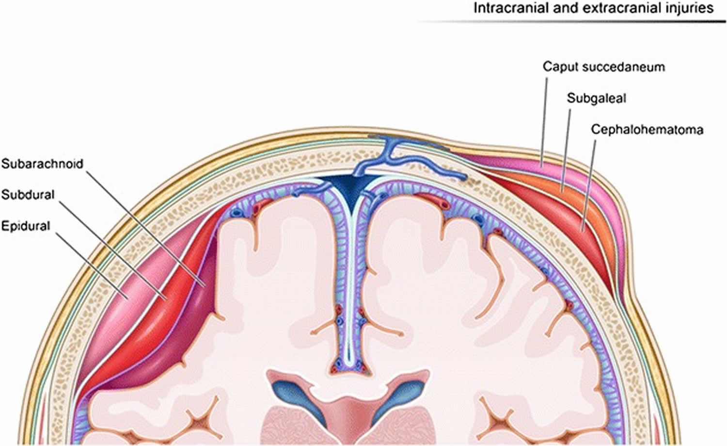
Footnote: Caput succedaneum is collection within the subcutaneous fibrofatty tissues superficial to galea aponeurosis. Cephalohematoma is bleeding into the fetal scalp that is located in the subperiosteal space.
Caput succedaneum vs Cephalohematoma
Cephalohematoma is bleeding into the fetal scalp that is located in the subperiosteal space and as such, is contained anatomically to a single skull bone 1. Cephalhematoma is a collection of blood between the skull and its periosteum. Cephalhematoma usually involves the parietal or occipital bone, sharply limited by the margins of the bone and does not cross the suture lines 2.
Cephalohematoma often appears several hours after birth as a raised lump on the baby’s head. Over time, the body resorbs the blood. Depending on the size, most cephalohematomas take two weeks to three months to disappear completely. If the area of bleeding is large, some babies may develop jaundice as the red blood cells break down.
Figure 4. Cephalohematoma
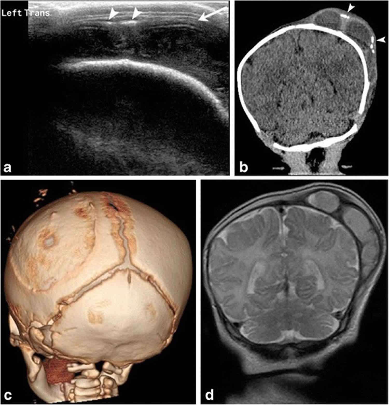
Footnote: One month old male baby with history of traumatic delivery presenting with right parietooccipital soft tissue swelling. Transverse grayscale ultrasound (a) image of the left parietooccipital scalp shows a complex fluid collection (arrow), with punctate linear echogenic foci along the superficial aspect (arrowheads), suggestive of calcifications. Relationship with the adjacent left lambdoid suture was difficult to evaluate by ultrasound. B. Coronal non-contrast head CT (b) image demonstrates a lobulated fluid collection with thick septations and peripheral calcifications (arrowheads) that does not cross the adjacent sagittal or the lambdoid suture, suggestive of cephalhematoma. 3-D volume (c) rendered images in bone algorithm shows cortical irregularity along the left parietal bone at the site of cephalhematoma as well as peripheral calcifications along the superficial aspect of the cephalhematoma. Coronal T2 image (d) from an MR exam obtained one week later in the setting of patient’s seizures re-demonstrates the large subperiosteal complex fluid collection with thick septations and isointense fluid signal consistent with evolving blood products in the known left parietal cephalhematoma
[Source 3 ]Caput succedaneum causes
A caput succedaneum is more likely to form during a long or hard vaginal delivery. Caput succedaneum is more common after the membranes have broken. This is because the fluid in the amniotic sac is no longer providing a cushion for the baby’s head. Vacuum extraction done during a difficult birth can also increase the chances of a caput succedaneum.
A caput succedaneum may be detected by prenatal ultrasound, even before labor or delivery begins. It has been found as early as 31 weeks of pregnancy. Very often, this is due to an early rupture of the membranes or too little amniotic fluid. It is less likely that a caput will form if the membranes stay intact.
Caput succedaneum symptoms
Caput succedaneum symptoms may include:
- Soft, puffy swelling on the scalp of a newborn infant
- Possible bruising or color change on the scalp swelling area
- Swelling that may extend to both sides of the scalp
- Swelling that is most often seen on the portion of the head which presented first
How long does caput succedaneum last?
The swelling usually disappears in a few days without problems.
Caput succedaneum treatment
No treatment is needed. A caput succedaneum most often goes away on its own within a few days. Complete recovery can be expected. The scalp will go back to a normal shape. Complications may include a yellow color to the skin (jaundice) if bruising is involved.
- Ali UA, Norwitz ER. Vacuum-assisted vaginal delivery. Rev Obstet Gynecol. 2009;2(1):5-17. https://www.ncbi.nlm.nih.gov/pmc/articles/PMC2672989/[↩][↩]
- Nicholson L. Caput succedaneum and cephalohematoma: the cs that leave bumps on the head. Neonatal Netw. 2007;26:277–81.[↩]
- Chaturvedi A, Chaturvedi A, Stanescu AL, Blickman JG, Meyers SP. Mechanical birth-related trauma to the neonate: An imaging perspective. Insights Imaging. 2018;9(1):103-118. https://www.ncbi.nlm.nih.gov/pmc/articles/PMC5825313/[↩]


