Contents
What is coagulopathy
Coagulopathy is a disease or condition that affects the blood’s ability to coagulate (clot) normally. Normally, if you get hurt, your body forms a blood clot to stop the bleeding. For blood to clot, your body needs cells called platelets and proteins known as clotting factors. If you have a coagulopathy or bleeding disorder, you either do not have enough platelets or clotting factors or they don’t work the way they should.
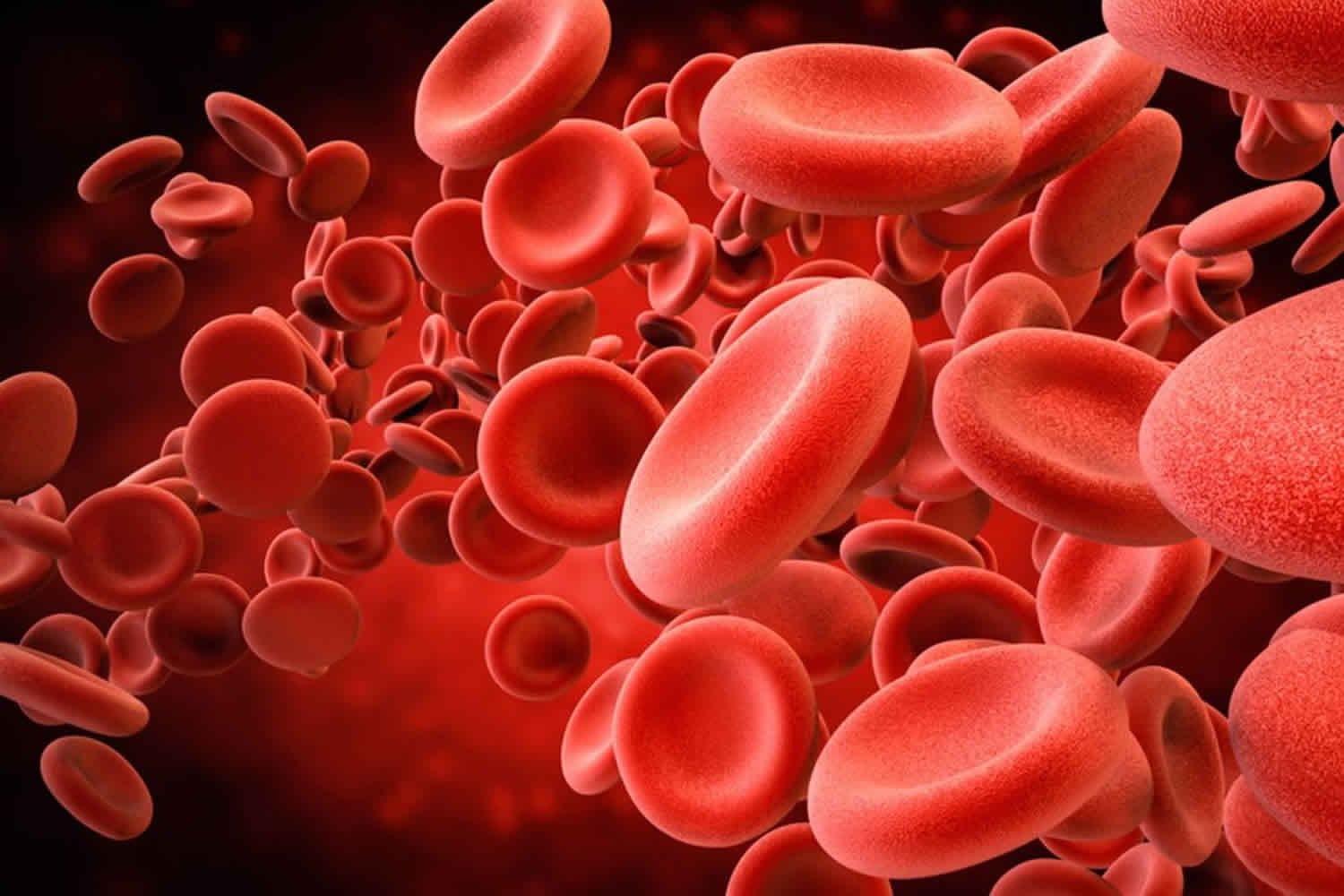
Normal blood clotting involves blood components, called platelets, and as many as 20 different plasma proteins. These are known as blood clotting or coagulation factors. These factors interact with other chemicals to form a substance that stops bleeding called fibrin.
Problems can occur when certain factors are low or missing. Coagulopathy can range from mild to severe.
Some coagulopathy are present at birth and are passed down through families (inherited). Others develop from:
- Illnesses, such as vitamin K deficiency or severe liver disease
- Treatments, such as the use of drugs to stop blood clots (anticoagulants) or the long-term use of antibiotics
Coagulopathy can also result from a problem with the number or function of the blood cells that promote blood clotting (platelets). Coagulopathy can also be either inherited or develop later (acquired). Hemophilia is an inherited bleeding disorder. Coagulopathy can also be a side effect of medicines such as blood thinners.
Various blood tests can check for a bleeding disorder. You will also have a physical exam and history. Treatments depend on the cause. They may include medicines and transfusions of blood, platelets, or clotting factor.
Coagulopathy causes
Coagulopathy are a group of conditions in which there is a problem with the body’s blood clotting process. These disorders can lead to heavy and prolonged bleeding after an injury. Bleeding can also begin on its own.
Specific coagulopathy include:
- Acquired platelet function defects
- Congenital platelet function defects
- Coagulopathy in liver disease
- Dilutional coagulopathy
- Disseminated intravascular coagulation (DIC)
- Factor II deficiency
- Factor V deficiency
- Factor VII deficiency
- Factor X deficiency
- Factor XI deficiency (hemophilia C)
- Glanzmann disease
- Hemophilia A
- Hemophilia B
- Idiopathic thrombocytopenic purpura (ITP)
- Coagulopathy in trauma
- Von Willebrand disease (types I, II, and III)
Disseminated intravascular coagulopathy
Disseminated intravascular coagulation (DIC) also known as consumptive coagulopathy, is a serious disorder, sometimes life-threatening condition in which the proteins in the blood involved in blood clotting become overactive. Blood clots form in small blood vessels throughout the body. This can disrupt normal blood flow to organs such as the kidneys and liver and can lead to organ failure. Because the clotting uses up coagulation proteins and platelets, excessive bleeding can occur. This abnormal activation of blood clotting mechanisms can develop as the result of a variety of diseases and conditions.
Typically, when a person has an injury to a blood vessel and bleeding occurs, the body stops the bleeding by initiating a process called hemostasis. First, platelets adhere to the injury site and clump together, forming a loose plug. Then coagulation factors are sequentially activated (see coagulation cascade) to produce a net of fibrin threads that weave through the platelet plug and form a stable clot. The clot stays in place until the injury is healed, then other factors break the clot down (fibrinolysis) and remove it. This clotting process is tightly regulated. Feedback mechanisms accelerate the clotting process, then slow it down, and control the volume of clot produced.
Normally, the body initiates hemostasis and forms a blood clot only when needed, i.e., when there is an injury and bleeding. The body detects a pro-coagulant, a substance such as tissue factor that is released from cells when they are damaged. Based on the extent of the injury, the body responds by stimulating sufficient clotting to stop the bleeding and confining it locally, that is, only at the site of injury.
With conditions that trigger disseminated intravascular coagulation, the response is exaggerated, clotting is activated throughout the body, and control mechanisms are inhibited. The result is the formation of a multitude of tiny clots that can block small blood vessels and prevent blood and oxygen from getting to tissues and organs, leading to multi-organ failure. Widespread clotting can use up platelets and coagulation factors at a rapid rate. This can overwhelm the system to the point that the body begins to bleed excessively because platelets and clotting factors have been depleted. Simultaneous clotting and bleeding can occur. disseminated intravascular coagulation can develop very quickly, becoming serious or even life-threatening in a short time.
Disseminated intravascular coagulation may occur with conditions such as:
- Infections, especially with severe or systemic infections (and sometimes resulting sepsis), primarily bacterial but also sometimes seen with fungal, viral, or parasitic infections
- Trauma, such as due to an accident or significant burns
- Major surgery, such as cardiopulmonary bypass surgery
- Pregnancy and childbirth, especially when a woman has retained placental material, or a fetus that has died (stillbirth)
- Cancers, such as acute promyelocytic leukemia or tumors that develop within glands (adenocarcinoma); cancer cells may release a pro-coagulant.
- Organ failure, such as liver, pancreas, or kidney failure
- Other less common causes, such as snake bite venom, toxic drug reaction, blood transfusion reaction, organ transplant, or frostbite
Most cases of disseminated intravascular coagulation that are diagnosed develop rapidly and suddenly (acute), but there are cases in which it develops gradually, occurring over a longer period of time. This chronic form of disseminated intravascular coagulation is difficult to recognize and is much less often diagnosed. Simultaneous clotting and bleeding can occur with chronic disseminated intravascular coagulation, but in most cases this is a lower grade, persistent clotting activation process and the body has sufficient capacity to compensate. With chronic disseminated intravascular coagulation, the predominant feature is typically increased clotting, not bleeding. Cancer is one of the most common causes of low-grade disseminated intravascular coagulation.
Disseminated intravascular coagulation causes
When you are injured, proteins in the blood that form blood clots travel to the injury site to help stop bleeding. If these proteins become abnormally active throughout the body, you could develop disseminated intravascular coagulation. The underlying cause is usually due to inflammation, infection, or cancer.
In some cases of disseminated intravascular coagulation, small blood clots form in the blood vessels. Some of these clots can clog the vessels and cut off the normal blood supply to organs such as the liver, brain, or kidneys. Lack of blood flow can damage and cause major injury to the organs.
In other cases of disseminated intravascular coagulation, the clotting proteins in your blood are consumed. When this happens, you may have a high risk of serious bleeding, even from a minor injury or without injury. You may also have bleeding that starts spontaneously (on its own). The disease can also cause your healthy red blood cells to fragment and break up when they travel through the small vessels that are filled with clots.
Risk factors for disseminated intravascular coagulation include:
- Blood transfusion reaction
- Cancer, especially certain types of leukemia
- Inflammation of the pancreas (pancreatitis)
- Infection in the blood, especially by bacteria or fungus
- Liver disease
- Pregnancy complications (such as placenta that is left behind after delivery)
- Recent surgery or anesthesia
- Severe tissue injury (as in burns and head injury)
- Large hemangioma (a blood vessel that is not formed properly)
Disseminated intravascular coagulation symptoms
Signs and symptoms of disseminated intravascular coagulation depend upon the underlying condition, such as infection, trauma or malignancy, and on the severity and the extent of disseminated intravascular coagulation. In addition to signs and symptoms of the underlying condition, those of disseminated intravascular coagulation are associated with bleeding and/or inappropriate clotting.
Bleeding
Significant bleeding usually occurs from at least three different sites.
- Blood in the stool or urine from internal bleeding
- Headaches and other symptoms associated with bleeding in the brain
- Bruising and the formation of small red dots on the skin (petechiae)
- Bleeding at the site of wounds, surgical sites, intravenous (IV) needle or catheter sites
- Mucosal bleeding – from the nose, gums, mouth, etc.
Blood Clotting
Symptoms depend on the location of blood clots and may include:
- Symptoms associated with organ dysfunction caused by blood clots blocking blood flow and oxygen to organs such as the liver and kidney, leading to liver and kidney failure
- Blackening of the skin caused by blockage from blood clots and poor blood flow to the skin
- Chest pain, coughing up blood, and/or difficulty breathing caused by blood clots in the lungs
- Chest pain and/or a heart attack caused by clotting in the heart
- Headaches and other symptoms associated with a stroke, caused by clotting in the brain
Disseminated intravascular coagulation possible complications
Complications from disseminated intravascular coagulation may include:
- Bleeding
- Lack of blood flow to the arms, legs, or vital organs
- Stroke
Disseminated intravascular coagulation diagnosis
The goals of testing are to identify disseminated intravascular coagulation, evaluate its severity, and to monitor its effects over time. There is not a single test that can be used to definitely diagnose disseminated intravascular coagulation. A healthcare practitioner will consider many factors when assessing a person who may have this condition, such as signs and symptoms, presence of an underlying condition, physical examination, and medical history.
The severity and extent of disseminated intravascular coagulation can change over time so laboratory testing is often performed at several intervals to monitor a person’s status. Some routine tests that may be performed include:
- CBC (complete blood count) – includes a platelet count; in disseminated intravascular coagulation, platelets are often low.
- Blood smears from individuals with disseminated intravascular coagulation often show decreased number of platelets and presence of large platelets and fragmented red cells (schistocytes).
- PT (prothrombin time) – often prolonged with disseminated intravascular coagulation as coagulation factors are consumed
- PTT (partial thromboplastin time) – may be prolonged
- D-dimer – a test that detects a protein that results from clot break-down; it is often markedly elevated with disseminated intravascular coagulation; if normal, then disseminated intravascular coagulation is unlikely.
- Fibrinogen – one of the clotting factors; is low with disseminated intravascular coagulation
A test scoring system developed by the International Society on Thrombosis and Haemostasis may be used to evaluate a group of test results to help determine if disseminated intravascular coagulation is present. The score is based on the results of a platelet count, PT, D-dimer (or fibrin degradation products) and fibrinogen. The higher the score, the more likely it is that disseminated intravascular coagulation is present.
As disseminated intravascular coagulation can affect the health and function of several organs, more general testing, such as a comprehensive metabolic panel (CMP), may be ordered to evaluate, for example, the functional status of kidneys and liver. Additionally, several other tests may be ordered to help detect the underlying disease or condition that is causing a person to develop disseminated intravascular coagulation.
Non-Laboratory Tests
An x-ray or other imaging scan is sometimes performed to help locate blood clots and evaluate organs.
Disseminated intravascular coagulation treatment
There is no specific treatment for disseminated intravascular coagulation. The goal is to determine and treat the underlying cause of disseminated intravascular coagulation. In most cases, disseminated intravascular coagulation will resolve when the disease or condition is treated. Acute disseminated intravascular coagulation is typically treated in a hospital setting.
Supportive measures may need to be taken to address bleeding and clotting. People primarily presenting with severe bleeding may be given platelets or fresh frozen plasma or cryoprecipitate that contains clotting factors, especially when surgical procedures are required to address the underlying condition – such as with a retained placental material.
Supportive treatments may include:
- Plasma transfusions to replace blood clotting factors if a large amount of bleeding is occurring.
- Blood thinner medicine (heparin) to prevent blood clotting if a large amount of clotting is occurring.
Disseminated intravascular coagulation prognosis
Outcome depends on what is causing the disorder. disseminated intravascular coagulation can be life threatening.
Dilutional coagulopathy
Dilutional coagulopathy is usually defined as loss, consumption, or dilution of coagulation factors and occurs when blood is replaced with fluids that do not contain adequate coagulation factors 1. Dilutional coagulopathy is one of the most common causes of bleeding after massive transfusion in the later phase after trauma 2. Hemorrhagic shock leads to a net flux of extracellular water into the vascular space because of a lowered intravascular hydrostatic pressure due to bleeding. This dilution of coagulation proteins is further enhanced by external fluid resuscitation. In addition to the dilutional effect, which may be calculated mathematically, side effects on coagulation from different fluids (e.g., crystalloids, colloids) with different compositions (e.g., unbalanced vs. balanced solutions; normotonic vs. hypertonic solutions) have to be considered 3.
Depending on the amount and the type of fluid replacement, dilution of cellular components and coagulation factors were inevitable. Based on the dynamic of blood loss and consequently loss and consumption of factors a clinical relevant dilution coagulopathy occurs constantly 4. Moreover, disturbance of hemostatic potential might be further deteriorated by hypothermia, acidosis, and fibrinolysis, thus leading to worsening of patient’s outcome 5. If a critical threshold of plasma concentration of coagulation factors was reached, bleeding will be further perpetuated. The incidence of dilution coagulopathy was decreased by preparation of modern (nearly plasma-free) autologous blood products 6. In contrast to congenital bleeding disorders based on single coagulation factor deficiency, dilutional coagulopathy is related to multifactorial changes that affects thrombin generation, clot firmness, and fibrinolysis 7. Therefore, it seems hard to predict if the complex interaction of cellular components, pro- and anticoagulant factors, and fibrinolytic activators and inhibitors will finally form a stable clot during moderate to severe hemodilution 6.
Fibrinogen is the first component to drop and is always present with other coagulation abnormalities in cases of dilutional coagulopathy and should be treated with substitution of fibrinogen concentrate, whereby minimal plasma fibrinogen levels need to be defined by further well-controlled studies 6. If not replaced adequately hypofibrinogenaemia following neurosurgical procedures leads to catastrophic post-operative complications as emphasized in recent intracranial surgery cases 8, where two of the patients needed recraniotomy to evacuate post-operative hematomas and this is evidence of the need for quick and adequate transfusion of cryoprecipitate.
Substitution with other coagulation factors (e.g. FXIII levels < 60%) should be based on laboratory findings. Fresh frozen plasma transfusion in a dose of 30 ml/kg might be useful for treatment of severe hemorrhage as adjunctive to basic treatment with coagulation factors but should only be considered after exclusion of any other factor that might influence hemostasis (i.e. fibrinogen deficiency, hyperfibrinolysis, acidosis, hypothermia, low platelet count/impaired platelet function) 6. Based on current literature, routine use of recombinant activated factor VII (rFVIIa) was not supported. Perioperative treatment with antifibrinolytic agents might be helpful in reducing the amount of bleeding and reducing transfusion requirements in noncardiac pediatric surgery with a high risk of bleeding. Further well-controlled studies are urgently needed to determine the effect of a targeted, laboratory-based coagulation management for treatment of dilutional coagulopathy 6.
Dilutional coagulopathy causes
The causes of dilutional coagulopathy are complex and both pro- and anticoagulant pathways are affected; but the underlying mechanisms are not fully understood 6. In terms of laboratory changes, dilutional coagulopathy seems to be comparable to hemostatic changes during major hemorrhage following trauma injury 9.
Dilutional coagulopathy diagnosis
Unfortunately, routine plasmatic coagulation tests are of limited help for timely management of perioperative bleeding due to long turnaround times, insufficient differential diagnosis of complex acquired intraoperative coagulopathy, and insensitivity for function of fibrinogen, hyperfibrinolysis and platelet dysfunction 10. Another meaningful limitation is that measurement of fibrinogen levels using the photometric Clauss assay can be considerably altered after massive fluid resuscitation and that colloids may induce erroneously increased levels of fibrinogen 11.
Rotation thrombelastometry (ROTEM®) offers an alternative approach to assess perioperative coagulation disorders by means of viscoelastic testing 12. First results are available only 10 min after starting with testing and clot formation can be online observed by a bedside monitor. Data on the effective use of the ROTEM were published from adult liver transplantation 13, adult trauma patients 14, and intraoperative coagulation management in children 15. Notably, a normal ROTEM trace showed high negative predictive value and was suggested to early identify surgical bleeding by distinguishing it from coagulopathic bleeding 16. Additionally, both devices were stated as golden standard for detecting hyperfibrinolysis, which may have deleterious impact on mortality. However, there is a lack of data proving the usefulness of a ROTEM-guided coagulation management in children and clinically meaningful trigger levels for initiating coagulation therapy.
Clinical observations have shown that intraoperative changes in hemostasis showed early signs of moderate to severe acquired hypofibrinogenemia as well as marked decrease in FXIII levels, while platelet count remained fairly stable 17. In a retrospective analysis of 150 children with craniofacial surgery 33 experienced severe hypofibrinogenemia with levels < 100 mg/dl, while only 2 children showed platelet count ≤ 50,000/µl 18. Throughout the surgical procedures children typically met the traditional laboratory criteria for dilutional coagulopathy (PT and activated partial thromboplastin time (aPTT) prolonged more than 1.5 times) while recent data showed no relevant changes as displayed in the ROTEM coagulation time (CT) 19 with standard plasmatic coagulation testing in paediatric surgery. Br J Anaesth. 2011;108:36–41)). Similar findings of other investigator groups 20 in children: age-related reference ranges and correlations with standard coagulation tests. Br J Anaesth. 2010;105:827–835)) supported the modest correlation between ROTEM CT and PT or aPTT. Therefore, results of the clotting time in the ROTEM and results of PT or aPTT cannot be interchangeably used for detecting intraoperative hemostatic disorders. Based on results of the ROTEM, it may be concluded that currently recommended thresholds for PT/aPTT might overestimate the need for coagulation therapy, or, in other words, coagulation measurement of the entire process of clot formation started with thrombin generation (PT/aPTT) with close interaction to clot strength (fibrinogen) and final lysis. In contrast, ROTEM FibTEM A10 and MCF showed very high correlation to plasma fibrinogen levels. In addition, recently published data support the fact that the mechanical detection principle of fibrinogen testing seems to be more reliable than photometric techniques 11. However, there are some important limitations that need to be considered when interpreting ROTEM results: inability to detect von Willebrand syndrome, platelet function disorders, or drug monitoring (e.g. vitamin K anatagonists) 10.
There was a clear advantage for ROTEM as compared to standard coagulation test concerning extensively shorter turnaround times, which will have impact on a timely and more targeted coagulation therapy 21.
Dilutional coagulopathy treatment
Administration of fresh frozen plasma was recommended for treatment of dilution coagulopathy and in massive transfusion scenarios 22. Remarkably, there is not a single randomized controlled trial published proving the beneficial use of fresh frozen plasma for treatment of perioperative bleeding in children with respect to improved clinical outcome. One of the rarely published randomized controlled trials using prophylactic fresh frozen plasma administration in preterm babies to prevent or reduce periventricular hemorrhage failed to provide evidence for routine early use of fresh frozen plasma 23. Observations of a pilot study in 30 children revealed that the use of fresh frozen plasma for intraoperative volume replacement had led to significantly higher fibrinogen levels (184.4 ± 9.2 mg/dl) as compared to colloid fluid resuscitation using albumin (106.4 ± 7.6 mg/dl) 24. However, there was no difference in terms of mean amounts of intraoperative or postoperative blood loss and transfusion requirements.
A meta-analysis by Stanworth et al. 23 revealed no significant improvement if fresh frozen plasma was used for treatment of coagulopathy bleeding. The British guideline stated a grade A recommendation to avoid the use of fresh frozen plasma as a simple volume replacement 25. Recommended dosages of 10–15 ml/kg fresh frozen plasma may not be adequate to achieve a clinically meaningful improvement in hemostatic potential 26, but higher doses were often be limited by the considerable necessary volume load.
Fibrinogen was specified to be the first clotting factor to fall to critically low levels during life-threatening hemorrhage in adults and children. Intraoperative substitution with human fibrinogen concentrate was effectively used to treat fibrinogen deficiency in a case report series in pediatric craniofacial surgery 27, by a prospective randomized trial in adult patients undergoing aortic valve/ascending aorta replacement 28, during adult radical cystectomy 29, in a clinical trial on adult major orthopedic surgery 30, and in a case report series in patients with massive hemorrhage 31. Fibrinogen concentrate offers a very good safety profile 32, but clinical evidence for recommendation of minimal tolerable fibrinogen levels by good-quality trials is still lacking. Recent European guidelines 33 recommend higher fibrinogen cut-off levels (150–200 mg/dl) as compared to international guidelines 34.
There is growing evidence that acquired FXIII deficiency is frequent in the surgical and acute care setting 35. A study of neurosurgical patients showed that postoperative fibrinogen levels of 150 mg/dl together with a marginal decrease of FXIII (<60%) was associated with a 12-fold increase in relevant bleeding (need for transfusion and/or surgical revision) 36. Results from a prospective randomized trial in adults 37 and other clinical investigations 38 underline the potential need for maintaining adequate FXIII levels (above 50–60%) during perioperative bleeding situations, but need further investigations.
Data from off-label treatment of recombinant activated factor VII (rFVIIa) was published from neurosurgical procedures in children 39. Results from a prospective randomized trial in pediatric cardiac surgery did not show significant differences in blood loss after administration of 40 µg/kg rFVIIa as compared to placebo 40. Likewise, two prospective randomized trials from blunt or penetrating injuries in adults failed to reduce transfusion of RBCs after administration of rFVIIa as compared to placebo 41. Thus, administration of rFVIIa for treatment of severe bleeding may only be efficacious if critical amounts of fibrinogen and platelets were established or if extremely high doses of rFVIIa were administered 42.
Antifibrinolytic agents have shown to significantly decrease blood loss and reduce allogeneic blood transfusion in pediatric cardiac and scoliosis surgery 43. Similar beneficial effects of tranexamic acid were also observed in a double-blind randomized trial in pediatric craniosynostosis surgery 44. However, optimum doses are unknown, and reported dosing ranges from 10 to 100 mg/kg loading doses and 1–10 mg/kg/h infusion rates.
Coagulopathy in trauma
Abnormal coagulation parameters can be found in 25% of trauma patients with major injuries 45. Furthermore, trauma patients presenting with coagulopathy on admission have worse clinical outcome 46. Tissue trauma and systemic hypoperfusion appear to be the primary factors responsible for the development of acute traumatic coagulopathy immediately after injury. As a result of overt activation of the protein C pathway, the acute traumatic coagulopathy is characterised by coagulopathy in conjunction with hyperfibrinolysis. This coagulopathy can then be exacerbated by subsequent physiologic and physical derangements such as consumption of coagulation factors, hemodilution, hypothermia, acidemia and inflammation, all factors being associated with ongoing haemorrhage and inadequate resuscitation or transfusion therapies. Knowledge of the different mechanisms involved in the pathogenesis of acute traumatic coagulopathy is essential for successful management of bleeding trauma patients. Therefore, early evidence suggests that treatment directed at aggressive and targeted haemostatic resuscitation can lead to reductions in mortality of severely injured patients.
There are two basic mechanisms by which patients may bleed after trauma:
- First, hemorrhage may occur as a direct result of major injuries (i.e., anatomical bleeding). This bleeding is life-threatening and leads to hemorrhagic shock and exsanguination if not treated readily. Bleeding may be stopped temporarily by external compression and tourniquets, however, surgical or interventional (e.g., arterial embolization) repair is required for final hemorrhage control.
- Second, a disturbed coagulation system with diffuse microvascular bleeding may result in a hemorrhage that is no longer localized on the site of injury (i.e., coagulopathic bleeding). This uncontrolled non-surgical hemorrhage may complicate life-saving surgery and force the early termination of operations 47.
The adverse outcomes of perturbations in blood coagulation are not limited to death from acute blood loss 48. Organ dysfunction and multiple organ failure are potential consequences of severe injury with subsequent activation of the inflammatory cascade and prolonged shock states 49. Coagulation is an integral part of the inflammatory system and widespread activation of coagulation results in a systemic inflammatory response syndrome (SIRS) and increased susceptibility to infections and sepsis 50. This is exacerbated by the adverse immunologic effects of blood transfusions 51. Coagulopathy further worsens outcomes in patients having sustained traumatic brain injury by an increased potential for progression of the intracranial hemorrhage and secondary neuronal loss 52.
Acute traumatic coagulopathy – early phase
Traditionally, the acute traumatic coagulopathy has been thought to be due to lost or inhibited coagulation proteases. Loss may be absolute due to widespread activation and consumption of coagulation factors, or relative due to dilution from intravenous fluid therapy. Inhibition can occur due to physical factors such as hypothermia and acidosis.22 This coagulopathy has also been described previously as systemic acquired coagulopathy 53.
More recently, it has been recognized that some trauma patients present with an early coagulopathy that is physiologically and mechanistically distinct from the above mentioned coagulopathy, systemic acquired coagulopathy. Two recent studies have identified an acute traumatic coagulopathy, present on arrival in the emergency department in 25% of patients with major trauma 54. This posttraumatic coagulopathy in the early phase, also called endogenous acute coagulopathy 53 or acute coagulopathy of trauma-shock 55 is associated with higher transfusion requirements, a greater incidence of
multiorgan dysfunction syndrome, longer ICU and hospital stays, and a 4-fold increased risk of mortality compared to those with normal coagulation 56.
In examining the mechanism for the coagulopathy in the early phase after trauma, our group recently reported that the combination of traumatic injury and tissue hypoperfusion resulted in a coagulopathy that was associated with a reduction in protein C levels 46. Protein C is a plasma serine protease and part of the natural anticoagulant pathways 57. The protein C anticoagulant pathway is initiated when thrombin binds to thrombomodulin on the surface of the endothelium. An endothelial cell protein C receptor (EPCR) further augments protein C activation by the thrombin–thrombomodulin complex more than 10-fold in vivo. Once activated, activated protein C (aPC) exerts its anticoagulant effects by irreversibly inactivating factors Va and VIIIa 57. In addition, activated protein C has further anticoagulant activity through its deactivation of plasminogen activator inhibitor 1 resulting in enhanced fibrinolysis 58.
Injury severity is closely associated with the degree of coagulopathy 54. However, severely injured patients with normal hemodynamics and no physiologic derangement (i.e. patients not being in shock) rarely present coagulopathic on admission and have a relatively low mortality rate 46. Systemic hypoperfusion (i.e., shock state) itself appears to play a central role in the pathogenesis of early traumatic coagulopathy. There is a dose-dependent association between the degree of tissue hypoperfusion and the level of admission coagulopathy as measured by prothrombin time (PT) and partial thromboplastin time (PTT) 46. As the shock progresses there is an increase in plasma levels of soluble thrombomodulin and a decrease in protein C levels 46. In the presence of tissue hypoperfusion, the endothelium seems to express thrombomodulin which complexes with thrombin to divert it to an anticoagulant function. Less thrombin is available to cleave fibrinogen and thrombin complexed to thrombomodulin activates protein C, which leads to coagulopathy (by inactivating factor Va and VIIIa) and hyperfibrinolysis (by inhibiting plasminogen activator inhibitor 1). Corroborating this, Ganter and Pittet 45 found that in the presence of tissue hypoperfusion and increased levels of thrombomodulin, platelet and fibrinogen levels remained normal, indicating that less thrombin was available to cleave fibrinogen (as it was complexed to thrombomodulin) and to consume platelets.
While highly suggestive of an activated protein C mediated early traumatic coagulopathy, the above mentioned human data was observational and correlative, requiring mechanistic confirmation. Therefore, Ganter and Pittet 45 developed a translational mouse model of acute traumatic coagulopathy. Using this animal model, their research group demonstrated that the combination of tissue injury and tissue hypoperfusion is required to produce an early traumatic coagulopathy 59. Furthermore, the data demonstrates that the anticoagulant function of activated protein C is the primary mechanism responsible for the development of the acute traumatic coagulopathy in the early phase after injury. Selective inhibition of protein C’s anticoagulant function in this animal model effectively prevented the development of the acute traumatic coagulopathy in response to injury and hypoperfusion 59. This result is consistent with the authors published clinical observations that activation of the protein C pathway correlates with the development of the early, acute traumatic coagulopathy in injured and hypoperfused trauma patients.
Hyperfibrinolysis is common after trauma and is a direct consequence of both tissue injury and shock. As mentioned above, it is mediated by de-inhibition of tissue plasminogen activator (tPA) through the consumption of plasminogen activator inhibitor 1 by activated protein C.
Low levels of plasminogen activator inhibitor 1, in combination with the increased release of tPA from the vessel wall will result in hyperfibrinolysis 60. Contrary to these findings, it has previously been suggested that the de-inhibition of fibrinolysis seen with protein C is not due to this mechanism (reduced plasminogen activator inhibitor 1 activity) but due to a competitive reduction in TAFI (thrombin activatable fibrinolysis inhibitor) activation. Thrombin activatable fibrinolysis inhibitor (TAFI) is the main driver of fibrinolysis inhibition, and reduction in thrombin activatable fibrinolysis inhibitor activation (by the competitive binding of protein C to thrombin-thrombomodulin) has been described as the primary mechanism for derepression of fibrinolysis with activation of protein C 61. In a previous study 60 however, the authors were able to demonstrate an increase in thrombin activatable fibrinolysis inhibitor levels with thrombomodulin, and a competition between thrombin activatable fibrinolysis inhibitor and protein C, but there was no observable correlation between TAFI and D-Dimer levels. Further confirmatory studies are required, but the consumption of plasminogen activator inhibitor 1 by activated protein C appears to be the more clinically important cause of hyperfibrinolysis in trauma patients. Besides the mechanism of inhibiting plasminogen activator inhibitor 1 through activated protein C, endothelial injury results in increased fibrinolysis because of the direct release of tPA 62. Tissue plasminogen activator (tPA) expression by the endothelium is also increased in the presence of thrombin 63. Fibrinolysis is exacerbated because of the combined effects of endothelial tissue plasminogen activator (tPA) release due to ischemia 64 and inhibition of plasminogen activator inhibitor-1 (PAI-1) in shock 46. Additionally, in the presence of reduced thrombin concentrations, fibrin monomers polymerize abnormally and are more susceptible to cleavage by plasmin 65. The purpose of this hyperfibrinolysis is presumably to limit clot propagation to the site of vascular injury. With widespread trauma, however, such localization may be lost 55.
Traumatic coagulopathy – later phase
In the later phase after trauma, acute traumatic coagulopathy mediated through tissue injury and shock may be worsened by the classically known causes of traumatic coagulopathy, i.e., consumption, dilution, acidosis and hypothermia. The inflammatory cascade thereby plays a further role, central but complex, as there is a significant cross-talk between the coagulation and inflammatory system 66.
Activation and consumption
Tissue injury leads to vascular damage with expression of subendothelial structures into the circulatory system thereby expressing tissue factor and initiating the clotting cascade. The severity of injury has been shown to directly correlate with the amount of thrombin generated and activation of the coagulation cascade results in consumption of coagulation factors 46. However, there is little evidence to support consumption of clotting factors as a relevant mechanism for the early phase of the acute traumatic coagulopathy: in patients without systemic hypoperfusion, coagulation parameters like PT and PTT were typically not prolonged regardless of the amount of thrombin formed 46. Furthermore, platelets and fibrinogen levels were within normal ranges in patients with acute traumatic coagulopathy 46. Specific injury patterns like severe traumatic brain injury or long bone fractures are frequently associated with the development of a coagulopathy 67. In patients with traumatic brain injury, it has been postulated that brain-specific thromboplastins are released into the circulation, with subsequent inappropriate consumption of coagulation factors causing a disseminated intravascular coagulation (DIC) like coagulopathy 68. There is, however, no evidence to support this disseminated intravascular coagulation hypothesis in the early phase after trauma. More recent data suggest that hyperfibrinolysis due to tissue damage and hypoperfusion with consecutive activation of the protein C pathway may be the predominant mechanism of coagulopathy in these patients 69. The same holds true for multiple long-bone fractures with marrow fat embolization. Here, it appears that the resulting coagulopathy is also caused by tissue injury, shock and inflammation, rather than through a bone marrow-specific pathogenesis 70. The use of disseminated intravascular coagulation to describe traumatic coagulopathy is therefore misleading and should be avoided.
Traumatic coagulopathy treatment
It is still unclear which type of fluid should be employed in the initial treatment of the bleeding trauma patient 71. Although several meta-analyses have shown an increased risk of death in patients treated with colloids compared with patients treated with crystalloids 72 and three of these studies showed that the effect was particularly significant in a trauma subgroup 72, a more recent meta-analysis showed no difference in mortality between colloids and crystalloids 73. The SAFE (Saline vs. Albumin Fluid Evaluation) study compared 4% albumin with 0.9% sodium chloride in a large number of ICU patients and showed that albumin administration was not associated with worse outcomes; however, there was a trend toward higher mortality in the trauma subgroup that received albumin 74.
Several studies have shown that infusion of colloid plasma expanders induces coagulopathy to a greater extent than simple dilution, thereby increasing the risk of bleeding 2. Several mechanisms have been proposed: some studies have shown that colloids may reduce levels of von Willebrand factor (vWF) and factor VIII 75, whereas other studies have reported on masked expression of glycoprotein IIb/IIIa on activated platelets 76. More recently, an acquired deficiency of fibrinogen with abnormal fibrin polymerization have been proposed as the main determinant of dilutional coagulopathy 77. The dilutional coagulopathy observed after administration of moderate amounts of hydroxyethyl starch solutions could be completely corrected by administration of fibrinogen concentrate. Additionally, bleeding and postoperative transfusion requirements were significantly reduced after reversing this dilutional coagulopathy by fibrinogen concentrate 78. Fibrinogen is the final substrate in the clotting cascade and has been described as the earliest coagulation factor to reach a critically low threshold level in bleeding patients 79. Furthermore, fibrinogen has been suggested as a key coagulation factor needed to ensure sufficient and stable hemostasis during severe hemorrhage 80.
Transfusion of packed red blood cells (pRBCs) also result in dilution of clotting factors and reduction in clotting ability 81. Packed red blood cells contain no functional platelets and only little plasma. By the time 10-12 units of packed red blood cells have been administered, at least two third of the patient’s original plasma has been lost. At that time, a prolongation of PT and PTT will be measurable by more than 1.5 times mid-range of normal 82. Platelets are lost more slowly than plasma proteins because a third of platelets are sequestered in the spleen and an additional fraction are adherent to the endothelium. As a result, platelet counts rarely fall below 100,000/mm³ before 10-20 units of packed red blood cells have been administered in situations of otherwise uncomplicated hemorrhage. In a study of 39 massively transfused patients, platelet counts below 50,000/mm³ were found in three of four patients who received 20 or more units of red blood cell products and in no patients who received less than 20 units 83. As a rule of thumb recognized for a long time, these data suggest that coagulation factor replacement is mandatory at the latest in patients who receive 12 or more units of packed red blood cells, and platelet replacement is necessary in patients who receive 20 or more units of packed red blood cells 84.
Coagulopathy in liver disease
Liver disease results in complex alterations of all 3 phases of hemostasis 85. It is now recognized that hemostasis is rebalanced in chronic liver disease. The fall in clotting factor levels is accompanied by a parallel fall in anticoagulant proteins. High von Willebrand factor levels counteract defects in primary hemostasis.
Liver parenchymal cells produce all of the coagulation factors involved in the generation of a fibrin clot except for FVIII, which is primarily synthesized by the hepatic endothelium and extrahepatic endothelial cells 86. Chronic liver disease is characterized by reduced synthesis of procoagulant proteins (FII, FV, FVII, FIX, FX, and FXI). Clotting factor levels usually fall in parallel with the progression of liver disease, although levels vary considerably 87. Fibrinogen levels are normal or increased in most patients with stable cirrhosis. An acquired dysfibrinogenemia develops in 50%-78% of patients with chronic liver disease. Regenerating hepatocytes synthesize an abnormal fibrinogen with increased sialic acid residues, which impair polymerization of fibrin monomers. FVIII levels are often increased several fold in patients with stable cirrhosis. High FVIII levels may result from increased synthesis in response to cytokine release from necrotic tissue, as well as reduced clearance due to impaired hepatic expression of low density lipoprotein receptor and stabilization by high levels of VWF characteristic of liver disease 88.
Natural anticoagulant protein levels fall progressively with increasing severity of liver disease 87. Antithrombin, protein C and protein S levels range from 10% to 65% of normal, similar to the range of values found in patients with inherited deficiencies 87. Tissue factor pathway inhibitor (TFPI) is synthesized by endothelial cells. Tissue factor pathway inhibitor levels are normal or elevated in patients with chronic liver disease. However, the tissue factor pathway inhibitor anticoagulant pathway is functionally impaired by low levels of protein S which functions as a cofactor for tissue factor pathway inhibitor-mediated downregulation of thrombin generation 89.
Fibrinolysis
All of the profibrinolytic and antifibrinolytic proteins are synthesized by the liver except tissue plasminogen activator (tPA) and plasminogen activator inhibitor-1 (PAI-1) which are synthesized by endothelial cells. Plasminogen activator inhibitor-1 (PAI-1) is produced by a variety of sources including adipose tissue. Levels of plasminogen, α2antiplasmin, thrombin-activatable fibrinolysis inhibitor (TAFI), and FXIII levels are often reduced in chronic liver disease 90. In contrast, plasma levels of tPA are usually elevated due to release by activated endothelial cells and reduced hepatic clearance. Plasminogen activator inhibitor-1 (PAI-1) levels are variable and may be normal or increased 90. Fibrinolytic activity varies considerably between individuals. Ascitic fluid in cirrhosis has fibrinolytic activity. Reabsorption of large volumes of ascitic fluid into the systemic circulation may contribute to accelerated fibrinolysis in some cases 91.
Platelet number and platelet function
Platelet number and platelet function may both be adversely affected by liver disease 85. Mild to moderate thrombocytopenia occurs in up to 76% of patients with cirrhotic liver disease although the platelet count is rarely < 30,000-40,000. The etiology of thrombocytopenia in liver disease is multifactorial. Platelet sequestration due to congestive splenomegaly reflects portal hypertension and is characterized by redistribution of circulating platelets to the splenic pool. Platelet counts correlate inversely with spleen size in some but not all studies. Bone marrow suppression by antiviral therapy, alcohol, or folate deficiency impairs platelet production. Impaired hepatic synthesis of thrombopoietin (TPO), the primary physiologic regulator of platelet production, may contribute to thrombocytopenia. Thrombopoietin mRNA levels are significantly reduced in cirrhotic liver tissue, and serum thrombopoietin levels are lower in patients with cirrhosis 92. Accelerated platelet turnover may contribute to thrombocytopenia in chronic liver disease 92. “Hypersplenism” reflects an exaggeration of the normal function of splenic macrophages to remove senescent cells. Immune-mediated platelet destruction occurs in patients with hepatitis C and primary biliary cirrhosis. Platelet consumption may also occur secondary to cirrhosis related hypercoagulability resulting in platelet activation. The widely variable extent of splenic sequestration, thrombopoietin levels, and platelet survival reported suggests that different mechanisms predominate in different patients.
There are conflicting data on platelet function in chronic liver disease. A variety of platelet function defects have been identified although their clinical significance is debated. Under normal conditions, platelets have a dual function. They adhere to the site of vascular injury by binding to the multimeric adhesive protein VWF. Platelets also support thrombin generation by assembling activated clotting factors on their surface. Platelet procoagulant activity assessed by thrombin generation assays using platelet rich plasma is preserved in patients with cirrhosis 93. Adhesion of platelets from patients with cirrhosis was similar to that of control platelets as long as platelet count and hematocrit were adjusted to normal levels 94. A substantial proportion of patients with cirrhosis have impaired platelet aggregation in vitro, although the pattern of abnormalities is not consistent. Elevated levels of markers of platelet activation were found in plasma from cirrhotic patients suggesting platelet hyperfunction possibly triggered by oxidant stress 95.
Management of active bleeding in coagulopathy of liver disease
Standard treatment for acute variceal hemorrhage includes a combination of a vasoconstrictor and endoscopic therapy. Recent United Kingdom consensus guidelines recommend a combination of pharmacologic (vasoconstrictor) and endoscopic therapy for initial management of active variceal bleeding 96.
Transfusion
Resuscitation with red cell transfusion should aim for a target hemoglobin of 7-8 g/dL, a policy supported by recent randomized trial evidence that a restrictive transfusion protocol improves control of variceal bleeding 96. Over transfusion of RBC or large volumes of fresh frozen plasma carries a risk of increased portal pressure and re-bleeding. There are no evidence-based guidelines for treatment of abnormal hemostasis during acute bleeding. Platelet transfusion is recommended for patients with thrombocytopenia and active bleeding. There is a general consensus that the platelet count should be maintained >50,000 during acute bleeding, the level shown to ensure adequate thrombin generation in vitro 93. The increment in platelet count is often poor in patients with hypersplenism, active bleeding and/or coexistent infection. Transfusion of cryoprecipitate to maintain a fibrinogen level >100 is usually recommended although a threshold hemostatic fibrinogen level is also not well-defined. There is no evidence that prophylactic plasma or platelet transfusions reduce the risk of re-bleeding in patients with varices.
Recombinant factor VIIa
Although recombinant factor VIIa (rfVIIa) normalizes a prolonged PT/INR, there is no evidence it reduces bleeding. Several randomized studies found the use of rFVIIa in addition to standard pharmacologic and endoscopic therapy no beneficial effect on clinically relevant outcomes in patients with cirrhosis and active variceal bleeding 97. A meta-analysis found that patients who received recombinant factor VIIa had a significantly lower re-bleeding rate during the first 5 days but there was no reduction in re-bleeding rate or mortality at 6 weeks 98. A Cochrane systematic analysis also found that recombinant factor VIIa did not reduce mortality in patients with liver disease and upper GI bleeding and concluded there is insufficient evidence to support the use of recombinant factor VIIa in this setting 99. Recombinant FVIIa is not approved for use in liver disease and off-label use is associated with an increased risk of arterial thromboembolism 98. Recombinant FVIIa is rarely used as a salvage therapy and bridge to more definitive treatment in select cases of uncontrolled bleeding requiring urgent control. This decision requires careful consideration of potential thrombotic risks, as well as likely benefit.
Prothrombin complex concentrates
Prothrombin complex concentrates are plasma-derived products that contain vitamin K-dependent coagulation factors (FVII, FIX, FX, prothrombin) and anticoagulant proteins (protein C and protein S). The composition of different prothrombin complex concentrates products varies considerably. Three-factor prothrombin complex concentrates contain very low concentrations of FVII and little/no protein C or protein S. Four-factor prothrombin complex concentrates contain clinically adequate amounts of all the vitamin K-dependent factors including protein C and protein S. prothrombin complex concentrates have several advantages over FFP including delivery of a smaller volume with a 25-fold higher concentration of coagulation factors and more rapid correction of hemostatic parameters. However there is no evidence that administration of prothrombin complex concentrates as adjunctive or rescue therapy in cirrhotic patients with active bleeding improves outcome. prothrombin complex concentrates may increase thrombotic risk in patients with liver disease. A multicenter randomized trial is currently investigating the efficacy of preoperative administration of prothrombin complex concentrates in patients undergoing liver transplantation. In the absence of evidence confirming benefit, the routine use of prothrombin complex concentrates for bleeding complications of chronic liver disease is not recommended.
Desmopressin
There is no evidence that desmopressin improves control of bleeding or clinical outcome in patients with variceal bleeding. Several studies found that desmopressin did not reduce blood loss in patients with acute variceal hemorrhage or undergoing hepatectomy or liver transplant. DDAVP may have minimal hemostatic benefit in cirrhotic patients with elevated baseline VWF and FVIII levels 100.
Antifibrinolytic agents
Although antifibrinolytic agents such as tranexamic acid were shown to reduce blood loss during liver transplantation, there is inadequate evidence of benefit outside of the transplant setting. A Cochrane systematic analysis of the use of tranexamic acid for upper GI bleeding suggested reduced mortality although the poor quality of the trials included precluded confirmation of benefit 101. The ongoing randomized controlled HALT-IT (hemorrhagic alleviation with tranexamic acid-intestinal system) trial is investigating the effectiveness and safety of tranexamic acid in patients with GI bleeding (https://clinicaltrials.gov/ct2/show/NCT01658124). There is currently insufficient evidence to support the routine use of tranexamic acid in the setting of acute variceal hemorrhage or other bleeding.
Coagulopathy symptoms
Coagulopathy may cause a wide range of signs and symptoms, depending on the cause. Several different combinations of signs and symptoms may be present with varying degrees of severity that changes over time. Bleeding may be severe, with episodes beginning in early childhood, or relatively mild, involving prolonged bleeding following surgery, dental procedures, or trauma. When bleeding episodes begin early in life and/or when a close relative has an inherited factor deficiency, an inherited bleeding disorder should be suspected.
Some signs and symptoms of coagulopathy may include:
- Unexplained or easy bruising
- Frequent nosebleeds
- Nosebleeds that do not stop easily
- Bleeding gums
- Bleeding into joints or muscles
- Heavy bleeding
- Prolonged bleeding from small cuts or after dental procedures
- Excessive bleeding with surgical procedures
- In women, heavy menstrual periods that last longer than average
- Heavy menstrual bleeding
- Umbilical cord bleeding after birth
- Joint and/or muscle pain or swelling after minor accident or injury
- Small red spots on the skin (petechiae); may sometimes look like a rash
- Small purplish spots on the skin (purpura) or large purplish lesions (ecchymosis) caused by bleeding under the skin
- Blood in the stool; bleeding from the digestive tract
- Arthritic-type symptoms from damage from bleeding into joints
- Loss of vision with bleeding in the eyes
- Chronic anemia (often iron deficiency anemia)
The problems that occur depend on the specific bleeding disorder, and how severe it is.
Coagulopathy possible complications
Coagulopathy complications may include:
- Bleeding in the brain
- Severe bleeding (usually from the gastrointestinal tract or injuries)
Other complications can occur, depending on the bleeding disorder.
Coagulopathy diagnosis
If a healthcare practitioner suspects that a person’s signs and symptoms are due to a bleeding disorder, the practitioner may order several laboratory tests. The investigation of a bleeding disorder is usually a step-by-step process. A healthcare provider may begin by ordering tests such as:
- Prothrombin Time (PT) and Partial Thromboplastin Time (PTT), which evaluate various components of the plasma protein clotting process (hemostasis)
- Complete blood count (CBC) to evaluate the number of platelets present and to determine if bleeding has led to anemia
- If the prothrombin time (PT) and/or the partial thromboplastin time (PTT) are prolonged, for example, further testing may be done to identify problems with coagulation factors and to see whether or not there may be factor specific inhibitor.
- Platelet Function Tests (aggregation study)
- Basic metabolic panels may be ordered to assess the liver and kidney functions. Patients with liver and/or kidney failure have increased risk of bleeding.
The following table summarizes some of the tests that may be done in the investigation of a bleeding disorder. Not all of the tests listed in the table are needed for each person with a suspected bleeding disorder.
Table 1. Tests for Coagulopathy or Bleeding Disorders
| Test | Description | Reason for testing | Possible significance of Abnormal Results |
|---|---|---|---|
| Blood Smear | Microscopic examination of blood; estimates the number and evaluates appearance and size of platelets | If a person is experiencing unexplained bleeding; if a platelet count is abnormal | Abnormal platelets may indicate a platelet disorder |
| CBC (Complete Blood Count) | Counts the different types of blood cells, including platelets, red blood cells (RBCs), and white blood cells (WBCs), types of WBCs; measures hemoglobin and hematocrit | Routine screen and general test to check for any abnormalities; detects anemia | Decreased platelets may indicate a platelet disorder; anemia may indicate excessive bleeding |
| Coagulation Factors, Activity | Individual tests to measure the activity (function) of specific coagulation factors | If PT or PTT results are abnormal | Decreased activity of one or more factors may indicate factor deficiency or specific factor inhibitor |
| Coagulation Factors, Antigen | Measures the amount of individual factors | When the activity of a specific factor is consistently low | Low level may indicate factor deficiency due to decreased production or increased consumption of a factor |
| D-dimer | Measures a specific type of cross-linked fibrin degradation product | Evaluate blood clot formation during bleeding and clotting episodes | If elevated, indicates recent clotting activity; may be due to conditions, such as a blood clot (thrombosis) or disseminated intravascular coagulation (DIC) |
| Factor Inhibitors | Detects antibodies directed against individual coagulation factors | If coagulation factor activity test is abnormal | If present, may cause specific factor deficiencies and excessive bleeding |
| Fibrinogen (activity) | Reflection of clotting ability and activity | Evaluate bleeding and clotting | If low, may indicate decreased production or increased use; may be elevated with infection and inflammation. |
| Partial Thromboplastin Time (PTT) | A general screen that evaluates factors XII, XI, IX, VIII, X, V, II (prothrombin), and I (fibrinogen) as well as prekallikrein (PK) and high molecular weight kininogen (HMWK) | Investigate bleeding | Prolonged PTT suggests need for further tests; may indicate coagulation factor deficiency, specific inhibitor (such as Factor VIII antibody), nonspecific inhibitor (such as lupus anticoagulant) |
| Platelet Function Tests (aggregation study) | Evaluate platelets’ ability to adhere and form clumps (aggregate) | Evaluate bleeding, especially when platelet count is normal | If abnormal, may indicate presence of one of several disorders including von Willebrand disease |
| Prothrombin Time (PT) | A general screen that evaluates factors VII, X, V, II, and I (fibrinogen) | Investigate bleeding | Prolonged PT may suggest need for further tests; may indicate coagulation factor deficiency or dysfunction, or specific factor inhibitor |
| Ristocetin Cofactor | Indirect measure of von Willebrand factor (VWF) activity/function | Evaluate bleeding episodes | If decreased, may indicate low VWF activity and decreased ability for platelets to adhere to injuries; may be due to von Willebrand disease |
| Thrombin Time (TT) | Thrombin activates fibrinogen to fibrin stands; TT detects presence of inhibitors to this process | Help evaluate bleeding episode; sometimes when PTT prolonged; when heparin contamination of sample suspected | If elevated, heparin may be contaminating blood sample; also elevated with presence of fibrin degradation products, with very low levels of fibrinogen, and with abnormal fibrinogen (e.g., dysfibrinogenemia) |
| von Willebrand Factor (VWF) Antigen | Measures amount of VWF | When activity (measured as Risocetin Cofactor) is low; evaluate bleeding episodes | If low, may indicate platelet-related acquired condition or von Willebrand disease, increased risk of bleeding |
| Thromboelastography (TEG) | Overall assessment of clotting abilities, including the function of platelets, coagulation factors, and clot breakdown | When there is need to assess the potential cause of excessive or prolonged bleeding during and after major surgery (e.g., cardiopulmonary bypass) | Abnormal result(s) could be due to quantitative or qualitative defect in platelets, coagulation factors, and/or fibrinolytic factors. The results are useful in guiding patient’s management, particularly blood product transfusion. |
Coagulopathy treatment
Treatment for a bleeding disorder depends on the cause, whether it is an acquired or inherited condition, as well as on the duration and severity of signs and symptoms. Typically, bleeding disorders are identified, monitored, and controlled both to prevent excessive blood loss and to prevent complications that may arise. The degree and frequency of treatment needed will depend on the severity of the deficiency or condition, whether or not there is a stimulus for bleeding such as surgery or trauma, and whether or not the condition progresses or worsens over time.
Sometimes simply avoiding injury, limiting physical contact sports for instance, may be enough to minimize bleeding episodes in those with mild conditions, and under most circumstances, in those with moderate bleeding tendencies.
If a bleeding disorder is due to an acquired condition, it may improve or worsen as the underlying condition is resolved or progresses. If, for instance, factor deficiencies are due to a lack of vitamin K, they may return to normal with vitamin supplementation. If they are due to liver disease or to a cancer, they will likely follow the course of the disease.
Treatment depends on the type of bleeding disorder. It may include:
- Clotting factor replacement
- Fresh frozen plasma transfusion
- Platelet transfusion
- Other treatments
- Ho AM, Karmakar MK, Dion PW. Are we giving enough coagulation factors during major trauma resuscitation? Am J Surg. 2005;190:479–484.[↩]
- Brummel-Ziedins K, Whelihan MF, Ziedins EG et al. The resuscitative fluid you choose may potentiate bleeding. J Trauma 2006; 61: 1350-1358.[↩][↩]
- Westphal M, James MF, Kozek-Langenecker S et al. Hydroxyethyl starches: different products–different effects. Anesthesiology 2009; 111: 187-202.[↩]
- Van der Linden PJ, Ickx BE. The effects of colloid solutions on hemostasis. Can J Anaesth. 2006;53(suppl):S30–S39[↩]
- Cosgriff N, Moore EE, Sauaia A, Kenny-Moynihan M, Burch JM, Galloway B. Predicting life-threatening coagulopathy in the massively transfused trauma patient: hypothermia and acidoses revisited. J Trauma. 1997;42:857–861 [↩]
- Haas T, Mauch J, Weiss M, Schmugge M. Management of Dilutional Coagulopathy during Pediatric Major Surgery. Transfus Med Hemother. 2012;39(2):114-119. https://www.ncbi.nlm.nih.gov/pmc/articles/PMC3364035/[↩][↩][↩][↩][↩][↩]
- Bolliger D, Gorlinger K, Tanaka KA. Pathophysiology and treatment of coagulopathy in massive hemorrhage and hemodilution. Anesthesiology. 2010;113:1205–1219.[↩]
- Hemostasis and intracranial surgery. van der Sande JJ, Veltkamp JJ, Bouwhuis-Hoogerwerf ML. J Neurosurg. 1983 May; 58(5):693-8.[↩]
- Meißner A, Schlenke P. Massive Bleeding and Massive Transfusion. Transfus Med Hemother. 2012;39(2):73-84. https://www.ncbi.nlm.nih.gov/pmc/articles/PMC3364037/[↩]
- Kozek-Langenecker SA. Perioperative coagulation monitoring. Best Pract Res Clin Anaesthesiol. 2010;24:27–40.[↩][↩]
- Fenger-Eriksen C, Moore GW, Rangarajan S, Ingerslev J, Sorensen B. Fibrinogen estimates are influenced by methods of measurement and hemodilution with colloid plasma expanders. Transfusion. 2010;50:2571–2576[↩][↩]
- Luddington RJ. Thrombelastography/thromboelastometry. Clin Lab Haematol. 2005;27:81–90.[↩]
- Wang SC, Shieh JF, Chang KY, Chu YC, Liu CS, Loong CC, Chan KH, Mandell S, Tsou MY. Thromboelastography-guided transfusion decreases intraoperative blood transfusion during orthotopic liver transplantation: randomized clinical trial. Transplant Proc. 2010;42:2590–2593.[↩]
- Schochl H, Nienaber U, Hofer G, Voelckel W, Jambor C, Scharbert G, Kozek-Langenecker S, Solomon C. Goal-directed coagulation management of major trauma patients using thromboelastometry (ROTEM)-guided administration of fibrinogen concentrate and prothrombin complex concentrate. Crit Care. 2010;14:R55.[↩]
- Romlin BS, Wahlander H, Berggren H, Synnergren M, Baghaei F, Nilsson K, Jeppsson A. Intraoperative thromboelastometry is associated with reduced transfusion prevalence in pediatric cardiac surgery. Anesth Analg. 2011;112:30–36.[↩]
- Cammerer U, Dietrich W, Rampf T, Braun SL, Richter JA. The predictive value of modified computerized thromboelastography and platelet function analysis for postoperative blood loss in routine cardiac surgery. Anesth Analg. 2003;96:51–57.[↩]
- Haas T, Korte W, Spielmann N, Mauch J, Madjdpour C, Schmugge M, Weiss M. Perioperative course of FXIII in children undergoing major surgery. Paediatric Anaesthesia. 2011 doi: 10.1111/ j.1460–9592.2011.03709.x.[↩]
- Stricker PA, Fiadjoe JE, Davis AR, Sussman E, Burgess BJ, Ciampa B, Mendelsohn J, Bartlett SP, Sesok-Pizzini DA, Jobes DR. Reconstituted blood reduces blood donor exposures in children undergoing craniofacial reconstruction surgery. Paed Anaesth. 2011;21:54–61.[↩]
- Haas T, Spielmann N, Mauch J, Madjdpour C, Speer O, Schmugge M, Weiss M. Comparison of thrombelastometry (ROTEM(r[↩]
- Oswald E, Stalzer B, Heitz E, Weiss M, Schmugge M, Strasak A, Innerhofer P, Haas T. Thromboelastometry (ROTEM(r[↩]
- Wegner J, Popovsky MA. Clinical utility of thromboelastography: one size does not fit all. Semin Thromb Hemost. 2010;36:699–706[↩]
- Fries D, Innerhofer P, Perger P, Gutl M, Heil S, Hofmann N, Kneifel W, Neuner L, Pernerstorfer T, Pfanner G, Schochl H, Ziegler B, Kolblinger C, Kozek-Langenecker S. Gerinnungsmanagement bei traumatisch bedingter Massivblutung – Empfehlungen der Arbeitsgruppe für perioperative Gerinnung der ÖGARI. Anasthesiol Intensivmed Notfallmed Schmerzther. 2010;45:552–561[↩]
- Stanworth SJ, Brunskill SJ, Hyde CJ, McClelland DB, Murphy MF. Is fresh frozen plasma clinically effective? A systematic review of randomized controlled trials. Br J Haematol. 2004;126:139–152.[↩][↩]
- Hildebrandt B, Machotta A, Riess H, Kerner S, Ahlers O, Haberl H, Dorken B, Kerner T. Intraoperative fresh-frozen plasma versus human albumin in craniofacial surgery – a pilot study comparing coagulation profiles in infants younger than 12 months. Thromb Haemost. 2007;98:172–177.[↩]
- Stainsby D, MacLennan S, Thomas D, Isaac J, Hamilton PJ. Guidelines on the management of massive blood loss. Br J Haematol. 2006;135:634–641[↩]
- Stanworth SJ, Grant-Casey J, Lowe D, Laffan M, New H, Murphy MF, Allard S. The use of fresh-frozen plasma in England: high levels of inappropriate use in adults and children. Transfusion. 2011;51:62–70[↩]
- Haas T, Fries D, Velik-Salchner C, Oswald E, Innerhofer P. Fibrinogen in craniosynostosis surgery. Anesth.Analg. 2008;106:725–731[↩]
- Rahe-Meyer N, Solomon C, Winterhalter M, Piepenbrock S, Tanaka K, Haverich A, Pichlmaier M. Thromboelastometry-guided administration of fibrinogen concentrate for the treatment of excessive intraoperative bleeding in thoracoabdominal aortic aneurysm surgery. J Thorac Cardiovasc Surg. 2009;138:694–702[↩]
- Fenger-Eriksen C, Jensen TM, Kristensen BS, Jensen KM, Tonnesen E, Ingerslev J, Sorensen B. Fibrinogen substitution improves whole blood clot firmness after dilution with hydroxyethyl starch in bleeding patients undergoing radical cystectomy: a randomized, placebo-controlled clinical trial. J Thromb Haemost. 2009;7:795–802[↩]
- Mittermayr M, Streif W, Haas T, Fries D, Velik-Salchner C, Klingler A, Oswald E, Bach C, Schnapka-Koepf M, Innerhofer P. Hemostatic changes following crystalloid or colloid fluid administration during major orthopedic surgery: role of fibrinogen administration. Anesth Analg. 2007;105:905–917[↩]
- Fenger-Eriksen C, Lindberg-Larsen M, Christensen AQ, Ingerslev J, Sorensen B. Fibrinogen concentrate substitution therapy in patients with massive haemorrhage and low plasma fibrinogen concentrations. Br J Anaesth. 2008;101:769–773.[↩]
- Dickneite G, Pragst I, Joch C, Bergman GE. Animal model and clinical evidence indicating low thrombogenic potential of fibrinogen concentrate (Haemocomplettan P) Blood Coagul Fibrinolysis. 2009;20:535–540[↩]
- Fries D, Innerhofer P, Perger P, Gutl M, Heil S, Hofmann N, Kneifel W, Neuner L, Pernerstorfer T, Pfanner G, Schochl H, Ziegler B, Kolblinger C, Kozek-Langenecker S. Gerinnungsmanagement bei traumatisch bedingter Massivblutung – Empfehlungen der Arbeitsgruppe für perioperative Gerinnung der ÖGARI. Anasthesiol Intensivmed Notfallmed Schmerzther. 2010;45:552–561.[↩]
- Practice Guidelines for Perioperative Blood Transfusion and Adjuvant Therapies an updated report by the American Society of Anesthesiologists Task Force on Perioperative Blood Transfusion and Adjuvant Therapies. Anesthesiology. 2006;105:198–208.[↩]
- Korte W. Fibrinmonomer und Faktor XIII. Neues Konzept bei ungeklärter intraoperativer Blutungsneigung. Hämostaseologie. 2006;26(suppl 1):S30–35.[↩]
- Gerlach R, Tolle F, Raabe A, Zimmermann M, Siegemund A, Seifert V. Increased risk for postoperative hemorrhage after intracranial surgery in patients with decreased factor XIII activity: implications of a prospective study. Stroke. 2002;33:1618–1623.[↩]
- Korte WC, Szadkowski C, Gahler A, Gabi K, Kownacki E, Eder M, Degiacomi P, Zoller N, Devay J, Lange J, Schnider T. Factor XIII substitution in surgical cancer patients at high risk for intraoperative bleeding. Anesthesiology. 2009;110:239–245[↩]
- Godje O, Gallmeier U, Schelian M, Grunewald M, Mair H. Coagulation factor XIII reduces postoperative bleeding after coronary surgery with extracorporeal circulation. Thorac Cardiovasc Surg. 2006;54:26–33[↩]
- Uhrig L, Blanot S, Baugnon T, Orliaguet G, Carli PA, Meyer PG. Use of recombinant activated factor VII in intractable bleeding during pediatric neurosurgical procedures. Pediatr Crit Care Med. 2007;8:576–579[↩]
- Ekert H, Brizard C, Eyers R, Cochrane A, Henning R. Elective administration in infants of low-dose recombinant activated factor VII (rFVIIa) in cardiopulmonary bypass surgery for congenital heart disease does not shorten time to chest closure or reduce blood loss and need for transfusions: a randomized, double-blind, parallel group, placebo-controlled study of rFVIIa and standard haemostatic replacement therapy versus standard haemostatic replacement therapy. Blood Coagul Fibrinolysis. 2006;17:389–395.[↩]
- Boffard KD, Riou B, Warren B, Choong PI, Rizoli S, Rossaint R, Axelsen M, Kluger Y. Recombinant factor VIIa as adjunctive therapy for bleeding control in severely injured trauma patients: two parallel randomized, placebo-controlled, double-blind clinical trials. J Trauma. 2005;59:8–15. discussion 15–8.[↩]
- Rossaint R, Bouillon B, Cerny V, Coats TJ, Duranteau J, Fernandez-Mondejar E, Hunt BJ, Komadina R, Nardi G, Neugebauer E, Ozier Y, Riddez L, Schultz A, Stahel PF, Vincent JL, Spahn DR. Management of bleeding following major trauma: an updated European guideline. Crit Care. 2010;14:R52.[↩]
- Schouten ES, van de Pol AC, Schouten AN, Turner NM, Jansen NJ, Bollen CW. The effect of aprotinin, tranexamic acid, and aminocaproic acid on blood loss and use of blood products in major pediatric surgery: a meta-analysis. Pediatr Crit Care Med. 2009;10:182–190[↩]
- Goobie SM, Meier PM, Pereira LM, McGowan FX, Prescilla RP, Scharp LA, Rogers GF, Proctor MR, Meara JG, Soriano SG, Zurakowski D, Sethna NF. Efficacy of tranexamic acid in pediatric craniosynostosis surgery: a double-blind, placebo-controlled trial. Anesthesiology. 2011;114:862–871.[↩]
- New insights into acute coagulopathy in trauma patients. Best Pract Res Clin Anaesthesiol. 2010 Mar;24(1):15-25. https://www.zora.uzh.ch/id/eprint/40840/1/Ganter_BestPract_2010.pdf[↩][↩][↩]
- Brohi K, Cohen MJ, Ganter MT et al. Acute traumatic coagulopathy: initiated by hypoperfusion: modulated through the protein C pathway? Ann Surg 2007; 245: 812-818.[↩][↩][↩][↩][↩][↩][↩][↩][↩]
- Schreiber MA. Damage control surgery. Crit Care Clin 2004; 20: 101-118.[↩]
- Gando S, Nanzaki S, Kemmotsu O. Disseminated intravascular coagulation and sustained systemic inflammatory response syndrome predict organ dysfunctions after trauma: application of clinical decision analysis. Ann Surg1999; 229: 121-127.[↩]
- Sauaia A, Moore FA, Moore EE et al. Epidemiology of trauma deaths: a reassessment. J Trauma 1995; 38: 185-193.[↩]
- Esmon CT. The interactions between inflammation and coagulation. Br J Haematol 2005; 131: 417-430.[↩]
- Charles A, Shaikh AA, Walters M et al. Blood transfusion is an independent predictor of mortality after blunt trauma. Am Surg 2007; 73: 1-5.[↩]
- Murray GD, Butcher I, McHugh GS et al. Multivariable prognostic analysis in traumatic brain injury: results from the IMPACT study. J Neurotrauma 2007; 24: 329-337.[↩]
- Chesebro BB, Rahn P, Carles M et al. Increase in activated protein C mediates acute traumatic coagulopathy in mice. Shock 2009.[↩][↩]
- Brohi K, Singh J, Heron M et al. Acute traumatic coagulopathy. J Trauma2003; 54: 1127-1130.[↩][↩]
- Hess JR, Brohi K, Dutton RP et al. The coagulopathy of trauma: a review of mechanisms. J Trauma 2008; 65: 748-754.[↩][↩]
- MacLeod JB, Lynn M, McKenney MG et al. Early coagulopathy predicts mortality in trauma. J Trauma 2003; 55: 39-44.[↩]
- Esmon CT. The protein C pathway. Chest 2003; 124: 26S-32S[↩][↩]
- Rezaie AR. Vitronectin functions as a cofactor for rapid inhibition of activated protein C by plasminogen activator inhibitor-1. Implications for the mechanism of profibrinolytic action of activated protein C. J Biol Chem 2001; 276: 15567-15570.[↩]
- Chesebro BB, Rahn P, Carles M et al. Increase in activated protein C mediates acute traumatic coagulopathy in mice. Shock 2009[↩][↩]
- Brohi K, Cohen MJ, Ganter MT et al. Acute coagulopathy of trauma: hypoperfusion induces systemic anticoagulation and hyperfibrinolysis. J Trauma 2008; 64: 1211-1217.[↩][↩]
- Binette TM, Taylor FB, Jr., Peer G et al. Thrombin-thrombomodulin connects coagulation and fibrinolysis: more than an in vitro phenomenon. Blood 2007; 110: 3168-3175.[↩]
- Hrafnkelsdottir T, Erlinge D, Jern S. Extracellular nucleotides ATP and UTP induce a marked acute release of tissue-type plasminogen activator in vivo in man. Thromb Haemost 2001; 85: 875-881.[↩]
- Di CE. Thrombin interactions. Chest 2003; 124: 11S-17S.[↩]
- Osterlund B, Andersson B, Haggmark S et al. Myocardial ischemia induces coronary t-PA release in the pig. Acta Anaesthesiol Scand 2002; 46: 271-278.[↩]
- Dhall TZ, Shah GA, Ferguson IA et al. Fibrin network structure: modification by platelets. Thromb Haemost 1983; 49: 42-46.[↩]
- Esmon CT. Crosstalk between inflammation and thrombosis. Maturitas 2008; 61: 122-131.[↩]
- Zehtabchi S, Soghoian S, Liu Y et al. The association of coagulopathy and traumatic brain injury in patients with isolated head injury. Resuscitation 2008; 76: 52-56.[↩]
- Stein SC, Smith DH. Coagulopathy in traumatic brain injury. Neurocrit Care2004; 1: 479-488.[↩]
- Cohen MJ, Brohi K, Ganter MT et al. Early coagulopathy after traumatic brain injury: the role of hypoperfusion and the protein C pathway. J Trauma 2007; 63: 1254-1261.[↩]
- Hauser CJ, Zhou X, Joshi P et al. The immune microenvironment of human fracture/soft-tissue hematomas and its relationship to systemic immunity. J Trauma 1997; 42: 895-903.[↩]
- Spahn DR, Cerny V, Coats TJ et al. Management of bleeding following major trauma: a European guideline. Crit Care 2007; 11: R17.[↩]
- Bunn F, Trivedi D, Ashraf S. Colloid solutions for fluid resuscitation. Cochrane Database Syst Rev 2008;CD001319.[↩][↩]
- Perel P, Roberts I. Colloids versus crystalloids for fluid resuscitation in critically ill patients. Cochrane Database Syst Rev 2007;CD000567[↩]
- Finfer S, Bellomo R, Boyce N et al. A comparison of albumin and saline for fluid resuscitation in the intensive care unit. N Engl J Med 2004; 350: 2247-2256.[↩]
- Treib J, Haass A, Pindur G et al. Highly substituted hydroxyethyl starch (HES200/0.62) leads to Type-I von Willebrand syndrome after repeated administration. Haemostasis 1996; 26: 210-213.[↩]
- Thaler U, Deusch E, Kozek-Langenecker SA. In vitro effects of gelatin solutions on platelet function: a comparison with hydroxyethyl starch solutions. Anaesthesia 2005; 60: 554-559.[↩]
- Fenger-Eriksen C, Tonnesen E, Ingerslev J et al. Mechanisms of hydroxyethyl starch induced dilutional coagulopathy. J Thromb Haemost 2009.[↩]
- Fenger-Eriksen C, Jensen TM, Kristensen BS et al. Fibrinogen substitution improves whole blood clot firmness after dilution with hydroxyethyl starch in bleeding patients undergoing radical cystectomy: a randomized, placebo-controlled clinical trial. J Thromb Haemost 2009; 7: 795-802.[↩]
- Hiippala ST, Myllyla GJ, Vahtera EM. Hemostatic factors and replacement of major blood loss with plasma-poor red cell concentrates. Anesth Analg 1995; 81: 360-365.[↩]
- Nielsen VG, Levy JH. Fibrinogen and bleeding: old molecule–new ideas. Anesth Analg 2007; 105: 902-903.[↩]
- Malone DL, Hess JR, Fingerhut A. Massive transfusion practices around the globe and a suggestion for a common massive transfusion protocol. J Trauma2006; 60: S91-S96.[↩]
- Reiss RF. Hemostatic defects in massive transfusion: rapid diagnosis and management. Am J Crit Care 2000; 9: 158-165.[↩]
- Leslie SD, Toy PT. Laboratory hemostatic abnormalities in massively transfused patients given red blood cells and crystalloid. Am J Clin Pathol1991; 96: 770-773.[↩]
- Miller RD. Massive blood transfusions: the impact of Vietnam military data on modern civilian transfusion medicine. Anesthesiology 2009; 110: 1412-1416.[↩]
- Coagulopathy in liver disease: a balancing act. Hematology 2015:243-249; doi:10.1182/asheducation-2015.1.243 http://asheducationbook.hematologylibrary.org/content/2015/1/243.long[↩][↩]
- Everett LA, Cleuren AC, Khoriaty RN, and Ginsburg D. (2014) Murine coagulation factor VIII is synthesized in endothelial cells. Blood 123(24):3697–3705.[↩]
- Tripodi A, Primignani M, Chantarangkul V, et al. (2009) An imbalance of pro- vs anti-coagulation factors in plasma from patients with cirrhosis. Gastroenterology 137(6):2105–2111.[↩][↩][↩]
- Tripodi A and Mannucci PM. (2011) The coagulopathy of chronic liver disease. N Engl J Med 365(2):147–156.[↩]
- Potze W, Arshad F, Adelmeijer J, et al. (2013) Decreased tissue factor pathway inhibitor (TFPI)-dependent anticoagulant capacity in patients with cirrhosis who have decreased protein S but normal TFPI plasma levels. Br J Haematol 162(6):819–826.[↩]
- Rijken DC, Kock EL, Guimaraes AH, et al. (2012) Evidence for an enhanced fibrinolytic capacity in cirrhosis as measured with two different global fibrinolysis tests. J Thromb Haemost 10(10):2116–2122.[↩][↩]
- Agarwal S, Joyner KA Jr.., and Swaim MW. (2000) Ascites fluid as a possible origin for hyperfibrinolysis in advanced liver disease. Am J Gastroenterol 95(11):3218–3224.[↩]
- Pradella P, Bonetto S, Turchetto S, et al. (2011) Platelet production and destruction in liver cirrhosis. J Hepatol 54(5):894–900.[↩][↩]
- Tripodi A, Primignani M, Chantarangkul V, et al. (2006) Thrombin generation in patients with cirrhosis: the role of platelets. Hepatology 44(2):440–445.[↩][↩]
- Lisman T, Bongers TN, Adelmeijer J, et al. (2006) Elevated levels of von Willebrand Factor in cirrhosis support platelet adhesion despite reduced functional capacity. Hepatology 44(1):53–61.[↩]
- Basili S, Raparelli V, Riggio O, et al. (2011) NADPH oxidase-mediated platelet isoprostane over-production in cirrhotic patients: implication for platelet activation. Liver Int 31(10):1533–1540.[↩]
- Tripathi D, Stanley AJ, Hayes PC, et al. (2015) UK guidelines on the management of variceal haemorrhage in cirrhotic patients. Gut, pii:gutjnl-2015-309262.[↩][↩]
- Bosch J, Thabut D, Albillos A, et al. (2008) Recombinant factor VIIa for variceal bleeding in patients with advanced cirrhosis: a randomized, controlled trial. Hepatology 47(5):1604–1614.[↩]
- Bendtsen F, D’Amico G, Rusch E, et al. (2014) Effect of recombinant Factor VIIa on outcome of acute variceal bleeding: an individual patient based meta-analysis of two controlled trials. J Hepatol 61(2):252–259.[↩][↩]
- Marti-Carvajal AJ, Karakitsiou DE, and Salanti G. (2012) Human recombinant activated factor VII for upper gastrointestinal bleeding in patients with liver diseases. Cochrane Database Syst Rev 3:CD004887.[↩]
- Arshad F, Stoof SC, Leebeek FW, et al. (2014) Infusion of DDAVP does not improve primary hemostasis in patients with cirrhosis. Liver Int 35(7):1809–1815.[↩]
- Marti-Carvajal AJ, Karakitsiou DE, and Salanti G. (2012) Human recombinant activated factor VII for upper gastrointestinal bleeding in patients with liver diseases. Cochrane Database Syst Rev 3:CD004887[↩]


