Contents
What are testicles ?
The testicles (also called the testes; a single testicle is called a testis) are ovoid structures about 5 centimeters in length and 3 centimeters in diameter. The testicles are part of the male reproductive system. These 2 organs are each normally a little smaller than a golf ball in adult males and are contained within a sac of skin called the scrotum. The scrotum hangs beneath the base of the penis.
Both testes are within the cavity of the saclike scrotum. A tough, white, fibrous capsule encloses each testis. Along the capsule’s posterior border, the connective tissue thickens and extends into the testis, forming thin septa that divide the testis into about 250 lobules.
A lobule contains one to four highly coiled, convoluted seminiferous tubules, each approximately 70 centimeters long uncoiled. These tubules course posteriorly and unite to form a complex network of channels that give rise to several ducts that join a tube called the epididymis. The epididymis is coiled on the outer surface of the testis and continues to become the ductus deferens. A specialized stratified epithelium with spermatogenic cells (germ cells), which give rise to sperm cells, lines the seminiferous tubules. Other specialized cells, called interstitial cells (cells of Leydig), lie in the spaces between the seminiferous tubules. Interstitial cells produce and secrete male sex hormones.
Scrotum
The scrotum is a pouch of skin and subcutaneous tissue that hangs from the lower abdominal region posterior to the penis. A medial septum divides the scrotum into two chambers, each of which encloses a testis. Each chamber also contains a serous membrane that covers the testis. The serous membrane secretes serous fluid, which reduces friction as the testis moves within the scrotum. The scrotum protects and helps regulate the temperature of the testes. These factors are important to sex cell production.
Exposure to cold stimulates the smooth muscle cells in the wall of the scrotum to contract, moving the testes closer to the pelvic cavity where they can absorb heat. Exposure to warmth stimulates the smooth muscle cells to relax and the scrotum to hang loosely, providing an environment 3°C (about 5°F) below body temperature, which is important to sperm production and survival.
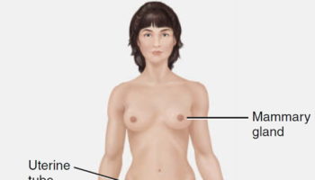
Figure 1. Male reproductive system
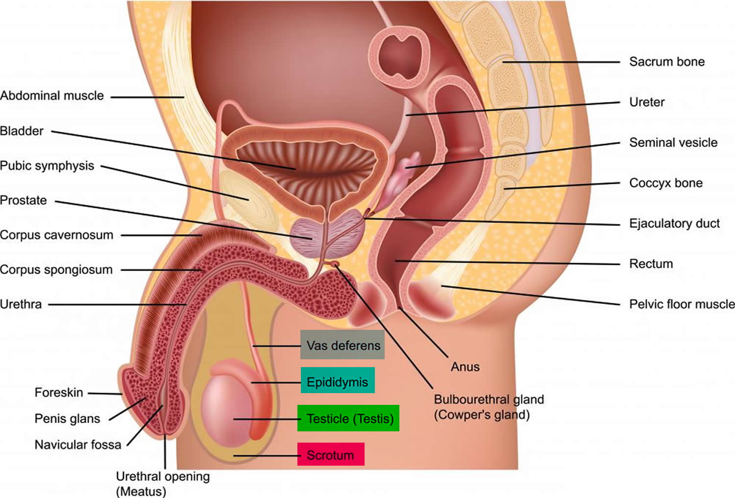
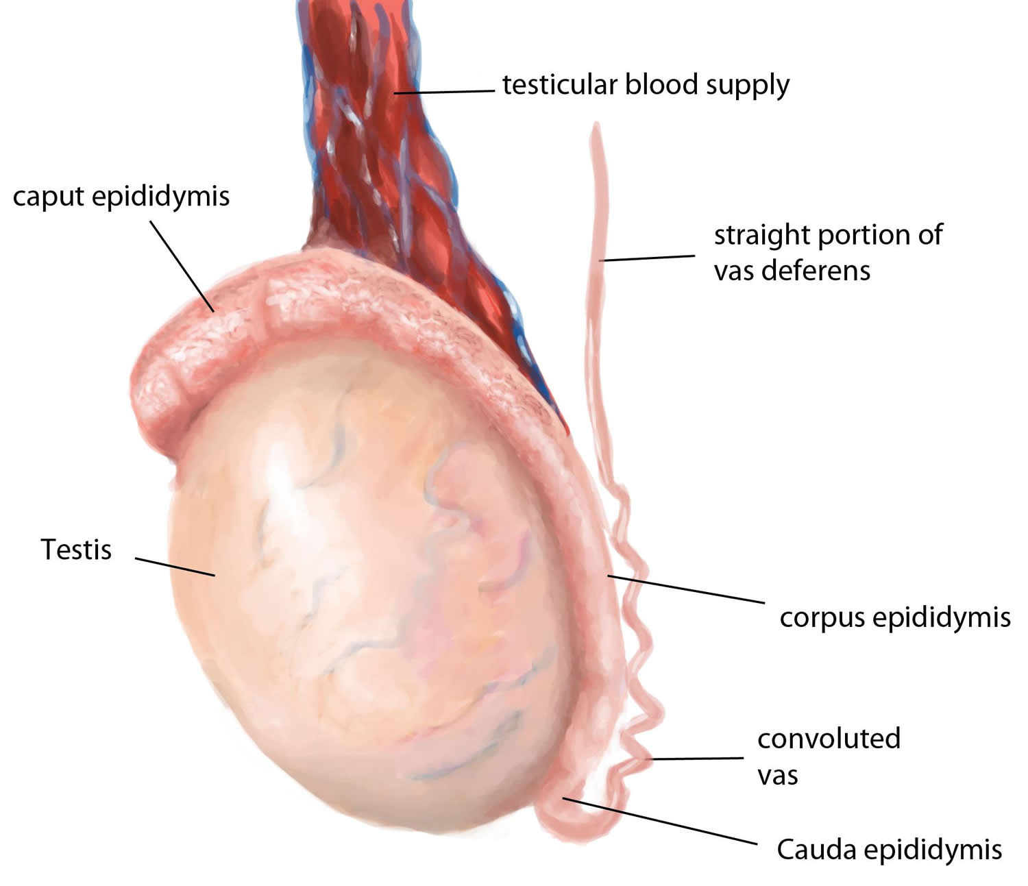
Figure 2. Testicle anatomy
Testis function
Testicles have 2 main functions:
- They make male hormones (androgens) such as testosterone.
- They make sperm, the male cells needed to fertilize a female egg cell to start a pregnancy.
Sperm cells are made in long, thread-like tubes inside the testicles called seminiferous tubules. They are then stored in a small coiled tube behind each testicle called the epididymis, where they mature.
During ejaculation, sperm cells are carried from the epididymis through the vas deferens to seminal vesicles, where they mix with fluids made by the vesicles, prostate gland, and other glands to form semen. This fluid then enters the urethra, the tube in the center of the penis through which both urine and semen leave the body.
The epithelium of the seminiferous tubules consists of sustentacular cells (Sertoli cells) and spermatogenic cells. Sustentacular cells support, nourish, and regulate the spermatogenic cells.
In the male embryo, the undifferentiated spermatogenic cells are called spermatogonia. Each spermatogonium contains 46 chromosomes (23 pairs) in its nucleus, the usual number for human body cells. Beginning during embryonic development, hormones stimulate spermatogonia to undergo mitosis. Each cell division gives rise to two new cells, one of which (type A) maintains the supply of undifferentiated cells, the other of which (type B) differentiates, becoming a
primary spermatocyte. Sperm cell production, or spermatogenesis, pauses at this stage.
Figure 3. Seminiferous tubule
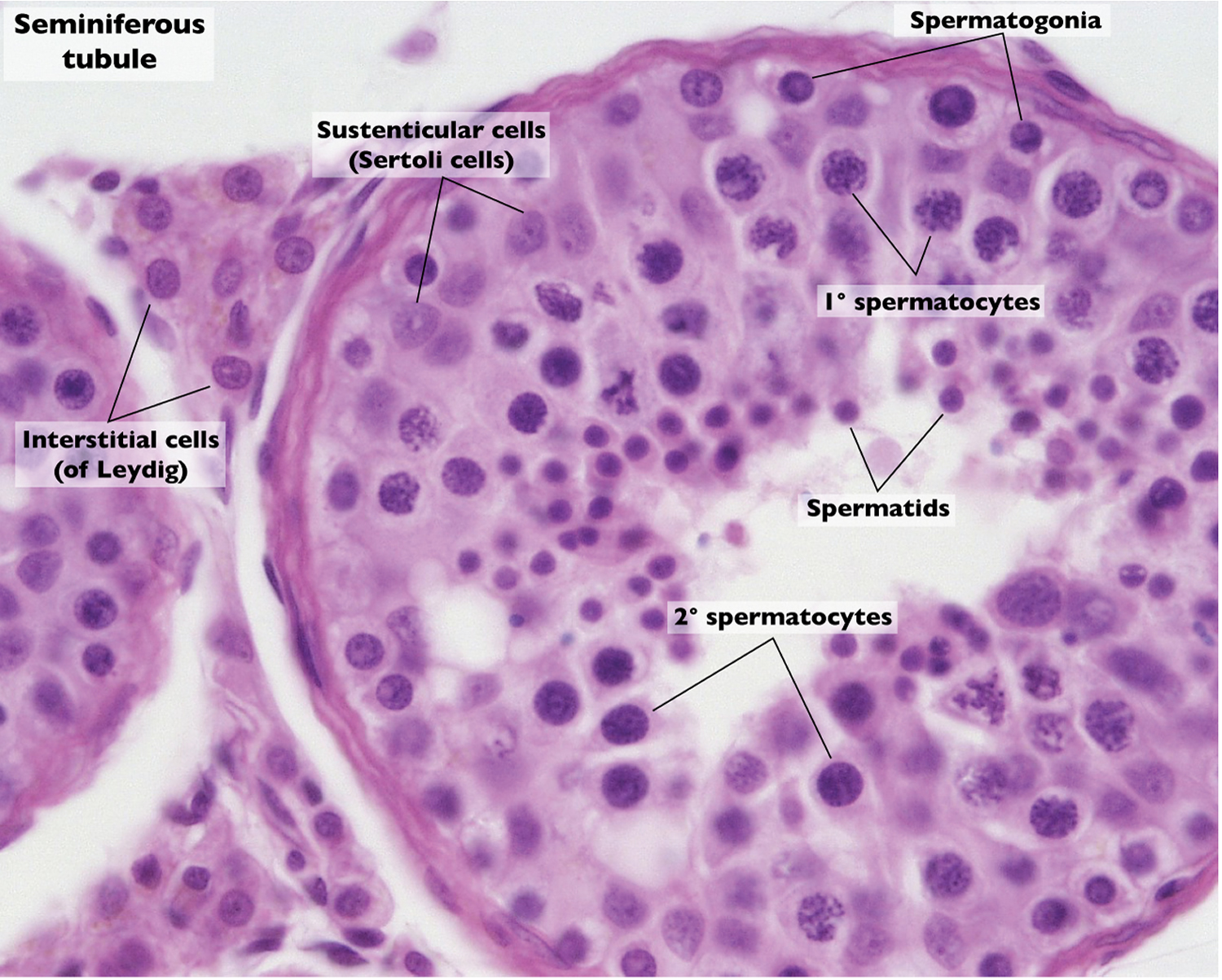
Figure 4. Spermatogenesis
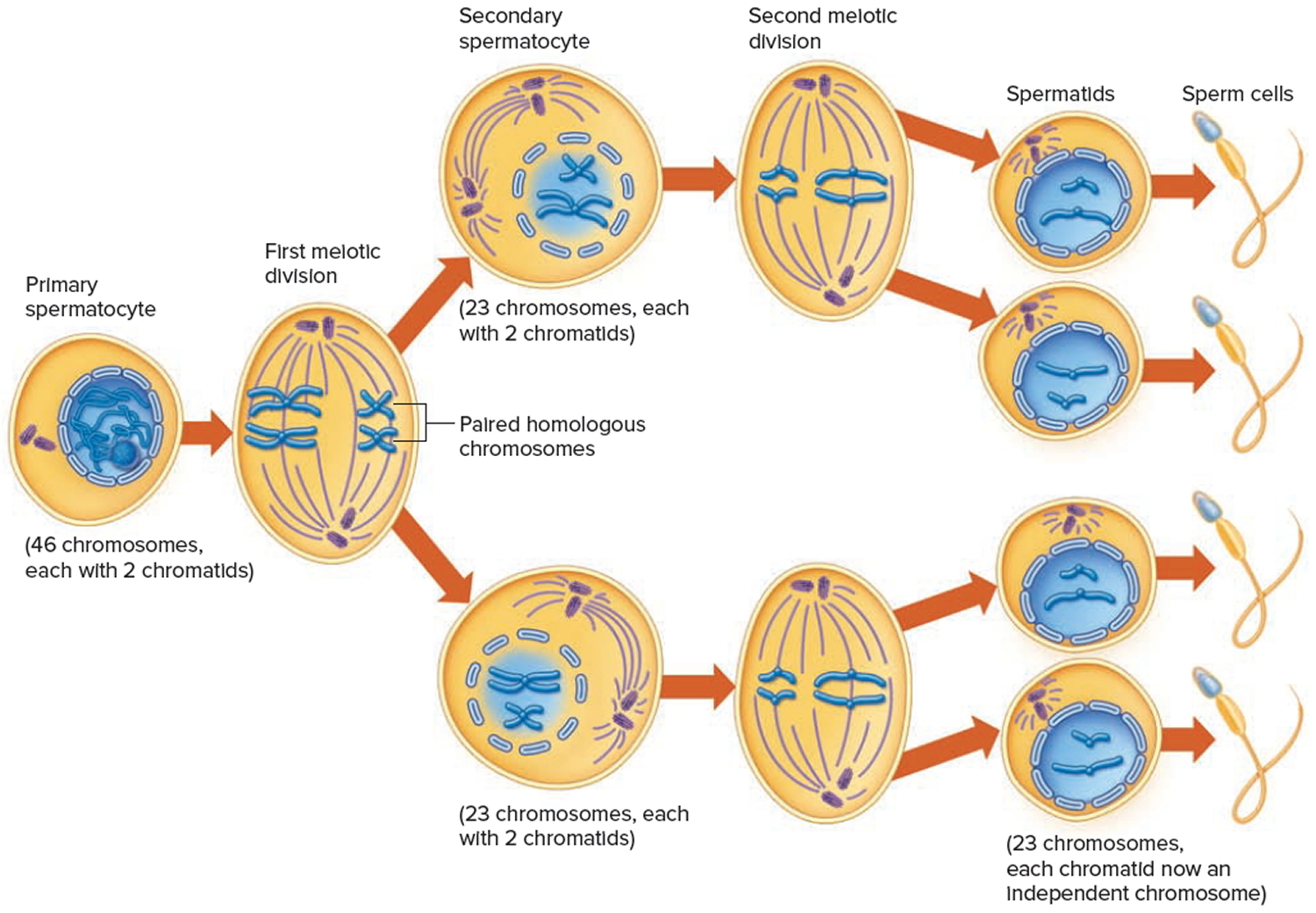
At puberty mitosis resumes, and new spermatogonia form. Testosterone secretion increases and the primary spermatocytes then reproduce by a special type of cell division called meiosis. Meiosis includes two successive divisions, called the first and second meiotic divisions.
Half of an individual’s 46 chromosomes are inherited from the mother (23), and half from the father (23). The 23 pairs of corresponding chromosomes, called homologous pairs, are the same, gene for gene. They may not be identical, however, because a gene may have variants, and a given chromosome that comes from the mother may carry a different variant of a gene than that chromosome from the father.
Before meiosis I, each homologous chromosome is replicated, so it consists of two identical DNA strands called chromatids. The chromatids of a replicated chromosome attach at regions called centromeres. The first meiotic division (meiosis I) separates homologous chromosome pairs. Thus, each of the secondary spermatocytes that undergoes the second meiotic division (meiosis II) begins with one member of each homologous pair, a condition termed haploid. This second division separates the chromatids, producing cells that are still haploid, but whose chromosomes are no longer in the replicated (two chromatids) form.
After meiosis II, each of the chromatids is an independent chromosome. The halving of chromosome number is accomplished as each primary spermatocyte divides to form two secondary spermatocytes. Each of these cells, in turn, divides to form two spermatids, which mature into sperm cells. Consequently, for the primary spermatocyte that undergoes meiosis, four sperm cells form, with 23 chromosomes each.
Spermatogenesis occurs continually in a male, starting at puberty. The resulting sperm cells collect in the lumen of each seminiferous tubule, and then pass to the epididymis, where they accumulate and mature.
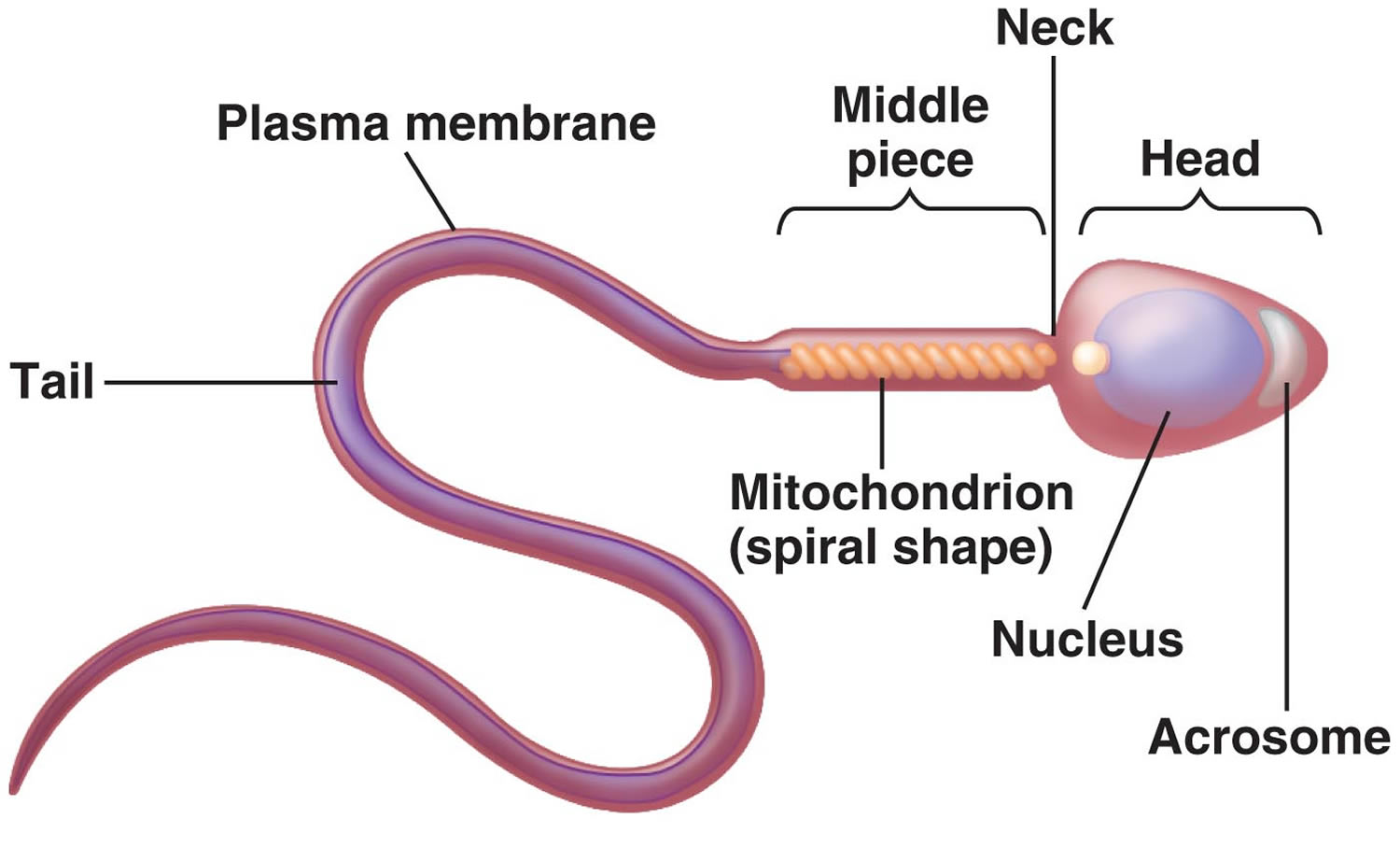
Structure of a Sperm Cell
A mature sperm cell is a tiny, tadpole-shaped structure about 0.06 millimeters long. It consists of a flattened head, a cylindrical midpiece (body), and an elongated tail. The oval head of a sperm cell is primarily composed of a nucleus and contains highly compacted chromatin consisting of 23 chromosomes. A small protrusion of its head, called the acrosome, contains enzymes that aid the sperm cell in penetrating the layers surrounding the oocyte (egg cell from the female ovary) during fertilization. One of the enzymes on the sperm cell membrane contributes to entering the oocyte.
The midpiece of a sperm cell has a central, filamentous core and many mitochondria organized in a spiral. The tail (flagellum) consists of several microtubules enclosed in an extension of the cell membrane. The mitochondria provide ATP for the tail’s lashing movement that propels the sperm cell through fluid.
Figure 5. Sperm Cell
Male Internal Accessory Reproductive Organs
The internal accessory organs of the male reproductive system are specialized to nurture and transport sperm cells. These structures include the two epididymides, two ductus deferentia, two ejaculatory ducts, and the urethra, as well as the two seminal vesicles, the prostate gland, and two bulbourethral glands.
Figure 6. Inside testicles
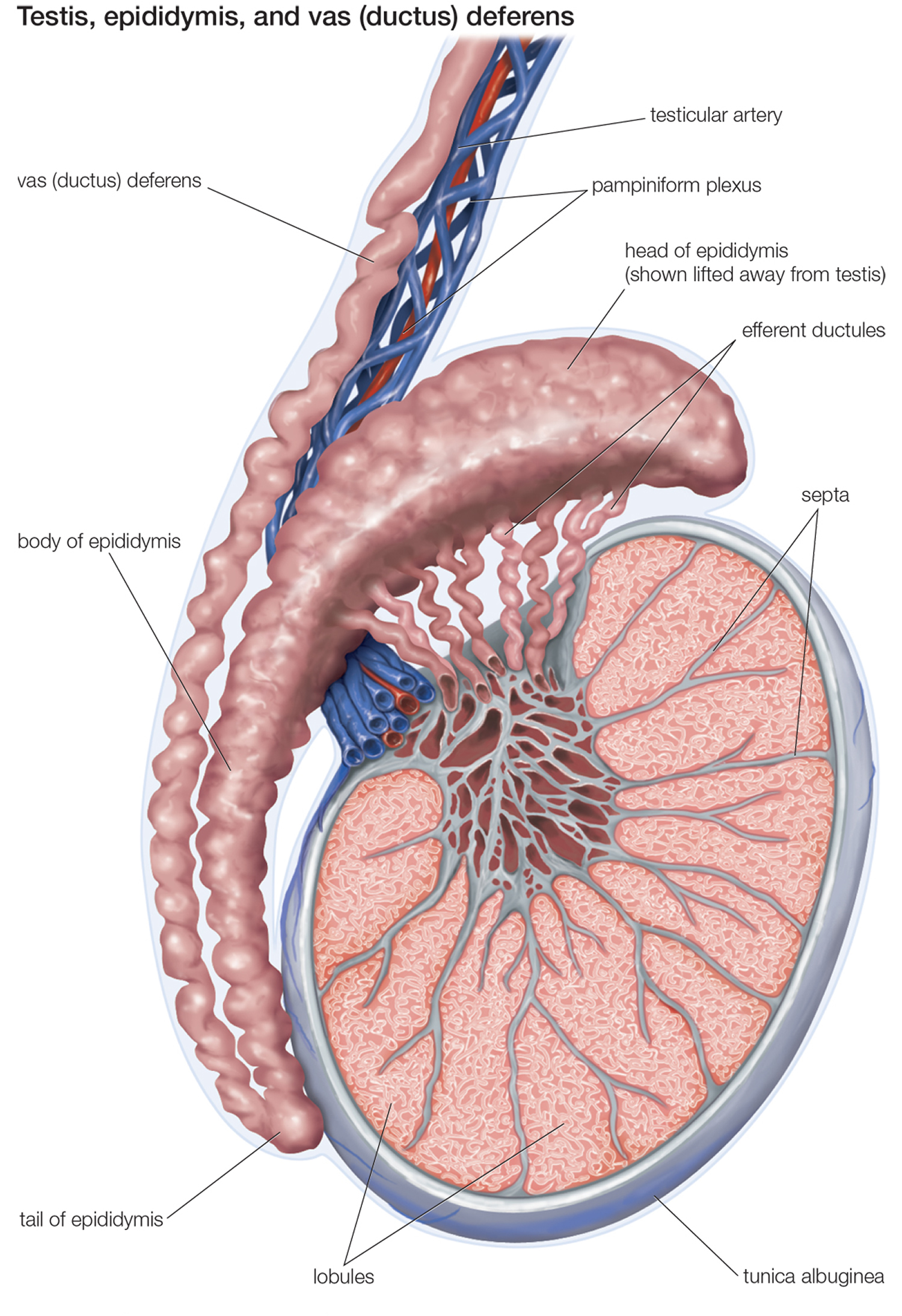
Epididymides
The epididymides (singular – epididymis) are tightly coiled tubes about 6 meters long. Each epididymis is connected to ducts within a testis. It emerges from the top of the testis, descends along the posterior surface of the testis, and then courses upward to become the ductus deferens.
When sperm cells reach the epididymis, they are nonmotile. As rhythmic peristaltic contractions help move these cells through the epididymis, the cells mature. Following this aging process, the sperm cells can move independently and fertilize oocytes. However, sperm cells usually do not move independently until after ejaculation.
Ductus Deferentia
The ductus deferentia (singular – ductus deferens), also called vasa deferentia, are muscular tubes about 45 centimeters long. Each ductus deferens passes upward along the medial side of a testis and through a passage in the lower abdominal wall (inguinal canal), enters the pelvic cavity, and ends behind the urinary bladder. Just outside the prostate gland, the ductus deferens unites with the duct of a seminal vesicle to form an ejaculatory duct, which passes through the prostate gland and empties into the urethra.
Seminal Vesicles
The seminal vesicles are convoluted, saclike structures about 5 centimeters long. Each attaches to a ductus deferens on the posterior surface and near the base of the urinary bladder. The glandular tissue lining the inner wall of a seminal vesicle secretes a slightly alkaline fluid. This fluid helps regulate the pH of the tubular contents as sperm cells travel to the outside. Additionally, seminal vesicle fluid neutralizes the acidic secretions of the vagina, helping to sustain sperm cells that enter the female reproductive tract. Seminal vesicle secretions also include fructose, a monosaccharide that provides energy to sperm cells, and prostaglandins which stimulate muscular contractions within the female reproductive organs, aiding the movement of sperm cells toward the oocyte.
