Contents
What is a hypertrophic scar
Hypertrophic scars are defined as visible and elevated scars that do not spread into surrounding tissues and that often regress spontaneously 1. Scar formation is a consequence of the wound healing process that occurs when body tissues are damaged by a physical injury. As wounds heal, scar tissue forms, which at first is often red and somewhat prominent. Over several months, a scar usually becomes flat and pale. If there is a lot of tension on a healing wound, the healing area is rather thicker and elevated than usual. This is known as a hypertrophic scar.
Hypertrophic scars are characterized by proliferation of the dermal tissue, with excessive deposition of fibroblast-derived extracellular matrix proteins and especially collagen, over long periods and by persistent inflammation and fibrosis 2.
The majority of individuals who develop hypertrophic scars and keloids are young, with ages ranging from 10 to 30 years old. The elderly rarely develop these lesions 3. This observation is partly attributed to the following facts: young individuals are more prone to trauma; their skin generally possesses more elastic fibers, resulting in greater tension; and the rate of collagen synthesis is greater in younger individuals 4. Keloids are more common in patients with darker skin, with an incidence of 4.5% to 16% in the black and Hispanic populations 4.
Hypertrophic scarring following surgical procedures, trauma and especially burns is a significant concern for patients and a challenging problem for clinicians because it can be painful, pruritic, erythematous, raised and cosmetically unacceptable. Hypertrophic scars are a common complication of burn injury. In the developed world, approximately four million patients acquire scars due to burns each year and the incidence is even greater in developing countries 5. Previous studies have reported diverging incidences of hypertrophic scarring, with incidence rates varying from 40% to 94% following surgery and from 30% to 91% following burns 6. A previous study reported that the most common and distressing complications in burn patients who developed hypertrophic scars were abnormal appearance (75.2%), pruritus (73.3%) and pain (67.6%) 7. The cause of pruritus (itchiness) in hypertrophic scars and keloid scars is not yet well characterized, but recent studies have indicated the probable involvement of direct activation of opioid receptors identified in the skin (46).
Numerous methods have been described for the treatment of hypertrophic scars, but to date, the optimal treatment method has not been established 1. A wide variety of treatments have been advocated for hypertrophic scars. Among these treatments are surgical excision with or without grafting 8, pressure therapy 9, intralesional interferon 5, topical and intralesional corticosteroids 10, intralesional bleomycin 11, laser therapy 12, silicone gel sheeting 13, onion extract gel and other therapies directed at collagen synthesis 14.
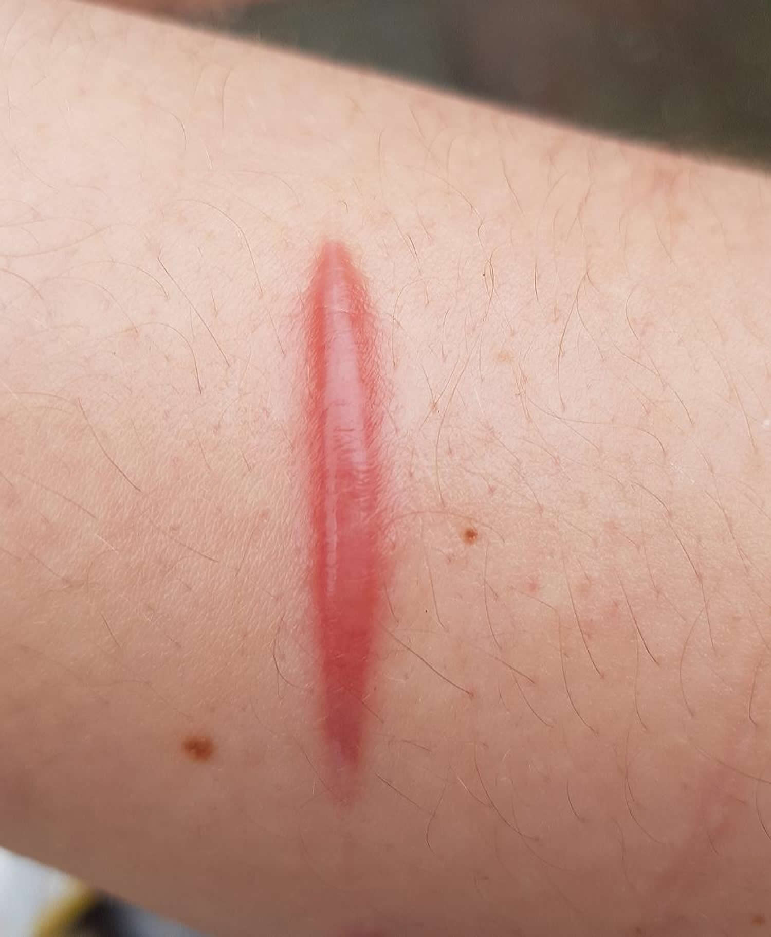
Figure 1. Hypertrophic scar
Hypertrophic scar vs Keloid
Keloid is a type of raised scar. A keloid scar is a firm, smooth, hard growth due to spontaneous scar formation. A keloid scar can arise soon after an injury, or develop months later. Keloids can grow to be much larger than the original injury that caused the scar. Keloids may be uncomfortable or itchy, and may be much larger than the original wound. Keloids may form on any part of the body, although the upper chest and shoulders are especially prone to them.
Anything that can cause a scar can cause a keloid. This includes being burned, cut, or having severe acne. Keloids can also develop after you get a body piercing or a tattoo, or have surgery. Keloids sometimes show up 3 months or more after your skin is injured. Some continue to grow for years.
The precise reason that wound healing sometimes leads to keloid formation is under investigation but is not yet clear.
While most people never form keloids, others develop them after minor injuries, burns, insect bites and acne spots. Dark skinned people form keloids more easily than Caucasians.
A keloid is harmless to general health and does not change into a skin cancer.
Keloids are not harmful to your health. But having keloids can be upsetting to you. You may be embarrassed about how they look. This can hurt your self-esteem. Most people who get treatment for keloids do so because they don’t like the way they look. Luckily, the treatments that are available can improve the way the keloids look, even if they don’t get rid of the scars completely.
Table 1. Hypertrophic Scars vs. Keloids
| Hypertrophic scars | Keloids |
|---|---|
Remain confined to border of original wound | Extend beyond border of original wound |
Arise in any location; commonly occur on extensor surfaces of joints | Commonly occur on the sternal skin, shoulders and upper arms, earlobes, and cheeks |
Regress with time | Grow for years |
Fewer thick collagen fibers | Thick collagen |
Scanty mucoid matrix | Mucoid matrix |
Flatten spontaneously in time | Remain elevated more than 4 mm |
Appear within one month | Appear at three months or later |
Less association with skin pigmentation | More common in darker skin types |
Can keloids be prevented or avoided?
People who are more likely to get keloids may decide not to get a body piercing or tattoo. If you get your ears pierced, you should wear special pressure earrings to reduce scarring on your earlobes.
Before any surgical procedure, patients should be asked if they have had previous problems with scarring. Discuss the potential for keloids as part of informed consent, and discourage ear piercing and other elective procedures in persons with dark skin. If ears are pierced despite this advice, pressure earrings are commercially available for reducing keloid risk. If surgery cannot be avoided in a high-risk patient, immediate silicone elastomer sheeting or corticosteroid injections should be instituted. Anything that expedites wound healing and diminishes skin tension (e.g., postsurgical taping for 12 weeks) will diminish risk 16. The cosmetic outcome of wounds closed with standard suture techniques appears to be similar to that of those closed with 2-octyl cyanoacrylate dermal adhesive (Dermabond). One small study showed that hypertrophic scars occurred in five out of 24 repairs with Dermabond versus three out of 28 repairs with traditional suture 17.
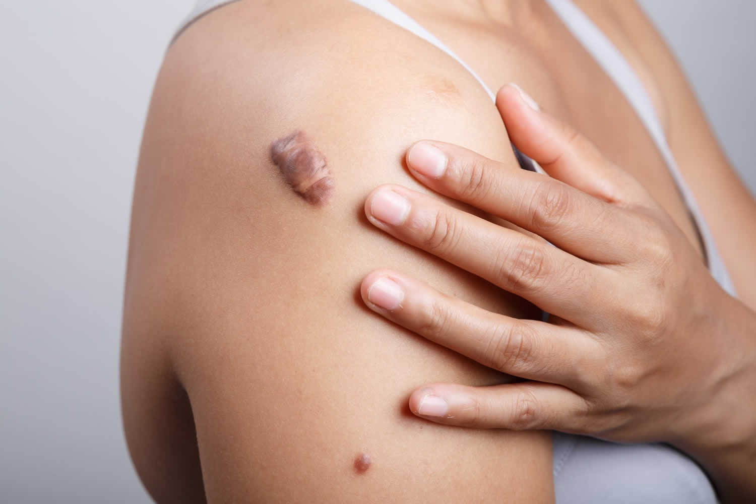
Figure 2. A keloid scar
What causes keloids?
After your skin is injured, your cells try to repair it by forming a scar. In some people, the scar tissue keeps forming long after the wound heals. This extra scar tissue causes the raised area on your skin that is called a keloid. Doctors still aren’t sure why some people’s skin scars this way.
Many different types of skin injuries can lead to a keloid. These include:
- cuts
- puncture wounds
- surgical scars
- severe acne
- chicken pox
- insect bites
- injection sites
- piercings
- tattoos
Some people are more likely to develop a keloid when they scar. You are more likely to develop a keloid if:
- You are black, Latino, or Asian.
- You are younger than 30 years of age.
- You are pregnant.
- You are a teenager going through puberty.
- You have a history of keloids in your family.
People who have darker skin are 15% to 20% more likely to develop keloids.
Risk factors for keloid
The primary risk factor for keloids is darkly pigmented skin, which carries a 15- to 20-fold increased risk, perhaps because of melanocyte-stimulating hormone anomalies 18. Familial predisposition, with autosomal dominant and recessive genetic variants is recognized 19. Black, Hispanic, and Asian persons are far more likely to develop keloids than white persons 20. Hypertrophic scars, however, are less likely to be associated with skin pigmentation.
Keloids are more common in persons younger than 30 years, with risk peaking between 10 to 20 years of age, and in patients with elevated hormone levels (e.g., during puberty or pregnancy) 21. Sternal skin, shoulders and upper arms, earlobes, and cheeks are most susceptible to developing keloids 22. Certain types of trauma and delayed healing (longer than three weeks) heighten keloid incidence even more, with burns carrying the highest risk. Acne, ear piercing, chickenpox, vaccinations (particularly bacille Calmette-Guérin [BCG] vaccination), biopsy procedures, and lacerations may cause abnormal scarring (Figure 2). Acne keloids are particularly common. Keloids are more than just cosmetically unacceptable; many are also pruritic and painful. They often result in severe emotional distress.
Symptoms of keloids
Keloids can have the following characteristics:
- Appear and grow slowly. It can take 3 months up to a year before you see the first signs of a keloid. Then it takes weeks or months for it to grow. Sometimes, they continue to grow slowly for years.
- Begin as a raised pink, red, or purple scar. A keloid is usually a raised scar with a flat surface. The color tends to darken with time. It usually ends up being darker than the person’s skin, with the border being darker than the center.
- Feel different than the surrounding skin. Some keloids feel soft and doughy. Others are hard and rubbery.
- Cause pain, itching, or tenderness. When they are growing, some keloids may be itchy, tender, or painful to the touch. These symptoms usually stop once the keloid stops growing.
Keloids can form anywhere on the body. They are most common on the neck, shoulders, chest, back, and ears. They can range in size from smaller than an inch to 12 inches or more.
How are keloids diagnosed?
Your doctor can diagnose a keloid by looking at your skin. Sometimes he or she may do a skin biopsy to rule out other types of skin growths.
How are keloids treated?
The goal of treatment is to flatten, soften, or shrink the keloid. Keloids can be hard to get rid of. Sometimes they return after treatment. Many doctors will use a combination of treatments for the best results. Treatments include the following:
- Corticosteroid shots. The medicine in these shots helps shrink the scar.
- Freezing the keloid scar. Called cryotherapy, this can be used to reduce the hardness and size of the keloid. It works best on small keloids.
- Wearing silicone sheets or gel over the keloid scar. This can help flatten the keloid.
- Laser therapy. This can help flatten the keloid. It also can fade the color.
- Surgical removal. This involves cutting out the keloid. Most keloids will return after this treatment.
- Pressure treatment. After keloid surgery, keeping pressure on the area reduces blood flow. This can help keep a keloid from returning.
Different treatments work for different people. Talk to your doctor about which treatment option is right for you.
How to distinguish between hypertrophic scar from keloid
When faced with patients seeking treatment for pathological scars, many physicians have difficulty in differentiating hypertrophic scars from keloids; therefore, it is crucial to establish criteria to distinguish them.
Hypertrophic scars are usually raised, although rarely elevated more than 4 mm above the skin; red or pink in color; hard; and itchy 1. Additionally, hypertrophic scars do not extend beyond the general geographic margins of the wound and tend to regress over time 23 (Figure 1). Hypertrophic scars primarily contain type III collagen, oriented parallel to the epidermal surface and with abundant nodules and large extracellular collagen filaments 24.
In contrast, keloids continue to evolve over time, without a quiescent or regressive phase and do infiltrate the surrounding tissue (Figure 2). Keloids appear as firm, mildly tender, bosselated scars with a shiny surface and occasional telangiectasia 1. The epithelium is thinned and there may be focal areas of ulceration. The color is pink to purple and may be accompanied by hyperpigmentation 25. The initial lesions are erythematous (reddish) and become brownish red, followed by paling as they age. Keloid lesions preferentially develop on the earlobes, shoulders and chest sternum skin; are void of hair follicles and other glands; and usually project above the level of the surrounding skin 26. Keloids are primarily composed of abnormally thick, irregularly branched and septal disorganized type I and III collagen bundles without nodules and with excess myofibroblasts 24 and overproduction of multiple fibroblast proteins, indicating the persistence of wound healing or even a failure to downregulate wound-healing cells. In addition, keloids are not triggered to enter the final phase of wound healing, or the “remodeling” phase, whereas hypertrophic scars will eventually do so 27.
Distinguishing hypertrophic scars from keloids histopathologically is occasionally difficult because thickened hyalinized collagen (keloidal collagen), the hallmark of keloids, is not always detectable and because α-smooth muscle actin (α-SMA), a differentiating marker of hypertrophic scars, is variably expressed in both types of scars 28.
The histopathological findings most commonly observed in hypertrophic scars are flattening of the epidermis and replacement of the papillary and reticular dermis by scar tissue with prominent vertically oriented blood vessels. In keloids, there is no flattening of the overlying epidermis, no scarring of the papillary dermis, the presence of a significant amount of keloidal collagen, an absence of prominent vertically oriented blood vessels and the presence of a significant disarray of fibrocollagenous fascicles 28.
Hypertrophic scar prevention
There is evidence suggesting that increased mechanical tension can initiate hypertrophic scar formation 29. Based on this hypothesis, it makes sense to minimize mechanical forces after surgery. Surgical excision scars should be positioned along, rather than across, relaxed skin tension lines whenever possible. An appropriate strength, depth and number of sutures should ensure that the risk of dehiscence is minimized.
Inflammation is also known to contribute to hypertrophic scarring 30 and every attempt to minimize the inflammatory response should be made by ascertaining clean surgery and good wound care to prevent infection thereafter. Using inert suture materials is also important in this context 31.
In patient candidates for skin grafts, the donor site must be well chosen by the surgeon in consultation with the patient to try to hide or avoid hypertrophic scars or keloids. In burn patients, the corresponding author believes that using the scalp as a source of thin skin grafts can reduce the level of visible aesthetic deformities at donor sites in patients who have already suffered the immense trauma that being a burn victim entails.
Conceptually and practically, treatment and prevention regimens can be similar and the following section presents the clinical data for both. Early diagnosis can considerably affect the outcome. There is evidence that the most successful non-surgical treatment of an hypertrophic scar or keloid is achieved when the scar is immature and the overlying epithelium is intact, although further studies are necessary to confirm this concept 32.
Hypertrophic scar treatment
Keloid and hypertrophic scar therapy is challenging and controversial (see Table 2). Both conditions respond to the same therapies, but hypertrophic scars are easier to treat. The large number of treatment options is a reflection of the poor quality of research on this topic, with no single proven best treatment or combination of treatments. First-line options include silicone sheeting, pressure treatment, and corticosteroid injections, but all of these require exemplary adherence and follow-up. Cryotherapy is useful, but only for smaller lesions, such as those resulting from acne. Cryotherapy may cause hypopigmentation in patients with dark skin. Surgical removal of keloids, although temporarily gratifying, is almost invariably followed (50 to 100 percent) by even more aggressive regrowth of scar tissue 21. Therefore, all surgical options should be followed by corticosteroid injections, silicone sheeting, or these options combined with pulsed dye laser. A variety of other choices are emerging, but are less well studied.
A hypertrophic scar generally settles in time or with treatment, but a keloid may persist and prove resistant to treatment. The following measures are helpful in at least some patients.
- Emollients (creams and oils)
- Polyurethane or silicone scar reduction patches
- Silicone gel
- Oral or topical tranilast (an inhibitor of collagen synthesis)
- Pressure dressings
- Surgical excision (but in keloids, excision may result in a new keloid even larger than the original one)
- Intralesional corticosteroid injection, repeated every few weeks
- Cryotherapy
- Superficial X-ray treatment soon after surgery.
- Pulsed dye laser
- Skin needling
- Subcision
Scar dressings should be worn for 12 to 24 hours per day, for at least 8 to 12 weeks, and perhaps for much longer.
Table 2. Prevention and Treatment Options for Keloids and Hypertrophic Scars
| Modality or treatment option | Response rate (%) | Recurrence rate (%) | Comments | Study design |
|---|---|---|---|---|
Prevention | ||||
Preventive silicone sheeting as postsurgery care | 0 to 75 | 25 to 36 | Multiple preparations available; tolerated by children | Review of multiple case studies 21 |
Expensive; should be avoided on open wounds; poor study designs | ||||
Postsurgical intralesional corticosteroid injection (triamcinolone acetonide [Kenalog] 10 to 40 mg per mL at six-week intervals) | NA | 0 to 100 (mean 50) | Patient acceptance and safety | Review of multiple case studies 22 |
May cause hypopigmentation, skin atrophy, telangiectasia | ||||
Postsurgical topical imiquimod 5% cream (Aldara) | NA | 28 | May cause hyperpigmentation, irritation | Case study 33 |
Postsurgical fluorouracil, triamcinolone acetonide, and pulsed dye laser (best outcomes) | 70 at 12 weeks | NA | Effective | Clinical trial 34 |
May cause hyperpigmentation, wound ulceration | ||||
First-line treatment | ||||
Cryotherapy | 50 to 76 | NA | Useful on small lesions; easy to perform | Review of multiple case studies 22 |
May cause hypopigmentation, pain | ||||
Intralesional corticosteroid injection (triamcinolone acetonide 10 to 40 mg per mL at six-week intervals) | 50 to 100 | 9 to 50 | Inexpensive; available in family physician’s office | Review of multiple case studies 22 |
Requires multiple injections | ||||
May cause discomfort, skin atrophy, telangiectasia | ||||
Silicone elastomer sheeting | 50 to 100 | NA | Multiple preparations available; tolerated by children | Review of multiple case studies 21 |
Expensive; poor study designs | ||||
Pressure dressing (24 to 30 mm Hg) worn for six to 12 months | 90 to 100 | NA | Inexpensive | Review of multiple case studies 22 |
Difficult schedule; poor adherence | ||||
Second-line and alternative treatment | ||||
Surgical excision | NA | 50 to 100 | Z-plasty option for burns | Review of multiple case studies 22 |
Immediate postsurgical treatment needed to prevent regrowth | ||||
Combined cryotherapy and intralesional corticosteroid injection | 84 | NA | See benefits of individual treatments | Case study 35 |
May cause hypopigmentation | ||||
“Triple keloid therapy” (surgery, corticosteroids, and silicone sheeting) | 88 at 13 months | 12.5 at 13 months | Tedious; time intensive; expensive | Case study 36 |
Pulsed dye laser | NA | NA | Specialist referral needed; expensive; variable results depending on trial (controversial) | Case studies 37 |
Verapamil 2.5 mg per mL intralesional injection combined with perilesional excision and silicone sheeting | 54 at 18 months | NA | Repeated injections; limited experience | Clinical trial 38 |
May cause discomfort | ||||
Fluorouracil 50 mg per mL intralesional injection two to three times per week | 88 | 0 | Effective | Review of multiple case studies 20 |
May cause hyperpigmentation, wound ulceration | ||||
Bleomycin tattooing 1.5 IU per mL | 92 | NA | Effective | Review 20 |
88 | May cause pulmonary fibrosis, cutaneous reactions | Case study 39 | ||
Control trial 40 | ||||
Postsurgical interferon alfa-2b 1.5 million IU intralesional injection twice daily for four days | 30 to 50 | 8 to 19 | Expensive | Review of multiple case studies 22 |
May cause pruritus, altered pigmentation, pain | ||||
Radiation therapy alone | 56 (mean) | NA | Local growth inhibition | Review of multiple case studies 22 |
May cause cancer, hyperpigmentation, paresthesias | ||||
Postsurgical radiation therapy | 76 | NA | Local growth inhibition | Review of multiple case studies 22 |
May cause cancer | ||||
Onion extract topical gels (e.g., Mederma) | NA | NA | Limited effect alone, better in combination with silicone sheeting | Prospective case study 41 |
Footnote: NA = not available
[Source 15 ]Hypertrophic scar home treatment
Many patients use topical vitamin E (alpha-tocopherol) hoping its antioxidant properties will prevent scars. However, there is little evidence that it is helpful, and some patients develop a contact dermatitis that may delay healing 42. Used early on, vitamin E may also reduce the tensile strength of the scar, and its use should be discouraged 15.
Another over-the-counter option is onion extract topical gels (e.g., Mederma), but limited clinical trials have failed to demonstrate any clinical improvement in scar height, erythema, or pruritis 43. Contractubex gel contains onion extract with heparin, which is thought to promote scar maturity. Although one trial compared this product favorably with corticosteroids, another showed that it was ineffective in improving scar height and itching 41.
Moist exposed burn ointment contains multiple herbs with betasitosterol, which provides hydration and possible benefits to wound healing 44. Another plant extract product contains Centella asiatica and Bulbine frutescens (Alpha Centella cream), which may increase wound strength if used in the first six to eight weeks 45. All of these commercially available products emphasize preventive use because they are unlikely to reverse well-established keloids.
Corticosteroid injections
Corticosteroid injections for prevention and treatment of keloids and hypertrophic scars are perhaps the first-line option for family physicians. Corticosteroids suppress inflammation and mitosis while increasing vasoconstriction in the scar. Triamcinolone acetonide suspension (Kenalog) 10 to 40 mg per mL (depending on the site) is injected intralesionally, which, although painful, will eventually flatten 50 to 100 percent of keloids, with a 9 to 50 percent recurrence rate 46. Lidocaine (Xylocaine) may be combined with the corticosteroid to lessen pain, whereas using adjunctive cryotherapy immediately before injection may make the procedure easier by softening the scar (based on expert opinion) 47. Combining cryotherapy and corticosteroid injections also improves outcomes more than either modality alone, although hypopigmentation is always a significant concern 48. Usually, two or three injections are given a month apart; however, therapy can continue for six months or longer 49. Newer keloids are more responsive to therapy than older, established lesions. Corticosteroid injections are more effective if combined with surgery; the sooner instituted, the greater the likelihood of success. Common adverse effects include atrophy, telangiectasias, and hypopigmentation.
Silicone sheeting
Silicone elastomer sheeting is a noninvasive and extensively studied approach to the prevention and treatment of keloids and hypertrophic scars. Silicone sheets are thought to work by increasing the temperature, hydration, and perhaps the oxygen tension of the occluded scar, causing it to soften and flatten 21. This technique should be avoided on open wounds, but can be applied as soon as the skin heals. More than 60 products have been marketed, including silicone sheets, strips, gels, sprays, and foams. Most are available over the counter, but can be expensive. To be effective, sheets must be worn over the scar for 12 to 24 hours per day for two to three months 21. The sheet and the scar should be washed daily with mild soap and water. The sheets can be reused until they start to disintegrate. Although most studies suggest silicone sheeting results in fewer scars in persons at risk, a recent Cochrane review concluded that most research in this area was of poor quality and highly susceptible to bias 50. Similar to silicone sheeting is the use of pressure dressings or garments, especially for the prevention of burn scars. However, pressure dressings (24 to 30 mm Hg) must be worn for six to 12 months, which is difficult and uncomfortable for most patients 51.
Pressure garments
Using mechanical compressive force exerted by pressure garments to treat hypertrophic scars in burn patients was first described in 1860 52. It was only in the 1960s that this treatment became standard in several burn centers to accelerate the remodeling phase of wound healing 52. Prophylactic pressure is recommended in burn patients if spontaneous closure of the wound takes longer than 10 to 14 days and in those requiring grafting 53.
Currently, elastic compression using elastic garments is the predominant means of both prophylaxis and treatment for hypertrophic scars 54, despite controversial evidence-based data about their value in reducing the prevalence or magnitude of scarring 54. In fact, studies investigating pressure garments have found no significant difference between treatments involving the use of high-pressure garments, lower-pressure garments, or no pressure at all 55. Others, however, claim that pressure therapy achieves hypertrophic scar regression success rates of 60% to 85% 54, without any conclusive evidence.
To date, the working mechanism of pressure and the way that pressure positively influences the development and maturation of hypertrophic scars are not fully understood and explanations remain hypothetical. However, many studies have been performed to try to explain the possible mechanisms of action, exploring theories based on hypoxia, biochemical changes, and cellular and collagenous influences. Certain valuable evidence suggests that pressure controls collagen synthesis by limiting the supply of blood, oxygen, and nutrients to the scar tissue 56; reduces collagen production to the levels found in normal scar tissue more rapidly than the natural maturation process does; encourages the realignment of collagen bundles that are already present 57; partly restores the extracellular matrix organization observed in normal scarring; and induces the disappearance of fibrogenic α-SMA-expressing myofibroblasts and vascular cells, most likely by apoptosis 58.
Additionally, certain studies have demonstrated that mechanical compression directly modulates the remodeling phase of wound healing, altering the release and activity of matrix metalloproteinase (MMP)-28 in hypertrophic scars and inducing a significant reduction in the protein’s presence in keratinocytes in hypertrophic scars 59. Moreover, it has been suggested that pressure acts by accelerating the remission phase of the postburn reparative process 57.
Currently, the recommendations for the clinical use of pressure garments are restricted to deep dermal wounds that have healed spontaneously over weeks, grafted wounds surrounded by a deep dermal wound that was permitted to heal spontaneously over weeks, wounds in children and young adults, wounds in individuals with dark skin and wounds in body locations where compression can be applied 60. The amount of effective pressure generated by a given pressure garment is also still unknown and remains controversial 61. Problems with pressure loss from the garments over time and problems with the compliance of the patients using the garments are yet other factors complicating the issue 60.
Hypertrophic scar removal with combination therapy
If neither silicone nor corticosteroids are effective over 12 months, second-line surgical treatment followed by corticosteroids and possibly silicone sheeting should be considered. The use of corticosteroid injections following keloid surgery reduces the recurrence rate to lless than 50 percent 62. Scar excision may be complete, or a minute remnant of scar may be left on the wound margin, which may reduce recurrence (based on expert opinion) 15. Immediate wound edge corticosteroid injection after the excision is followed by weekly injections for two to five weeks and monthly injections for three to six months 22. A “triple keloid therapy” combining surgery, corticosteroids, and silicone sheeting has been shown to be even more effective, with only a 12.5 percent recurrence rate after 13 months 36. However, this approach was described as tedious and time intensive by the author of the study and requires a motivated patient.
Imiquimod 5% cream
Imiquimod 5% cream (Aldara), an immune response modifier that enhances healing, has also been used to help prevent keloid recurrence after surgical excision 33. Imiquimod 5% cream is applied on alternate nights for eight weeks after surgery. Although the trials have been small, the postsurgical recurrence rate averaged only 28 percent over a six- to nine-month follow-up period, with best results (2.9 percent recurrence) in low skin tension areas such as earlobes 33. Adverse effects include irritation and hyperpigmentation.
Pulsed dye laser
Treatment of keloids with short-pulsed, 585-nm pulsed dye laser has shown limited promise, with a 57 to 83 percent improvement rate 63. It is more vascular-specific than other laser therapies and appears to be most effective if used early and in conjunction with other techniques. Laser-treated portions of keloidal median sternotomy scars showed significant improvement in erythema, pruritus, and scar height compared with untreated portions of the same scars, and these improvements persisted for at least six months 37. The principal effect of a pulsed dye laser is on scar microvasculature, reducing erythema and pruritus and improving skin texture. The effectiveness of this therapy remains controversial, however, with other studies showing insignificant reduction in scar thickness 64. Disadvantages include significant expense and availability only through a specialist.
Other therapies
Other therapies with limited studies include intralesional verapamil, fluorouracil, bleomycin, and interferon alfa-2b injections. Although all of these have results comparable or sometimes superior to corticosteroid injection and silicone sheeting, the optimal keloid therapy remains undefined. Combinations of therapies have proved superior to individual approaches.
Intralesional verapamil (2.5 mg per mL) in conjunction with silicone sheeting reduced keloid postsurgical recurrence by 90 percent at 18 months (54 percent of patients were keloid-free; 36 percent had partial success) compared with only 18 percent showing any improvement with silicone sheeting alone (no patients were keloid-free) 38. Calcium antagonists appear to work by reducing collagen production and may be a reasonable and safe alternative to corticosteroid injection in the future.
Intralesional fluorouracil (50 mg per mL, two to three times per week) appears to shrink keloids safely while avoiding the tissue atrophy and telangiectasia that may occur with repeated corticosteroid injections 65. Combining fluorouracil with corticosteroid injections and pulsed dye laser produced superior results more rapidly than corticosteroid injections alone or corticosteroids with fluorouracil 34. Good to excellent responses at 12 weeks as rated by a blinded observer were 15 percent for triamcinolone acetonide, 40 percent for triamcinolone plus fluorouracil, and 70 percent for all three modalities (all significant). Combining corticosteroids and fluorouracil diminished the adverse effects of corticosteroids. Rare skin complications of fluorouracil may include hyperpigmentation and wound ulceration. No systemic adverse effects (e.g., anemia, leucopenia, thrombocytopenia) occurred in this study.
Bleomycin is another useful chemotherapeutic agent; a standard approach is bleomycin tattooing 0.1 mL (1.5 IU per mL) over two to six sessions, with a maximal dose of 6 mL 66. Results of one study showed a total regression of 84 percent 39. Multiple intralesional punctures are probably safe because it is likely that less than 5 percent of the dose ever reaches the bloodstream.18 Compared with triamcinolone injections combined with cryotherapy, bleomycin tattoo performed significantly better for keloids larger than 100 mm² 40. Systemically administered bleomycin is capable of causing pulmonary fibrosis (at doses greater than 400 U) and various cutaneous reactions (at doses of 200 to 300 U), including hair loss, hyperpigmentation, fibrosis, and vasospasm, any of which warrants cessation of treatment 67.
Intralesional interferon alfa-2b (1.5 million IU twice daily for four days) reduced keloid size by 50 percent over nine days, proving superior to intralesional corticosteroids 66. Interferon alfa-2b was also more effective than corticosteroids for preventing keloid recurrence after excision. Injection pain and expense (about $100 per treatment) are the main concerns. A liposome-encapsulated interferon alfa-2b cream is also being investigated for scar reduction 68.
Radiation, alone or (more commonly) after keloid excision, is a much more controversial option. It may pose a risk of local growth inhibition in children and possibly subsequent cancer. Commondoses range between 1,500 to 2,000 rads over five to six sessions following surgery 62. The success rate for radiation alone is 56 percent (range of 10 to 94 percent), but this increases to 76 percent (range of 25 to 100 percent) if administered immediately after surgery 22. Another study showed a 67 percent success rate with radiation, increasing to 75 percent if delivered within 48 hours of surgery 69. Most physicians would reserve radiation as a last resort for keloids refractory to all other approaches.
- Rabello FB, Souza CD, Farina Júnior JA. Update on hypertrophic scar treatment. Clinics (Sao Paulo). 2014;69(8):565-73. https://www.ncbi.nlm.nih.gov/pmc/articles/PMC4129552/[↩][↩][↩][↩]
- Atiyeh BS. Nonsurgical management of hypertrophic scars: evidence-based therapies, standard practices, and emerging methods. Aesthetic Plast Surg. 2007;31(5):468–92[↩]
- Rockwell WB, Cohen IK, Ehrlich HP. Keloids and hypertrophic scars: a comprehensive review. Plast Reconstr Surg. 1989;84(5):827–37[↩]
- Aarabi S, Longaker MT, Gurtner GC. Hypertrophic scar formation following burns and trauma: new approaches to treatment. PLoS Med. 2007;4(9):e234[↩][↩]
- Leventhal D, Furr M, Reiter D. Treatment of keloids and hypertrophic scars: a meta-analysis and review of the literature. Arch Facial Plast Surg. 2006;8(6):362–8.[↩][↩]
- Miller MC, Nanchahal J. Advances in the modulation of cutaneous wound healing and scarring. BioDrugs. 2005;19(6):363–81.[↩]
- Forbes-Duchart L, Cooper J, Nedelec B, Ross L, Quanbury A. Burn therapists’ opinion on the application and essential characteristics of a burn scar outcome measure. J Burn Care Res. 2009;30(5):792–800.[↩]
- English RS, Shenefelt PD. Keloids and hypertrophic scars. Dermatol Surg. 1999;25(8):631–8.[↩]
- Anzarut A, Olson J, Singh P, Rowe BH, Tredget EE. The effectiveness of pressure garment therapy for the prevention of abnormal scarring after burn injury: a meta-analysis. J Plast Reconstr Aesthet Surg. 2009;62:77–84.[↩]
- Jalali M, Bayat A. Current use of steroids in management of abnormal raised skin scars. Surgeon. 2007;5(3):175–80.[↩]
- Naeini FF, Najafian J, Ahmadpour K. Bleomycin tattooing as apromising therapeutic modality in large keloids and hypertrophicscars. Dermatol Surg. 2006;32(8):1023–9. discussion 1029-30.[↩]
- Leclère FM, Mordon SR. Twenty-five years of active laser prevention of scars: what have we learned. J Cosmet Laser Ther. 2010;12:227–34.[↩]
- O′Brien L, Pandit A. Silicon gel sheeting for preventing and treating hypertrophic and keloid scars. Cochrane Database Syst Rev. 2013;9:CD003826[↩]
- Shih R, Waltzman J, Evans GR. Plastic Surgery Educational Foundation Technology Assessment Committee Review of over-the-counter topical scar treatment products. Plast Reconstr Surg. 2007;119(3):1091–5.[↩]
- Management of Keloids and Hypertrophic Scars. Am Fam Physician. 2009 Aug 1;80(3):253-260. https://www.aafp.org/afp/2009/0801/p253.html[↩][↩][↩][↩]
- Atkinson JA, McKenna KT, Barnett AG, McGrath DJ, Rudd M. Randomized controlled trial to determine the efficacy of paper tape in preventing hypertrophic scar formation in surgical incisions that transverse Langer’s skin tension lines. Plast Reconstr Surg. 2005;116(6):1648–1656.[↩]
- Bernard L, Doyle J, Friedlander SF, Eichenfield LF, Gibbs NF, Cunningham BB. Prospective comparison of octyl cyanoacrylate tissue adhesive (Dermabond) and suture for the closure of excisional wounds in children and adolescents. Arch Dermatol. 2001;137(9):1177–1180.[↩]
- Kelly AP. Keloids and hypertrophic scars. In: Parish LC, Lask GP, eds. Aesthetic Dermatology. New York, NY: McGraw-Hill; 1991:8–69.[↩]
- Omo-Dare P. Genetic studies on keloid. J Natl Med Assoc. 1975;67(6):428–432.[↩]
- Butler PD, Longaker MT, Yang GP. Current progress in keloid research and treatment. J Am Coll Surg. 2008;206(4):731–741.[↩][↩][↩]
- Berman B, Perez OA, Konda S, et al. A review of the biologic effects, clinical efficacy, and safety of silicone elastomer sheeting for hypertrophic and keloid scar treatment and management. Dermatol Surg. 2007;33(11):1291–1303.[↩][↩][↩][↩][↩][↩]
- Niessen F, Spauwen P, Schalkwijk J, Kon M. On the nature of hypertrophic scars and keloids: a review. Plast Reconstr Surg. 1999;104(5):1435–1458.[↩][↩][↩][↩][↩][↩][↩][↩][↩][↩][↩]
- Ehrlich HP, Desmouliere A, Diegelmann RF, Cohen IK, Compton CC, Garner WL, et al. Morphological and immunochemical differences between keloid and hypertrophic scar. Am J Pathol. 1994;145(1):105–13.[↩]
- Gauglitz GG, Korting HC, Pavicic T, Ruzicka T, Jeschke MG. Hypertrophic Scarring and Keloids: Pathomechanisms and Current and Emerging Treatment Strategies. Mol Med. 2011;17(1-2):113–25.[↩][↩]
- Al-Attar A, Mess S, Thomassen JM, Kauffman CL, Davison SP. Keloid pathogenesis and treatment. Plast Reconstr Surg. 2006;117:286–300.[↩]
- Burd A, Huang L. Hypertrophic response and keloid diathesis: two very different forms of scar. Plast Reconstr Surg. 2005;116(7):150e–57e.[↩]
- Lane JE, Waller JL, Davis LS. Relationship between age of ear piercing and keloid formation. Pediatrics. 2005;115(5):1312–4[↩]
- Lee JY, Yang CC, Chao SC, Wong TW. Histopathological differential diagnosis of keloid and hypertrophic scar. Am J Dermatopathol. 2004;26(5):379–84.[↩][↩]
- Aarabi S, Bhatt KA, Shi Y, Paterno J, Chang EI, Loh SA, et al. Mechanical load initiates hypertrophic scar formation through decreased cellular apoptosis. FASEB J. 2007;21(12):3250–61.[↩]
- Wang J, Hori K, Ding J, Huang Y, Kwan P, Ladak A, et al. Toll-like receptors expressed by dermal fibroblasts contribute to hypertrophic scarring. J Cell Physiol. 2011;226(5):1265–73.[↩]
- Tziotzios C, Profyris C, Sterling J. Cutaneous scarring: pathophysiology, molecular mechanisms and scar reduction therapeutics Part II. Strategies to reduce scar formation after dermatologic procedures. J Am Acad Dermatol. 2012;66(1):13–24. Jaad 2012.[↩]
- Mustoe TA, Cooter RD, Gold MH, Hobbs FD, Ramelet AA, Shakespeare PG, et al. International clinical recommendations on scar management. Plast Reconstr Surg. 2002;110(2):560–7.[↩]
- Chuangsuwanich A, Gunjittisomrarn S. The efficacy of 5% imiquimod cream in the prevention of recurrence of excised keloids. J Med Assoc Thai. 2007;90(7):1363–1367.[↩][↩][↩]
- Asilian A, Daroughheh A, Shariati F. New combination of triamcinolone, 5-fluorouracil, and pulsed-dye laser for treatment of keloid and hypertrophic scars. Dermatol Surg. 2006;32(7):907–915.[↩][↩]
- Jackson IT, Bhageshpur R, DiNick V, Khan A, Bhaloo S. Investigation of recurrence rates among earlobe keloids utilizing various postoperative therapeutic modalities. Eur J Plast Surg. 2001;24(2):88–95.[↩]
- Agbenorku P. Triple keloid therapy: a combination of steroids, surgery and silicone gel strip/sheet for keloid treatment. Eur J Plast Surg. 2000;23(3):150–151.[↩][↩]
- Alster TS, Williams CM. Treatment of keloid sternotomy scars with 585 nm flashlamp-pumped pulsed-dye laser. Lancet. 1995;345(8959):1198–1200.[↩][↩]
- D’Andrea F, Brongo S, Ferraro G, Baroni A. Prevention and treatment of keloids with intralesional verapamil. Dermatology. 2002;204(1):60–62.[↩][↩]
- Espana A, Solano T, Quintanilla E. Bleomycin in the treatment of keloids and hypertrophic scars by multiple needle punctures. Dermatol Surg. 2001;27(1):23–27.[↩][↩]
- Naeini FF, Najafian J, Ahmadpour K. Bleomycin tattooing as a promising therapeutic modality in large keloids and hypertrophic scars. Dermatol Surg. 2006;32(8):1023–1029.[↩][↩]
- Hosnuter M, Payasli C, Isikdemir A, Tekerekoglu B. The effects of onion extract on hypertrophic and keloid scars. J Wound Care. 2007;16(6):251–254.[↩][↩]
- Khoosal D, Goldman R. Vitamin E for treating children’s scars. Does it help reduce scarring? Can Fam Physician. 2006;52:855–856.[↩]
- Saulis AS, Mogford JH, Mustoe TA. Effect of Mederma on hypertrophic scarring in a rabbit ear model. Plast Reconstr Surg. 2002;110(1):177–183.[↩]
- Jurjus A, Atiyeh BS, Abdallah IM, et al. Pharmacological modulation of wound healing in experimental burns. Burns. 2007;33(7):892–907.[↩]
- Widgerow AD, Chait LA, Stals R, Stals PJ. New innovations in scar management. Aesthetic Plast Surg. 2000;24(3):227–234.[↩]
- Niessen F, Spauwen P, Schalkwijk J, Kon M. On the nature of hypertrophic scars and keloids: a review. Plast Reconstr Surg. 1999;104(5):1435–1458[↩]
- Lahiri A, Tsiliboti D, Gaze NR. Experience with difficult keloids. Br J Plast Surg. 2001;54(7):633–635.[↩]
- Layton AM. A review on the treatment of acne vulgaris. Int J Clin Pract. 2006;60(1):64–72.[↩]
- Sherris DA, Larrabee WF Jr, Murakami CS. Management of scar contractures, hypertrophic scars, and keloids. Otolaryngol Clin North Am. 1995;28(5):1057–1068.[↩]
- O’Brien L, Pandit A. Silicon gel sheeting for preventing and treating hypertrophic and keloid scars. Cochrane Database Syst Rev. 2006;(1):CD003826.[↩]
- McIntyre L, Baird M. Pressure garments for use in the treatment of hypertrophic scars—a review of the problems associated with their use. Burns. 2006;32(1):10–15.[↩]
- Linares HA, Larson DL, Willis-Galstaun BA. Historical notes on the use of pressure in the treatment of hypertrophic scars or keloids. Burns. 1993;19(1):17–21.[↩][↩]
- Atiyeh BS. Nonsurgical management of hypertrophic scars: evidence-based therapies, standard practices, and emerging methods. Aesthetic Plast Surg. 2007;31(5):468–92.[↩]
- Esselman PC, Thombs BD, Magyar-Russell G, Fauerbach JA. Burn rehabilitation: State of the science. Am J Phys Med Rehabil. 2006;85(4):383–413.[↩][↩][↩]
- Macintyre L, Baird M. Pressure garments for use in the treatment of hypertrophic scars: A review of the problems associated with their use. Burns. 2006;32(1):10–5.[↩]
- Puzey G. The use of pressure garments on hypertrophic scars. J Tissue Viability. 2002;12(1):11–5.[↩]
- Costa AM, Peyrol S, Porto LC, Comparin JP, Foyatier JL, Desmouliere A. Mechanical forces induce scar remodeling. Study in non–pressure- treated versus pressure-treated hypertrophic scars. Am J Pathol. 1999;155(5):1671–9.[↩][↩]
- Reno F, Sabbatini M, Lombardi F, Stella M, Pezzuto C, Magliacani G, et al. In vitro mechanical compression induces apoptosis and regulates cytokines release in hypertrophic scars. Wound Rep Reg. 2003;11(5):331–6.[↩]
- Reno F, Sabbatini M, Stella M, Magliacani G, Cannas M. Effect of in vitro mechanical compression on Epilysin (matrix metalloproteinase-28) expression in hypertrophic scars. Wound Repair Regen. 2005;13(3):255–61.[↩]
- Engrav LH, Heimbach DM, Rivara FP, Moore ML, Wang J, Carrougher GJ, et al. 12-Year within-wound study of the effectiveness of custom pressure garment therapy. Burns. 2010;36(7):975–83.[↩][↩]
- Van den Kerckhove E, Stappaerts K, Fieuws S, Laperre J, Massage P, Flour M. The assessment of erythema and thickness on burn related scars during pressure garment therapy as a preventive measure for hypertrophic scarring. Burns. 2005;31(6):696–702.[↩]
- Mustoe T, Cooter RD, Gold MH, et al., for the International Advisory Panel on Scar Management. International clinical recommendations on scar management. Plast Reconstr Surg. 2002;110(2):560–571.[↩][↩]
- Alster TS. Improvement of erythematous and hypertrophic scars by the 585-nm flashlamp-pumped pulsed dye laser. Ann Plast Surg. 1994;32(2):186–190.[↩]
- Allison KP, Kiernan MN, Waters RA, Clement RM. Pulsed dye laser treatment on burn scars. Alleviation or irritation? Burns. 2003;29(3):207–213.[↩]
- Fitzpatrick RE. Treatment of inflamed hypertrophic scars using intralesional 5-FU. Dermatol Surg. 1999;25(3):224–232.[↩]
- Atiyah BS. Nonsurgical management of hypertrophic scars: evidence-based therapies, standard practices, and emerging methods. Aesthetic Plast Surg. 2007;31(5):468–492.[↩][↩]
- Yamamoto T. Bleomycin and the skin. Br J Dermatol. 2006;155(5):869–875.[↩]
- Lee J, Jalili R, Tredget E, Demare JR, Ghahary A. Antifibrogenic effects of liposome-encapsulated IFN-alpha2b cream on skin wounds in a fibrotic rabbit ear model. J Interferon Cytokine Res. 2005;25(10):627–631.[↩]
- Darzi MA, Chowdri NA, Kail SK, Kahn M. Evaluation of various methods of treating keloids and hypertrophic scars: a 10-year follow-up study. Br J Plast Surg. 1992;45(5):374–379.[↩]


