Contents
What is a liver biopsy
Liver biopsy is a procedure to remove a small piece of liver tissue, so it can be examined under a microscope for signs of damage or disease. Your doctor may recommend a liver biopsy if blood tests or imaging studies suggest you might have a liver problem. A liver biopsy is also used to determine the severity of liver disease. This information helps guide treatment decisions. A liver biopsy procedure is performed by a radiologist (doctor who specialises in x-rays and scans).
Liver biopsy is used to diagnose diseases of the liver such as inflammation (hepatitis), cirrhosis, infections and cancer. Liver biopsy can also be used to examine a transplanted liver for signs of transplant rejection. This is a frequent indication in children. Liver biopsies are typically performed percutaneously by inserting a needle through the skin. The liver can also be biopsied via a catheter inserted through the jugular vein (a large neck vein) to capture a tissue sample, or can be biopsied surgically.
The most common type of liver biopsy is called percutaneous liver biopsy. It involves inserting a thin needle through your abdomen into the liver and removing a small piece of tissue. Two other types of liver biopsy — one using a vein in the neck (transjugular) and the other using a small abdominal incision (laparoscopic) — also remove liver tissue with a needle.
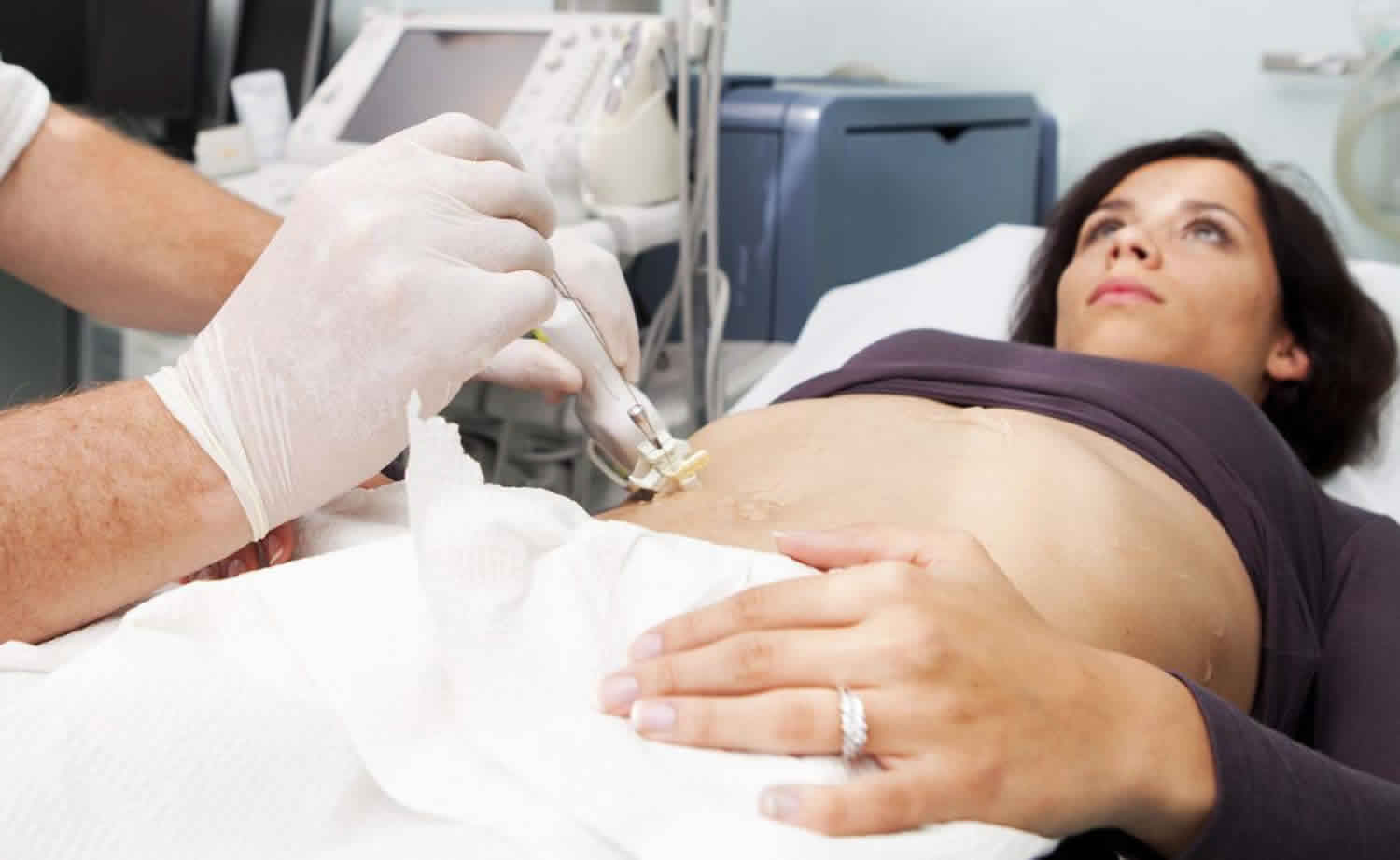
Ultrasound-guided liver biopsy
Your doctor will make a small cut on the skin on your right side, usually between your lower ribs. They will insert the needle through the cut and into your liver to remove a small piece of tissue. They will often use an ultrasound scan to guide them while they perform the biopsy.
Transjugular liver biopsy
Transjugular liver biopsy (also called transvenous liver biopsy) indications:
- massive ascites
- coagulopathy
- hepatic peliosis
- morbid obesity
- failed percutaneous liver biopsy or in a patient with a contraindication to percutaneous biopsy
Your doctor will make a small cut on your skin on the right side of your neck and then insert a catheter (tube) into your jugular vein. They will use x-rays to help them guide the tube through your veins. When the tube reaches your liver, your doctor will pass a needle down the tube to remove a small piece of tissue.
Does a liver biopsy hurt?
You may feel a brief sting or burn when the numbing medicine (anesthetic) goes in your skin. When the biopsy needle is inserted, you may again feel a sharp pain for a few seconds.
You may feel deep pressure and a dull pain in your belly when the biopsy needle is inserted. After the anesthetic wears off, you may feel a dull pain in your right shoulder. This is called referred pain and generally goes away in about 12 hours. You can take a non-prescription medicine, such as acetaminophen (Tylenol), for the pain. Call your doctor if your pain gets worse or lasts longer than 2 days.
A small amount of bleeding from the biopsy site can be expected. Ask your doctor how much drainage to expect.
Reasons for liver biopsy
There are many reasons for liver biopsy. They include diagnosis of the cause of chronic hepatitis, cirrhosis, storage diseases, unexplained hepatomegaly or enzyme elevations, post-liver transplant liver-enzyme abnormalities, space-occupying lesions, intrahepatic cholestasis, and drug-induced liver injury.
A liver biopsy may be done to:
- Diagnose a liver problem that can’t be otherwise identified
- Obtain a sample of tissue from an abnormality found by an imaging study
- Determine the severity of liver disease — a process called staging
- Help develop treatment plans based on the liver’s condition
- Determine how well treatment for liver disease is working
- Monitor the liver after a liver transplant
Your doctor may recommend a liver biopsy if you have:
- Abnormal liver test results that can’t be explained
- A mass (tumor) or other abnormalities on your liver as seen on imaging tests
- Ongoing, unexplained fevers
A liver biopsy also is commonly performed to help diagnose and stage certain liver diseases, including:
- Nonalcoholic fatty liver disease
- Chronic hepatitis B or C
- Autoimmune hepatitis
- Alcoholic liver disease
- Primary biliary cirrhosis
- Primary sclerosing cholangitis
- Hemochromatosis
- Wilson’s disease
Indications for liver biopsy fall into three broad categories:
- Diagnosis: Liver biopsy is crucial for diagnostic dilemmas: e.g., differentiating autoimmune hepatitis from nonalcoholic steatohepatitis (NASH) in obese patients with abnormal liver function tests and positive autoimmune serology. Wilson disease is traditionally called as the “great masquerader,” due to its varied presentations. Quantifying copper on the liver biopsy specimen helps clinch the diagnosis. It is very useful in overlap syndromes, e.g., autoimmune hepatitis with primary biliary cholangitis. Its role in post liver transplant setting cannot be overemphasized. It is very helpful in the evaluation of abnormal liver function tests in the immediate post-transplant setting. Vascular pathologies or infections helps guide management, differentiating rejection from a recurrence of underlying diseases such as hepatitis C infection. Diagnostic challenges like differentiating cholangiocarcinoma from hepatocellular cancer can be made with liver biopsy in atypical cases.
- Prognosis: Liver biopsy can be used as a prognostic tool for several diseases. In NASH, the presence of advanced fibrosis or cirrhosis has important prognostic considerations. Similarly, in diseases such as hemochromatosis, the presence of cirrhosis predicts increased risk for developing hepatocellular cancer. Although now largely replaced by noninvasive markers, the presence of fibrosis has very important prognostic implications for patients with chronic liver diseases such as hepatitis C infection.
- Treatment: Liver biopsy plays a sentinel role in patients with autoimmune hepatitis being treated with steroids and immunomodulators. The presence of histologically active disease is associated with high risk of relapse if treatment is stopped. There is the marked improvement in liver histology if patients are taking treatment, and therefore, can be helpful in monitoring compliance.
Liver biopsy contraindications
There are relatively few contraindications to liver biopsy. Most of the contraindications are relative as they are both technique and operator dependent.
Absolute contraindications
- Uncooperative patient: Patient should be counseled thoroughly about the procedure and informed consent taken. Uncooperative patients can increase the risk of complications. If a liver biopsy is required, it can be done under anesthesia.
- Increased risk of bleeding: In general liver biopsy is not attempted if INR greater than 1.5 or platelet count is less than 60,000. Such patients may require correction of abnormal parameters before attempting biopsy.
- Vascular tumors of the liver: Liver biopsy is associated with increased risk of bleeding in presumed vascular tumors.
Relative contraindications
- Ascites: Percutaneous biopsy is difficult and associated with increased risk of complications in the setting of ascites. The transvenous route is preferred.
- Morbid obesity: The procedure is much more challenging in morbidly obese patients due to interference by adipose tissue. The transvenous route is preferred.
How you prepare for liver biopsy
Before your liver biopsy, you’ll meet with your doctor to talk about what to expect during the biopsy. This is a good time to ask questions about the procedure and make sure you understand the risks and benefits.
Stop taking certain medications
When you meet with your doctor, bring a list of all medications you take, including over-the-counter medications, vitamins and herbal supplements. Before your liver biopsy, you’ll likely be asked to stop taking medications and supplements that can increase the risk of bleeding, including:
- Aspirin, ibuprofen (Advil, Motrin IB, others) and certain other pain relievers
- Blood-thinning medications (anticoagulants), such as warfarin (Coumadin)
- Certain dietary supplements that may increase risk of uncontrolled bleeding
Your doctor or nurse will let you know if you need to temporarily avoid any of your other medications.
Undergo blood tests
Before your biopsy, you’ll have a blood test to check your blood’s ability to clot. If you have blood-clotting problems, you may be given a medication before your biopsy to reduce the risk of bleeding.
Stop eating and drinking before the procedure
You may be asked not to drink or eat for six to eight hours before the liver biopsy. Some people can eat a light breakfast.
Prepare for your recovery
You may receive a sedative before your liver biopsy. If this is the case, arrange for someone to drive you home after the procedure. Have someone stay with you or check on you during the first night. Many doctors recommend that people spend the first evening within an hour’s driving distance of the hospital where the biopsy is done, in case a complication develops.
Liver biopsy procedure
A liver biopsy is done at a hospital or outpatient center. You’ll likely arrive early in the morning. Your health care team will review your medical history, including the medications you take.
Just before your liver biopsy you will:
- Have an IV line placed, usually into a vein in your arm, so that you can be given medications if you need them
- Possibly be given a sedative to help you relax during the procedure
- Use the toilet if needed because you’ll need to remain in bed for a few hours after the procedure
What you can expect during your liver biopsy will depend on the type of procedure you’ll undergo. A percutaneous liver biopsy is the most common type of liver biopsy, but it isn’t an option for everyone. Your doctor may recommend a different form of liver biopsy if you:
- Have trouble holding still during the procedure
- Have a history of or likelihood of bleeding problems or blood-clotting disorders
- Might have a tumor involving blood vessels in your liver
- Have an abnormal amount of fluid in your abdomen (ascites)
- Are very obese
- Have a liver infection
How is a liver biopsy done?
The steps involved in liver biopsy vary according to the type:
Percutaneous liver biopsy
To begin your procedure, your doctor will locate your liver by tapping on your abdomen or using ultrasound images. In certain situations, ultrasound might be used during the biopsy to guide the needle into your liver. You’ll lie on your back and position your right hand above your head on the table. Your doctor will apply a numbing medication to the area where the needle will be inserted. The doctor then makes a small incision near the bottom of your rib cage on your right side typically between the 6 and 9 intercostals spaces between the anterior and the midclavicular line and inserts the biopsy needle. The biopsy itself takes just a few seconds. As the needle passes quickly in and out of your liver, you’ll be asked to hold your breath.
Transjugular liver biopsy
You’ll lie on your back on an X-ray table. Your doctor applies a numbing medication to one side of your neck, makes a small incision and inserts a flexible plastic tube into your jugular vein. The tube is threaded down the jugular vein and into the large vein in your liver (hepatic vein). Your doctor then injects a contrast dye into the tube and makes a series of X-ray images. The dye shows up on the images, allowing the doctor to see the hepatic vein. A biopsy needle is then threaded through the tube, and one or more liver samples are removed. The catheter is carefully removed, and the incision on your neck is covered with a bandage.
Laparoscopic liver biopsy
During a laparoscopic biopsy, you’ll likely receive general anesthetics. You’ll be positioned on your back on an operating table and your doctor will make one or more small incisions in your abdomen. Special tools are inserted through the incisions, including a tiny video camera that projects images on a monitor in the operating room. The doctor uses the video images to guide the tools to the liver to remove tissue samples. The tools are removed and the incisions are closed with stitches.
Plugged liver biopsy
This is a modification of the percutaneous approach that can be used in patients who are at high risk for bleeding (coagulopathy or thrombocytopenia). Although a transjugular biopsy can be obtained in this subset of patients, the plugged approach is used when a larger specimen size is desirable.
The technique is similar to the percutaneous approach except the biopsy tract is plugged with gel foam, collagen or thrombin while the sheath is being removed.
Liver biopsy aftercare
The patient is usually kept in the right decubitus position. The duration of observation varies across centers ranging from 1 hour to 6 hours. The American Association for the Study of Liver Diseases guidelines recommends observation for 2 to 4 hours. The vital signs are monitored every 15 minutes for the first hour, every 30 minutes for the next hour and hourly till discharge. Most complications of liver biopsy usually occur within the first 1 to 3 hours after biopsy or within 24 hours. Hence the patient should have a dependent individual to stay overnight after the procedure. The patient should be hospitalized if there are any complications associated with the procedure, including pain that requires more than one dose of analgesia within the first 4 hours of observation following the procedure.
Liver biopsy recovery
After your liver biopsy, you can expect to:
- Be taken to a recovery room, where a nurse will monitor your blood pressure, pulse and breathing
- Rest quietly for two to four hours, or longer if you had a transjugular liver biopsy procedure
- Feel some soreness where the needle was inserted, which may last as long as a week
- Have someone drive you home, since you won’t be able to drive until the sedative wears off
- Avoid lifting more than 10 to 15 pounds for one week
- Be able to get back to your usual activities gradually over a period of a week
Liver biopsy recovery time
You should be able to go home the same day.
You should be able to return to work the next day unless you are told otherwise. Do not do strenuous exercise for 1 to 2 days.
Your heathcare team will discuss with you any treatment or follow-up you need.
Liver biopsy results
Your liver tissue goes to a laboratory to be examined by a doctor who specializes in diagnosing disease (pathologist). The pathologist will look for signs of disease and damage to the liver. Your biopsy report should come back from the pathology lab within a few days to a week. Test results are generally ready in 2 to 4 days. If tests are done to find infections, it may take several weeks for the results to be ready.
Occasionally, a liver biopsy may not provide helpful results because not enough tissue is sampled to make a clear diagnosis.
Normal results:
- The liver tissue looks normal under a microscope. No signs of infection, inflammation, cancer, or cirrhosis are present.
Abnormal results:
- Abnormal cells or liver tissue are present. This may be caused by an infection such as hepatitis, liver disease such as cirrhosis, or cancer. If liver cancer is present, the biopsy can help find the type of cancer. If hepatitis is present, the test can be used to see the chance of developing cirrhosis. Test results may also show the severity of cirrhosis.
At a follow-up visit, your doctor will explain the results. You may be diagnosed with a liver disease, or your liver disease may be given a stage or grade number based on the severity — mild, moderate or severe. Your doctor will discuss what treatment, if any, you need.
Liver biopsy complications
A liver biopsy is a safe procedure when performed by an experienced doctor. The overall rate of serious complications was approximately 1% in two large series while in another; overall mortality risk was estimated to be 0.2% 1. The usual indicators of complications requiring overnight hospital observation are severe abdominal or shoulder tip pain not relieved with one dose of parenteral analgesic, hypotension, or tachycardia following the procedure.
Possible complications include:
- Pain. Pain at the biopsy site is the most common complication after a liver biopsy. Pain can be seen in up to 84% of the patients. It is most common at the site of the biopsy or the right shoulder (this often indicates a subcapsular hematoma), or both. Pain after a liver biopsy is usually a mild discomfort. If pain makes you uncomfortable, you may be given a narcotic pain medication, such as acetaminophen with codeine (Tylenol with Codeine). Severe persistent pain should alert your physician to investigate serious causes of pain like bile peritonitis, hemorrhage. The patient may require admission and radiological assessment.
- Bleeding. Bleeding can occur after a liver biopsy. Excessive bleeding may require you to be hospitalized for a blood transfusion or surgery to stop the bleeding. The risk of fatal bleeding in patients without malignant disease is 0.04%, and the risk of nonfatal hemorrhage is 0.16% 1. In those with cancer, the risk of nonfatal hemorrhage is 0.4% and 0.57% for nonfatal hemorrhage.
- Three types of bleeding can be seen:
- Free intraperitoneal bleed: This can be secondary to liver laceration with deep breathing during the intercostal procedure, perforation of distended portal or hepatic veins or aberrant arteries or inadvertent puncture of a major intrahepatic blood vessel. The patients usually present with hemodynamic instability, severe abdominal pain and rapid drop in hemoglobin. It is imperative to recognize this complication early. The patient should be admitted, resuscitated and both interventional radiology and surgeon should be consulted. Angiographic embolization is usually successful in controlling the bleeding, but in rare cases, surgical intervention might be required particularly in the transplant patient who carries a higher risk for major bile duct injury with arterial embolization. Rarely bleeding can be intrathoracic from an intercostal artery.
- Intrahepatic/subcapsular hematoma: These can be seen even in asymptomatic patients. Bleeding usually presents with pain, tachycardia and mild drop in hemoglobin with the rise in serum transaminases. If large, they can cause right upper quadrant tenderness and hepatomegaly, and appear as triangular hyper dense segments in the arterial phase of CT scan. Most patients can be managed with conservative treatment, and radiological or surgical intervention is rarely required.
- Hemobilia: It usually presents with the classical triad of gastrointestinal bleeding, biliary pain, and jaundice. The bleeding is usually arterial in origin but can be venous in patients with preexisting portal hypertension. It can vary in severity from the occult to exsanguinating hemorrhage. Hemobilia rarely presents acutely and most commonly presents after a median of 5 days with the gradual erosion of a biopsy-induced hematoma or pseudoaneurysm into the bile duct. The presentation can vary from hemodynamically significant bleeding to chronic anemia. Imaging or endoscopy can make a diagnosis. Treatment depends on the severity of bleed. Hemodynamically significant radiologic intervention may be required. ERCP might be needed in some cases to remove clotted blood from the bile duct causing obstruction and cholangitis.
- Three types of bleeding can be seen:
- Infection. Rarely, bacteria may enter the abdominal cavity or bloodstream. This is usually clinically insignificant except in patients with obstructive jaundice like primary sclerosing cholangitis or in the post-transplant setting. Currently, there is no recommendation for treating with prophylactic antibiotics and treatment can be offered on a case by case basis.
- Accidental injury to a nearby organ. In rare instances, the needle may stick another internal organ, such as the gallbladder or a lung, during a liver biopsy.
- Bile peritonitis: This can occur with inadvertent puncture of the gallbladder or in patients with obstructive jaundice and dilated bile ducts. It usually presents with abdominal pain, fever, leukocytosis. It can also be painless in some patients. Biliary scintigraphy demonstrates the leak. Treatment is usually with fluids and antibiotics. Very rarely endoscopic procedures like ERCP or surgery may be required.
- Miscellaneous: Cardiovascular complications, especially in patients with preexisting heart disease, arteriovenous fistula, pneumothorax, are other rare reported complications. Carcinoid crisis can occur after the percutaneous biopsy.
In a transjugular procedure, a thin tube is inserted through a large vein in your neck and passed down into the vein that runs through your liver. If you have a transjugular liver biopsy, other infrequent risks include:
- Collection of blood (hematoma) in the neck. Blood may pool around the site where the catheter was inserted, potentially causing pain and swelling.
- Temporary problems with the facial nerves. Rarely, the transjugular procedure can injure nerves and affect the face and eyes, causing short-term problems, such as a drooping eyelid.
- Temporary voice problems. You may be hoarse, have a weak voice or lose your voice for a short time.
- Puncture of the lung. If the needle accidentally sticks your lung, the result may be a collapsed lung (pneumothorax).


