Contents
- What is a mammogram
- How do mammograms work?
- What are the types of mammograms?
- Having a Mammogram After You’ve Had Breast Cancer Surgery
- Mammograms for Women with Breast Implants
- Mammogram prep
- What doctors look for on a mammogram
- How are screening and diagnostic mammograms different?
- Limitations of Mammograms
- Mammogram screening
- Mammogram screening guidelines
- Mammogram results
- Getting Called Back After a Mammogram
What is a mammogram
A mammogram is a low-dose x-ray that allows doctors called radiologists to look for changes in breast tissue that may be signs of breast cancer. The results are recorded on x-ray film or directly into a computer for a doctor called a radiologist to examine.
A mammogram can find breast changes that could be cancer years before physical symptoms develop. Results from many decades of research clearly show that women who have regular mammograms are more likely to have breast cancer found early, are less likely to need aggressive treatment like surgery to remove the breast (mastectomy) and chemotherapy, and are more likely to be cured.
Women ages 50 to 74 years should get a mammogram every 2 years. Women younger than age 50 should talk to a doctor about when to start and how often to have a mammogram.
Mammograms are not perfect. They miss some cancers. And sometimes a woman will need more tests to find out if something found on a mammogram is or is not cancer. There’s also a small possibility of being diagnosed with a cancer that never would have caused any problems had it not been found during screening. It’s important that women getting mammograms know what to expect and understand the benefits and limitations of screening.
Why do I need mammograms ?
A mammogram can often find or detect breast cancer at an early stage, when it’s small and even before a lump can be felt. This is when treatment is most successful.
A mammogram allows the doctor to have a closer look for changes in breast tissue that cannot be felt during a breast exam. It is used for women who have no breast complaints and for women who have breast symptoms, such as a change in the shape or size of a breast, a lump, nipple discharge, or pain. Breast changes occur in almost all women. In fact, most of these changes are not cancer and are called “benign,” but only a doctor can know for sure. Breast changes can also happen monthly, due to your menstrual period.
What is the best method of screening for breast cancer?
Regular high-quality screening mammograms and clinical breast exams are the most sensitive ways to screen for breast cancer.
Research has not shown a clear benefit of regular physical breast exams done by either a health professional (clinical breast exams) or by yourself (breast self-exams). There is very little evidence that these tests help find breast cancer early when women also get screening mammograms. Most often when breast cancer is detected because of symptoms (such as a lump), a woman discovers the symptom during usual activities such as bathing or dressing. Women should be familiar with how their breasts normally look and feel and report any changes to a health care provider right away.
Regular breast self-exam — that is, checking your own breasts for lumps or other unusual changes—is not specifically recommended for breast cancer screening. In clinical trials, breast self-exam alone was not found to help reduce the number of deaths from breast cancer.
However, many women choose to examine their own breasts. Women who do so should remember that breast changes can occur because of pregnancy, aging, or menopause; during menstrual cycles; or when taking birth control pills or other hormones. It is normal for breasts to feel a little lumpy and uneven. Also, it is common for breasts to be swollen and tender right before or during a menstrual period. Whenever you notices any unusual changes in your breasts, you should contact your health care provider.
What do mammograms show?
Mammograms can often show abnormal areas in the breast. They can’t prove that an abnormal area is cancer, but they can help health care providers decide whether more testing is needed. The 2 main types of breast changes found with a mammogram are calcifications and masses.
How much does a mammogram cost?
Insurance plans governed by the federal Affordable Care Act must cover screening mammography as a preventive benefit every 1–2 years for women age 40 and over without requiring copayments, coinsurance, or deductibles. In addition, many states require that Medicaid and public employee health plans cover screening mammography. Women should contact their mammography facility or health insurance company for confirmation of the cost and coverage.
Medicare pays for annual screening mammograms for all female Medicare beneficiaries who are age 40 or older. Medicare will also pay for one baseline mammogram for female beneficiaries between the ages of 35 and 39. There is no deductible requirement for this benefit. Information about coverage of mammograms is available on the Medicare website or through the Medicare Hotline at 1–800–MEDICARE (1–800–633–4227). For the hearing impaired, the telephone number is 1–877–486–2048.
Women who need a diagnostic mammogram should check with their health insurance provider about coverage.
How can uninsured or low-income women obtain a free or low-cost screening mammogram?
Some state and local health programs and employers provide mammograms free or at low cost. For example, the Centers for Disease Control and Prevention (CDC) coordinates the National Breast and Cervical Cancer Early Detection Program. This program provides screening services, including clinical breast exams and mammograms, to low-income, uninsured women throughout the United States and in several U.S. territories. Contact information for local programs is available on the CDC website or by calling 1–800–CDC–INFO (1–800–232–4636).
Information about free or low-cost mammography screening programs is also available from National Cancer Institute’s Cancer Information Service at 1–800–4–CANCER (1–800–422–6237) and from local hospitals, health departments, women’s centers, or other community groups.
Where can I get a high-quality mammogram?
Women can get high-quality mammograms in breast clinics, hospital radiology departments, mobile vans, private radiology offices, and doctors’ offices. The Food and Drug Administration (FDA) certifies mammography facilities that meet strict quality standards for their x-ray machines and staff and are inspected every year. You can ask your doctor or the staff at the mammography center about FDA certification before making your appointment. A list of FDA-certified facilities can be found on the Internet.
Your doctor, local medical clinic, or local or state health department can tell you where to get no-cost or low-cost mammograms. You can also call the National Cancer Institute’s Cancer Information Service toll free at 1-800-422-6237.
How do mammograms work?
A mammogram uses a machine designed to look only at breast tissue. You stand in front of a special x-ray machine. The machine takes x-rays at lower doses than usual x-rays. Because these x-rays don’t go through tissue easily, the machine has 2 plates that compress or flatten the breast to spread the tissue apart. This gives a better picture and allows less radiation to be used.
The person who takes the x-rays, called a radiologic technician, places your breasts, one at a time, between an x-ray plate and a plastic plate. These plates are attached to the x-ray machine and compress the breasts to flatten them. This spreads the breast tissue out to obtain a clearer picture. You will feel pressure on your breast for a few seconds. It may cause you some discomfort; you might feel squeezed or pinched. This feeling only lasts for a few seconds, and the flatter your breast, the better the picture. Most often, two pictures are taken of each breast — one from the side and one from above. A screening mammogram takes about 20 minutes from start to finish.
In the past, mammograms were typically printed on large sheets of film. Today, digital mammograms (also known as full-field digital mammography or FFDM) are much more common. Digital images are recorded and saved as files in a computer.
A newer type of mammogram is known as breast tomosynthesis or 3D mammography also known as digital breast tomosynthesis (DBT). For this, the breast is compressed once, and a machine takes many low-dose x-rays as it moves over the breast. A computer then puts the images together into a 3-dimensional picture. In some cases, this uses more radiation than standard 2-view mammograms, but it may allow doctors to see the breast tissues more clearly. Some studies have suggested it might lower the chance of being called back for follow-up testing. It may also be able to find more cancers. But not all health insurance providers may cover tomosynthesis. Furthermore, digital breast tomosynthesis (DBT) has not yet been determined conclusively whether it is superior to 2-D mammography at identifying early cancers and avoiding false-positive results.
Digital 3D mammography or digital breast tomosynthesis (DBT) may offer these benefits:
- Long-distance consultations with other doctors may be easier because the images can be shared by computer.
- Slight differences between normal and abnormal tissues may be more easily noted.
- The number of follow-up tests needed may be fewer.
- Fewer repeat images may be needed, reducing exposure to radiation.
The United States Preventive Services Task Force concludes that the current evidence is insufficient to assess the benefits and harms of digital breast tomosynthesis (DBT) as a primary screening method for breast cancer 1.
A large-scale randomized breast screening trial expected to open in mid-2017 will compare 3-D mammography with 2-D mammography. The Tomosynthesis Mammography Imaging Screening Trial (TMIST) will compare the number of advanced cancers detected in women screened for 4 years with DBT with that detected in women screened with standard digital mammography.
What other technologies or strategies are being developed for breast cancer screening?
National Cancer Institute is supporting the development of several new technologies to detect breast tumors. This research ranges from methods being developed in research labs to those that are being studied in clinical trials. Efforts to improve conventional mammography include digital mammography, magnetic resonance imaging (MRI), positron emission tomography (PET) scanning, and diffuse optical tomography, which uses light instead of x-rays to create pictures of the breast.
The Women Informed to Screen Depending on Measures of Risk (https://wisdom.secure.force.com/portal/WsdSiteHome) study is a randomized trial that is testing a personalized approach to breast cancer screening. This 5-year study, which will involve about 100,000 women in California and the Midwest, aims to determine if risk-based screening—that is, screening at intervals that are based on each woman’s risk as determined by her genetic makeup, family history, and other risk factors—is as safe, effective, and accepted as annual screening.
Are mammograms safe?
Mammograms expose the breasts to small amounts of radiation. But the benefits of mammography outweigh any possible harm from the radiation exposure. Modern machines use low radiation doses to get breast x-rays that are high in image quality. On average the total dose for a typical mammogram with 2 views of each breast is about 0.4 millisieverts, or mSv. A millisievert (mSv) is a measure of radiation dose.
To put the dose into perspective, people in the US are normally exposed to an average of about 3 millisievert (mSv) of radiation each year just from their natural surroundings. This is called background radiation. The dose of radiation used for a screening mammogram of both breasts is about the same amount of radiation a woman would get from her natural surroundings over about 7 weeks.
If there’s any chance you might be pregnant, let your health care provider and x-ray technologist know. Although the risk to the fetus is likely very small, screening mammograms aren’t routinely done in pregnant women.
What are the types of mammograms?
Screening mammograms
A screening mammogram is used to look for signs of breast cancer in women who don’t have any breast symptoms or problems of the disease. Screening mammograms usually involve two or more x-ray pictures, or images, of each breast. The x-ray images often make it possible to detect tumors that cannot be felt. Screening mammograms can also find microcalcifications (tiny deposits of calcium) that sometimes indicate the presence of breast cancer.
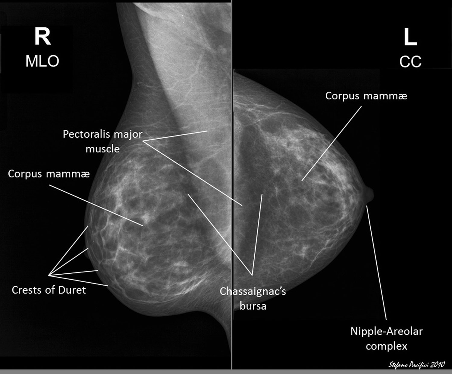
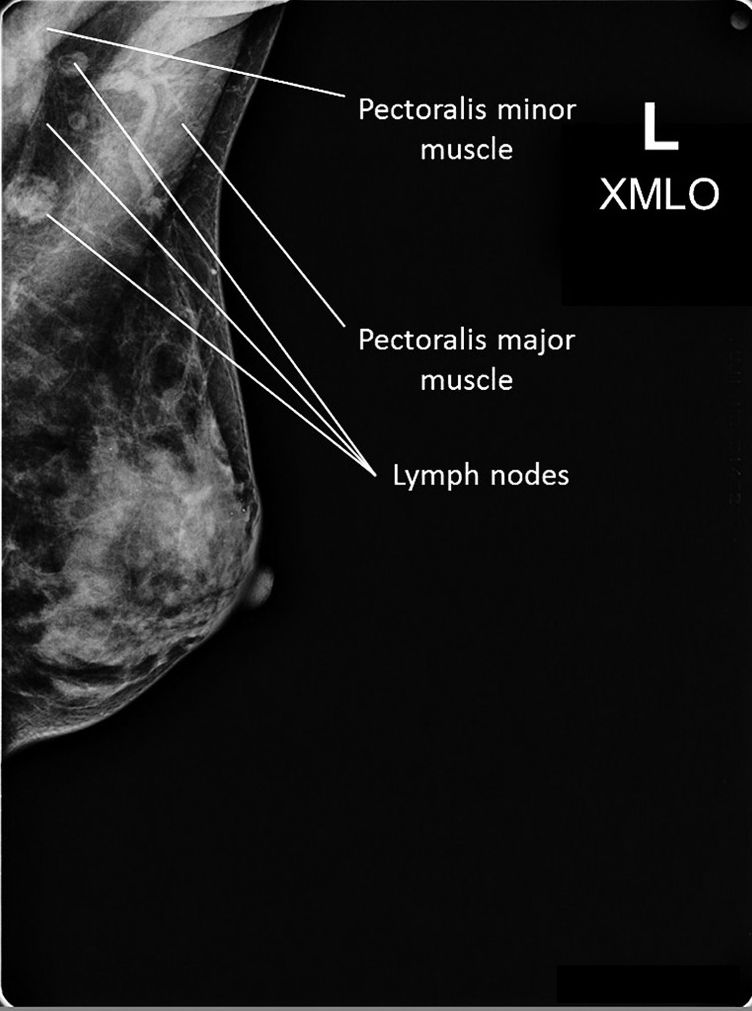
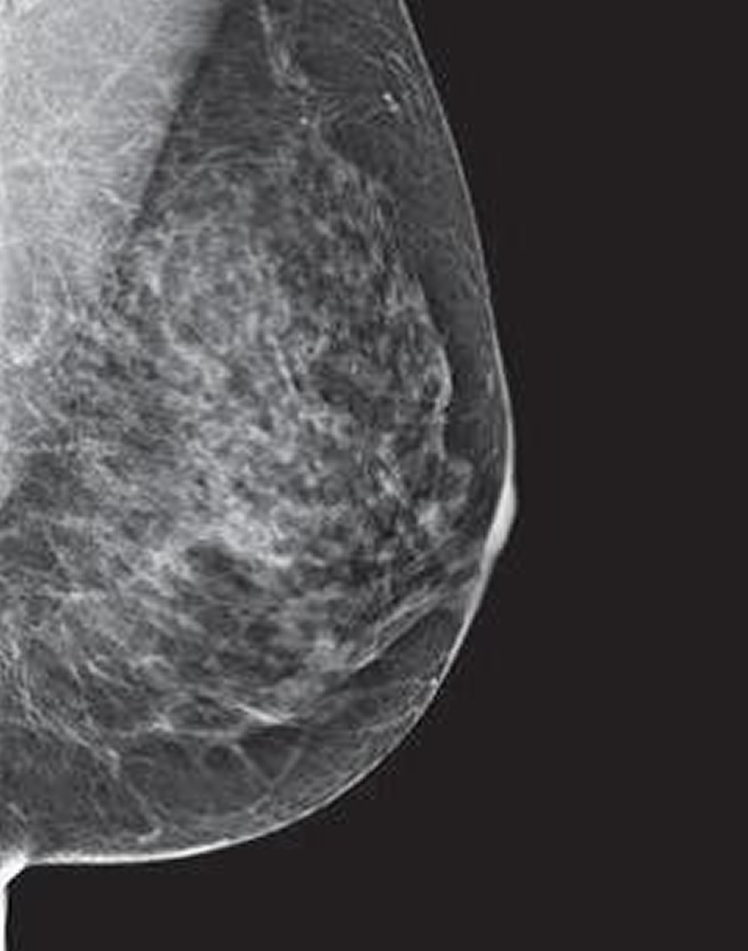
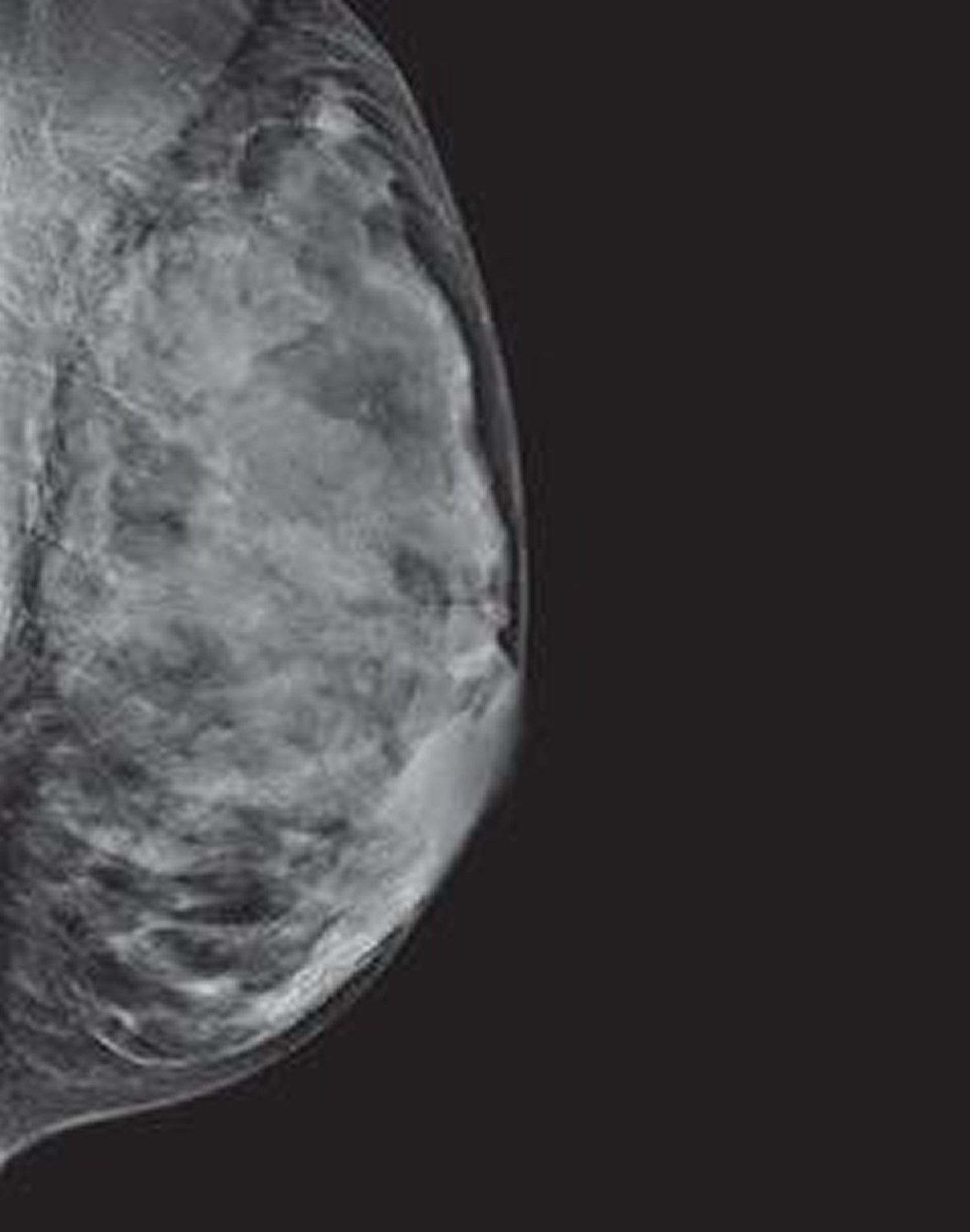
Radiologists prefer the Mediolateral Oblique (MLO) view from an angle or oblique view view or ‘from the side-at an angle‘, view to a 90 degree projection. This is because the Mediolateral Oblique (MLO) view allows imaging of more of the breast in the upper-outer quadrant, and also the axilla (armpit area).
With the top-down or Cranial Caudal (CC) view, the entire breast is depicted. Fat tissue closest to the breast muscle should appear as a dark strip on the X-ray. Also, the Cranial Caudal (CC) view also tends to clearly depict the nipple.
The Mediolateral (ML) view is very important because the lateral side of the breast is probably the most common place for pathological changes to occur. The view from the center of the chest, outward to the side, Mediolateral (ML) view gives the best view of the lateral side of the breast. In this view the chest muscle (pectoral) shows on mammogram as a narrow light band on about half of the picture. Again, imaging of the nipple is also clear in profile.
With the Latero-Medial view (LM) the breast is X-rayed from the side towards the middle, and this gives the best view of the medial (mid-body) side of the breast.
What if my screening mammogram shows a problem?
If you have a screening test result that suggests cancer, your doctor must find out whether it is due to cancer or to some other cause. Your doctor may ask about your personal and family medical history. You may have a physical exam. Your doctor also may order some of these tests:
- Diagnostic mammogram, to focus on a specific area of the breast
- Ultrasound, an imaging test that uses sound waves to create a picture of your breast. The pictures may show whether a lump is solid or filled with fluid. A cyst is a fluid-filled sac. Cysts are not cancer. But a solid mass may be cancer. After the test, your doctor can store the pictures on video or print them out. This exam may be used along with a mammogram.
- Magnetic resonance imaging (MRI), which uses a powerful magnet linked to a computer. MRI makes detailed pictures of breast tissue. Your doctor can view these pictures on a monitor or print them on film. MRI may be used along with a mammogram.
- Biopsy, a test in which fluid or tissue is removed from your breast to help find out if there is cancer. Your doctor may refer you to a surgeon or to a doctor who is an expert in breast disease for a biopsy.
Diagnostic mammograms
Mammograms can also be used to look at a woman’s breast for breast cancer after a lump or other sign or symptom of the disease has been found or if she has breast symptoms or if a change is seen on a screening mammogram. When used in this way, they are called diagnostic mammograms. They may include extra views (images) of the breast that aren’t part of screening mammograms. Sometimes diagnostic mammograms are used to screen women who were treated for breast cancer in the past.
Besides a lump, signs of breast cancer can include breast pain, thickening of the skin of the breast, nipple discharge, or a change in breast size or shape; however, these signs may also be signs of benign conditions. A diagnostic mammogram can also be used to evaluate changes found during a screening mammogram or to view breast tissue when it is difficult to obtain a screening mammogram because of special circumstances, such as the presence of breast implants.
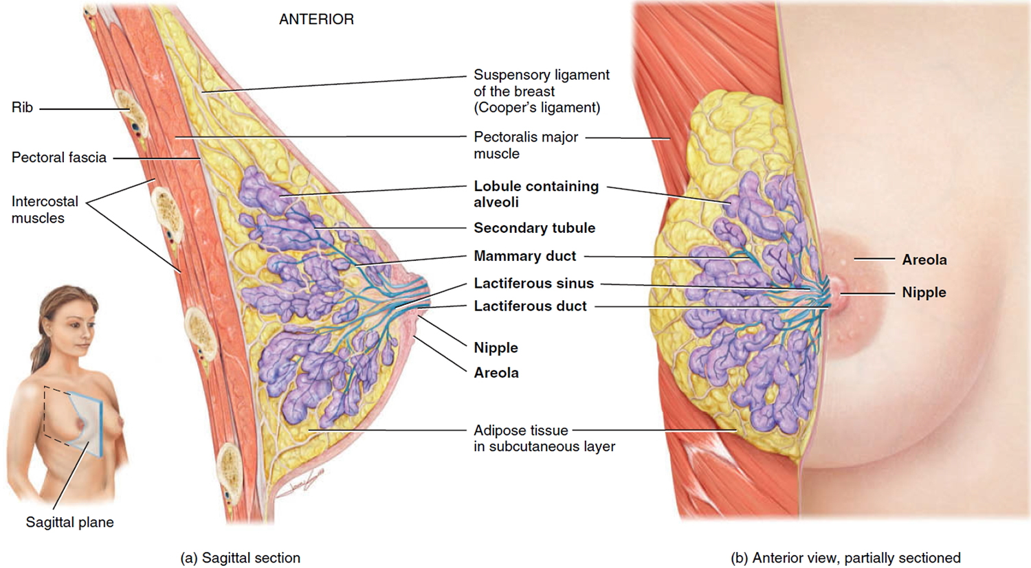
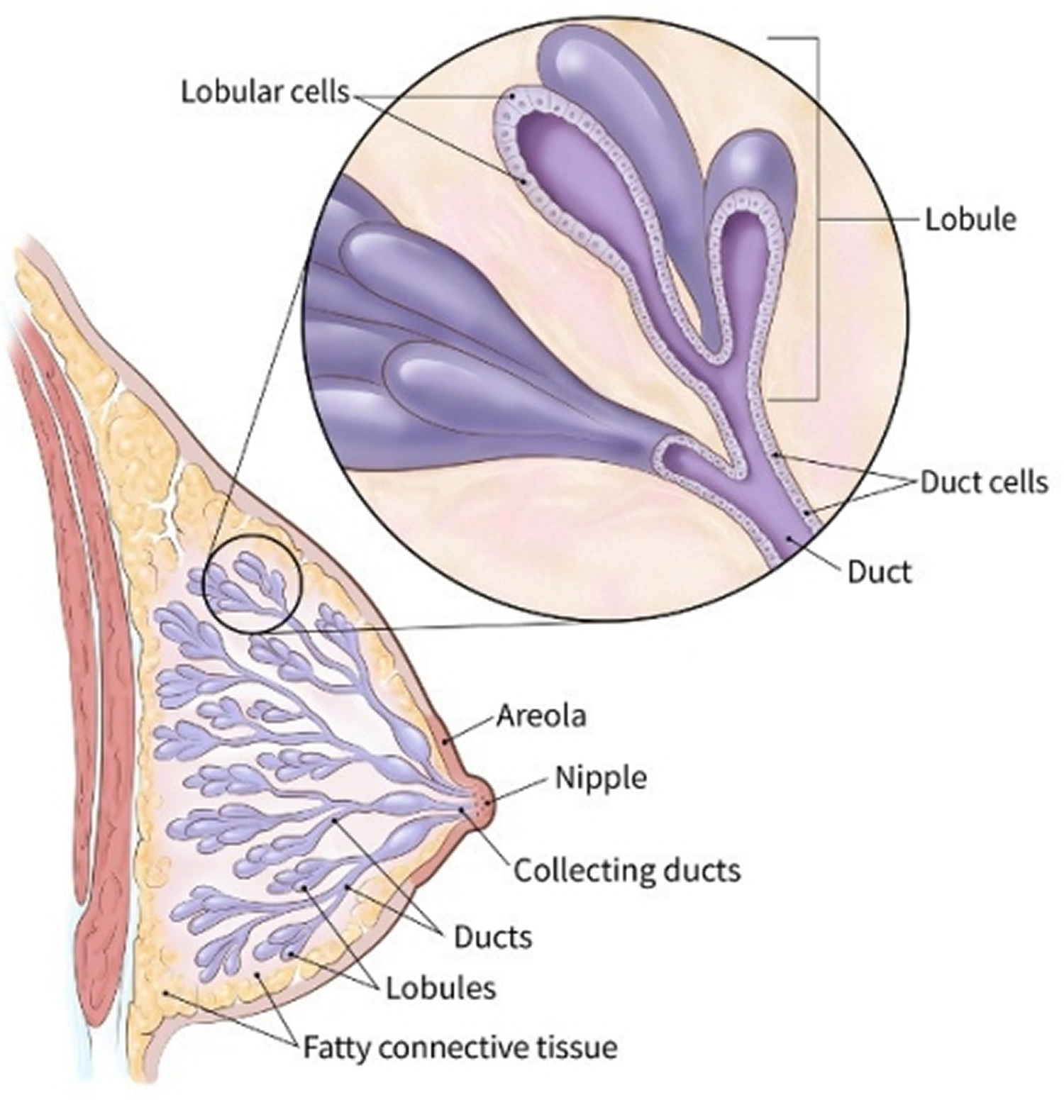
Figure 1. Normal breast (female)
Figure 2. Normal mammograms – Note screening mammogram with Mediolateral oblique (MLO) view from an angle or oblique view and Cranial-Caudal (CC) view from above the breast. Fat, being relatively radiolucent, appears as black on the film. ‘Radiolucent’ refers to anything that permits the penetration and passage of X-rays whereas ‘radiopaque’ refers to anything that blocks the penetration of X-rays.
Having a Mammogram After You’ve Had Breast Cancer Surgery
There are many different kinds of breast cancer surgery. The type of surgery you have had will determine whether you need to get mammograms in the future. If you have had breast-conserving surgery (BCS), you need to continue to get mammograms. If you have had a mastectomy, you may not need a mammogram.
If you had surgery (of any type) on only one breast, you will still need to get mammograms of the unaffected breast. This is very important, because women who have had one breast cancer are at higher risk of developing a new cancer in the other breast.
Mammograms after breast-conserving surgery
Most experts recommend that women who have had breast-conserving surgery or BCS (sometimes called a partial mastectomy or lumpectomy) get a mammogram of the treated breast 6 to 12 months after radiation treatment ends. Surgery and radiation both cause changes in the skin and breast tissues that will show up on the mammogram, making it harder to read. The mammogram done at this time serves as a new baseline for the affected breast. Future mammograms will be compared with this one, to help the doctor check on healing and look for signs that the cancer has come back (recurred).
You should have follow-up mammograms of the treated breast at least yearly after that, but some doctors may recommend that you have mammograms more often. You will still need to have routine mammograms on the opposite (untreated) breast as well.
Mammograms after mastectomy
Women who have had a mastectomy (including simple mastectomy, modified radical mastectomy, and radical mastectomy) to treat breast cancer need no further routine screening mammograms on the affected side. If both breasts are removed (a double or bilateral mastectomy), they don’t need mammograms at all. There isn’t enough tissue remaining after these kinds of mastectomies to do a mammogram. Cancer can come back in the skin or chest wall on that side, but it can be found on a physical exam.
It’s possible for women with reconstructed breasts to get mammograms, but experts agree that women who have breast reconstruction after a simple, modified radical, or radical mastectomy don’t need routine mammograms. Still, if an area of concern is found during a physical exam on a woman who has had breast reconstruction, a diagnostic mammogram may be done. Breast ultrasound or MRI may also be used to look at the area closely.
Women who have had a subcutaneous mastectomy, also called skin-sparing mastectomy, still need follow-up mammograms. In this surgery, the woman keeps her nipple and the tissue just under the skin. Often, an implant is put under the skin. This surgery leaves behind enough breast tissue to require yearly screening mammograms in these women.
Any woman who’s not sure what type of mastectomy she has had or whether she needs to get mammograms should ask her doctor.
Mammograms for Women with Breast Implants
If you have breast implants, you should still get regular screening mammograms as recommended.
It’s important to tell the technologist you have implants before your mammogram is started. In fact, it’s best to mention this when you make the appointment to have your mammogram done. This way you can find out if the facility has experience doing mammograms in women with breast implants.
You should be aware that it might be hard for the doctor to see certain parts of your breast. The x-rays used in mammograms cannot go through silicone or saline implants well enough to show the breast tissue under them. This means that part of the breast tissue can be hard to see on a mammogram.
To help the doctor can see as much breast tissue as possible, women with implants have 4 extra pictures done (2 on each breast), as well as the 4 standard pictures taken during a screening mammogram. In these extra pictures, called implant displacement (ID) views, the implant is pushed back against the chest wall and the breast is pulled forward over it. This allows better imaging of the front part of each breast.
Implant displacement views are harder to do and can be uncomfortable if the woman has had a lot of scar tissue (called contractures) form around the implants. implant displacement (ID) views are easier in women whose implants were placed underneath (behind) the chest muscles.
Very rarely, mammograms can rupture an implant. This is another important reason to make sure the mammography facility knows you have implants.
Mammogram prep
How to prepare for your mammogram
- If you have a choice, use a facility that specializes in mammograms and does many mammograms a day.
- Try to go to the same facility every time so that your mammograms can easily be compared from year to year.
- If you’re going to a facility for the first time, bring a list of the places and dates of mammograms, biopsies, or other breast treatments you’ve had before.
- If you’ve had mammograms at another facility, try to get those records to bring with you to the new facility (or have them sent there) so the old pictures can be compared to the new ones.
- Schedule your mammogram when your breasts are not tender or swollen to help reduce discomfort and get good pictures. Try to avoid the week just before your period.
- If you have breast implants, be sure to tell your mammography facility that you have them when you make your appointment.
- On the day of the exam, don’t wear deodorant, antiperspirant, perfume, lotion, or powder under your arms or on your breasts. Some of these contain substances that can show up on the x-ray as white spots. If you’re not going home afterward, you might want to take your deodorant with you to put on after your exam.
- You might find it easier to wear a skirt or pants, so that you’ll only need to remove your top and bra for the mammogram.
- Discuss any recent changes or problems in your breasts with your health care provider before getting the mammogram.
Don’t be afraid of mammograms. Remember that only 2 to 4 screening mammograms in 1,000 lead to a diagnosis of breast cancer. Moreover, breast cancer that’s found early, when it’s small and has not spread, is easier to treat successfully. Getting regular screening mammograms is the most reliable way to find breast cancer early.
Tips for getting a mammogram
These tips can help you have a good quality mammogram:
- Always describe any breast changes or problems you’re having to the technologist doing the mammogram. Also describe any medical history that could affect your breast cancer risk—such as surgery, hormone use, breast cancer in your family, or if you’ve had breast cancer before.
- Before getting any type of imaging test, tell the technologist if you’re breastfeeding or if you think you might be pregnant.
What to expect when getting a screening mammogram
- You’ll have to undress above the waist to get a mammogram. The facility will give you a wrap to wear.
- A technologist will position your breasts for the mammogram. You and the technologist are the only ones in the room during the mammogram.
- To get a high-quality picture, your breast must be flattened. The technologist places your breast on the machine’s plate. The plastic upper plate is lowered to compress your breast for a few seconds while the technologist takes a picture.
- The whole procedure takes about 20 minutes. The actual breast compression only lasts a few seconds.
- You might feel some discomfort when your breasts are compressed, and for some women it can be painful. Tell the technologist if it hurts.
- Two views of each breast are taken for a screening mammogram. But for some women, such as those with breast implants or large breasts, more pictures may be needed.
What to expect when getting a diagnostic mammogram
A diagnostic mammogram is often done if a woman has breast symptoms or if a change is seen on a screening mammogram.
- More pictures are taken during a diagnostic mammogram with a focus on the area that looked different on the screening mammogram.
- During a diagnostic mammogram, the images are checked by the radiologist while you’re there so that more pictures can be taken if needed to look more closely at any area of concern.
- In some cases, special images known as spot views or magnification views are used to make a small area of concern easier to see.
What doctors look for on a mammogram
A radiologist will look at your mammogram. Radiologists are doctors who diagnose diseases and injuries using imaging tests such as x-rays.
When possible, the doctor reading your mammogram will compare it to your old mammograms. This can help show if any findings are new, or if they were already there on previous mammograms. Findings that haven’t changed from older mammograms aren’t likely to be cancer, which might mean you won’t need further tests.
The doctor reading your mammogram will be looking for different types of breast changes, such as small white spots called calcifications, lumps or tumors called masses, and other suspicious areas that could be signs of cancer.
Calcifications
Calcifications are tiny calcium deposits within the breast tissue. They look like small white spots on a mammogram. They may or may not be caused by cancer.
There are 2 types of calcifications
Macrocalcifications
Macrocalcifications are larger calcium deposits that are most likely due to changes caused by aging of the breast arteries, old injuries, or inflammation. These deposits are typically related to non-cancerous conditions and don’t need to be checked for cancer with a biopsy. Macrocalcifications become more common as women get older (especially after age 50).
Microcalcifications
Microcalcifications are tiny specks of calcium in the breast. When seen on a mammogram, they are more of a concern than macrocalcifications, but they don’t always mean that cancer is present. The shape and layout of microcalcifications help the radiologist judge how likely it is that the change is due to cancer.
In most cases, microcalcifications don’t need to be checked with a biopsy. But if they have a suspicious look and pattern, a biopsy will be recommended to check for cancer.
A mass
A mass is the same as a lump or a tumor. With or without calcifications, it’s another important change seen on a mammogram. Masses can be many things, including cysts (non-cancerous, fluid-filled sacs) and non-cancerous solid tumors (such as fibroadenomas), but they may also be a sign of cancer.
Cysts are fluid-filled sacs. Simple cysts (fluid-filled sacs with thin walls) are not cancer and don’t need to be checked with a biopsy. If a mass is not a simple cyst, it’s of more concern, so a biopsy might be needed to be sure it isn’t cancer.
Solid tumors can be more concerning, but most breast tumors are not cancer.
A cyst and a solid tumor can feel the same. They can also look the same on a mammogram. The doctor must be sure it’s a cyst to know it’s not cancer. To be sure, a breast ultrasound is often done because it is a better tool to see fluid-filled sacs. Another option is to use a thin needle to remove (aspirate) fluid from the area.
If a mass is not a simple cyst (that is, if it’s at least partly solid), more imaging tests might be needed to decide if it could be cancer. Some masses can be watched over time with regular mammograms or ultrasound to see if they change, but others may need to be checked with a biopsy. The size, shape, and margins (edges) of the mass may help the radiologist decide how likely it is to be cancer.
Breast density
Your mammogram report will also contain an assessment of your breast density. Breast density is based on how fibrous and glandular tissues are distributed in your breast, as opposed to how much of your breast is made up of fatty tissue.
Dense breasts are not abnormal, but they are linked to a higher risk of breast cancer. Dense breast tissue can also make it harder to find cancers on a mammogram. Still, experts don’t agree what other tests, if any, should be done along with mammograms in women with dense breasts who aren’t in a breast cancer high-risk group (based on gene mutations, breast cancer in the family, or other factors).
Why is breast density important?
Women who have dense breast tissue seem to have a slightly higher risk of breast cancer compared to women with less dense breast tissue. It’s unclear at this time why dense breast tissue is linked to breast cancer risk.
Scientists do know that dense breast tissue makes it harder for radiologists to see cancer. On mammograms, dense breast tissue looks white. Breast masses or tumors also look white, so the dense tissue can hide some tumors. In contrast, fatty tissue looks almost black. On a black background it’s easier to see a tumor that looks white. So, mammograms can be less accurate in women with dense breasts.
How do I know if I have dense breasts?
Breast density is seen only on mammograms. Some women think that because their breasts feel firm, they are dense. But breast density isn’t based on how your breasts feel. It’s not related to breast size or firmness.
If I have dense breasts, do I still need mammograms?
Yes. Most breast cancers can be seen on a mammogram even in women who have dense breast tissue, so it’s still important to get regular mammograms. Mammograms can help save women’s lives.
Even if you have a normal mammogram result (regardless of how dense your breasts are), you should know how your breasts normally look and feel. Anytime there’s a change, you should report it to a health care provider right away.
Should I have any other screening tests if I have dense breast tissue?
At this time, experts do not agree what other tests, if any, women with dense breasts should get in addition to mammograms.
Studies have shown that breast ultrasound and magnetic resonance imaging (MRI) can help find some breast cancers that can’t be seen on mammograms. But MRI and ultrasound both show more findings that turn out not to be cancer. This can lead to more tests and unnecessary biopsies. And the cost of ultrasound and MRI may not be covered by insurance.
Talk to your health care provider about whether you should have other tests.
What should I do if I have dense breast tissue?
If your mammogram report says that you have dense breast tissue, talk with your provider about what this means for you. Be sure that your doctor or nurse knows your medical history and if there’s anything else in your history that increases your risk for breast cancer.
Any woman who’s already in a high-risk group (based on gene mutations, a strong family history of breast cancer, or other factors) should have an MRI along with her yearly mammogram.
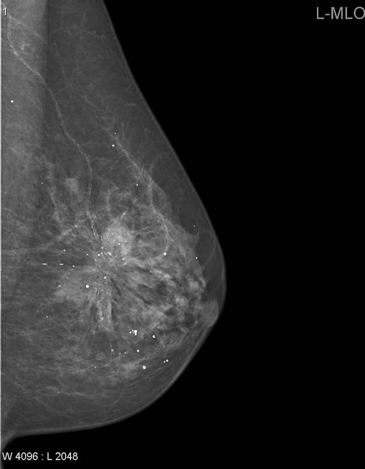
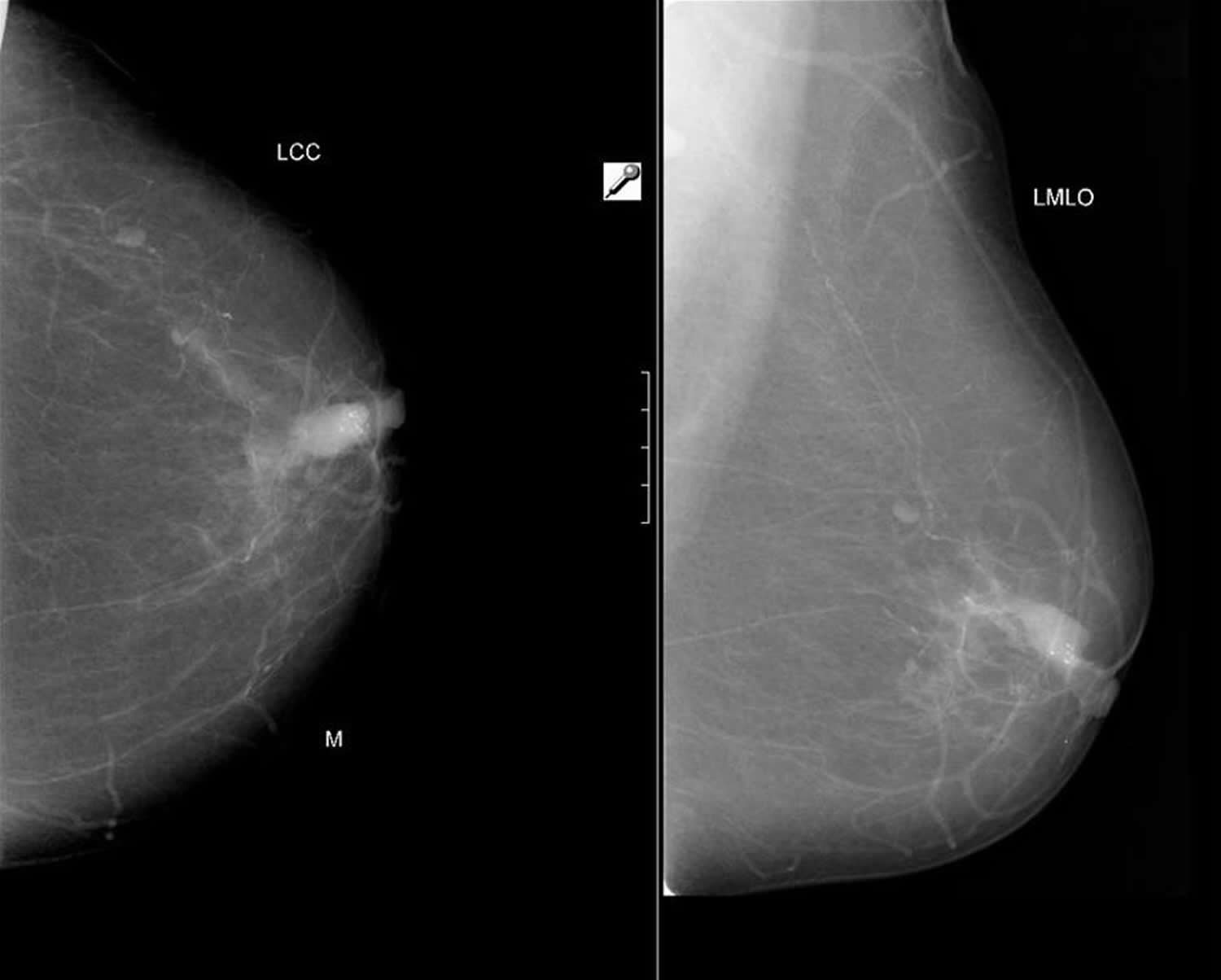
Figure 3. Spiculated breast cancer mammogram – Note standard mammographic mediolateral oblique (MLO) view of a left breast demonstrating a large spiculated mass in the upper outer quadrant consistent with a breast cancer is present.
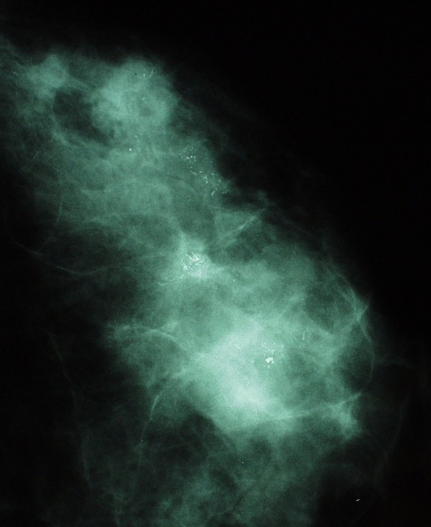
Figure 4. Multifocal breast cancer mammogram
Note: The density far lateral in the left breast was a palpable infiltrating ductal carcinoma. A second smaller infiltrating ductal carcinoma is visible deeper to the first. Also demonstrated are multiple foci of ductal carcinoma in situ (DCIS) calcification at sites in the breast. Typical ductal carcinoma in situ (DCIS) calcifications.
Figure 5. Metaplastic carcinoma of the breast
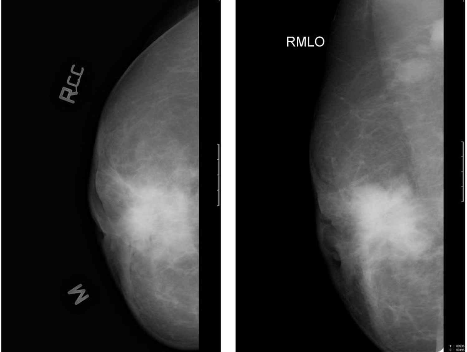
Figure 6. Locally advanced breast cancer
Figure 7. Intraductal carcinoma
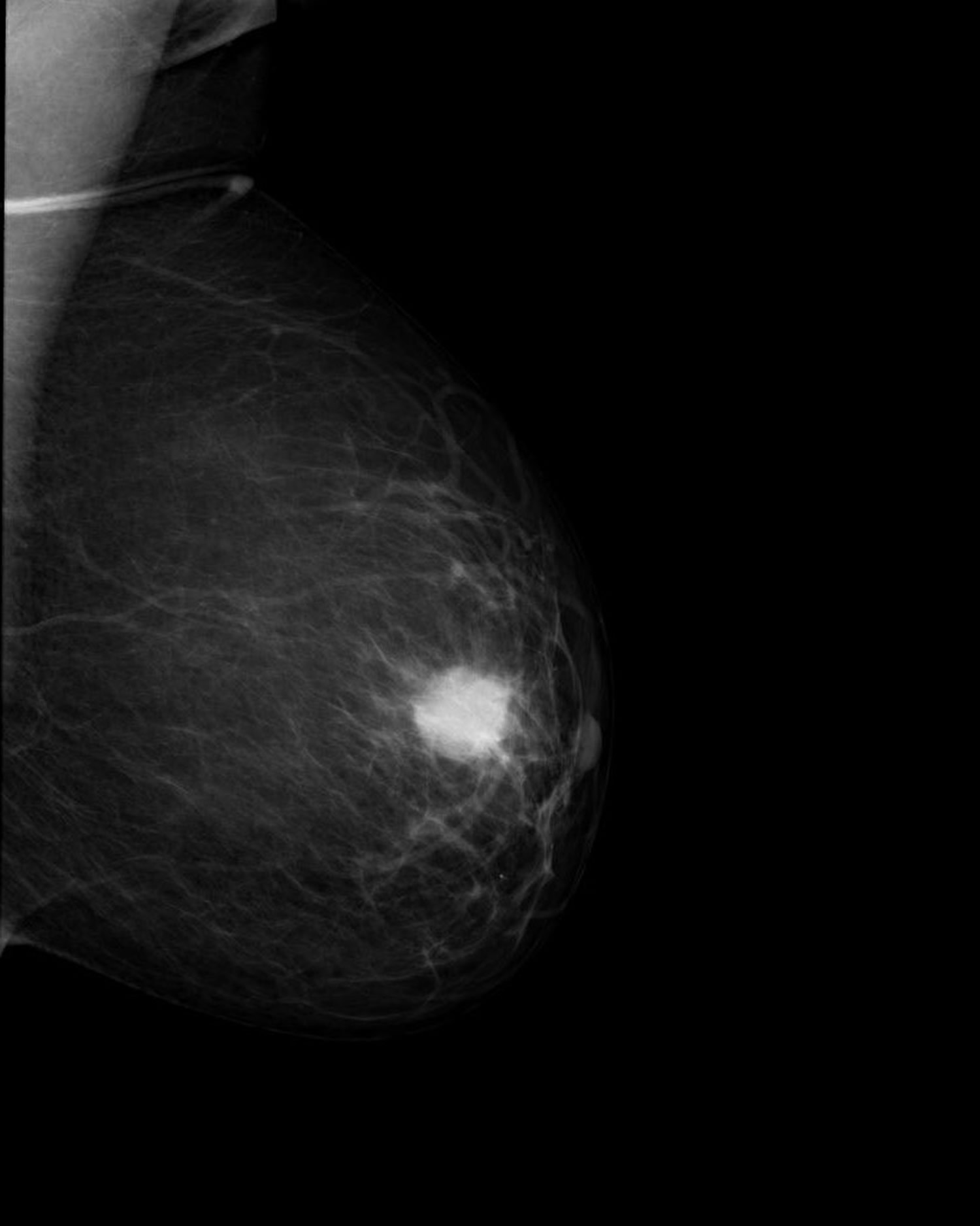
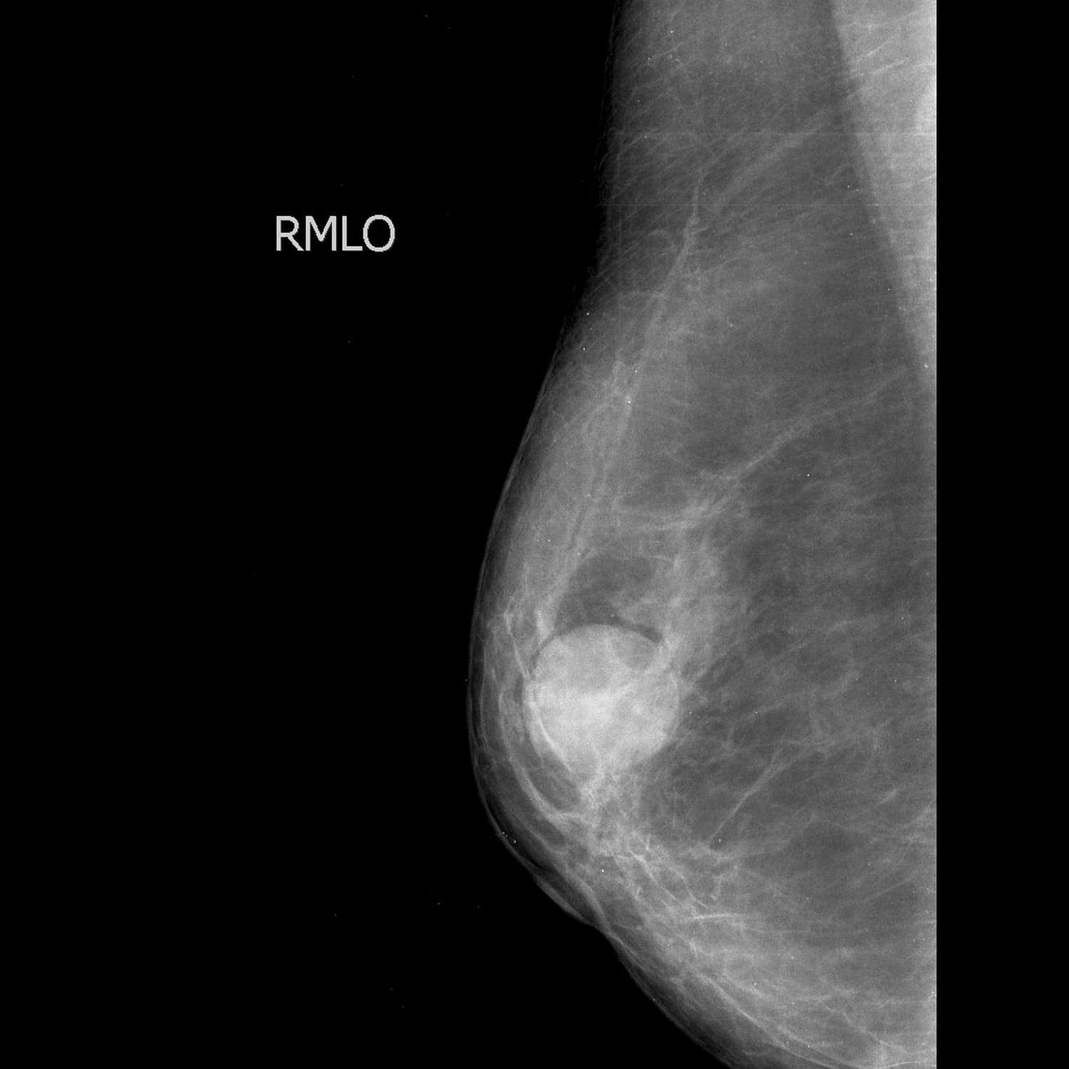
Figure 8. Malignant epithelial neoplasm of breast – Note: Well defined radio-opacity with microlobulation in the upper outer quadrant of right breast, compressing the surrounding tissue resulting in a lucent halo around.
Figure 9. Tubular carcinoma of the breast – a subtype of of invasive ductal carcinoma (IDC).
How are screening and diagnostic mammograms different?
The same machines are used for both types of mammograms. However, diagnostic mammogram takes longer to perform than screening mammogram and the total dose of radiation is higher because more x-ray images are needed to obtain views of the breast from several angles. The technologist may magnify a suspicious area to produce a detailed picture that can help the doctor make an accurate diagnosis.
What are the benefits and potential harms of screening mammograms?
Early detection of breast cancer with screening mammogram means that treatment can be started earlier in the course of the disease, possibly before it has spread. Results from randomized clinical trials and other studies show that screening mammogram can help reduce the number of deaths from breast cancer among women ages 40 to 74, especially for those over age 50 2. However, studies to date have not shown a benefit from regular screening mammogram in women under age 40 or from baseline screening mammograms (mammograms used for comparison) taken before age 40.
The benefits of screening mammogram need to be balanced against its harms, which include:
- False-positive results. False-positive results occur when radiologists see an abnormality (that is, a potential “positive”) on a mammogram but no breast cancer is actually present. All abnormal mammograms should be followed up with additional testing (diagnostic mammograms, ultrasound, and/or biopsy) to determine whether cancer is present. False-positive mammogram results can lead to anxiety and other forms of psychological distress in affected women. The additional testing required to rule out cancer can also be costly and time consuming and can cause physical discomfort. False-positive results are more common for younger women, women with dense breasts, women who have had previous breast biopsies, women with a family history of breast cancer, and women who are taking estrogen (for example, menopausal hormone therapy). The chance of having a false-positive result increases with the number of mammograms a woman has. More than 50% of women screened annually for 10 years in the United States will experience a false-positive result, and many of these women will have a biopsy.
- Overdiagnosis and overtreatment. Screening mammograms can find breast cancers and cases of ductal carcinoma in situ (DCIS, a noninvasive tumor in which abnormal cells that may become cancerous build up in the lining of breast ducts) that need to be treated. However, they can also find cases of ductal carcinoma in situ (DCIS) and small breast cancers that would never cause symptoms or threaten a woman’s life. This phenomenon is called “overdiagnosis.” Treatment of overdiagnosed breast cancers and overdiagnosed cases of DCIS is not needed and results in “overtreatment.” Because doctors cannot easily distinguish cancers and cases of ductal carcinoma in situ (DCIS) that need to be treated from those that do not, they are all treated.
- False-negative results. In breast cancer screening, a negative result means no abnormality is present. False-negative results occur when mammograms appear normal even though breast cancer is present. Overall, screening mammograms miss about 20% of breast cancers that are present at the time of screening. False-negative results can lead to delays in treatment and a false sense of security for affected women. One cause of false-negative results is high breast density. Breasts contain both dense tissue (i.e., glandular tissue and connective tissue, together known as fibroglandular tissue) and fatty tissue. Fatty tissue appears dark on a mammogram, whereas fibroglandular tissue appears as white areas. Because fibroglandular tissue and tumors have similar density, tumors can be harder to detect in women with denser breasts. False-negative results occur more often among younger women than among older women because younger women are more likely to have dense breasts. As a woman ages, her breasts usually become more fatty, and false-negative results become less likely. Some breast cancers grow so quickly that they appear within months of a normal (negative) screening mammogram. This situation does not represent a false-negative result, because the negative result of the screening was correct. But it means that a negative result can give a false sense of security. Some of the cancers missed by screening mammograms can be detected by clinical breast exams (physical exams of the breast done by a health care provider).
- Finding breast cancer early may not reduce a woman’s chance of dying from the disease. Even though mammograms can detect malignant tumors that cannot be felt, treating a small tumor does not always mean that the woman will not die from the cancer. A fast-growing or aggressive cancer may have already spread to other parts of the body before it is detected. Instead, women with such tumors live a longer period of time knowing that they likely have a potentially fatal disease. In addition, finding breast cancer early may not help prolong the life of a woman who is suffering from other, more life-threatening health conditions.
- Radiation exposure. Mammograms require very small doses of radiation. The risk of harm from this radiation exposure is low, but repeated x-rays have the potential to cause cancer. Although the potential benefits of mammography nearly always outweigh the potential harm from the radiation exposure, women should talk with their health care providers about the need for each x-ray. In addition, they should always let their health care provider and the x-ray technologist know if there is any possibility that they are pregnant, because radiation can harm a growing fetus.
Limitations of Mammograms
Mammograms are the best breast cancer screening tests we have at this time. But mammograms have their limits. For example, they aren’t 100% accurate in showing if a woman has breast cancer:
- A false-negative mammogram looks normal even though breast cancer is present.
- A false-positive mammogram looks abnormal even though there’s no cancer in the breast.
False-negative results
A false-negative mammogram looks normal even though breast cancer is present. Overall, screening mammograms do not find about 1 in 5 breast cancers.
- Women with dense breasts have more false-negative results.
- Breasts often become less dense as women age, so false negatives are more common in younger women.
- False-negative mammograms can give women a false sense of security, thinking that they don’t have breast cancer when in fact they do.
False-positive results
A false-positive mammogram looks abnormal even though no cancer is actually present. Abnormal mammograms require extra testing (diagnostic mammograms, ultrasound, and sometimes MRI or even a breast biopsy) to find out if the change is cancer.
- False-positive results are more common in women who are younger, have dense breasts, have had breast biopsies, have breast cancer in the family, or are taking estrogen.
- About half of the women getting annual mammograms over a 10-year period will have a false-positive finding.
- The odds of a false-positive finding are highest for the first mammogram. Women who have past mammograms available for comparison reduce their odds of a false-positive finding by about 50%.
- False-positive mammograms can cause anxiety. They can also lead to extra tests to be sure cancer isn’t there, which cost time and money and maybe even physical discomfort.
Mammograms might not be helpful for all women
The value of a screening mammogram depends on a woman’s overall health. Finding breast cancer early may not help her live longer if she has other serious or life-threatening health problems, such as serious heart disease, or severe kidney, liver, or lung disease. The American Cancer Society breast cancer screening guidelines emphasize that women with serious health problems or short life expectancies should discuss with their doctors whether they should continue having mammograms. Our guidelines also stress that age alone should not be the reason to stop having regular mammograms.
It’s important to know that even though mammograms can often find breast cancers that are too small to be felt, treating a small tumor does not always mean it can be cured. A fast-growing or aggressive cancer might have already spread.
Overdiagnosis and overtreatment
Screening mammograms can find invasive breast cancer and ductal carcinoma in situ (DCIS, cancer cells in the lining of breast ducts) that need to be treated. But it’s possible that some of the invasive cancers and ductal carcinoma in situ (DCIS) found on mammograms would never grow or spread. (Finding and treating cancers that would never cause problems is called overdiagnosis.) These cancers are not life-threatening, and never would have been found or treated if the woman had not gotten a mammogram. The problem is that doctors can’t tell these cancers from those that will grow and spread.
Overdiagnosis leads to some women getting treatment that’s not really needed (overtreatment), because the cancer never would have caused any problems. Doctors don’t know which women fall into this group when the cancer is found because they can’t tell which cancers will be life-threatening and which won’t ever cause problems. Because of this, all cases are treated. This exposes some women to the adverse effects of cancer treatment that’s really not needed.
Still, overdiagnosis is not thought to happen that often. There’s a wide range of estimates of the percentage of breast cancers that might be overdiagnosed by mammography, but the most credible estimates range from 1% to 10%.
Radiation exposure
Because mammograms are x-ray tests, they expose the breasts to radiation. The amount of radiation from each mammogram is low, but it can still add up over time.
Mammogram screening
Benefit of Screening
The results of the meta-analysis of clinical trials from the systematic evidence review commissioned by the United States Preventive Services Task Force are summarized in Table 1. Over a 10-year period, screening 10,000 women aged 60 to 69 years will result in 21 (95% CI, 11 to 32) fewer breast cancer deaths. The benefit is smaller in younger women: screening 10,000 women aged 50 to 59 years will result in 8 (CI, 2 to 17) fewer breast cancer deaths, and screening 10,000 women aged 40 to 49 years will result in 3 (CI, 0 to 9) fewer breast cancer deaths.2, 3 Most of these trials began enrollment more than 30 years ago, and these estimates may not reflect the current likelihood of avoiding a breast cancer death with contemporary screening mammography technology. Mammography imaging has since improved, which may result in more tumors being detected at a curable stage today than at the time of these trials. However, breast cancer treatments have also improved, and as treatment improves, the advantage of earlier detection decreases, so that some of the women who died of breast cancer in the nonscreened groups in these trials would survive today.
Table 1. Breast Cancer Deaths Avoided per 10,000 Women Screened by Repeat Screening Mammography Over 10 Years: Data From Randomized, Controlled Trials*
| Ages 40–49 y | Ages 50–59 y | Ages 60–69 y | Ages 70–74 y | |
|---|---|---|---|---|
| Breast cancer deaths avoided | 3 (0–9) | 8 (2–17) | 21 (11–32) | 13 (0–32) |
* All women did not have 100% adherence to all rounds of screening offered in the randomized, controlled trials.
[Source 1]Harms of Screening
The most important harm of screening is the detection and treatment of invasive and noninvasive cancer that would never have been detected, or threaten health, in the absence of screening (overdiagnosis and overtreatment). Existing science does not allow for the ability to determine precisely what proportion of cancer diagnosed by mammography today reflects overdiagnosis, and estimates vary widely depending on the data source and method of calculation used 3. In the United States, the rate of diagnosis of invasive plus noninvasive breast cancer increased by 50% during the era of mammography screening 4. It is not possible to know with certainty what proportion of that increase is due to overdiagnosis and what proportion reflects other reasons for a rising incidence. If overdiagnosis is the only explanation for the increase, 1 in 3 women diagnosed with breast cancer today is being treated for cancer that would never have been discovered or caused her health problems in the absence of screening. The best estimates from randomized, controlled trials (RCTs) evaluating the effect of mammography screening on breast cancer mortality suggest that 1 in 5 women diagnosed with breast cancer over approximately 10 years will be overdiagnosed 5. Modeling studies conducted in support of this recommendation by the Cancer Intervention and Surveillance Modeling Network (CISNET) provide a range of estimates that reflect different underlying assumptions; the median estimate is that 1 in 8 women diagnosed with breast cancer with biennial screening from ages 50 to 74 years will be overdiagnosed. The rate increases with an earlier start age or with annual mammography 6. Even with the conservative estimate of 1 in 8 breast cancer cases being overdiagnosed, for every woman who avoids a death from breast cancer through screening, 2 to 3 women will be treated unnecessarily.
The other principal harms of screening are false-positive results, which require further imaging and often breast biopsy, and false-negative results. Table 2 summarizes the rates of these harms per screening round using registry data for digital mammography from the Breast Cancer Surveillance Consortium, a collaborative network of 5 mammography registries and 2 affiliated sites with linkages to tumor registries across the United States 7. (Note that Table 2 describes a different time horizon than Table 1 [per screening round rather than per decade]).
Table 2. Harms of One-Time Mammography Screening per 10,000 Women Screened: Breast Cancer Surveillance Consortium Registry Data
| Ages 40–49 y | Ages 50–59 y | Ages 60–69 y | Ages 70–74 y | |
|---|---|---|---|---|
| False-positive mammograms (false alarms), n | 1212 | 932 | 808 | 696 |
| Breast biopsies, n | 164 | 159 | 165 | 175 |
| False-negative mammograms (missed cancers), n | 10 | 11 | 12 | 15 |
When to Start Screening
Clinical trials, observational studies, and modeling studies all demonstrate that the likelihood of avoiding a breast cancer death with regular screening mammography increases with age, and this increase in benefit likely occurs gradually rather than abruptly at any particular age. In contrast, the harms of screening mammography either remain constant or decrease with age. For example, about the same number of breast biopsies are performed as a result of screening mammography in women aged 40 to 49 years as in those aged 60 to 69 years, but many more of these biopsies will result in a diagnosis of invasive cancer in the older age group. Thus, the balance of benefit and harms improves with age (Table 3).
Table 3. Lifetime Benefits and Harms of Biennial Screening Mammography per 1000 Women Screened: Model Results Compared With No Screening*
| Variable | Ages 40–74 y | Ages 50–74 y |
|---|---|---|
| Fewer breast cancer deaths, n | 8 (5–10) | 7 (4–9) |
| Life-years gained, n | 152 (99–195) | 122 (75–154) |
| False-positive tests, n | 1529 (1100–1976) | 953 (830–1325) |
| Unnecessary breast biopsies, n | 213 (153–276) | 146 (121–205) |
| Overdiagnosed breast tumors, n | 21 (12–38) | 19 (11–34) |
* Values reported are medians (ranges).
[Source 1]The United States Preventive Services Task Force concludes that while there are harms of mammography, the benefit of screening mammography outweighs the harms by at least a moderate amount from age 50 to 74 years and is greatest for women in their 60s. For women in their 40s, the number who benefit from starting regular screening mammography is smaller and the number experiencing harm is larger compared with older women. For women in their 40s, the benefit still outweighs the harms, but to a smaller degree; this balance may therefore be more subject to individual values and preferences than it is in older women. Women in their 40s must weigh a very important but infrequent benefit (reduction in breast cancer deaths) against a group of meaningful and more common harms (overdiagnosis and overtreatment, unnecessary and sometimes invasive follow-up testing and psychological harms associated with false-positive test results, and false reassurance from false-negative test results). Women who value the possible benefit of screening mammography more than they value avoiding its harms can make an informed decision to begin screening.
Neither clinical trials nor models can precisely predict the potential benefits and harms that an individual woman can expect from beginning screening at age 40 rather than 50 years, as these data represent population effects. However, model results may be the easiest way for women to visualize the relative tradeoffs of beginning screening at age 40 versus 50 years. Cancer Intervention and Surveillance Modeling Network (CISNET) conducted modeling studies to predict the lifetime benefits and harms of screening with contemporary digital mammography at different starting and stopping ages and screening intervals. The models varied their assumptions about the natural history of invasive and noninvasive breast cancer and the effect of detection by digital mammography on survival. The models assumed the ideal circumstances of perfect adherence to screening and current best practices for therapy across the life span. Table 3 compares the median and range across the models for predicted lifetime benefits and harms of screening biennially from ages 50 to 74 years with screening biennially from ages 40 to 74 years. (Note that Table 3 differs from Tables 1 and 2 in terms of population metrics [per 1000 vs. 10,000 women] and time horizon considered [lifetime vs. 10-year or single event]).
It is, however, a false dichotomy to assume that the only options are to begin screening at age 40 or to wait until age 50 years. As women advance through their 40s, the incidence of breast cancer rises. The balance of benefit and harms may also shift accordingly over this decade, such that women in the latter half of the decade likely have a more favorable balance than women in the first half. Indeed, the Cancer Intervention and Surveillance Modeling Network (CISNET) models suggest that most of the benefit of screening women aged 40 to 49 years would be realized by starting screening at age 45 6.
Risk Factors That May Influence When to Start Screening
Advancing age is the most important risk factor for breast cancer in most women, but epidemiologic data from the Breast Cancer Surveillance Consortium suggest that having a first-degree relative with breast cancer is associated with an approximately 2-fold increased risk for breast cancer in women aged 40 to 49 years 3. Further, the Cancer Intervention and Surveillance Modeling Network (CISNET) models suggest that for women with about a 2-fold increased risk for breast cancer, starting annual digital screening at age 40 years results in a similar harm-to-benefit ratio (based on number of false-positive results or overdiagnosed cases per 1000 breast cancer deaths avoided) as beginning biennial digital screening at age 50 years in average-risk women 6. This approach has not been formally tested in a clinical trial; therefore, there is no direct evidence that it would result in net benefit similar to that of women aged 50 to 74 years. However, given the increased burden of disease and potential likelihood of benefit, women aged 40 to 49 years who have a known first-degree relative (parent, child, or sibling) with breast cancer may consider initiating screening earlier than age 50 years. Many other risk factors have been associated with breast cancer in epidemiologic studies, but most of these relationships are weak or inconsistent and would not likely influence how women value the tradeoffs of the potential benefits and harms of screening. Risk calculators, such as the National Cancer Institute’s Breast Cancer Risk Assessment Tool (available at https://www.cancer.gov/BCRISKTOOL/Default.aspx), have good calibration between predicted and actual outcomes in groups of women but are not accurate at predicting an individual woman’s risk for breast cancer 8.
How Often to Screen
Once a woman has decided to begin screening, the next decision is how often to undergo screening. No clinical trials compared annual mammography with a longer interval in women of any age. In the randomized trials that demonstrated the effectiveness of mammography in reducing breast cancer deaths in women aged 40 to 74 years, screening intervals ranged from 12 to 33 months 3. There was no clear trend for greater benefit in trials of annual mammography, but other differences between the trials preclude certainty that no difference in benefit exists. Available observational evidence evaluating the effects of varying mammography intervals found no difference in the number of breast cancer deaths between women aged 50 years or older who were screened biennially versus annually 3.
Regardless of the starting age for screening, the models consistently predict a small incremental increase in the number of breast cancer deaths averted when moving from biennial to annual mammography, but also a large increase in the number of harms (Table 4) 6. The United States Preventive Services Task Force concludes that for most women, biennial mammography screening provides the best overall balance of benefit and harms.
Table 4. Lifetime Benefits and Harms of Annual Versus Biennial Screening Mammography per 1000 Women Screened: Model Results Compared With No Screening*
| Variable | Ages 50–74 y, Annual Screening | Ages 50–74 y, Biennial Screening |
|---|---|---|
| Fewer breast cancer deaths, n | 9 (5–10) | 7 (4–9) |
| Life-years gained, n | 145 (104–180) | 122 (75–154) |
| False-positive tests, n | 1798 (1706–2445) | 953 (830–1325) |
| Unnecessary breast biopsies, n | 228 (219–317) | 146 (121–205) |
| Overdiagnosed breast tumors, n | 25 (12–68) | 19 (11–34) |
* Values reported are medians (ranges).
[Source 1]When to Consider Stopping Screening
Clinical trial data for women aged 70 to 74 years are inconclusive. In its 2009 recommendation 9, the United States Preventive Services Task Force extended the recommendation for screening mammography to age 74 years based on the extrapolation that much of the benefit seen in women aged 60 to 69 years should continue in this age range, and modeling done at the time supported this assumption. Current Cancer Intervention and Surveillance Modeling Network (CISNET) models suggest that women aged 70 to 74 years with moderate to severe comorbid conditions that negatively affect their life expectancy are unlikely to benefit from mammography 6. Moderate comorbid conditions include cardiovascular disease, paralysis, and diabetes. Severe comorbid conditions include (but are not limited to) AIDS, chronic obstructive pulmonary disease, liver disease, chronic renal failure, dementia, congestive heart failure, and combinations of moderate comorbid conditions, as well as myocardial infarction, ulcer, and rheumatologic disease 10.
Screening in Women Aged 75 Years or Older
The United States Preventive Services Task Force found insufficient evidence to assess the balance of benefits and harms of screening mammography in women aged 75 years or older. Current Cancer Intervention and Surveillance Modeling Network (CISNET) models suggest that biennial mammography screening may potentially continue to offer a net benefit after age 74 years among women with no or low comorbidity 6, but no randomized trials of screening included women in this age group 3.
Mammogram screening guidelines
Many organizations and professional societies, including the United States Preventive Services Task Force (which is convened by the Agency for Healthcare Research and Quality, a federal agency), have developed guidelines for mammography screening. All recommend that women talk with their doctor about the benefits and harms of mammography, when to start screening, and how often to be screened.
United States Preventive Services Task Force screenings recommendations for women
Women aged 50 to 74 years
The United States Preventive Services Task Force recommends biennial (every 2 years) screening mammography for women aged 50 to 74 years 1.
Women aged 40 to 49 years
The decision to start screening mammography in women prior to age 50 years should be an individual one 1. Women who place a higher value on the potential benefit than the potential harms may choose to begin biennial screening between the ages of 40 and 49 years.
- For women who are at average risk for breast cancer, most of the benefit of mammography results from biennial screening during ages 50 to 74 years. Of all of the age groups, women aged 60 to 69 years are most likely to avoid breast cancer death through mammography screening. While screening mammography in women aged 40 to 49 years may reduce the risk for breast cancer death, the number of deaths averted is smaller than that in older women and the number of false-positive results and unnecessary biopsies is larger. The balance of benefits and harms is likely to improve as women move from their early to late 40s.
- In addition to false-positive results and unnecessary biopsies, all women undergoing regular screening mammography are at risk for the diagnosis and treatment of noninvasive and invasive breast cancer that would otherwise not have become a threat to their health, or even apparent, during their lifetime (known as “overdiagnosis”). Beginning mammography screening at a younger age and screening more frequently may increase the risk for overdiagnosis and subsequent overtreatment.
- Women with a parent, sibling, or child with breast cancer are at higher risk for breast cancer and thus may benefit more than average-risk women from beginning screening in their 40s.
Women aged 75 years or older
The United States Preventive Services Task Force concludes that the current evidence is insufficient to assess the balance of benefits and harms of screening mammography in women aged 75 years or older.
Women with dense breasts
The United States Preventive Services Task Force concludes that the current evidence is insufficient to assess the balance of benefits and harms of adjunctive screening for breast cancer using breast ultrasonography, magnetic resonance imaging (MRI), Digital Breast Tomosynthesis (DBT), or other methods in women identified to have dense breasts on an otherwise negative screening mammogram.
All women
The United States Preventive Services Task Force concludes that the current evidence is insufficient to assess the benefits and harms of digital breast tomosynthesis (DBT) as a primary screening method for breast cancer.
American Cancer Society screenings recommendations for women at average breast cancer risk
These guidelines are for women at average risk for breast cancer 11. For screening purposes, a woman is considered to be at average risk if she doesn’t have a personal history of breast cancer, a strong family history of breast cancer, or a genetic mutation known to increase risk of breast cancer (such as in a BRCA gene), and has not had chest radiation therapy before the age of 30. (See below for guidelines for women at high risk.)
- Women between 40 and 44 have the option to start screening with a mammogram every year.
- Women 45 to 54 should get mammograms every year.
- Women 55 and older can switch to a mammogram every other year, or they can choose to continue yearly mammograms. Screening should continue as long as a woman is in good health and is expected to live 10 more years or longer.
All women should understand what to expect when getting a mammogram for breast cancer screening – what the test can and cannot do.
American Cancer Society screening recommendations for women at high risk
Women who are at high risk for breast cancer based on certain factors should get an MRI and a mammogram every year, typically starting at age 30. This includes women who:
- Have a lifetime risk of breast cancer of about 20% to 25% or greater, according to risk assessment tools that are based mainly on family history (see below)
- Have a known BRCA1 or BRCA2 gene mutation (based on having had genetic testing)
- Have a first-degree relative (parent, brother, sister, or child) with a BRCA1 or BRCA2 gene mutation, and have not had genetic testing themselves
- Had radiation therapy to the chest when they were between the ages of 10 and 30 years
- Have Li-Fraumeni syndrome, Cowden syndrome, or Bannayan-Riley-Ruvalcaba syndrome, or have first-degree relatives with one of these syndromes
The American Cancer Society recommends against MRI screening for women whose lifetime risk of breast cancer is less than 15%.
There’s not enough evidence to make a recommendation for or against yearly MRI screening for women who have a higher lifetime risk based on certain factors , such as:
- Having a personal history of breast cancer, ductal carcinoma in situ (DCIS), lobular carcinoma in situ (LCIS), atypical ductal hyperplasia (ADH), or atypical lobular hyperplasia (ALH)
- Having “extremely” or “heterogeneously” dense breasts as seen on a mammogram
If MRI is used, it should be in addition to, not instead of, a screening mammogram. This is because although an MRI is more likely to detect cancer than a mammogram, it may still miss some cancers that a mammogram would detect.
Most women at high risk should begin screening with MRI and mammograms when they are 30 and continue for as long as they are in good health. But a woman at high risk should make the decision to start with her health care providers, taking into account her personal circumstances and preferences.
Tools used to assess breast cancer risk
Several risk assessment tools are available to help health professionals estimate a woman’s breast cancer risk. These tools give approximate, rather than precise, estimates of breast cancer risk based on different combinations of risk factors and different data sets.
Because the different tools use different factors to estimate risk, they may give different risk estimates for the same woman. Two models could easily give different estimates for the same person.
Risk assessment tools that include family history in first-degree relatives (parents, siblings, and children) and second-degree relatives (such as aunts and cousins) on both sides of the family should be used with the American Cancer Society guidelines to decide if a woman should have MRI screening. The use of any of the risk assessment tools and its results should be discussed by a woman with her health care provider.
Mammogram results
How will I get my mammogram results?
If you don’t hear from your health care provider within 10 days, do not assume that your mammogram was normal. Call your provider or the facility where the mammogram was done.
A full report of the results of your mammogram will be sent to your health care provider. Mammography clinics also must mail women an easy-to-understand summary of their mammogram results within 30 days—or “as quickly as possible” if the results suggest cancer is present. This means you could get the results before your provider calls you. If you want the full written mammogram report as well as the summary, you’ll need to ask for it.
A doctor called a radiologist will categorize your mammogram results using a number system of 0 through 6. You should talk to your doctor about your mammogram’s category and what you need to do next.
Breast Imaging Reporting and Data System Assessment Category
Doctors use a standard system to describe mammogram findings and results. This system (called the Breast Imaging Reporting and Data System or BI-RADS) sorts the results into categories numbered 0 through 6.
By sorting the results into these categories, doctors can describe what they find on a mammogram using the same words and terms. This makes accurately communicating about these test results and following up after the tests much easier.
Table 5. Breast Imaging Reporting and Data System Categories
| Category | Definition | What it means |
| 0 | Additional imaging evaluation and/or comparison to prior mammograms is needed. | This means the radiologist may have seen a possible abnormality, but it was not clear and you will need more tests, such as another mammogram with the use of spot compression (applying compression to a smaller area when doing the mammogram), magnified views, special mammogram views, or ultrasound. This may also suggest that the radiologist wants to compare your new mammogram with older ones to see if there have been changes in the area over time.
|
| 1 | Negative
| There’s no significant abnormality to report. Your breasts look the same (they are symmetrical) with no masses (lumps), distorted structures, or suspicious calcifications. In this case, negative means nothing bad was found. |
| 2 | Benign (non-cancerous) finding | This is also a negative mammogram result (there’s no sign of cancer), but the radiologist chooses to describe a finding known to be benign, such as benign calcifications, lymph nodes in the breast, or calcified fibroadenomas. This ensures that others who look at the mammogram will not misinterpret the benign finding as suspicious. This finding is recorded in your mammogram report to help when comparing to future mammograms. |
| 3 | Probably benign finding – Follow-up in a short time frame is suggested
| The findings in this category have a very high chance (greater than 98%) of being benign (not cancer). The findings are not expected to change over time. But since it’s not proven to be benign, it’s helpful to see if the area in question does change over time. You will likely need follow-up with repeat imaging in 6 months and regularly after that until the finding is known to be stable (usually at least 2 years). This approach helps avoid unnecessary biopsies, but if the area does change over time, it still allows for early diagnosis. |
| 4 | Suspicious abnormality – Biopsy should be considered | Findings do not definitely look like cancer but could be cancer. The radiologist is concerned enough to recommend a biopsy. The findings in this category can have a wide range of suspicion levels. For this reason, some, but not all, doctors divide this category further: 4A: Finding with a low suspicion of being cancer 4B: Finding with an intermediate suspicion of being cancer 4C: Finding of moderate concern of being cancer, but not as high as Category 5 |
| 5 | Highly suggestive of malignancy – Appropriate action should be taken | The findings look like cancer and have a high chance (at least 95%) of being cancer. Biopsy is very strongly recommended. |
| 6 | Known biopsy-proven malignancy – Appropriate action should be taken | This category is only used for findings on a mammogram that have already been shown to be cancer by a previous biopsy. Mammograms may be used in this way to see how well the cancer is responding to treatment. |
Your mammogram report will also include an assessment of your breast density, which is a description of how much fibrous and glandular tissue is in your breasts, as opposed to fatty tissue. The denser your breasts, the harder it can be to see abnormal areas on mammograms.
Breast Imaging Reporting and Data System (BI-RADS) classifies breast density into 4 groups. Regular mammograms are the best way to find breast cancer early. But if your mammogram report says that you have dense breast tissue, you may be wondering what that means. Having dense breasts is very common and is not abnormal.
Breast density categories
Radiologists use the Breast Imaging Reporting and Data System (BI-RADS), to classify breast density into 4 categories. They go from almost all fatty tissue to extremely dense tissue with very little fat.
Some mammogram reports sent to women mention breast density. Your health care provider can also tell you if your mammogram shows that you have dense breasts.
In some states, women whose mammograms show heterogenously dense or extremely dense breasts must be told that they have dense breasts in the summary of the mammogram report that is sent to patients (sometimes called the lay summary).
The radiologist who reads the mammogram chooses the category that best describes the level of breast density seen on the mammogram film. The categories, from the least amount of breast density to the highest, are as follows:
- The breasts are almost entirely fatty
- There are scattered areas of dense glandular tissue and fibrous connective tissue (together known as fibroglandular density)
- The breasts are heterogeneously dense, which means they have more of these areas of fibroglandular density. This may make it hard to see small masses in the breast tissue on a mammogram.
- The breasts are extremely dense, which makes it hard to see tumors in the breast tissue on a mammogram.
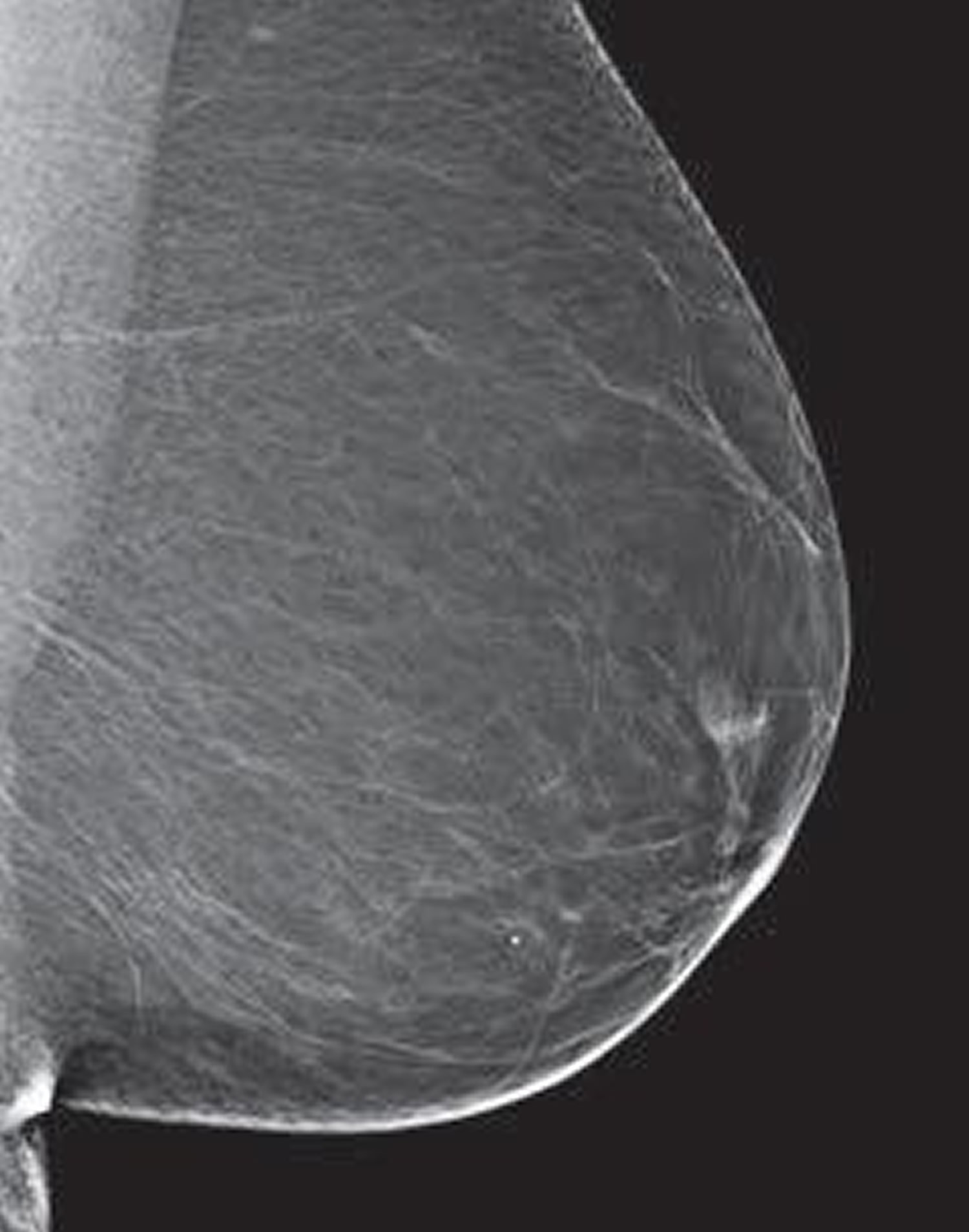
Figure 10. Breasts are almost all fatty tissue
Figure 11. Scattered areas of dense glandular and fibrous tissue
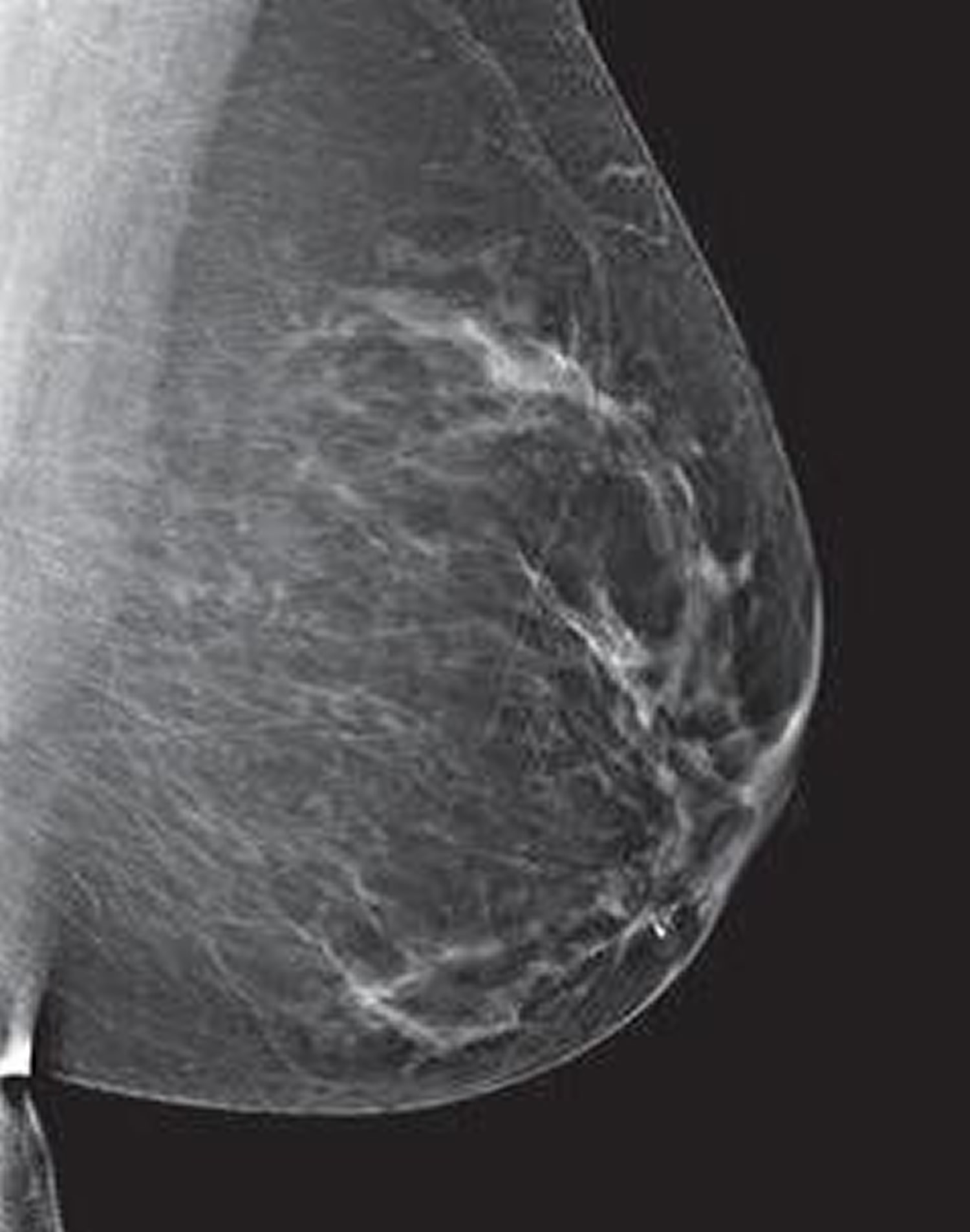
Figure 12. Heterogenously dense – Note: More of the breast is made of dense glandular and fibrous tissue. This can make it hard to see small tumors in or around the dense tissue.
Figure 13. Breasts are extremely dense making it hard to see tumors in the breast tissue
Getting Called Back After a Mammogram
Getting called back after a screening mammogram is fairly common and doesn’t mean you have breast cancer. In fact, fewer than 1 in 10 women called back for more tests are found to have breast cancer. Often, it just means more x-rays or an ultrasound needs to be done to get a closer look at an area of concern.
Getting called back is more common after a first mammogram, or when there’s no previous mammogram to compare the new mammogram with. It’s also more common in women who haven’t gone through menopause.
What else could it be?
You could be called back after your mammogram because:
- The pictures weren’t clear or didn’t show some of your breast tissue and need to be retaken.
- You have dense breast tissue, which can make it hard to see some parts of your breasts.
- The radiologist sees calcifications or a mass (a cyst or solid tumor).
- The radiologist sees an area that just looks different from other parts of the breast.
Sometimes when more x-rays are taken of the area or mass, or the area is compressed more, it no longer looks suspicious. In fact, most repeat mammograms do not find cancer.
What will happen at the follow-up appointment?
- You’ll likely have another mammogram called a diagnostic mammogram. (Your previous mammogram was called a screening mammogram.) A diagnostic mammogram is done just like a screening mammogram, but more pictures are taken so that any areas of concern can be carefully studied. A radiologist is on hand to advise the technologist (the person who operates the mammogram machine) to be sure they have all the images that are needed.
- You may also have an ultrasound test, which uses sound waves to make pictures of the inside of your breast at the area of concern.
- Some women may need a breast MRI. For this test, you’ll lie face down inside a narrow tube for up to an hour while the machine creates more detailed images of the breast tissues. MRI is painless, but it can be uncomfortable for people who don’t like small, tight spaces.
You can expect to learn the results of your tests during the visit. You are likely to be told one of the following:
- The suspicious area turned out to be nothing to worry about and you can return to your normal mammogram schedule.
- The area is probably nothing to worry about, but you should have your next mammogram sooner than normal – usually in 6 months – to watch it closely and make sure it’s not changing over time.
- The changed area could be cancer, so you will need to have a biopsy to know for sure.
You’ll also get a letter with a summary of the findings that will tell you if you need more tests and/or when you should schedule your next mammogram.
What if I need a biopsy?
During a breast biopsy, a small piece of breast tissue is removed and checked for cancer under a microscope. Even if you need a biopsy, it doesn’t mean you have cancer. Most biopsy results are not cancer, but a biopsy is the only way to find out.
There are several different types of biopsies, some of which are done using a needle and some that are done through a cut in the skin. The type you have depends on things like how suspicious the tumor looks, how big it is, where it is in the breast, how many tumors there are, other medical problems you might have, and your personal preferences.
How can I stay calm while waiting?
Waiting for appointments and the results of tests can be frightening. Many women have strong emotions including disbelief, anxiety, fear, anger, and sadness during this time. Here are some things to remember:
- It’s normal to have these feelings.
- Most breast changes are not cancer and are not life-threatening.
- Talking with a loved one or a counselor about your feelings may help.
- Talking with other women who have been through a breast biopsy may help.
- The American Cancer Society is available at https://www.cancer.org/ or the National Cancer Institute at https://www.cancer.gov/ around the clock to answer your questions and provide support.
What if it’s cancer?
If you do have cancer and you’re referred to a breast specialist, use these tips to make your appointment as useful as possible:
- Make a list of questions to ask.
- Take a family member or friend with you. They can serve as an extra pair of ears, take notes, help you remember things later, and give you support.
- Ask if you can record the conversations.
- Take notes. If someone uses a word you don’t know, ask them to spell it and explain it.
- Ask the doctors or nurses to explain anything you don’t understand.
Coping with the shock, fear and sadness that come with a cancer diagnosis can take time. You may feel overwhelmed just when you need to make crucial decisions. With time, each person finds a way of coping and coming to terms with the diagnosis.
Until you find what brings you the most comfort, consider trying to:
- Find out enough about the cancer to make decisions about your care. Ask your doctor for the specifics about your cancer, such as its type and stage. And ask for recommended sources of information where you can learn more about your treatment options. The National Cancer Institute at https://www.cancer.gov/ and the American Cancer Society at https://www.cancer.org/ are good places to start.
- Stay connected to friends and family. Your friends and family can provide a crucial support network for you during your cancer treatment. As you begin telling people about your cancer diagnosis, you’ll likely get offers for help. Think ahead about things you may like help with, whether it’s having someone to talk to if you’re feeling low or getting help preparing meals.
- Find someone to talk to. You might have a close friend or family member who’s a good listener. Or talk to a counselor, medical social worker, or pastoral or religious counselor.
Consider joining a support group for people with cancer. You may find strength and encouragement in being with people who are facing the same challenges you are. Ask your doctor, nurse or social worker about groups in your area. Or try online message boards, such as those available through the American Cancer Society 13.
- Breast Cancer: Screening. Final Recommendation Statement. https://www.uspreventiveservicestaskforce.org/Page/Document/RecommendationStatementFinal/breast-cancer-screening1[↩][↩][↩][↩][↩][↩][↩]
- Mandelblatt JS, Cronin KA, Bailey S, et al. Effects of mammography screening under different screening schedules: model estimates of potential benefits and harms. Annals of Internal Medicine 2009;151(10):738-747. https://www.ncbi.nlm.nih.gov/pmc/articles/PMC3515682/[↩]
- Nelson HD, Cantor A, Humphrey L, Fu R, Pappas M, Daeges M, Griffin J. Screening for Breast Cancer: A Systematic Review to Update the 2009 U.S. Preventive Services Task Force Recommendation. Evidence Synthesis No. 124. AHRQ Publication No. 14-05201-EF-1. Rockville, MD: Agency for Healthcare Research and Quality; 2016.[↩][↩][↩][↩][↩]
- Howlader N, Noone AM, Krapcho M, Garshell J, Miller D, Altekruse SF, et al, eds. SEER Cancer Statistics Review, 1975-2011. Bethesda, MD: National Cancer Institute; 2014 https://seer.cancer.gov/archive/csr/1975_2011/[↩]
- Marmot MG, Altman DG, Cameron DA, Dewar JA, Thompson SG, Wilcox M. The benefits and harms of breast cancer screening: an independent review. Br J Cancer. 2013;108:2205-40.[↩]
- Mandelblatt JS, Stout NK, Schechter CB, van den Broek JJ, Miglioretti DL, Krapcho M, et al. Collaborative modeling of the benefits and harms associated with different U.S. breast cancer screening strategies. Ann Intern Med. 2016. [Epub ahead of print][↩][↩][↩][↩][↩][↩]
- Nelson HD, O’Meara ES, Kerlikowske K, Balch S, Miglioretti D. Factors associated with rates of false-positive and false-negative results from digital mammography screening: an analysis of registry data. Ann Intern Med. 2016. [Epub ahead of print].[↩]
- Nelson HD, Smith ME, Griffin JC, Fu R. Use of medications to reduce risk for primary breast cancer: a systematic review for the U.S. Preventive Services Task Force. Ann Intern Med. 2013;158(8):604-14.[↩]
- U.S. Preventive Services Task Force. Screening for breast cancer: U.S. Preventive Services Task Force recommendation statement. Ann Intern Med. 2009;151(10):716-26.[↩]
- Lansdorp-Vogelaar I, Gulati R, Mariotto AB, Schechter CB, de Carvalho TM, Knudsen AB, et al. Personalizing age of cancer screening cessation based on comorbid conditions: model estimates of harms and benefits. Ann Intern Med. 2014;161(2):104-12.[↩]
- American Cancer Society Recommendations for the Early Detection of Breast Cancer. https://www.cancer.org/cancer/breast-cancer/screening-tests-and-early-detection/american-cancer-society-recommendations-for-the-early-detection-of-breast-cancer.html[↩]
- Understanding Your Mammogram Report. https://www.cancer.org/cancer/breast-cancer/screening-tests-and-early-detection/mammograms/understanding-your-mammogram-report.html[↩]
- American Cancer Society. https://www.cancer.org/[↩]

