Contents
Cancrum oris
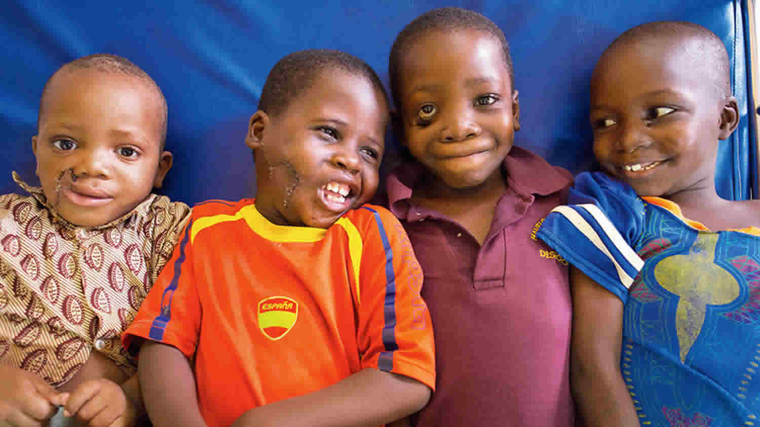
Cancrum oris also known as Noma or gangrenous stomatitis is a severe, preventable and often fatal rapidly progressing infection of the mouth and face that requires urgent and intensive medical and surgical care 1, 2, 3, 4, 5, 6, 7. The term noma originates from the Greek word “nomein” which means “to devour” or “to graze” 8, 6. Cancrum oris or noma usually begins as an ulcer on gums (the tissues that support your teeth) and rapidly spreads into the jawbone, cheek, and soft tissues of the face. This is followed by death of the facial tissues and fatal sepsis. Cancrum oris or Noma is associated with a reported 90% mortality rate within weeks after onset, if left untreated 2. Survivors are left with severe facial disfigurement and impairments in breathing, swallowing, speaking and vision 1, 2, 5, 7.
The exact cause is cancrum oris or noma is still poorly understood, but noma may be due to a certain kind of bacteria 1, 2. Previous observational studies have suggested that risk factors for development of cancrum oris or noma include severe malnutrition, low birthweight, absence of breastfeeding, poor oral hygiene, co-morbidities, proximity of livestock to area of residence, large family size, access to unsafe drinking water and living in a village with a high prevalence of acute necrotizing gingivitis 9, 10, 11, 12. Recently, an increased incidence of cancrum oris or noma has been reported in higher resource settings in patients with immunosuppressive diseases such as human immunodeficiency virus (HIV) 13, 14, 15, 16, 17.
Cancrum oris or noma mostly affects children aged 2 to 6 years old living in poor communities who lack access to basic nutrition, hygiene services, and health care, although cases are reported in immunocompromised adults 1, 2. One theory for the higher incidence of noma in children aged two to six years, is that this is the teething age when deciduous teeth are formed 18. This formation slows down the circulatory flow to the gums due to compression, leaving the oral cavity more susceptible to infections 18. A Zambian study theorized that during the weaning period from breastfeeding, children eat more solid food, which was less nutritious and less sterile than breast milk, and this placed them at potential risk for noma development 19. Another study showed that if weaning foods are prepared under unhygienic conditions, they are frequently contaminated with pathogens and are a major factor in the cause of diarrheal diseases 20, a further reported risk factor for noma 21.
In recent years, noma has been reported in many countries around the world, but primarily in low and middle income countries in Africa and Asia 3. Epidemiological data on cancrum oris or noma are scarce, but a current estimate of the global incidence is 30,000 to 40,000 cases per year, with a mortality rate of approximately 85% and a burden of disease calculated to be a loss of 1–10 million disability-adjusted life years 7.
Cancrum oris or noma is diagnosed using World Health Organization (WHO) clinical criteria that differ according to the different stages of disease progression. There is currently no point of care diagnostic test. The World Health Organization (WHO) classifies cancrum oris or Noma into 5 clinical stages 1, 2:
- Stage 0 – Gum inflammation (simple gingivitis);
- Stage 1 – Acute necrotizing gingivitis;
- Stage 2 – Edema;
- Stage 3 – Gangrene;
- Stage 4 – Scarring;
- Stage 5 – Complications.
The first 4 stages (Stage 0 to 4) are the acute stages of noma lasting only a few weeks. Deaths in noma patients are primarily reported to be due to starvation, aspiration pneumonia, respiratory insufficiency or sepsis 22, 23, 7.
Early detection is essential, as therapy is most effective at the early reversible stages of noma when it appears as aggressively swollen gums (acute necrotizing gingivitis). Commonly available broad-spectrum antibiotics with proper nutrition together with practices to improve oral hygiene, disinfectant mouthwash (salt water or chlorhexidine could be used) helps stop the disease from getting worse 1, 2. Once cancrum oris or noma progresses past early reversible stages (Stage 0 to 4), the complications of noma are numerous and include difficulty in eating, drinking, seeing and breathing 1, 2, 24, 25. Plastic surgery may be necessary to remove destroyed tissues and reconstruct facial bones. This will improve facial appearance and the function of the mouth and jaw 26. For those who seek care for these complications, it can mean hospital stays of many months with multistage surgical treatments that can take years to complete. Survivors often need complex surgical reconstruction to restore form and function 26. Trismus (a restriction in mouth opening) is one of the most disabling complication 27, 28 and can lead to complications such as aspiration, malnutrition, poor oral hygiene, speech deficits, a compromised airway, and pain 29.
Cancrum oris causes
The exact cause is cancrum oris or noma is still poorly understood, but noma may be due to a certain kind of bacteria 1, 2, 30, 7. Previous observational studies have suggested that risk factors for development of cancrum oris or noma include severe malnutrition, low birthweight, absence of breastfeeding, poor oral hygiene, co-morbidities, proximity of livestock to area of residence, large family size, access to unsafe drinking water and living in a village with a high prevalence of acute necrotizing gingivitis 9, 10, 11, 12. Even though noma primarily affects young children 24, noma cases in adults, mostly in conjunction with other severe infections like human immunodeficiency virus (HIV), cancer or oral myiasis have been reported 31, 32, 33, 34. Recently, an increased incidence of cancrum oris or noma has been reported in higher resource settings in patients with immunosuppressive diseases such as human immunodeficiency virus (HIV) 13, 14, 15, 16, 17.
A range of organisms have been identified in the oral flora of noma patients, but none have been consistently present, casting doubt on a specific organism’s role in the development of noma 35, 36, 37, 38, 39, 40, 41. Other studies have noted that the characteristics of noma are similar to that of an opportunistic infection, implicating a change in the equilibrium of commensal bacteria due to a derailment of host defences 42, 43, 39, 44. Evidence that supports noma being an opportunistic infection rests in the fact that most cases have concurrent infections or occur in immunocompromised individuals 42, 19, 36, 18, 45, 46, 47, 48.
It has been speculated that Borrelia vincentii and Fusobacterium are prominent bacteria in noma lesions 49, 50. Symbiotic relationships between fusiform bacilli and non-hemolytic streptococci and staphylococci have been considered significant factors in the development of noma 51. Recent reports suggest that besides fusiform bacilli and spirochetes, other anaerobic bacteria are present in a relatively high proportion of noma lesions 50. Fusobacterium necrophorum is considered a key component; this organism produces dermatotoxins, which could explain the rapid progression of the disease 52. Fusobacterium necrophorum is acquired by impoverished children through fecal contamination of water, which occurs when residential facilities are shared with animals and sanitation is very poor 8. Prevotella intermedia has the ability to break down lipid structures, which contributes to tissue destruction 8. It also produces proteolytic enzymes
capable of breaking down immunoglobulin G (IgG), which impedes elimination of microorganisms 51. Some reports have suggested that these microorganisms are resistant to penicillin 50, which emphasizes the need for culture and sensitivity tests before administration of antibiotics.
Recent studies using genomic approaches showed an imbalance in the normal oral flora with acute noma seems to be characterized by the diminution of Capnocytophaga and Fusobacteria genera and by the increase of Prevotella genus 53, 54, 55. Prevotella intermedia was already reported in previous studies undertaken with classical cultures 49. Prevotella intermedia is a well-known periodontal pathogen in adults, but it has been detected also in the primary dentition of small children 56, 57. It is frequently recovered from various oral purulent infections in children and also in secondary nosocomial infections 58, 59. Of note, Prevotella intermedia is always associated with other pathogens and it has never been reported as a monoinfecting agent. Therefore, the proliferation of Prevotella intermedia in noma lesions seems to be the consequence of changes in local ecological conditions due to the development of the disease. These findings invalidate the existence of a single noma pathogen and tend to reconsider noma as a multifactorial, opportunistic disease developing in a relatively normal oral flora in children suffering from chronic malnutrition 53.
Risk factors for cancrum oris or noma
Several risk factors are associated with the pathogenesis of cancrum oris or noma. Reported risk factors for the development of cancrum oris or noma is severe malnutrition, especially protein-calorie malnutrition 19, 42, 36, 46, 40, 60, 61, 62. Other factors that significantly contribute to the pathogenesis of cancrum oris or noma include:
- Comorbidities either at the time of noma diagnosis or in the three months leading up to diagnosis 19, 42, 36, 18, 45, 46, 48, 40, 60, 61, 63, 62.
- Reported comorbidities in the primary studies (case control, cohort, retrospective chart reviews) include malnutrition, respiratory disease 47, 40, diarrhea 42, 40, malaria 47, 61 and vaccine preventable diseases, specifically measles 42, 48, 61.
- Recently, an increased incidence of noma has been reported in higher resource settings in patients with immunosuppressive diseases such as human immunodeficiency virus (HIV) 13, 14, 15, 16, 17, 64, 47, 63.
- Most of the case reports and case series (n = 68) list at least one comorbidity (103 comorbidities listed in these case reports and case series). The most widely reported comorbidities in the case reports and case series included in a review were malnutrition, HIV, anemia and measles 3.
- Low vitamin A and vitamin C levels 35
- Being between two and five years of age 19, 42, 18, 45, 46, 43, 48, 61
- Not being breastfed 60, 65
- Lack of access to basic health care 18
- Lack of childhood vaccinations 66, 65
- Poor oral hygiene practices leading to gingivitis (Stage 0 noma) 66
- Low socioeconomic status 47
- Lack of variety in the diet 65
- Mother being unmarried, not the primary caretaker 65 and having a high number of previous pregnancies 40 and the absence of chickens at home 40
Other studies have hypothesized further risk factors for noma development include:
- Proximity of livestock to living areas and poor sanitation 66, which is thought to lead to possible contamination of water and food sources and consequently increasing the risk of infections 67, 68.
However; caution is needed when interpreting these findings as they are based solely on case reports and case series, primarily reported from health care centers, no causal link or strength of association can be measured. Infections are usually the product of a compromised host and a single offending agent or multiple offending agents. Due to challenges with conducting scientifically robust risk factor analysis for noma, it is difficult to separate comorbidities from predisposing conditions and true causative factors 3, 69.
Cancrum oris symptoms
Cancrum oris or noma causes sudden tissue destruction that rapidly gets worse. First, the gums and lining of the cheeks become inflamed, swollen and develop sores (ulcers) 19, 70, 26, 1, 2. These ulcers spread rapidly to the skin, and the tissues in the lips and cheeks die. This can eventually destroy the soft tissue and bone. The destruction of the bones around the mouth causes deformity of the face and loss of teeth 19, 70, 26, 1, 2. The color of the oral cavity may also change into a greyish color. The ulcers develop a foul-smelling drainage, causing bad breath and skin odor. Affected children that survive cancrum oris or noma are often left with serious aesthetic and functional consequences including disfigurement and impairments in breathing, swallowing, speaking and vision that further contribute to their social isolation, stigmatization and discrimination.
Noma can also affect the genitals, spreading to the genital skin this is sometimes called noma pudendi.
Noma stages
For the purposes of early detection, the World Health Organization (WHO) divides cancrum oris or noma into 5 clinical stages 1, 2:
- Stage 0 – Gum inflammation (simple gingivitis);
- Stage 1 – Acute necrotizing gingivitis;
- Stage 2 – Edema;
- Stage 3 – Gangrene;
- Stage 4 – Scarring;
- Stage 5 – Complications.
The first 4 stages (Stage 0 to 4) are the acute stages of noma lasting only a few weeks. Deaths in noma patients are primarily reported to be due to starvation, aspiration pneumonia, respiratory insufficiency or sepsis 22, 23, 7.
Stage 0 Warning Signs
Before the development of noma, there may be simple gingivitis: inflammation and reddening of the gums which bleed when touched or during brushing of the teeth. The WHO recommends disinfectant mouthwash; if not available, use warm, salted water that has been boiled 1, 2. A high-protein diet, Vitamin A supplements, and patient education on oral hygiene are also recommended to prevent noma from progressing to the acute stages 1, 2.
Stage 1 Acute necrotizing gingivitis
Acute necrotizing gingivitis (ANG) is the first stage of noma 71, 72. The gums are red or reddish-purple and bleed spontaneously 2. Acute necrotizing gingivitis (ANG) is characterized by spontaneous bleeding, ulceration of the gingival papillae, pain and sometimes, greyish pseudomembranes 73, 74, 1. The child has foul smelling breath and may drool. Painful ulcers of the gums develop, causing trouble eating. If the patient is malnourished and has recently been sick with an infectious disease, such as measles or chickenpox, they are at more risk for developing noma. Fever may develop at this stage, which can persist indefinitely. Without treatment, acute necrotizing gingivitis (ANG) may evolve into a necrotizing stomatitis and result in the destruction of the attached gingival mucosa, the surrounding oral mucosa, and the underlying bone (Figure 1B). This stage requires antibiotic treatment can halt the disease 1, 2. If no treatment is undertaken, the risk of progression toward noma is very high 75.
Figure 1. Acute necrotizing gingivitis
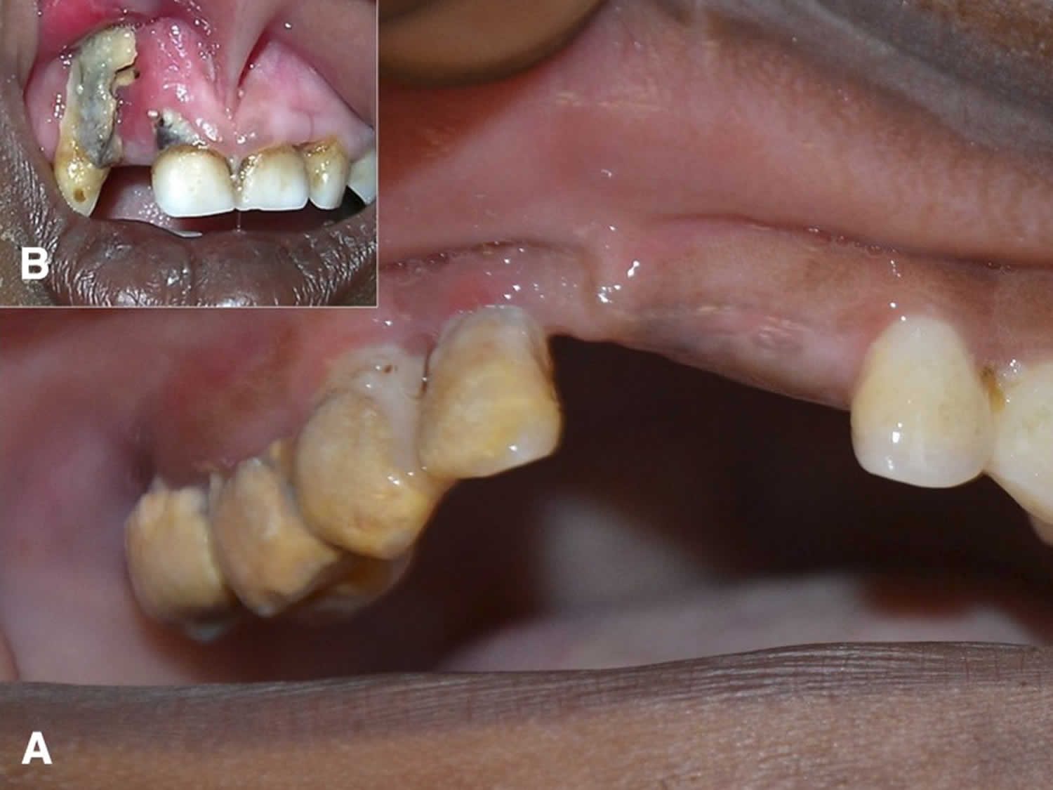
Footnotes: (A) Acute necrotizing gingivitis and (B) necrotizing stomatitis. (A) Acute necrotizing gingivitis (ANG) is considered as an important precursor of noma and is characterized by spontaneous bleeding, ulceration of the papillae, and gingival pain. In this patient, acute necrotizing gingivitis (ANG) of the upper right dental arch is accompanied by an abundance of calculus and plaque, greyish pseudomembranes and disappearance of gingival papillae. (B) Upper right dental arch: necrotizing stomatitis with destruction of gingival papillae and attached mucosa. Presence of greyish pseudomembranes and exposure of alveolar bone. The lesion shows destruction of gingival mucosa and underlying necrotic bone, a likely precursor of noma.
[Source 7 ]Stage 2 Edema
The starting point of noma is defined when facial edema appears in addition to intraoral necrotizing stomatitis (see Figure 2 ), accompanied by a typical halitosis often considered as pathognomonic. This stage is short and lasts only a few days.
This stage begins the acute phase of noma. The telltale sign is facial edema (swelling) of the lips, cheeks, eyes, etc. Ulceration of the gums worsens during this stage; ulceration may spread to the mucosa (soft, mucus-producing tissue) of the mouth and nose. The patient may feel pain or soreness in their mouth and cheeks. Other symptoms at this stage include fever, drooling, fetid breath, lymphadenopathy (swollen lymph nodes), and difficulty eating. Progression of the disease can be halted with appropriate treatment 1, 2.
Figure 2. Stage 2 Noma edema stage
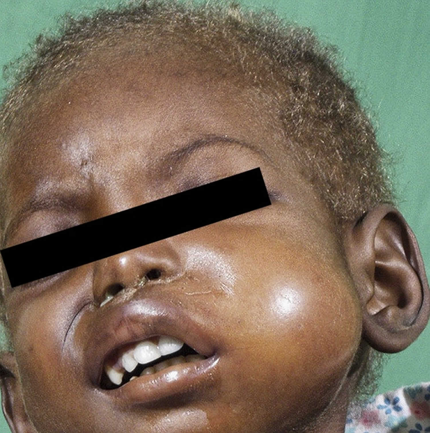
Footnote: Stage 2 edema stage in a child with discolored and brittle hair, a secondary sign of malnutrition.
[Source 7 ]Stage 3 Gangrene
At this and subsequent stages, although the disease can still be treated, complications will inevitably set in. In this stage, the infection eats away at the soft tissue of the patient’s face. The gangrene may affect the cheeks, lips, nose, mouth, and nasal and oral cavities. A peculiarity of this gangrene is that it appears to have a self-limiting character ending in a well-demarcated necrosis. Dead tissue sloughs away over time, leaving holes in the face and the soft tissue, possibly exposing bones and teeth. The patient is apathetic, has little appetite, and has great difficulty eating 1, 2. At this stage, there is a high risk of sepsis leading to death 22, 23, 7.
On some occasions, the body appears to be able to resist and halt the expansion of the gangrene at a certain stage 7. Interestingly, some children do not receive treatment and develop relatively small lesions, while others experience huge facial destruction despite appropriate medical treatment 7. This may be due to differences in the degree of impairment of their immune systems 7.
Stage 4 Scarring
The acute phase is over by this point, but the patient’s life is still at risk and treatment is still recommended. This stage lasts one to two weeks. The patient may experience trismus (difficulty moving/opening the jaw), scars will form, and any exposed teeth will set in place 1, 2.
Stage 5 Complications
The disease is over by this point, but consequences from the gangrenous and scarring stages remain. Tissue may be missing, teeth may still be exposed, and the face is disfigured. The patient may have difficulty eating, drinking, and speaking. Teeth may become set in the wrong places, or be lost altogether. There may still be problems with drooling and with opening/closing the jaw. Reconstructive surgery is an option at this phase. Social reintegration is also very important 1, 2.
Cancrum oris diagnosis
A physical examination is done initially to check for inflamed mucus membranes, oral cavity ulcers, and skin ulcers. This is followed by taking a detailed medical history of the patient.
X-ray, MRI, or CT scans of the jaw, head, and neck can also be done to check the extent and severity of the damage.
Bacterial culture analysis using oral swabs can be done to detect the causative species. In some special cases, biopsy of the oral tissue is also performed.
Blood tests are recommended to determine the immune system functioning.
Cancrum oris treatment
Treatment with broad-spectrum antibiotics, wound cleaning and debridement, hydration and nutritional support and treatment of concomitant diseases such as pulmonary and gastrointestinal infections, malaria, and HIV in the early, reversible stages of cancrum oris or noma can reduce the duration and severity of the acute phase of noma and the extent of tissue damage, therefore reducing mortality and morbidity of noma 24, 76, 43, 77, 78, 79, 80, 36, 77, 22, 76, 43. A range of antibiotics were reported in the studies include amoxicillin 81, 82, 77, 8, metronidazole 83, 80, 37, 82, 84, lincomycin 85 and cefotaxime 83, 86. No studies comparing the relative efficacy of these antibiotics were identified.
Acute phase treatment (Stages 0 to 4)
The current WHO guidelines for the management of the acute stages of noma in clinical settings includes 1, 2: oral hygiene (mouth wash Chlorhexidine 0,2%, 10 ml), antibiotic treatment (amoxicillin and metronidazole), nutritional support (high protein), wound cleaning (compresses soaked in hydrogen peroxide) and dressing (honey for local dressing and for antibacterial action and regeneration) 1, 2.
A range of antibiotics were reported in the studies include amoxicillin 81, 82, 77, 8, metronidazole 83, 80, 37, 82, 84, lincomycin 85 and cefotaxime 83, 86. No studies comparing the relative efficacy of these antibiotics were identified.
Complications treatment
If the patient survives the acute illness, they can live into adulthood but often require extensive reconstructive surgery and intensive physiotherapy to improve the resulting structural and functional defects that often require a number of surgical treatments 87, 88. Studies have highlighted the fact that the time between acute illness and surgical care can be decades 25, 89, 28. The clinical manifestation of each noma case is unique, and as such, the surgical procedures used to treat each noma case differ 3.
Reported surgical techniques include pedicled supraclavicular flaps for the treatment of large unilateral facial defects 90, 91; myocutaneous submental artery flaps, bony and/or soft tissue trismus releases 92, forehead flaps 92, 28 and lower lid ectropion release 92. In one study, extra-articular ankylosis due to noma was treated using soft tissue reconstruction with large free flaps 93. Trismus was treated using a bone distractor in one study 94 and in another mouth opening was performed by bone-bridge excision, sometimes associated with contralateral coronoidectomy 93. In a further study, the reconstruction of an upper lip defect was conducted using Gillies fan flaps 95. A 2006 book on noma surgical techniques includes information on the reconstruction of the lips and corner of the mouth using Abbe, Estlander and fan flaps; and the reconstruction of the cheek using temporo-parietal fascia and deltopectoral flaps; and the reconstruction of central defects using radial forearm and local turnover flaps 26. Challenges with anaesthesia for noma survivors have been reported, particularly in patients with severe trismus 96, 97.
Physical therapy is an essential part of noma treatment, especially to prevent or minimize trismus 98 and can lead to improvements in eating, chewing and speaking 99.
Noma often leads to stigmatization and resultant social isolation of the patients and their family members from their communities 24. Several studies have highlighted the importance of including social and psychological support for noma patients and their families 25, 88, 100, 101.
Outcomes of noma treatment are difficult to ascertain due to inconsistent patient follow-up in most studies 27. This is mostly due to the remote locations of the home villages of patients and difficulties in accessing health care facilities 22, 102, 78. However, there have been evaluations of noma survivors after surgery which have shown that surgical treatment for noma survivors greatly improves their quality of life, even if functional improvements (specifically mouth opening) are not pronounced 90, 25, 89, 100. The extent of long-term consequences and their impact on quality of life of noma patients depends on the severity of the disease at initial presentation, efficacy of antibiotic treatment, wound debridement and the facial structures affected 27, 76, 43, 77.
Cancrum oris prognosis
In some cases, cancrum oris or noma can be deadly if it is not treated. Other times, cancrum oris or noma may heal over time, even without treatment. However, once noma reaches the gangrenous stage of the disease with a visible hole in the face, it is likely that children who survive will suffer severe facial disfigurement, have severe complications including difficulty eating, seeing, breathing, face social stigma and isolation 1, 2, 24, 25. Only approximately 15% of children survive acute noma 103, 104, 105. Most survivors present with facial deformities and trismus or ankylosis of the mandible, resulting in eating problems, oral incontinence, speech difficulties, and social isolation 7.
Survivors often need complex reconstructive surgery to restore form and function 26. Trismus (a restriction in mouth opening) is one of the most disabling complication 27, 28 and can lead to complications such as aspiration, malnutrition, poor oral hygiene, speech deficits, a compromised airway, and pain 29.
- Noma. https://www.who.int/news-room/fact-sheets/detail/noma[↩][↩][↩][↩][↩][↩][↩][↩][↩][↩][↩][↩][↩][↩][↩][↩][↩][↩][↩][↩][↩][↩]
- World Health Organization Regional Office for Africa. Information brochure for early detection and management of noma. 2017. https://iris.who.int/handle/10665/254579[↩][↩][↩][↩][↩][↩][↩][↩][↩][↩][↩][↩][↩][↩][↩][↩][↩][↩][↩][↩][↩][↩][↩]
- Farley E, Mehta U, Srour ML, Lenglet A. Noma (cancrum oris): A scoping literature review of a neglected disease (1843 to 2021). PLoS Negl Trop Dis. 2021 Dec 14;15(12):e0009844. doi: 10.1371/journal.pntd.0009844[↩][↩][↩][↩][↩]
- Noma (cancrum oris). https://www.dental.umaryland.edu/media/sod/microbial-pathogenesis/noma_publication_1.pdf[↩]
- Enwonwu CO, Falkler WA, Idigbe EO. Oro-facial gangrene (noma/cancrum oris): pathogenetic mechanisms. Crit Rev Oral Biol Med. 2000;11(2):159-71. https://doi.org/10.1177/10454411000110020[↩][↩]
- Noma. https://msf.org.uk/issues/noma[↩][↩]
- Srour ML, Marck K, Baratti-Mayer D. Noma: Overview of a Neglected Disease and Human Rights Violation. Am J Trop Med Hyg. 2017 Feb 8;96(2):268-274. doi: 10.4269/ajtmh.16-0718[↩][↩][↩][↩][↩][↩][↩][↩][↩][↩][↩][↩][↩]
- Auluck A, Pai KM. Noma: life cycle of a devastating sore – case report and literature review. J Can Dent Assoc. 2005 Nov;71(10):757. http://www.cda-adc.ca/jcda/vol-71/issue-10/757.pdf[↩][↩][↩][↩][↩]
- Ashok N, Tarakji B, Darwish S, Rodrigues JC, Altamimi MA. A Review on Noma: A Recent Update. Glob J Health Sci. 2015 Jul 30;8(4):53-9. doi: 10.5539/gjhs.v8n4p53[↩][↩]
- Srour ML, Marck K, Baratti-Mayer D. Noma: Overview of a neglected disease and human rights violation. Am J Trop Med Hyg. 2017;96(2):268–74. 10.4269/ajtmh.16-0718[↩][↩]
- Baratti-Mayer Denise, Gayet-Ageron Angèle, Hugonnet Stéphane, François Patrice, Pittet-Cuenod Brigitte, Huyghe Antoine, Bornand Jacques-Etienne, Gervaix Alain, Montandon Denys, Schrenzel Jacques, Mombelli Andrea P D. Risk factors for noma disease: A 6-year, prospective, matched case-control study in Niger. Lancet Glob Heal. 2013;1(2):87–96. 10.1016/s2214-109x(13)70015-9[↩][↩]
- Baratti-Mayer D, Gayet-Ageron A, Cionca N, Mossi MA, Pittet D, Mombelli A. Acute necrotising gingivitis in young children from villages with and without noma in Niger and its association with sociodemographic factors, nutritional status and oral hygiene practices: results of a population-based survey. BMJ Glob Heal. 2017;2(3). 10.1136/bmjgh-2016-000253[↩][↩]
- Hatcher J, Williamson L. Noma in a patient with HIV. Lancet Infect Dis. 2017;17(6):672 10.1016/S1473-3099(17)30263-3[↩][↩][↩]
- Maley A, Desai M, Parker S. Noma: A disease of poverty presenting at an urban hospital in the United States. JAAD Case Reports. 2015;1(1):18–20. 10.1016/j.jdcr.2014.10.001[↩][↩][↩]
- Lee YJ, Kim YJ, Won CH, Chang SE, Lee MW, Choi JH. Nomalike Necrotizing Stomatitis in a Child with Crohn’s Disease. Pediatr Dermatol. 2017;1–2. 10.1111/pde.13212[↩][↩][↩]
- Ashok N, Tarakji B, Darwish S, Rodrigues J, Altamimi M. A Review on Noma: A Recent Update. Glob J Health Sci. 2015;8(4):53–9. 10.5539/gjhs.v8n4p53[↩][↩][↩]
- Feller L, Altini M, Chandran R, Khammissa RAG, Masipa JN, Mohamed A, et al. Noma (cancrum oris) in the South African context. J Oral Pathol Med. 2014;43(1):1–6. 10.1111/jop.12079[↩][↩][↩]
- Ndiaye F, Bourgeois D, Leclercq M, Berthe O. Noma: public health problem in Senegal and epidemiological surveillance. Oral Dis. 1999;5: 163–166. doi: 10.1111/j.1601-0825.1999.tb00083.x[↩][↩][↩][↩][↩][↩]
- Nath S, Jovic G. Cancrum Oris: Management, Incidence, and Implications of Human Immunodeficiency Virus in Zambia. Plast Reconstr Surg. 1998;102: 350–357. doi: 10.1097/00006534-199808000-00008[↩][↩][↩][↩][↩][↩][↩]
- Motarjemi Y, Käferstein F, Moy G, Quevedo F. Contaminated weaning food: a major risk factor for diarrhoea and associated malnutrition. Bull World Health Organ. 1993;71(1):79-92. https://www.ncbi.nlm.nih.gov/pmc/articles/PMC2393433/pdf/bullwho00035-0091.pdf[↩]
- Feller L, Altini M, Chandran R, Khammissa RAG, Masipa JN, Mohamed A, et al. Noma (cancrum oris) in the South African context. J Oral Pathol Med. 2014;43: 1–6. doi: 10.1111/jop.12079[↩]
- Baratti-Mayer D, Pittet B, Montandon D, Bolivar I, Bornand JE, Jaquinet A, et al. Noma: an “infectious” disease of unknown aetiology. Lancet Infect. 2003;3: 419–431. doi: 10.1016/S1473-3099(03)00670-4[↩][↩][↩][↩][↩]
- Silva K, Twaddell S, Powers D. A 40-year-old man with a perforated cheek. Am J Med Sci. 2011;341: 399–403. doi: 10.1097/MAJ.0b013e3181e2ed3e[↩][↩][↩]
- Ashok N, Tarakji B, Darwish S, Rodrigues JC, Altamimi MA. A Review on Noma: A Recent Update. Glob J Health Sci. 2016;8: 53–59. doi: 10.5539/gjhs.v8n4p53[↩][↩][↩][↩][↩]
- Rickart AJ, Rodgers W, Mizen K, Merrick G, Wilson P, Nishikawa H, et al. Facing Africa: Describing noma in Ethiopia. Am J Trop Med Hyg. 2020;103: 613–618. doi: 10.4269/ajtmh.20-0019[↩][↩][↩][↩][↩]
- Marck K and Bos K. The surgical treatment of Noma. Bathgate R, van Knippenberg M, editors. Amsterdam, The Netherlands: Mart Spruijt bv, Amsterdam; 2006.[↩][↩][↩][↩][↩][↩]
- Bisseling P, Bruhn J, Erdsach T, Ettema AM, Sautter R, Berge SJ. Long-term results of trismus release in noma patients. Int J Oral Maxillofac Surg. 2010;39: 873–877. doi: 10.1016/j.ijom.2010.05.002[↩][↩][↩][↩]
- Ghorui T, Ray A. Release of Extra Articular Ankylosis of Jaws as a Sequelae of Cancrum Oris with Extensive Gingival Myasis in a Scoliosis Patient: A Rare Case Report. Indian J Otolaryngol Head Neck Surg. 2019;71: 734–736. doi: 10.1007/s12070-018-1526-x[↩][↩][↩][↩]
- Walker M, Burns K. Trismus: diagnosis and management considerations for the speech pathologist. American Speech-Language-Hearing Association. 2006. pp. 1–55.[↩][↩]
- Baratti-Mayer D, Pittet B, Montandon D, Bolivar I, Bornand JE, Hugonnet S, Jaquinet A, Schrenzel J, Pittet D; Geneva Study Group on Noma. Noma: an “infectious” disease of unknown aetiology. Lancet Infect Dis. 2003 Jul;3(7):419-31. doi: 10.1016/s1473-3099(03)00670-4[↩]
- Lathigara MD. An Unusual Case of Cancrum Oris. Ind Med Gaz. 1935 Nov;70(11):626-627. https://www.ncbi.nlm.nih.gov/pmc/articles/PMC5169462/pdf/indmedgaz72289-0028b.pdf[↩]
- Koech K. Cancrum oris in an adult with human immunodeficiency virus infection: case report. East Afr Med J. 2010;87: 38–40. doi: 10.4314/eamj.v87i1.59953[↩]
- Aguiar M, Enwonwu C, Pires F. Noma (cancrum oris) associated with oral myiasis in an adult. Oral Dis. 2003;9: 158–159. doi: 10.1034/j.1601-0825.2003.03942.x[↩]
- Pedro K, Smit D, Morkel J. Cancrum Oris (noma) in an HIV-positive adult: a case report and literature review. SADJ. 2016;71: 248–252.[↩]
- Phillips RS, Enwonwu CO, Falkler WA. Pro- versus anti-inflammatory cytokine profile in African children with acute oro-facial noma (cancrum oris, noma). Eur Cytokine Netw. 2005 Jan-Mar;16(1):70-7. https://www.jle.com/fr/revues/ecn/e-docs/pro_versus_anti_inflammatory_cytokine_profile_in_african_children_with_acute_oro_facial_noma_cancrum_oris_noma__266020/article.phtml[↩][↩]
- Enwonwu C, Falkler W, Idigbe E, Afolabi B, Ibrahim M, Onwujekwe D, et al. Pathogenesis of cancrum oris (noma): Confounding interactions of malnutrition with infection. Am J Trop Med Hyg. 1999;60: 223–232. doi: 10.4269/ajtmh.1999.60.223[↩][↩][↩][↩][↩]
- Falkler W, Enwonwu C, Idigbe E. Isolation of fusobacterium necrophorium from cancrum oris (noma). Am J Trop Med Hyg. 1999;60: 150–156. doi: 10.4269/ajtmh.1999.60.150[↩][↩][↩]
- Falkler W, Enwonwu C, Idigbe E. Microbiological understandings and mysteries of noma (cancrum oris). Oral Dis. 1999;5: 150–155. doi: 10.1111/j.1601-0825.1999.tb00081.x[↩]
- Whiteson K, Lazarevic V, Tangomo-bento M, Girard M, Maughan H, Pittet D, et al. Noma affected children from Niger have distinct oral microbial communities based on high-throughput sequencing of 16S rRNA gene fragments. PLOS NTD. 2014;8: 1–13. doi: 10.1371/journal.pntd.0003240[↩][↩]
- Baratti-Mayer D, Gayet-Ageron A, Hugonnet S, François P, Pittet-Cuenod B, Huyghe A, et al. Risk factors for noma disease: a 6-year, prospective, matched case-control study in Niger. Lancet Glob Heal. 2013;1: 87–96. doi: 10.1016/S2214-109X(13)70015-9[↩][↩][↩][↩][↩][↩][↩]
- Feller L, Khammissa RAG, Altini M, Lemmer J. Noma (cancrum oris): An unresolved global challenge. Periodontol 2000. 2019;80: 189–199. doi: 10.1111/prd.12275[↩]
- Lazarus D, Hudson DA. Cancrum oris–a 35-year retrospective study. S Afr Med J. 1997 Oct;87(10):1379-82.[↩][↩][↩][↩][↩][↩][↩]
- Chidzonga M, Mahomva L. Noma (Cancrum Oris) in Human Immunodeficiency Virus Infection and Acquired Immunodeficiency Syndrome (HIV and AIDS): Clinical Experience in Zimbabwe. J Oral Maxillofac Surg. 2008;66: 475–485. doi: 10.1016/j.joms.2007.09.024[↩][↩][↩][↩][↩]
- Kimura T. An oro-facial disease ‘noma (cancrum oris)’ in a Japanese monkey (Macaca fuscata): clinical signs, clinicopathological features, and response to treatment. J Med Primatol. 2008;37: 217–222. doi: 10.1111/j.1600-0684.2008.00312.x[↩]
- Osuji O. Necrotizing ulcerative gingivitis and Cancrum Oris (noma) in Ibadan, Nigeria. J Periodontol. 1990;61: 769–772. doi: 10.1902/jop.1990.61.12.769[↩][↩][↩]
- Oginni F, Oginni A, Ugboko V, Otuyemi O. A survey of cases of cancrum oris seen in Ile-Ife, Nigeria. Int J Paediatr Dent. 1999;9: 75–80. doi: 10.1046/j.1365-263x.1999.00110.x[↩][↩][↩][↩]
- Konsem T, Millogo M, Assouan C, Ouedraogo D. Evoluting form of cancrum oris, about 55 cases collected at the Academic Hospital Yalgado Ouedraogo of Ouagadougou. Bull Soc Pathol Exot. 2014;107: 74–78. doi: 10.1007/s13149-014-0338-9[↩][↩][↩][↩][↩]
- Adeniyi S, Awosan K. Pattern of Noma (Cancrum Oris) and its risk factors in northwestern Nigeria: a hospital-based retrospective study. Ann Afr Med. 2019;18: 17–22. doi: 10.4103/aam.aam_5_18[↩][↩][↩][↩]
- Falkler WA Jr, Enwonwu CO, Idigbe EO. Isolation of Fusobacterium necrophorum from cancrum oris (noma). Am J Trop Med Hyg. 1999 Jan;60(1):150-6. doi: 10.4269/ajtmh.1999.60.150[↩][↩]
- Falkler WA Jr, Enwonwu CO, Idigbe EO. Microbiological understandings and mysteries of noma (cancrum oris). Oral Dis. 1999 Apr;5(2):150-5. doi: 10.1111/j.1601-0825.1999.tb00081.x[↩][↩][↩]
- Berthold P. Noma: a forgotten disease. Dent Clin North Am. 2003 Jul;47(3):559-74. doi: 10.1016/s0011-8532(03)00020-x[↩][↩]
- Enwonwu CO, Falkler WA Jr, Idigbe EO, Afolabi BM, Ibrahim M, Onwujekwe D, Savage O, Meeks VI. Pathogenesis of cancrum oris (noma): confounding interactions of malnutrition with infection. Am J Trop Med Hyg. 1999 Feb;60(2):223-32. doi: 10.4269/ajtmh.1999.60.223[↩]
- Baratti-Mayer D, Gayet-Ageron A, Hugonnet S, François P, Pittet-Cuenod B, Huyghe A, Bornand JE, Gervaix A, Montandon D, Schrenzel J, Mombelli A, Pittet D; Geneva Study Group on Noma (GESNOMA). Risk factors for noma disease: a 6-year, prospective, matched case-control study in Niger. Lancet Glob Health. 2013 Aug;1(2):e87-e96. doi: 10.1016/S2214-109X(13)70015-9. Epub 2013 Jul 5. Erratum in: Lancet Glob Health. 2013 Aug;1(2):e76.[↩][↩]
- Bolivar I, Whiteson K, Stadelmann B, Baratti-Mayer D, Gizard Y, Mombelli A, Pittet D, Schrenzel J; Geneva Study Group on Noma (GESNOMA). Bacterial diversity in oral samples of children in niger with acute noma, acute necrotizing gingivitis, and healthy controls. PLoS Negl Trop Dis. 2012;6(3):e1556. doi: 10.1371/journal.pntd.0001556[↩]
- Huyghe A, François P, Mombelli A, Tangomo M, Girard M, Baratti-Mayer D, Bolivar I, Pittet D, Schrenzel J; Geneva Study Group on Noma. Microarray analysis of microbiota of gingival lesions in noma patients. PLoS Negl Trop Dis. 2013 Sep 26;7(9):e2453. doi: 10.1371/journal.pntd.0002453[↩]
- Kamma JJ, Diamanti-Kipioti A, Nakou M, Mitsis FJ. Profile of subgingival microbiota in children with primary dentition. J Periodontal Res. 2000 Feb;35(1):33-41. doi: 10.1034/j.1600-0765.2000.035001033.x[↩]
- Könönen E. Development of oral bacterial flora in young children. Ann Med. 2000 Mar;32(2):107-12. doi: 10.3109/07853890009011759[↩]
- Brook I. Aerobic and anaerobic microbiology of infections after trauma in children. J Accid Emerg Med. 1998 May;15(3):162-7. https://www.ncbi.nlm.nih.gov/pmc/articles/PMC1343057/pdf/jaccidem00024-0022.pdf[↩]
- Brook I. Microbiology of nosocomial sinusitis in mechanically ventilated children. Arch Otolaryngol Head Neck Surg. 1998 Jan;124(1):35-8. doi: 10.1001/archotol.124.1.35[↩]
- Enwonwu C, Phillips R, Ferrell C. Temporal relationship between the occurrence of fresh noma and the timing of linear growth retardation in Nigerian children. Trop Med Int Heal. 2005;10: 65–73. doi: 10.1111/j.1365-3156.2004.01351.x[↩][↩][↩]
- Denloye O, Aderinokun G, Lawoyin J, Bankole O. Reviewing trends in the incidence of cancrum oris in Ibadan, Nigeria. West Afr J Med. 2003;22: 26–29. doi: 10.4314/wajm.v22i1.27974[↩][↩][↩][↩][↩]
- Braimah R, Adeniyi A, Taiwo A, Ibikunle A, Gbotolorun M, Aregbesola S, et al. Risk factors and mortality rate of acute cancrum oris (noma) in Sokoto North-West Nigeria: A 13-year survey. J Pediatr Dent. 2017;5: 1. doi: 10.4103/jpd.jpd_58_16[↩][↩]
- Millogo M, Konsem T, Ouedraogo D, Ouoba K, Zwetyenga N. HIV and noma in Burkina Faso. Rev Stomatol Chir Maxillofac. 2012;113: 433–6. doi: 10.1016/j.stomax.2012.07.004[↩][↩]
- Ghosh K. Use of penicillin in cancrum oris. Indian J Pediatr. 1947;14: 171–172. https://link.springer.com/article/10.1007/BF02812636[↩]
- Farley E, Lenglet A, Ariti C, Jiya NM, Adetunji AS, van der Kam S, et al. Risk factors for diagnosed noma in northwest Nigeria: A case-control study, 2017. PLoS Negl Trop Dis. 2018;12: 1–11. doi: 10.1371/journal.pntd.0006631[↩][↩][↩][↩]
- Barrera J, Connor M. Noma in an Afghani child: A case report. Int J Pediatr Otorhinolaryngol. 2012;76: 742–744. doi: 10.1016/j.ijporl.2012.01.034[↩][↩][↩]
- Garrett V, Ogutu P, Mabonga P, Ombeki S, Mwaki A, Aluoch G, et al. Diarrhoea prevention in a high-risk rural Kenyan population through point-of-use chlorination, safe water storage, sanitation, and rainwater harvesting. Epidemiol Infect. 2008;136: 1463–1471. doi: 10.1017/S095026880700026X[↩]
- Tumwine JK, Thompson J, Katua-Katua M, Mujwajuzi M, Johnstone N, Porras I. Diarrhoea and effects of different water sources, sanitation and hygiene behaviour in East Africa. Trop Med Int Health. 2002 Sep;7(9):750-6. doi: 10.1046/j.1365-3156.2002.00927.x[↩]
- Farley E, Lenglet A, Ariti C, Jiya NM, Adetunji AS, van der Kam S, Bil K. Risk factors for diagnosed noma in northwest Nigeria: A case-control study, 2017. PLoS Negl Trop Dis. 2018 Aug 23;12(8):e0006631. doi: 10.1371/journal.pntd.0006631[↩]
- Adeniyi S, Taiwo A, Ibikunle A, Braimah R, Gbotolorun O, Ogbeide M, et al. Pattern of tissue destruction among patients diagnosed with cancrum oris (Noma) at a Northwestern Nigerian Hospital, Sokoto. Saudi J Oral Sci. 2017;4: 101. doi: 10.4103/sjos.sjoralsci_55_16[↩][↩]
- Horning GM. Necotizing gingivostomatitis: NUG to noma. Compend Contin Educ Dent. 1996 Oct;17(10):951-4, 956, 957-8 passim; quiz 964.[↩]
- Idigbe EO, Enwonwu CO, Falkler WA, Ibrahim MM, Onwujekwe D, Afolabi BM, Savage KO, Meeks VI. Living conditions of children at risk for noma: Nigerian experience. Oral Dis. 1999 Apr;5(2):156-62. doi: 10.1111/j.1601-0825.1999.tb00082.x[↩]
- Goldhaber P, Giddon DB. Present concepts concerning the etiology and treatment of acute necrotizing ulcerative gingivitis. Int Dent J. 1963;14:468–496.[↩]
- Giddon DB, Zackin SJ, Goldhaber P. Acute necrotizing ulcerative gingivitis in college students. J Am Dent Assoc. 1964;68:380–386.[↩]
- Taiwo JO. Oral hygiene status and necrotizing ulcerative gingivitis in Nigerian children. J Periodontol. 1993 Nov;64(11):1071-4. doi: 10.1902/jop.1993.64.11.1071[↩]
- Ahlgren M, Funk T, Marimo C, Ndiaye C. Management of noma: practice competence and knowledge among healthcare workers in a rural district of Zambia. Glob Health Action. 2017;10: 1–9. doi: 10.1080/16549716.2017.1340253[↩][↩][↩]
- Masipa J, Baloyi A, Khammissa R, Altini M, Lemmer J, Feller L. Noma (Cancrum Oris): A report of a case in a young AIDS patient with a review of the pathogenesis. Head Neck Pathol. 2013;7: 188–192. doi: 10.1007/s12105-012-0393-0[↩][↩][↩][↩][↩]
- Srour ML, Watt B, Phengdy B, Khansoulivong K, Harris J, Bennett C, Strobel M, Dupuis C, Newton PN. Noma in Laos: stigma of severe poverty in rural Asia. Am J Trop Med Hyg. 2008 Apr;78(4):539-42.[↩][↩]
- Vaidya S, Agrawal A, Sharma VK. Cancrum Oris: A case report. Indian J Otolaryngol Head Neck Surg. 2006 Oct;58(4):411-3. https://www.ncbi.nlm.nih.gov/pmc/articles/PMC3450384/pdf/12070_2009_Article_BF03049618.pdf[↩]
- Oji C. Cancrum oris: its incidence and treatment in Enugu, Nigeria. Br J Oral Maxillofac Surg. 2002;40: 406–409. doi: 10.1016/S0266-4356(02)00192-4[↩][↩][↩]
- Weledji E, Njong S. Cancrum Oris (noma): the role of nutrition in management. J Am Coll Clin Wound Spec. 2016;7: 50–52. doi: 10.1016/j.jccw.2016.08.003[↩][↩]
- Behanan A, Auluck A, Pai K. Cancrum oris. Br J Oral Maxillofac Surg. 2004;42: 267–269. doi: 10.1016/j.bjoms.2004.02.018[↩][↩][↩][↩]
- Yuca K, Yuca S, Çankaya H, Çaksen H, Calka O, Kir M. Report of an infant with Noma (Cancrum Oris). J Dermatol. 2004;31: 488–491. doi: 10.1111/j.1346-8138.2004.tb00539.x[↩][↩][↩][↩]
- Maley A, Desai M, Parker S. Noma: A disease of poverty presenting at an urban hospital in the United States. JAAD Case Reports. 2015;1: 18–20. doi: 10.1016/j.jdcr.2014.10.001[↩][↩]
- Lembo S, Leonibus C, Francia M, Lembo C, Ayala F. Cancrum oris in a boy with Down syndrome. J Am Dermatology. 2011;64: 1200–1202. doi: 10.1016/j.jaad.2009.08.048[↩][↩]
- Evans L, Lane H, Jones M. Cancrum oris in a Caucasian male with Type 2 diabetes mellitus. Diabet Med. 2001;18: 246–248. doi: 10.1046/j.1464-5491.2001.00375.x[↩][↩]
- Enwonwu C, Falkler W, Phillips R. Noma (cancrum oris). Lancet. 2006;368: 147–156. doi: 10.1016/S0140-6736(06)69004-1[↩]
- Shafi’u I, Amirtharajah M, Farley E, Semiyu Adetunji A, Samuel J, Oluyide B, et al. Model of care, Noma Children’s Hospital, northwest Nigeria. Trop Med Int Heal. 2021; 1–10. 10.1111/tmi.13630[↩][↩]
- Farley E, Amirtharajah M, Winters R, Taiwo A, Oyemakinde M, Fotso A, et al. Outcomes at 18 mo of 37 noma (cancrum oris) cases surgically treated at the Noma Children’s Hospital, Sokoto, Nigeria. Trans R Soc Trop Med Hyg. 2020;0: 1–8. doi: 10.1093/trstmh/traa061[↩][↩]
- Shaye D, Winters R, Rabbels J, Adentunje A, Magee A, Vo D. Noma Surgery. Laryngoscope. 2019;129: 96–99. doi: 10.1002/lary.27230[↩][↩]
- Hartman E, Van Damme P, Sauter H, Suominen S. The use of the pedicled supraclavicular flap in noma reconstructive surgery. J Plast Reconstr Aesthetic Surg. 2006;59: 337–342. doi: 10.1016/j.bjps.2005.10.005[↩]
- Saleh D, Fourie L, Mizen K. Reconstruction of complex oro-facial defects using the myocutaneous sub-mental artery flap. J Cranio-Maxillofacial Surg. 2014;42: 668–673. doi: 10.1016/j.jcms.2013.09.013[↩][↩][↩]
- Ruegg E, Baratti-Mayer D, Jaquinet A, Montandon D, Pittet-Cuenod B. The surgical management of extra-articular ankylosis in noma patients. Int J Oral Maxillofac Surg. 2018;47: 1527–1533. doi: 10.1016/j.ijom.2018.07.012[↩][↩]
- Holle J, Kubiena H, Issa OH. Distraction Therapy to Correct Trismus Following Noma. J Craniofac Surg. 2020;31: 488–491. doi: 10.1097/SCS.0000000000006082[↩]
- Bello S. Gillies fan flap for the reconstruction of an upper lip defect caused by noma: case presentation. Clin Cosmet Investig Dent. 2012; 17–20. doi: 10.2147/CCIDEN.S31190[↩]
- Sund GC, Muvunyi P, Harling MJ. Airway Management Through a Facial Defect Resulting From Noma (Orofacial Gangrene): A Case Report. A&A Pract. 2020;14: e01319. doi: 10.1213/XAA.0000000000001319[↩]
- Braun U, Wiese KG, Merten HA, Timmermann A. Anaesthetic care for noma (cancrum oris)—the disease, the airway and how to provide anaesthetic care without a clinical safety infrastructure. Trends Anaesth Crit Care. 2020;31: 16–20. doi: 10.1016/j.tacc.2020.02.002[↩]
- Mukerji SN. A Case of Stiff Jaw after Cancrum Oris-Surgical Interference-Cure. Ind Med Gaz. 1927 Jul;62(7):387. https://www.ncbi.nlm.nih.gov/pmc/articles/PMC5197680/pdf/indmedgaz72103-0030a.pdf[↩]
- García-Guilarte F, Urcelay R, Frohner B, Meli G, González D, Celada E. Mandibular ankylosis: a Noma frequent sequel. Cirugía Plástica Ibero-Latinoamericana -. 2009;35: 2–6.[↩]
- Lafferty N. Changing the face of Africa. Estimating the burden of noma in rural Ethiopia and identifying options for prevention and improvement in its diagnosis and management. Liverpool School of Tropical Medicine. 2012. doi: 10.1146/annurev.soc.28.110601.140938[↩][↩]
- Wali IM, Regmi K. People living with facial disfigurement after having had noma disease: A systematic review of the literature. J Health Psychol. 2016;September: 1243–1255. doi: 10.1177/1359105315624751[↩]
- Eip N, Neuhoefer E, La Rosee G, Choudhrie R, Samman N, Kreusch T. Case report Submental intubation for cancrum oris: a case report. Pediatr Anesth. 2005;15: 1009–1012. doi: 10.1111/j.1460-9592.2005.01573.x[↩]
- Fieger A, Marck KW, Busch R, Schmidt A. An estimation of the incidence of noma in north-west Nigeria. Trop Med Int Health. 2003 May;8(5):402-7. doi: 10.1046/j.1365-3156.2003.01036.x[↩]
- Barmes DE, Enwonwu CO, Leclercq MH, Bourgeois D, Falkler WA. The need for action against oro-facial gangrene (noma). Trop Med Int Health. 1997 Dec;2(12):1111-4. doi: 10.1046/j.1365-3156.1997.d01-220.x[↩]
- Bourgeois DM, Diallo B, Frieh C, Leclercq MH. Epidemiology of the incidence of oro-facial noma: a study of cases in Dakar, Senegal, 1981-1993. Am J Trop Med Hyg. 1999 Dec;61(6):909-13. doi: 10.4269/ajtmh.1999.61.909[↩]
