Contents
What is a papule
Papule is an elevated, solid, palpable lesion that is ≤ 1 cm in diameter. Papule may be solitary or multiple.
Papules may be:
- Acuminate (pointed)
- Dome-shaped (rounded)
- Filiform (thread-like)
- Flat-topped
- Oval or round
- Pedunculated (with a stalk)
- Sessile (without a stalk)
- Umbilicated (with a central depression)
- Verrucous (warty)
Fibrous papule
A fibrous papule is also known as adenoma sebaceum, fibrous papule of the face or fibrous papule of the nose, is a firm benign (harmless) bump that most often occurs on the nose and less commonly, on the cheeks, chin, neck, and, rarely, on the lip or forehead 1. Fibrous papules are benign small skin colored lesions (usually less than 5mm in size). Fibrous papule is very common with most of the patients are in their third to fifth decade of life 1. Fibrous papules of the nose are seen in equal numbers of middle-aged men and women, regardless of race or ethnicity. Fibrous papules on the face usually arise as single lesions; however, occasionally multiple (i.e., normally less than 10) lesions may be present 2. However, patients with multiple fibrous papules should be examined for other signs of systemic diseases including tuberous sclerosis, neurofibromatosis type 2, multiple endocrine neoplasia type 1, and Hornstein-Knickenberg syndrome 3. Tuberous sclerosis should be considered in children with multiple fibrous papules 3. Inquire about medical and family history including seizures, mental impairment or developmental delay, behavioral disorders, visual problems, and tumors of the brain, heart, kidneys, lungs, and skin 3.
Occurring predominantly on the nose, fibrous papules are generally 1-5 mm, shiny, skin-colored, firm, dome-shaped papules. The fibrous papule has a characteristic appearance under the microscope. Fibrous papule is considered a nevus (a birthmark), and develops spontaneously as part of human development. The precise reason why fibrous papule form is unknown. Fibrous papule is best considered a variant of angiofibroma 4. A study from 2014 showed expression of antibodies against p-mTOR in dermal stromal cells and epidermal keratinocytes in fibrous papules, similar to tuberous-sclerosis complex–associated facial angiofibromas, suggesting topical rapamycin may be a treatment option 5.
The fibrous papule will not cause any symptoms like pain or itching. However, papules may become inflamed or bleed when irritated or scratched.
Histologically, angiofibromas are characterized by a proliferation of numerous hyperplastic blood vessels with perivascular and periadnexal fibrosis 6. This fibrous tissue often consists of stellate fibroblasts, some of which may be multinucleated 6. Periodically, scattered lymphocytes and a sparse inflammatory cell infiltrate can be appreciated 2. Numerous different histological subtypes of angiofibroma have been described 2. These subtypes all share the aforementioned histologic features, which are found in all angiofibromas, but, in addition, each described subtype has a certain unique quality that renders it distinct.
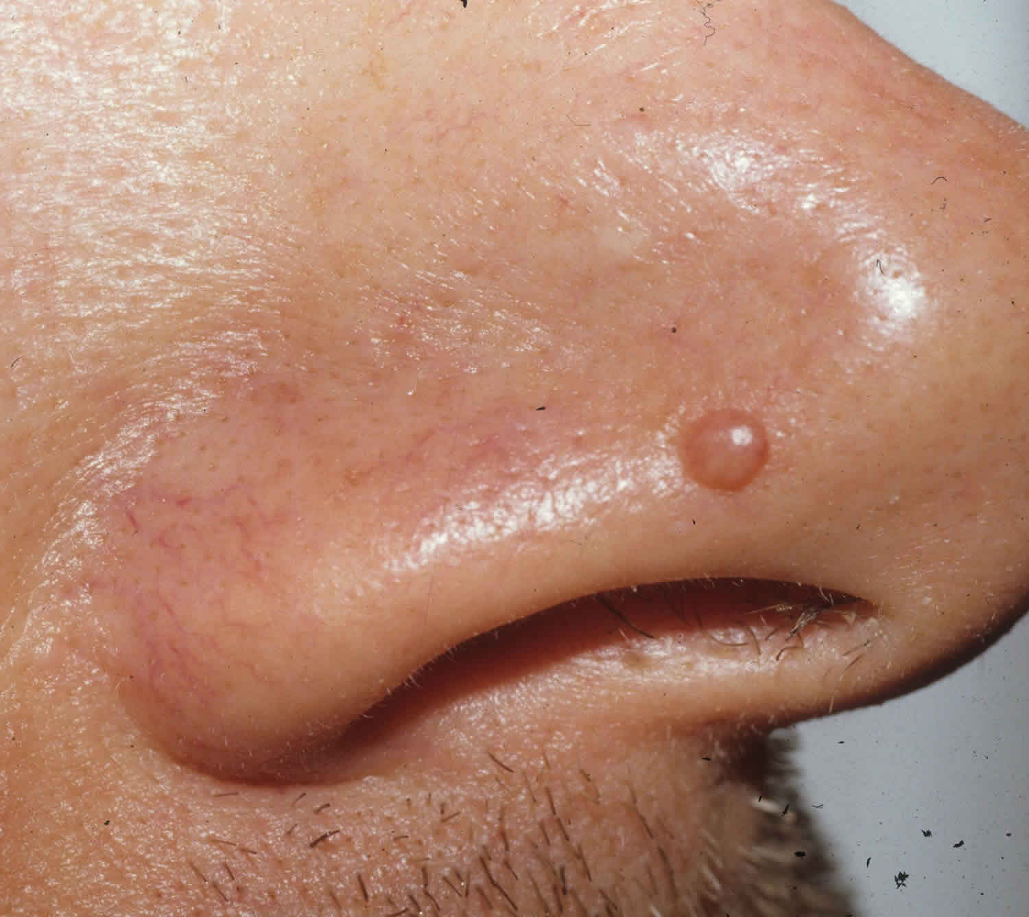
Figure 1. Fibrous papule
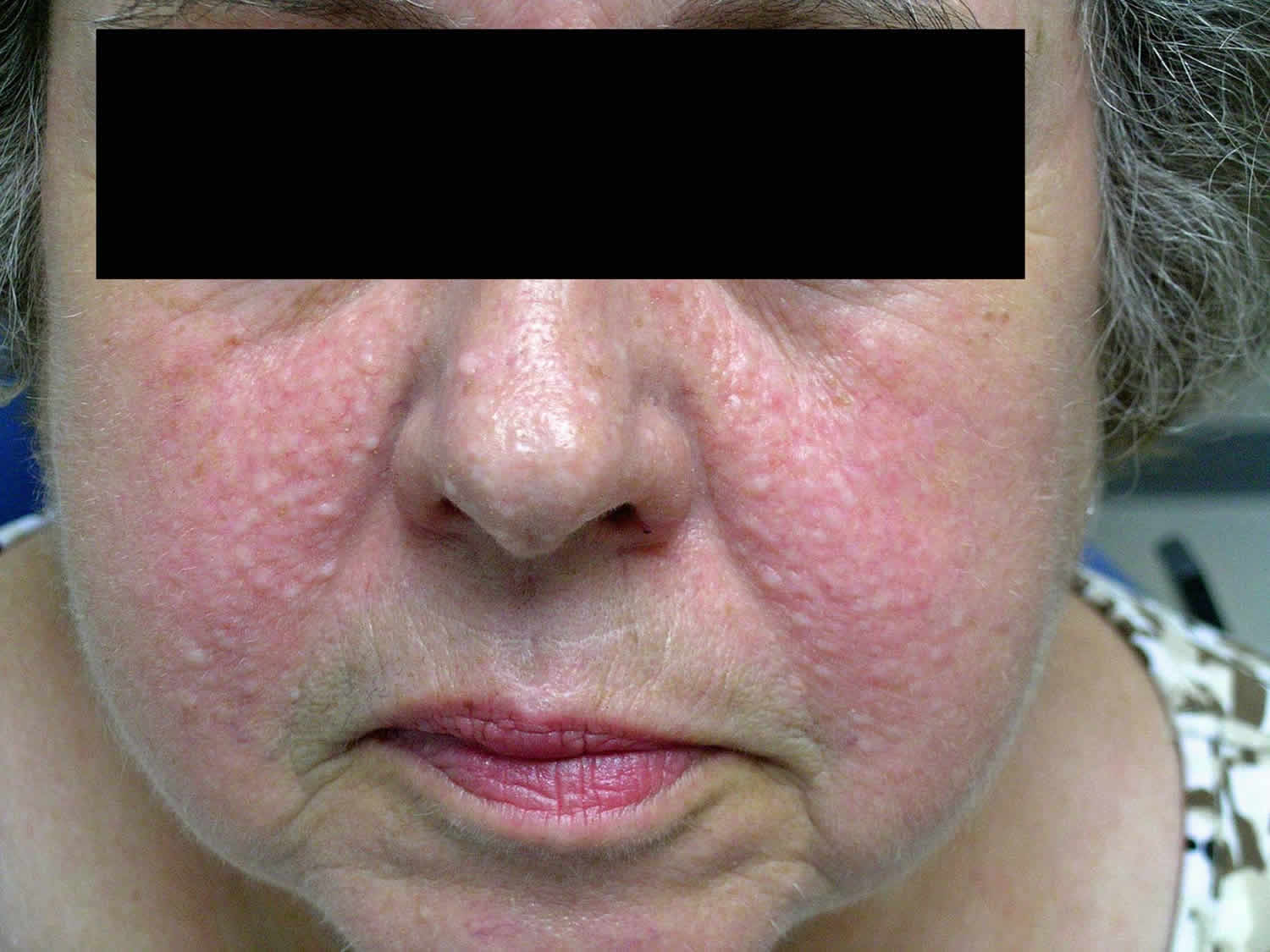
Footnote: A 66-year-old healthy woman with multiple skin-colored papules on her face. The papules first appeared 50 years earlier. At the time the lesions had first appeared, the patient had been told by her dermatologist that the lesions were trichoepitheliomas. She had never had any skin biopsies, but some of the lesions were removed with curettage and light hyfrecation. She never applied any medications to her face with the exception of intermittent pimecrolimus cream, which her primary care physician had prescribed for occasional, mild pruritus that was associated with the lesions. On physical examination, she was noted to have approximately 100-150 1-10 millimeter skin-colored papules predominantly located on the cheeks, but also present on the nose, forehead, and cutaneous portion of the lip. Biopsies were taken from four of the lesions for histopathologic examination. Representative findings include fibrosis and an increased number of blood vessels in the upper dermis. Based on these findings, a diagnosis of angiofibroma was made.
There are four main clinical variants of angiofibromas: fibrous papules, adenoma sebaceum, Koenen tumors, and pearly penile papules 7. These lesions are clinically distinct, yet histologically similar.
[Source 8 ]Papule causes
Fibrous papules are a result of overgrowth of dilated blood vessels, fibroblasts and collagen in a localized area. What triggers this is not well understood However, patients with multiple fibrous papules should be examined for other signs of systemic diseases including tuberous sclerosis, neurofibromatosis type 2, multiple endocrine neoplasia type 1, and Hornstein-Knickenberg syndrome 3. Tuberous sclerosis should be considered in children with multiple fibrous papules 3. Inquire about medical and family history including seizures, mental impairment or developmental delay, behavioral disorders, visual problems, and tumors of the brain, heart, kidneys, lungs, and skin 3.
Fibrous papule of the nose
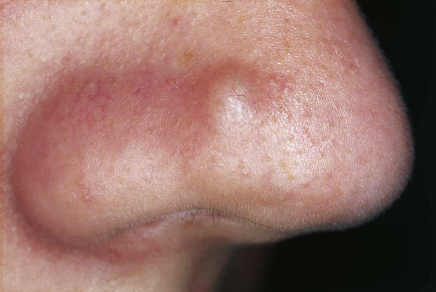
A fibrous papule develops during late adolescence or early adult life on the nose, or less often, elsewhere on the face. It is a dome shaped shiny lesion 2-6 mm in diameter, sometimes bearing a central hair. Although it looks similar to a skin-coloured mole (dermal naevus), it is more firm in texture. It is harmless but persists unchanged lifelong.
It is important to distinguish fibrous papule from the common skin cancer, basal cell carcinoma, which may also present as a firm shiny bump. Basal cell carcinoma most often arises later in life. It slowly grows, and tends to bleed and ulcerate.
Figure 2. Fibrous papule of the nose
What other problems can occur with fibrous papules?
There are no other problems associated with a fibrous papules. Multiple fibrous papules may sometimes be associated with rare genetic conditions including tuberous sclerosis complex, multiple endocrine neoplasia syndrome type 1 (MEN-1) and Birt-Hogg-Dube syndrome. Tuberous sclerosis is caused by mutations in the genes tuberous sclerosis complex 1 (TSC 1) that encodes the protein hamartin and tuberous sclerosis complex 2 (TSC 2) that encodes the protein tuberin 9. Multiple endocrine neoplasia type 1 (MEN-1) is due to a mutation in the MEN1 gene which encodes menin. Birt-Hogg-Dube syndrome is caused by a mutation in the FLCN gene which encodes folliculin. Clinical findings in Birt-Hogg-Dube include fibrofolliculomas, perifollicular fibromas (some authorities consider perifollicular fibroma to be related to angiofibroma), and trichodiscomas. These are all present as skin colored to hypopigmented papules on the head and neck or upper trunk.
Fibrous papule diagnosis
Fibrous papule is diagnosed by skin biopsy. There are distinctive features on histopathology (proliferation of fibroblasts, fibrotic stroma, and dilated blood vessels).
How is fibrous papule treated?
Fibrous papule dose not require any treatment. Several options are available for the removal of fibrous papules. Some dermatologists routinely employ surgical procedures such as curettage, shave excision, or elliptical excision with excellent cosmetic results. Successful treatment with various lasers, including the pulsed dye laser 10, carbon dioxide (CO2) laser 11, potassium titanyl phosphate (KTP) laser 12, and argon laser 13.
Papule vs Pustule
Pustules are small, inflamed, pus-filled, blister-like sores (lesions) on the skin surface.
Pustules are common in acne and folliculitis (inflammation of the hair follicle). They may occur anywhere on the body, but are most commonly seen in these areas:
- Back
- Face
- Over the breastbone
- Shoulders
- Sweaty areas, such as the groin or armpit
Pustules may be a sign of an infection. They should be checked by a health care provider and may need to be tested (cultured) for bacteria or fungus.
Papule vs Nodule
Nodule is an elevated skin lesion more than 0.5 cm in diameter.
Papule treatment
Fibrous papules do not require any treatment but if their appearance is of concern, there are a number of options that can be considered on an individual basis. These include:
- cosmetic camouflage
- excision (the fibrous papule is completely removed surgically)
- shave or punch biopsy (removing part of the fibrous papule)
- electrosurgery (using an electrical device to destroy the fibrous papule)
- topical application of sirolimus (also called rapamycin which is an mTOR inhibitor) for multiple fibrous papules associated with tuberous sclerosis complex.
Rapamycin (mTOR inhibitor) has recently gained popularity in the treatment of angiofibromas. After binding to mTOR, rapamycin inhibits its activity which downregulates cell proliferation. Rapamycin also decreases vascular endothelial growth factor (VEGF) production by downregulating VEGF- stimulated endothelial cell proliferation 14. Several case series, case reports, and one randomized controlled trial have been published verifying the effectiveness of topical rapamycin used as 0.1% once or twice daily, as well as 0.2%, used 5 times a week and 0.4% used 3 times a week 9. The angiofibromas cleared as long as the medication was being used. The longest reported follow up has been 3 years. Many have used crushed rapamycin tablets and mixed them in Vaseline to obtain the desired concentration which was not a standardized dose. DeKlotz et al. 15 proposed a standardized formulation on how to make .1% topical rapamycin in 2011. Few if any side effects occur from the topical medication including mild irritation and erythema. Park et al. 16 showed that topical rapamycin was enough to treat the lesions when they were small in size, that is less than 4 mm. However, if they were larger than 4 mm, ablative resurfacing was needed in conjunction with rapamycin. It can also be costly to use topical rapamycin in the treatment of angiofibromas due to the length of treatment that is necessary to obtain sufficient results costing several hundred to several thousand dollars out of pocket.
Successful treatment with various lasers, including the pulsed dye laser 10, carbon dioxide (CO2) laser 11, potassium titanyl phosphate (KTP) laser 12, and argon laser 13.
- Fibrous Papule of the Face. https://emedicine.medscape.com/article/1057309-overview[↩][↩]
- Bansal C, Stewart D, Li A, Cockerell CJ. Histologic variants of fibrous papule. J Cutan Pathol. 2005 Jul;32(6):424-8. https://www.ncbi.nlm.nih.gov/pubmed/15953376[↩][↩][↩]
- Fibrous papule of nose – Skin. https://www.visualdx.com/visualdx/diagnosis/fibrous+papule+of+nose?diagnosisId=51572&moduleId=11[↩][↩][↩][↩][↩][↩]
- Nemeth AJ, Penneys NS, Bernstein HB. Fibrous papule: a tumor of fibrohistiocytic cells that contain factor XIIIa. J Am Acad Dermatol. 1988 Dec. 19(6):1102-6.[↩]
- Chan JY, Wang KH, Fang CL, Chen WY. Fibrous papule of the face, similar to tuberous sclerosis complex-associated angiofibroma, shows activation of the mammalian target of rapamycin pathway: evidence for a novel therapeutic strategy?. PLoS One. 2014. 9 (2):e89467.[↩]
- Requena L, Sangueza OP. Cutaneous vascular proliferations. Part III. Malignant neoplasms, other cutaneous neoplasms with significant vascular component, and disorders erroneously considered as vascular neoplasms. J Am Acad Dermatol. 1998 Feb;38(2 Pt 1):143-75; quiz 176-8.[↩][↩]
- Meigel WN, Ackerman AB. Fibrous papule of the Face. Am J Dermatopathol. 1979 Winter;1(4):329-40. https://www.ncbi.nlm.nih.gov/pubmed/396811[↩]
- Numerous fibrous papules of the face unassociated with any genodermatosis. Dermatology Online Journal 2007, 13 (4): 12 https://escholarship.org/uc/item/0pc1r638[↩]
- Macri A, Tanner LS. Angiofibroma, Cutaneous. [Updated 2018 Oct 27]. In: StatPearls [Internet]. Treasure Island (FL): StatPearls Publishing; 2018 Jan-. Available from: https://www.ncbi.nlm.nih.gov/books/NBK482470[↩][↩]
- Sharma VK, Khandpur S, Khanna N. An interesting case of unilateral angiofibromas successfully treated with pulsed dye laser. J Eur Acad Dermatol Venereol. 2004 Sep;18(5):641-2.[↩][↩]
- Papadavid E, Markey A, Bellaney G, Walker NP. Carbon dioxide and pulsed dye laser treatment of angiofibromas in 29 patients with tuberous sclerosis. Br J Dermatol. 2002 Aug;147(2):337-42[↩][↩]
- Tope WD, Kageyama N. “Hot” KTP-laser treatment of facial angiofibromata. Lasers Surg Med. 2001;29(1):78-81. https://www.ncbi.nlm.nih.gov/pubmed/11500867[↩][↩]
- Boixeda P, Sanchez-Miralles E, Azana JM, Arrazola JM, Moreno R, Ledo A. CO2, argon, and pulsed dye laser treatment of angiofibromas. J Dermatol Surg Oncol. 1994 Dec;20(12):808-12.[↩][↩]
- Habib SL, Al-Obaidi NY, Nowacki M, Pietkun K, Zegarska B, Kloskowski T, Zegarski W, Drewa T, Medina EA, Zhao Z, Liang S. Is mTOR Inhibitor Good Enough for Treatment All Tumors in TSC Patients? J Cancer. 2016;7(12):1621-1631[↩]
- Dramatic improvement of facial angiofibromas in tuberous sclerosis with topical rapamycin: optimizing a treatment protocol. DeKlotz CM, Ogram AE, Singh S, Dronavalli S, MacGregor JL. Arch Dermatol. 2011 Sep;147(9):1116-7. doi: 10.1001/archdermatol.2011.254. https://jamanetwork.com/journals/jamadermatology/article-abstract/1105295[↩]
- Treatment of Angiofibromas in Tuberous Sclerosis Complex: The Effect of Topical Rapamycin and Concomitant Laser Therapy. Dermatology. 2014;228(1):37-41. doi: 10.1159/000357033. Epub 2013 Dec 21. https://doi.org/10.1159/000357033[↩]


