Contents
What is pityriasis alba
Pityriasis alba is a low-grade type of eczema or dermatitis that mostly affects children aged 3–16, with 90% of cases occurring in children younger than 12 years 1. An estimated 5% of children in the United States may be affected. Studies have demonstrated a higher prevalence in Egypt (18%) and Mali (20%) 1. The name refers to its appearance: pityriasis refers to its characteristic fine scale, and alba to its pale color (hypopigmentation). The patches are often found on the cheeks and upper arms and are more noticeable in darker skinned children. Pityriasis alba is generally asymptomatic.
Pityriasis alba is common worldwide with a prevalence in children of around 5%.
- Pityriasis alba mainly affects children and adolescents aged 3 to 16 years, but may also arise in older and younger people
- Pityriasis alba affects boys and girls equally.
- Pityriasis alba is more prominent, and may also be more prevalent, in dark skin compared to white skin.
Pityriasis alba looks like scaly or flaky patches that are lighter than the surrounding skin, and the affected areas usually occur on the face or cheeks. Pityriasis alba can be bothersome because of its appearance, but the condition is not a serious one.
The cause of pityriasis alba is not well known, but it does seem to be more common in children and teens who also have allergies or asthma, so the things that trigger these other conditions may also trigger pityriasis alba. Most people also notice that the condition is more severe in the summertime; this may be because it is more noticeable in the summer as the surrounding skin becomes tan. The development or prominence of pityriasis alba can be reduced by avoiding exposure to sunlight.
There is no certain treatment for pityriasis; it usually goes away on its own within a year or in some cases persists for up to two or three years. Your skin color will gradually return completely to normal.
No treatment is necessary for asymptomatic pityriasis alba.
- A moisturizing cream may improve the dry appearance
- A mild topical steroid (hydrocortisone) cream may reduce redness and itch
- Sunscreens (broad spectrum SPF 30+) in summer may prevent the patches becoming more noticeable by reducing the tanning of the non-affected skin.
Is pityriasis alba contagious?
Pityriasis alba is not contagious.
Is pityriasis alba hereditary?
No.
Can pityriasis alba be cured?
Pityriasis alba usually resolves spontaneously, and the skin color gradually returns back to normal with no scarring. However, it may persist for a few years, and may come and go during this period, particularly in summer when the patches become prominent due to the tanning of non-affected skin.
What are the clinical features of pityriasis alba?
Classic pityriasis alba usually presents with 1 to 20 patches or thin plaques.
- Most lesions occur on the face, especially on cheeks and chin.
- They may also arise on neck, shoulders and upper arm and are uncommon on other sites of the body.
- Size varies from 0.5 to 5 cm in diameter.
- They are round, oval or irregular in shape.
- Pityriasis alba may have well-demarcated or poorly defined edges.
- Itch is minimal or absent.
- Hypopigmentation is more noticeable in summer, especially in dark-skinned children.
- Dryness and scaling is more noticeable in winter, when environmental humidity tends to be lower.
Typically, each area of pityriasis alba goes through several stages.
- Slightly scaly pink plaque with just palpable papular surface
- Hypopigmented plaque with fine surface scale
- Then post-inflammatory hypopigmented macule without scale
- Resolution
Figure 1. Pityriasis alba
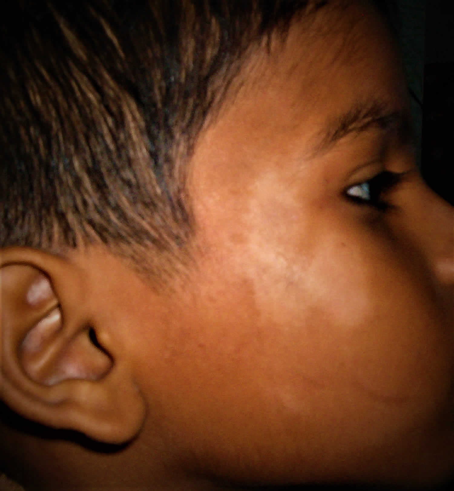
Figure 2. Pityriasis alba face

What causes pityriasis alba
The cause of pityriasis alba is unknown but is thought to be a type of dermatitis. Pityriasis alba is not contagious, and no infectious cause has been reported. Pityriasis alba is most common in individuals with a history of atopy, although it may occur in nonatopic individuals as well. In many cases, pityriasis alba is considered to be a minor manifestation of atopic dermatitis. It is thought to represent nonspecific dermatitis with residual post-inflammatory hypopigmentation. Histopathology shows decreased melanin production in the affected areas. Associated findings in some studies include atrophic sebaceous glands, iron-deficiency anemia, and low levels of serum copper. The significance of these findings and their relationship to pityriasis alba is uncertain.
The dry component may appear more obvious in winter months. The hypopigmentation is more obvious in summer because of relative tanning of unaffected skin.
The cause of pityriasis alba is unknown.
- It often coexists with dry skin and atopic dermatitis.
- It often presents following sun exposure, perhaps because tanning of surrounding skin makes affected areas more prominent.
Researchers have not reached any conclusions about the relationship of pityriasis alba to the following:
- Ultraviolet radiation
- Excessive or inadequate bathing
- Low levels of serum copper
- Malassezia yeasts (which produce a metabolite, pityriacitrin, that inhibits tyrosinase thus causing hypopigmentation)
After some weeks or months the dry surface resolves leaving a smooth pale patch, which slowly disppears over a year or so.
Differential diagnosis
The differential diagnosis of pale dry patches on the face or limb of a child includes:
- Postinflammatory hypopigmentation, in which there is a history of another inflammatory skin disorder;
- Atopic dermatitis, characterised by very itchy symmetrical plaques that respond to topical steroids;
- Psoriasis, in which there are symmetrical scaly plaques in typical sites including scalp;
- Seborrheic dermatitis, which is uncommon in children aged 2-12;
- Pityriasis versicolor, which affects upper trunk of adolescents and adults and has positive microscopy;
- Tinea corporis, which also has positive mycology and there is often a history of contact with a kitten;
- Nummular dermatitis, in which there are dry or crusted itchy round patches;
- Nevus depigmentosus, a stable pale mark present since birth;
- Vitiligo, in which there are progressive macules with complete pigment loss and no scale most often arising around eyes or mouth;
- Hypopigmentation secondary to topical medications such as retinoic acid, benzoyl peroxide, and corticosteroids;
- Hypopigmented mycosis fungoides (rare cutaneous T-cell lymphoma), in which there are persistent progressive infiltrated plaques.
In the proper geographic and clinical setting, the diagnosis of leprosy also should be considered.
Pityriasis alba signs and symptoms
The most common locations for pityriasis alba include:
- Cheeks, around the mouth, chin
- Forehead
- Neck
- Shoulders, upper chest, and upper arms
Pityriasis alba appears as several (2–20) light-colored (hypopigmented) patches ranging in size from 1–4 cm. The patches may have slight and subtle surface patches (scale). Occasionally, pityriasis alba begins as mildly itchy, pink patches that develop into lightened patches.
People often think that pityriasis alba gets worse in the summer, but it just becomes more obvious as the normal surrounding skin becomes darker with sun exposure.
Pityriasis alba diagnosis
Pityriasis alba can be confused with several other disorders that cause hypopigmentation.
To exclude these, investigations may include:
- Wood lamp examination: hypopigmentation does not enhance, and there is no fluorescence in pityriasis alba. This finding is in contrast to vitiligo which will fluoresce more brightly and have edges with sharper demarcation.
- Scrapings for mycology: microscopy and fungal culture are negative in pityriasis alba. Potassium hydroxide (KOH) preparation of a skin scraping will be negative for fungal elements. This result is in contrast to tinea versicolor or tinea corporis which will be positive for fungal elements.
- Skin biopsy: skin biopsy is rarely required, but may reveal mildly spongiotic dermatitis and reduction in melanin. Skin biopsy when performed it can distinguish pityriasis alba from mycosis fungoides (cutaneous T-cell lymphoma).
Mycology is negative in pityriasis alba, but may be necessary to rule out a dermatophyte infection or pityriasis versicolor.
Skin biopsy is rarely necessary and may be reported as nonspecific dermatitis.
Pityriasis alba treatment
Patients and their parents can be reassured regarding the benign and self-limited nature of pityriasis alba. However, they also should be made aware that its slow resolution may take several months to a few years; although most cases resolve within one year.
No specific treatment is necessary in most children as the pityriasis alba patches are asymptomatic and resolve by themselves.
A trial of mild topical steroid (1% hydrocortisone cream or ointment) is sometimes warranted for a couple of weeks. It can be expected to reduce the redness, scaling and itch (if present). Long term, a moisturizer may prove as effective. Mild emollients, such as petroleum jelly and Eucerin cream, may reduce scaling.
Calcineurin inhibitors, pimecrolimus cream and tacrolimus ointment, may be as effective as hydrocortisone and have been reported to speed recovery of skin color.
Treatment with topical calcineurin inhibitors, such as 0.1% tacrolimus ointment and 1% pimecrolimus cream, have also been reported to be effective; however, because of their high cost, they are seldom indicated. Calcitriol, a topical vitamin D analog, showed comparable efficacy compared with tacrolimus. Other treatment options, usually reserved for extensive cases, include psoralen plus ultraviolet-A (PUVA) photochemotherapy and targeted phototherapy with a 308-nm excimer laser.
Sunscreens may be useful to reduce tanning of surrounding skin.
Pityriasis alba home remedies
If you suspect that you have pityriasis alba, the most important self-care measure is to keep the skin well moisturized. Try the following:
- Use non-soap cleansers or moisturizing soaps.
- Apply moisturizers such as petroleum jelly (Vaseline®) or fragrance-free ointments and creams.
- Avoid sun exposure and wear sunscreen.
- Apply over-the-counter hydrocortisone cream sparingly for 3–7 days.
See your doctor for evaluation if the condition does not improve with self-care measures, if it seems to be getting worse, or if it spreads to other areas.


