Contents
What is ptosis
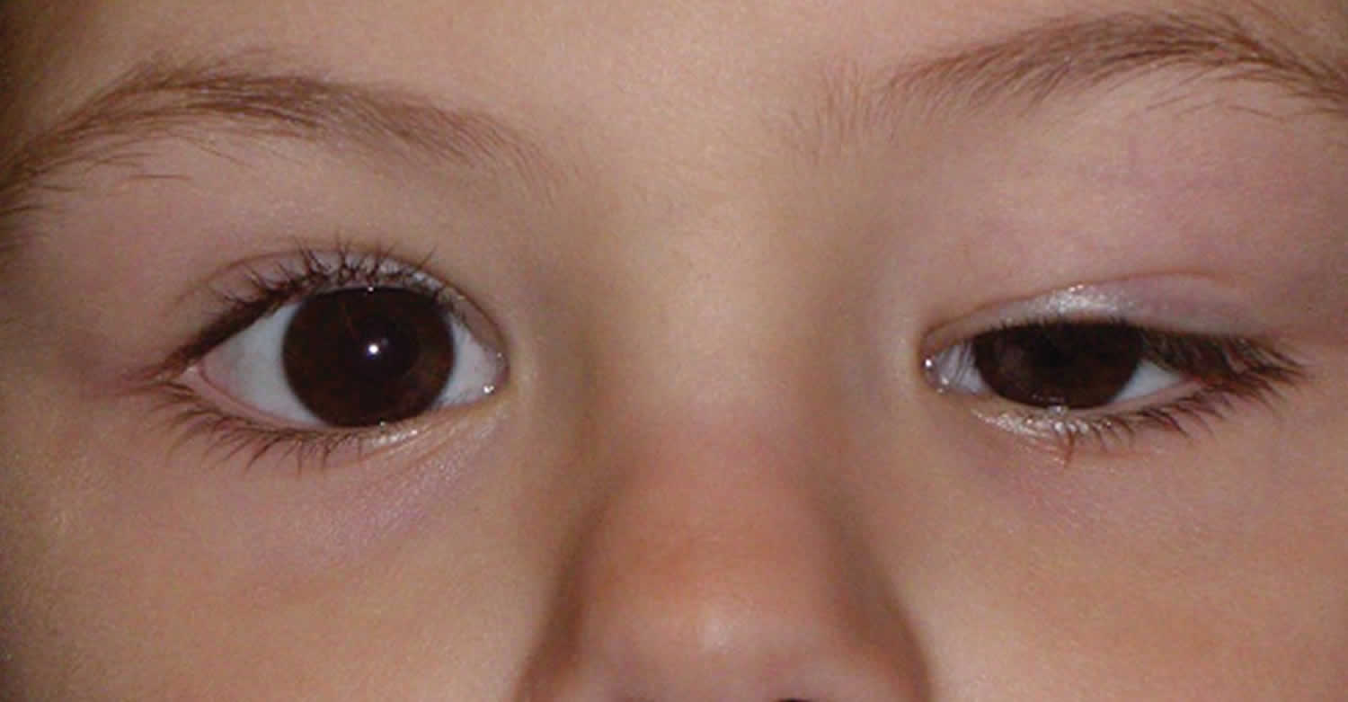
Ptosis is the involuntary drooping of the upper eyelid that can be present at birth (congenital) or can occur later in life (acquired). The upper eyelid may droop just a little, or so much that it covers the pupil (the black dot at the center of your eye that lets light in) (see Figure 1). Normally, the upper eyelid covers 1.0-2.0mm of the superior part of the cornea. Ptosis can limit or even completely block normal vision. The levator palpebrae superioris muscle holds the upper eyelid in proper position and moves it up and down. Any condition that affects this muscle will also affect the eyelid position. Poor development of the levator palpebrae superioris muscle in the upper eyelid can lead to an inability to properly open the eye. It is the most common cause of congenital ptosis. Acquired ptosis has many possible causes. Acquired ptosis is usually because of a mechanical factor, such as the lid is too heavy for the muscle to lift or it may be associated with a neurological disease or paralytic disease. Less common causes include injury, previous eye surgery or diabetes. Most cases of ptosis in an adult come on gradually during the later years of life, as part of the normal aging process. The levator tendon stretches, thins, or loosens its attachment to the eyelid, causing it to sag. This age-related ptosis is called involutional ptosis, aponeurotic ptosis or senile ptosis. Ptosis can involve one or both upper eyelids, with or without symmetry (see Figure 3).
Acquired ptosis can be caused by neurologic conditions that affect the nerves and/or muscles of the eye. These include myasthenia gravis, progressive external ophthalmoplegia, Horner syndrome, and third cranial nerve palsy. The ptosis may be combined with an eye movement disorder with resultant double vision. An eyelid mass can also cause ptosis.
If a disease is found, it will be treated. Most cases of drooping eyelids are due to aging and there is no disease involved.
Eyelid lift surgery (blepharoplasty) is done to repair sagging or drooping upper eyelids.
- In milder cases, it can be done to improve the appearance of the eyelids.
- In more severe cases, surgery may be needed to correct interference with vision.
- In children with ptosis, surgery may be needed to prevent amblyopia, also called “lazy eye.”
When amblyopia is present, appropriate treatment is initiated. When astigmatism is significant enough to potentially cause amblyopia, glasses are prescribed. Early eyelid surgery is usually indicated for a drooping eyelid that blocks vision (which may cause delayed vision development), or leads to a significant chin-up head position (which may cause neck problems and/or delay of developmental skills). Children are usually monitored regularly for vision abnormalities. Surgery may also be indicated during preschool years if the ptosis does not improve with normal growth and maturation of the face.
Figure 1. Ptosis (severe affecting both eye and the upper eyelids covering both pupils blocking vision)
Ptosis in children
Ptosis in children can affect one eye or both eyes. Ptosis may be present at birth, or may be acquired later in life. If a droopy eyelid is present at birth or within the first year of life, the condition is called congenital ptosis. In most cases of congenital ptosis, the problem is isolated and does not affect the vision. Ptosis in children can be caused by problems with the muscle that lifts the eyelid (called the levator muscle). Any ptosis that develops over a period of days or weeks can signal a serious medical problem and needs further neurologic and physical evaluation.
In most cases of congenital ptosis, a droopy eyelid results from a localized myogenic dysgenesis. Rather than normal muscle fibers, fibrous and adipose tissues are present in the muscle belly, diminishing the ability of the levator to contract and relax. Therefore, the condition is commonly called congenital myogenic ptosis.
Congenital ptosis can also occur when the innervation to the levator is interrupted through neurologic or neuromuscular junction dysfunction.
Congenital ptosis occurs equally among the different races.
Congenital ptosis occurs equally between males and females.
Congenital ptosis is usually present at birth but may manifest within the first year of life.
The most obvious sign of ptosis is a drooping eyelid. Another sign is when the upper eyelid creases do not line up evenly with each other. A child with ptosis may tip their head back, lift up their chin, or raise their eyebrows to try to see better. Over time, these movements can cause head and neck problems.
Sometimes, a child born with ptosis can also have other eye-related problems. They can include eye movement issues, eye muscle disease, tumors (on the eyelid or elsewhere) and other problems.
Having ptosis puts a child at risk for vision problems. If the child’s eyelid droops so much that it blocks vision, amblyopia (also called “lazy eye”) can develop. One eye will have better vision than the other. A child with ptosis can also have astigmatism, where they see blurry images. The child may also develop misaligned (crossed) eyes. Development of amblyopia is an indication for immediate surgical correction.
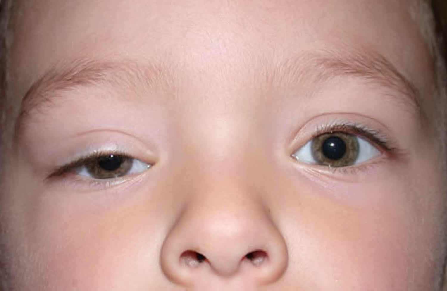
Figure 2. Ptosis in children
Ptosis in children causes
In most cases of congenital ptosis, the cause is idiopathic.
Histologically, the levator muscles of patients with congenital ptosis are dystrophic. The levator muscle and aponeurosis tissues appear to be infiltrated or replaced by fat and fibrous tissue. In severe cases, little or no striated muscle can be identified at the time of surgery. This suggests that congenital ptosis is secondary to local developmental defects in muscle structure.
Congenital ptosis may occur through autosomal dominant inheritance. Common familial occurrences suggest that genetic or chromosomal defects are likely.
Other potential causes of congenital blepharoptosis include 1:
- Blepharophimosis syndrome: This condition consists of short palpebral fissures, congenital ptosis, epicanthus inversus, and telecanthus.
- Third cranial nerve palsy: Signs of aberrant regeneration are usually present. The pupil may be paradoxically small and nonreactive.
- Horner syndrome: Ipsilateral findings of mild ptosis, miosis, and anhidrosis characterize this syndrome. The ipsilateral lower eyelid may be elevated. Also, because of the lack of sympathetic innervation to the iris melanocyte development, a difference in the iris color between the eyes may result (called heterochromia).
- Marcus Gunn jaw-winking syndrome: The motor nerve to the external pterygoid muscle is misdirected to the ipsilateral levator muscle. Lid elevation occurs with mastication or with movement of the jaw to the opposite side.
- Birth trauma
- Duane syndrome: In this condition, the sixth cranial nerve fails to innervate a lateral rectus muscle. Then, the muscle acquires an innervation of the third cranial nerve. Although the synkinesis produced does not involve lid innervation, enophthalmos with apparent ptosis may result. In Esotropic Duane syndrome, the upper eyelid droops further and the lower lid elevates when the eye is adducted because of a co-contraction of the horizontal rectus muscles.
- Periorbital tumor: Neuroblastoma, plexiform neuromas, lymphomas, leukemias, rhabdomyosarcomas, neuromas, neurofibromas, or other deep orbital tumors may produce ptosis or proptosis.
- Kearns-Sayre syndrome: This mitochondrial deletion disorder is characterized by progressive external ophthalmoplegia, heart block, retinitis pigmentosa, and central nervous system manifestations. This condition begins in childhood but is rarely present at birth. The conditions are most likely to become symptomatic in the first or second decade of life. Bilateral ptosis is a prominent feature of this syndrome.
- Myotonic dystrophy: Patients with this condition may present with polychromatic cataracts, gonadal atrophy, or premature thinning and/or loss of hair. Myotonic dystrophy is an autosomal dominant disorder that is characterized clinically by myotonia and progressive muscular weakness.
- Blepharochalasis: This condition is characterized by infiltrative processes that thicken the lids and produce ptosis.
- Myasthenia gravis: A defect at the neuromuscular junction produces relative unresponsiveness to released acetylcholine, resulting in ptosis.
- Pseudotumor of the orbit: Patients with this condition may present with ptosis due to inflammation and edema of the eyelid.
- Pseudoptosis: Less tissue in the orbit (e.g., unilateral smaller eye, fat atrophy, blowout fracture) produces the appearance of ptosis secondary to the decreased volume of orbital contents.
Ptosis in children diagnosis
All pediatric patients presenting with either unilateral droopy eyelid or bilateral droopy eyelids need a thorough examination that includes a medical history, a family history, a history of drug or allergic reactions, and a review of systems.
- Family photographs can help determine onset or variability of the ptosis. Providing photographs also gives the surgeon a chance to examine the other family members. A patient with a strong family history of congenital ptosis may not need an extensive workup.
- In severe cases of congenital ptosis in which surgery is needed, historical emphasis should be placed on any anticoagulant use or bleeding disorder to avoid potential complications during surgery. The surgeon should also inquire about a family history of malignant hyperthermia and cardiac disorders. Patients with ptosis and Kearns-Sayre syndrome or chronic progressive external ophthalmoplegia may also have a cardiac conduction disorder.
- A history of fluctuating ptosis with strabismus may indicate myasthenia gravis.
- A careful medical history regarding cancer should be obtained. Metastatic or primary orbital tumors can result in malpositioning of the eyelid.
- A history of trauma with orbital wall fractures can result in pseudoptosis with enophthalmos. Additionally, third cranial nerve palsy from trauma may result in ptosis.
- A history of drug or allergic reactions may be helpful. Allergic reactions can result in eyelid edema and droopy eyelid.
- A history of difference in the size of the pupil may be helpful in diagnosing Horner syndrome. Patients with Horner syndrome have ptosis and miosis on the same side. Cervical or apical thoracic tumors can cause damage to the sympathetic chain and result in this condition. Neuroblastoma, which is one of the most common childhood cancers, should be ruled out.
- A history of dry eyes, intermittent epiphora, or chronic conjunctivitis can indicate a dry eye disorder or corneal surface disease.
Physical examination
All pediatric patients presenting with either unilateral droopy eyelid or bilateral droopy eyelids need a thorough physical evaluation.
- Visual acuity, refractive error, and cycloplegic refraction should be recorded. In infants, the surgeon should make sure that the baby can fixate and follow objects with each eye individually.
- The patient should be evaluated for strabismus (misalignment) and undergo a dilated fundus examination.
- Serial external photographs of the eyes and the face may be included in the patient’s record for documentation.
- Tear function should be evaluated if any doubt exists about the adequacy of tear production. This evaluation would include a slit-lamp examination with fluorescein stain to examine the cornea, tear meniscus, and tear break-up time. The Schirmer test can also be performed for dry eye syndrome; to do so, a filter paper is applied at the junction of the middle and lateral one third of the lower eyelid.
- Corneal sensitivity should be tested if possible. This may be a difficult test in young pediatric patients.
- An exophthalmometer can be used to assess relative proptosis or enophthalmos of each eye. In pseudoptosis, a proptosis of the contralateral eye gives the false impression that the normal upper eyelid is droopy.
- The pupillary size and the iris color differences between the eyes should be examined for Horner syndrome.
- The lid height (palpebral fissure distance) should be observed and measured in millimeters with each eye fixating on a distant target. The distance is the measurement of the greatest width of the palpebral fissure with the patient’s eyes in straight gaze. The lid position in downgaze should be noted. In congenital ptosis, the ptotic lid appears higher in downgaze.
- After the palpebral fissure distance is measured, the levator function should be evaluated. The patient looks downward as a ruler is positioned with a mark adjacent to the upper lid margin. With the examiner’s hand eliminating any brow action by the patient, the patient looks upward as far as possible without a change in head position. Lid elevation is measured directly from the ruler and is recorded in millimeters of levator function.
- The patient should be examined for Bell phenomenon. The patient closes both eyes tightly as the examiner holds the upper and lower lids apart. If the globe elevates during the forced lid closure, a normal Bell phenomenon is present. This evaluation can help the surgeon to determine the risk of exposure keratopathy following the eyelid surgery.
- Careful external examination along with palpation of the eyelids and the orbital rim should be performed. A lid mass can cause extra weight in the lid, resulting in ptosis. Plexiform neuromas, lymphoma, or leukemia can result in an eyelid mass. Rhabdomyosarcoma may present with a mass that is palpable through the lid.
Laboratory test
If myasthenia gravis is suspected, check serum acetylcholine receptor antibody levels 2.
Imaging Studies
The following are indications to perform neuroimaging studies (e.g., MRI, CT) of the orbit and brain:
- History not consistent and onset not clear
- Other neurologic findings along with ptosis
- Orbital wall fracture suspected with history of trauma
- Visible or palpable lid mass
- Orbital tumors (eg, lymphoma, leukemia, rhabdomyosarcoma) suspected
- New onset of Horner syndrome with or without other neurologic findings
- New onset of third cranial nerve palsy with or without other neurologic findings
- Globe displacement with either enophthalmos or proptosis
Other Tests
If myasthenia gravis is suspected, the following tests are recommended:
- Single fiber electromyography (EMG)
- Tensilon or Ice test
- Serum Acetylcholine receptor antibody
If a mitochondrial disorder is suspected, a muscle biopsy should be performed.
What problems can occur as a result of childhood ptosis?
One or more of the following abnormalities may accompany ptosis in childhood: astigmatism (refractive error), obstruction of the visual axis (the path that light takes into the eye), a chin-up head position, and amblyopia. The abnormal resting position of the eyelid on the cornea may result in astigmatism (a misshaping of the cornea) or other refractive errors, and is a risk factor for developing amblyopia (refractive amblyopia). Another risk factor for amblyopia is an eyelid drooping so low that it actually prevents light from entering the eye and creating an image on the retina at the back of the eye (deprivation amblyopia) Also, a chin-up head position may be present. This head position is adopted in order to be able to see beneath the edge of the drooping upper eyelid. Contraction of the frontalis muscle (in the forehead) to further elevate the upper eyelid is a very common compensatory mechanism.
When can my infant have ptosis surgery?
If the ptosis is mild we recommend waiting until your 4-month-old is preschool age. If the ptosis is moderate or severe or causing a refractive error eye surgeons recommend surgery as early as six months. Please consult your ophthalmologist to determine the correct treatment for your child.
Ptosis in adults
Adults get ptosis (called involutional ptosis or aponeurotic ptosis or senile ptosis) when the levator muscle stretches or separates away from their eyelid. This can be caused by aging or an eye injury. Sometimes ptosis happens as a side effect after certain eye surgery. Rarely, diseases or tumors can affect the eyelid muscle, causing ptosis.
Senile ptosis (involutional ptosis or aponeurotic ptosis) was fist described by Jones Quickert and Wobig in 1975 who demonstrated that the levator aponeurosis appeared dehisced or disinserted from the tarsus. This disinsertion may be congenital or acquired. Most patients with acquired ptosis (involutional ptosis or aponeurotic ptosis or senile ptosis) develop the condition secondary to involutional changes in the levator aponeurosis. Gradual stretching or dehiscence of this structure causes slowly progressive ptosis.
Most patients with acquired ptosis develop the condition secondary to involutional changes in the levator aponeurosis. Gradual stretching or dehiscence of this structure causes slowly progressive ptosis.
Some patients have revealed a normal levator aponeurosis but a myogenic degeneration of the muscle itself, characterized by a fatty degeneration in the area of the Whitnall’s ligament. This fatty infitration has been confimed by light microscopy and appears to be a degenerative change found in adults with acquired ptosis. Müller’s muscle appeared to be grossly intact, but microscopic fibrosis with plentiful collagen fibers was observed in Müller’s muscles of patients with acquired blepharoptosis induced by prolonged hard contact lens wear.
Your ophthalmologist will find the cause of your ptosis in order to recommend treatment. They will do a complete eye exam, and may also want you to have blood tests and imaging tests. The ophthalmologist will likely recommend surgery to help the eyelid muscle work better.
Risk factors for senile ptosis
Congenital aponeurotic ptosis is uncommon but this could be secondary to trauma by forceps delivery, vacuum extraction, fetal rotation, and shoulder distocia. There are multiple factors that can cause disinsertion of the levator aponeurosis, such as continuous rubbing of the eye, chronic use of contact lenses, inflammatory diseases, trauma or following eyelid or intraocular surgery. Approximately 6% of patients following cataract surgery develops ptosis.
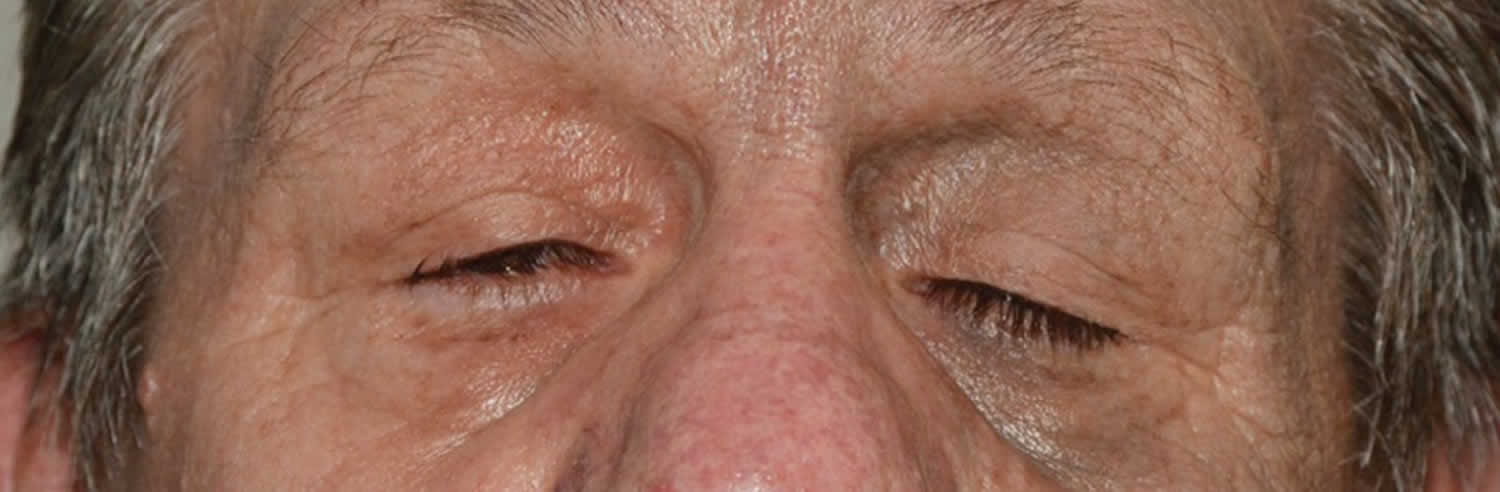
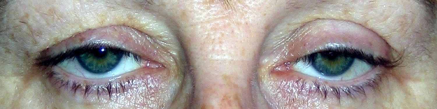
Figure 3. Age-related ptosis (involutional ptosis)
Ptosis in adults symptoms
Patients with aponeurotic ptosis may present with a spectrum of symptoms ranging from visually significant obstruction to minor, visually asymptomatic cosmetic eyelid asymmetry. Visual field obstruction results in functional blockage of the superior visual field. Symptoms are often worse when reading or downgaze. Patients tend to compensate with overaction of the frontalis muscle. Persistent brow elevation may lead to frontalis fatigue or even cephalgia.
Also it´s important to look for fluctuation in symptoms of fatigue throughout the day that may indicate myasthenia gravis. In these cases, patients should also be asked about their use of statin medications, cause there have been recent reports of myasthenialike syndromes that resulted in ptosis.
Ptosis in adults diagnosis
Physical examination
Here are some clinical key points that should be noticed, some measurements must be interpreted with regard and thinking in each patient as unique, cause besides exist a “normal” range all are variable depending of age, ethnicity, gender, genetic, etc.
Margin reflex distance. Defined as the distance from the upper eyelid margin to the corneal light reflex in primary position. Normal distance 4-5 mm.
Vertical palpebral fissure height. Defined as the widest point between the lower eyelid and the upper eyelid. This measurement is taken with the patient fixating on a distant object in primary gaze. Normal palpebral fissure measures between 12–15 mm.
Upper eyelid crease position. Defined as distance from upper eyelid crease to the eyelid margin Normal distance is 8-9 mm in males and 9-11 mm in females. Aponeurotic defects characteristically have a high or an absent upper eyelid crease, also any asymmetry, altered contour, lack of continuity or thinning of the eyelid superior to the upper tarsal plate may indicate the presence of levator aponeurosis disinsertion.
Levator function (upper eyelid excursion). It is estimated by measuring from downgaze to upgaze with frontalis muscle function negated. Crowell Beard reported normal eyelid excursion to be between 12-17 mm
The levator function is classified as:
- Good >= 8 mm
- Fair 5 -7 mm
- Poor 4 mm
The levator muscles obey Hering’s law of equal innervation. wich means that are innervated symmetrically, resulting in equal central neural output, so in cases of bilateral asymmetrical ptosis, the less affected eyelid may maintain a normal level of elevation due to excessive innervational stimulation determined by the more ptotic eyelid. This condition can be detected prior to surgery by manually elevating the ptotic eyelid. An immediate fall of the contralateral eyelid confims the presence of bilateral, asymmetrical ptosis masked by levator “overaction.”
Myasthenia gravis should be considered in all patients with ptosis, and one may check for levator fatigability by asking the patient to alternate between upgaze and downgaze repeatedly, or by asking the patient to look in extreme upgaze for up to 1–2 min.
Visual acuity and refraction: This is important because it could change after ptosis surgery. This occurs because the weight of the upper eyelid on the cornea may affect the shape of the cornea, and hence refractive error may change after surgical repositioning of the eyelid. Corneal topography has usually demonstrated an increase in against the rule astigmatism. These changes tend to be temporary, with a decrease in refractive shift by 12 months after surgery. That´s why, one should avoid prescribing glasses to patients prior to and up to 3 months following ptosis surgery.
Elderly patients who have dermatochalasis must be assessed carefully to notice if the lower position of the eyelid is a mechanical effect of the redundant skin and/or due to an aponeurotic defect.
Pupil abnormalities, ocular, extraocular and facial movements should be normal.
Head position, chin elevation , brow position, and brow action in attempted upgaze must be appreciated as a sign of ptosis.
Bell phenomenon, corneal sensation, lagophthalmos and poor tear film quantity or quality may be explored cause a faillure in this ones may predispose a patient to complications of ptosis repair such as dryness and exposure keratitis,
Diagnostic procedures
Visual field testing with the eyelids untaped (in the natural, ptotic state) and taped (artificially elevated) helps determine the patient’s level of functional visual impairment.
External full-face photography is helpful to compare past and actual state of ptosis.
Pharmacologic testing may be helpful to exclude some clinical diagnosis like Horner síndrome (Phenyleprine 10%) or myasthenia gravis (edrophonium chloride, ice- pack, or acetylcholine receptor antibody tests)
Ptosis of eyelid prognosis
A drooping eyelid can stay constant, worsen over time (be progressive), or come and go (be intermittent).
The expected outcome depends on the cause of the ptosis. In most cases, surgery is very successful in restoring appearance and function.
In children, more severe drooping eyelids may lead to lazy eye or amblyopia. This may result in long-term vision loss.
Droopy eyelid symptoms
Drooping of one or both eyelids is present. The lid may cover only the upper eye, or the entire pupil will be covered.
Problems with vision will also be present.
- At first, just a sense that the very upper field of vision is being blocked.
- When the drooping eyelid covers the pupil of the eye, the upper part of vision becomes blocked.
- Children may tip their head back to help them see under the eyelid.
- Tiredness and achiness around the eyes may also be present.
Increased tearing despite a feeling of dry eyes may be noticed.
Ptosis causes
Eyelid drooping can develop at any time, regardless of age. It is often seen in older people due to the aging process, but can also signal an underlying medical condition, some of which may be neurologic, mechanical or muscular in nature.
A drooping eyelid is most often due to:
- Weakness of the muscle that raises the eyelid
- Damage to the nerves that control that muscle
- Looseness of the skin of the upper eyelids
Drooping eyelid can be:
- Caused by the normal aging process
- Present before birth
- The result of an injury or disease
Diseases or illnesses that may lead to eyelid drooping include:
- Tumor around or behind the eye
- Eyelid tumor
- Diabetes
- Horner syndrome
- Myasthenia gravis
- Stroke
- Brain aneurysm
- Brain tumor
- Cancer
- Horner’s syndrome
- Migraines
- Myotonic dystrophy
- Nerve injury
- Swelling in the eyelid, such as with a chalazion or stye
A physical examination and tests are necessary to determine if any of these conditions are the cause of the eyelid droop.
Droopy eyelid causes in infants and children
Ptosis in infants and children is often due to a problem with the muscle that raises the eyelid. A nerve problem in the eyelid can also cause it to droop.
Ptosis may also occur due to other conditions. Some of these include:
- Trauma at birth (such as from the use of forceps)
- Eye movement disorders
- Brain and nervous system problems
- Eyelid tumors or growths
Other conditions to consider when evaluating a drooping eyelid
Dermatochalasis
Dermatochalasis, often referred to as “baggy eyes”, is excess skin and/or fat on the upper or lower eyelid. If there is enough excess skin and/or fat on the upper eyelid, eyelid drooping can occur. Patients may experience varying degrees of this condition. A mild to moderate case of dermatochalasis can result in an appearance that is sad or tired looking. More severe cases can result in changes in vision and may require surgical correction.
Brow ptosis
Brow ptosis, or a drooping of the eyebrow, is also a consideration when evaluating drooping eyelids. When the brow descends to a lower position, a drooping eyelid can result. Brow ptosis condition can coexist with both dermatochalasis and ptosis.
Droopy eyelid diagnosis
After completing a medical history and physical exam, your doctor may want to run tests, such as the slit-lamp test, Tensilon test, and visual field test, to find the cause of the eyelid drooping.
Slit-lamp Exam
The slit-lamp uses a low-power microscope and a high-intensity light source that can be focused to provide a thin beam. This test allows your doctor to examine the eyes, especially the eyelids, cornea, conjunctiva, sclera, and iris. Drops may be placed in the eyes to dilate the pupils, which may cause some discomfort. The slit-lamp examination is then repeated using another small lens held close to the eye, so the back of the eye can be examined.
Tensilon Test
The drug Tensilon is injected into your veins. The doctor will ask the patient to perform eye movements to access if the drug improves muscle strength. This test will help determine whether the eyelid drooping is due to neuromuscular issues from an autoimmune process.
Visual Field Test
The visual field refers to the total area in which objects can be seen in the side (peripheral) vision while you focus your eyes on a central point. This eye exam uncovers any vision deficiencies in your visual field and helps determine the cause. Abnormal results may be due to diseases or central nervous system disorders, such as tumors that damage or press on (compress) the parts of the brain that deal with vision.
Droopy eyelid treatment
Ptosis treatment for children
Ophthalmologists consider the following factors when deciding the best way to treat ptosis in children:
- The child’s age
- Whether one or both eyelids are involved
- The eyelid height
- The strength of the eyelid’s muscle
- The eye’s movements
Although not all patients with congenital ptosis need surgical intervention, patients need to be closely monitored for the possible development of deprivational amblyopia. Since amblyopia may not be reversed after age 7-10 years, appropriate and timely medical and surgical treatment of congenital ptosis is critical to preserve the child’s vision.
- Uncorrected congenital ptosis can result in amblyopia secondary to deprivation or uncorrected astigmatism.
- An abnormal eyelid position can have negative psychosocial effects.
- Uncorrected acquired blepharoptosis results in decreased field of vision and frontal headaches.
General treatment
- Early consultation to avoid amblyopia
- Must be able to rule out and document other possible causes of ptosis (eg, Horner syndrome, third cranial nerve palsy)
Patients with congenital ptosis may have other conditions that need to be addressed. These conditions include amblyopia, strabismus, craniofacial abnormalities, and other neurologic findings. Appropriate consultation may be needed depending on the associated findings.
- Pediatric ophthalmologist
- Pediatric oculoplastic service
- Pediatric neurologist
- Cardiologist (if mitochondrial disorder suspected)
In most cases, ophthalmologists recommend surgery to treat ptosis in children. This is to either tighten the levator muscle or attach the eyelid to other muscles that can help lift the eyelid. The goal is to improve vision.
If the child also has amblyopia, that condition must be treated as well. Amblyopia may be treated by wearing an eye patch or special eyeglasses, or using certain eye drops, to strengthen the weaker eye.
All children with ptosis—whether or not they have surgery—should see their ophthalmologist regularly for eye exams. Ask your child’s ophthalmologist how often exams are needed. Because kids’ eyes grow and change shape, they need to be checked for amblyopia, refractive disorders, and other eye problems.
Medical therapy
Observation is only required in mild cases of congenital ptosis if no signs of amblyopia, strabismus, and abnormal head posture are present.
- Depending on the severity of the congenital ptosis, patients should be monitored every 3-12 months for signs of amblyopia due to congenital ptosis.
- External photographs can be helpful in monitoring patients.
- Head posture should be carefully examined. If the patient acquires a chin-up posture due to the worsening of ptosis, surgery may be indicated.
- The patient should be checked for astigmatism due to the compression of the droopy eyelid.
Medical follow up
- Patients who underwent surgery for congenital ptosis are initially monitored every 2-4 weeks for signs of exposure keratopathy, infection, granuloma formation, and overcorrection and undercorrection. External photographic documentation can be helpful in monitoring patients.
- Following the surgery, visual acuity, head posture, and refractive error should be carefully monitored. Any residual amblyopia should be treated aggressively.
Congenital ptosis surgery
Congenital ptosis has physical, functional, and psychological consequences. The method of repair depends on treatment goals, the underlying diagnosis, and the degree of levator function. Although the primary reason for the repair is functional, the surgeon has an opportunity through this procedure to produce symmetry in lid height, contour, and eyelid crease for better cosmesis 3.
Surgical correction of congenital ptosis can be undertaken at any age depending on the severity of the disease. Earlier intervention may be required if significant amblyopia or ocular torticollis is present. Severe cases of ocular torticollis may delay mobility in infants and toddlers because of the balance problems from extreme chin-up head posture. If intervention is not urgent, surgery is often delayed until age 3-4 years. Waiting until this age allows for more accurate measurements preoperatively 4.
Surgery for ptosis in patients with a history of dry eyes, seventh cranial nerve palsy, or significant extraocular muscle abnormalities, such as severe Graves ophthalmopathy, double elevator palsy, or progressive external ophthalmoplegia, should be approached with great caution to avoid exposure keratopathy following the surgery.
Levator muscle resection
This procedure is the shortening of the levator-aponeurosis complex through a lid-crease incision. The skin incision is hidden either in the existing lid fold or in a new lid fold created to match that of the contralateral eyelid.
- Indications: Moderate levator function must be present to offer a chance for correction with a levator resection. If the levator function is greater than 4 mm but less than 6 mm, a levator resection of greater than or equal to 22 mm is recommended. If the levator function is 6-8 mm, a levator resection of 16-18 mm is indicated. If the levator function is greater than 8 mm, a levator resection of 10-13 mm is indicated.
- Contraindications: An external levator resection is not indicated when the levator function is less than 4 mm. In such cases, a long-term surgical outcome may result in undercorrection. Poor Bell phenomenon (limited elevation of the eye), reduced corneal sensitivity, or poor tear production can produce exposure keratopathy.
Frontalis suspension procedure
This procedure is designed to augment the patient’s lid elevation through brow elevation. Frontalis suspension procedures produce lagophthalmos in most cases. Some surgeons prefer to perform a bilateral suspension procedure for severe unilateral congenital ptosis to obtain symmetry.
- Indications: The procedure is indicated when the levator function is less than 4 mm.
- Relative contraindications: Poor Bell phenomenon (limited elevation of the eye), reduced corneal sensitivity, or poor tear production can produce exposure keratopathy. If surgery is still indicated, these patients need close postoperative follow-up care to avoid corneal exposure, infection, corneal ulcer and amblyopia.
- Surgical technique: Several materials are available to secure the lids to the frontalis muscles 5. These materials include:
- Autogenous fascia lata: Autogenous fascia lata can be obtained from the leg of patients older than 3 years.
- Preserved (tissue bank) fascia lata
- Nonabsorbable suture material (eg, 2-0 Prolene, Nylon (Supramid) or Mersilene)
- Silicone bands, silicone rods
- ePTFE (expanded Poly Tetra Fluoro Ethylene), Gore-Tex
- Autogenous materials used less frequently include palmaris longus tendon and temporalis fascia.
- Surgical outcome: Patients may not be able to close their eyelids during sleep from a few weeks to several months following surgery. Families must be warned of this outcome before the operation. The problem of open lids during sleep improves with time; however, aggressive lubrication is needed to avoid exposure keratopathy.
Fasanella-Servat procedure
The upper lid is elevated by removing a block of tissue from the underside of the lid. This tissue includes the tarsus, conjunctiva, and Müller muscle. This procedure is not commonly performed for cases of congenital ptosis.
Müller muscle–conjunctival resection
This surgery is chosen if the eyelid has had a good response to phenylephrine.
- The conjunctiva and the Müller muscle are marked off, clamped, and sutured. The tissues are resected. Then, the conjunctival layer is closed.
- This procedure is not commonly performed for cases of congenital ptosis, although its use has been well-documented and its utility has increased in recent literature.
Congenital ptosis surgical follow up
Close follow up is necessary in the first few weeks following surgery to make sure that exposure keratoconjunctivitis doesn’t develop and is controlled if it does develop. This also allows for evaluation of the wound itself and for signs of infection or inflammation of any foreign implanted material.
Congenital ptosis surgery complications
Complications associated with the frontalis suspension procedure for congenital ptosis repair include the following:
- Granuloma: If suspension materials are not placed well beneath the skin, granuloma formation may occur. Granulomas should be treated conservatively because they tend to eventually resolve.
- Lid asymmetry
- Overcorrection with exposure keratopathy and dry eyes
- Undercorrection: Suspension materials may dissolve or break. Suture material may tear through soft tissue. Undercorrected congenital ptosis repair may require repeat surgery.
- Infection
Congenital ptosis surgery prognosis
- The repair of congenital ptosis can produce excellent functional and cosmetic results.
- With careful observation and treatment, amblyopia can be treated successfully.
- Of patients who require surgical intervention, 50% or more may require repeat surgery in 8-10 years following the initial surgery.
Adult ptosis surgery
Once the diagnosis of aponeurotic ptosis is made, different surgical options are available. The goal of surgery is to reattach a disinserted or dehisced aponeurosis to the superior anterior surface of the tarsus, or shorten and tighten a weak levator muscle, is usually done under local anesthesia, with or without IV sedation. Ptosis surgery is done as an outpatient procedure in your ophthalmologist’s office. A local anesthesia will be used to numb your eye and the area around it.
Sometimes, the surgeon may only need to make a small adjustment to the lid’s lifting muscle. For more severe ptosis, the levator muscle may need to be strengthened and reattached to the eyelid.
Anterior levator aponeurotic-muscle reattachment or resection is effective. Some surgeons perform a posterior resection of Müller’s muscle in patients who demonstrate adequate elevation of the eyelid following instillation of topical phenylephrine. Also a small anterior, minimal dissection procedure was compared with a traditional, anterior aponeurotic approach, and results of the less invasive surgery were as efficacious as the traditional approach. The small incision procedure uses a surgical opening of about 4 mm, in contrast to the traditional 10 mm. In the study, those in the small incision group also experienced better eyelid contour outcome. Moreover, with the minimal dissection technique, operating time was significantly less overcorrection or undercorrection.
As with any type of surgery, there are possible risks and complications with ptosis repair. Your ophthalmologist will discuss these with you.
Before eyelid surgery, be sure to tell your ophthalmologist about all the medicines you take. Include all prescription and over-the-counter medications, vitamins, and supplements. It is important for your eye surgeon to know if you take aspirin (or aspirin-containing drugs) or blood thinners, or if you have a bleeding problem.
Ptosis surgery complications
The most common complication of ptosis surgery is undercorrection which is seen after about 10–15% of cases. This patients should be observed until edema has resolved and the eyelid position has stabilized. Overcorrection should be observed until the lid position has become stable. Digital massage or “squeezing” exercises occasionally lower the eyelid, improving mild overcorrection. Surgical revision is considered in patients with persistent symptomatic ptosis or overcorrection.
Changes in corneal astigmatism can be seen in up to 72% of patients undergoing ptosis repair. It is generally with-the-rule, and in most cases regress back toward the preoperative level within 1 year.
Other potential complications are hemorrhage, infection, poor wound healing traumatize the superior oblique muscle or lacrimal gland ductules. unsatisfactory or asymmetric eyelid contour, scarring, wound dehiscence, eyelid crease asymmetry, conjunctival prolapse, tarsal eversion, and lagophthalmos with exposure keratitis.
Ptosis surgery prognosis
The majority of ptosis procedures are successful and restore a normally functioning eyelid.
- Ptosis, Congenital. http://eyewiki.org/Ptosis,_Congenital[↩]
- Wabbels B, Schroeder JA, Voll B, Siegmund H, Lorenz B. Electron microscopic findings in levator muscle biopsies of patients with isolated congenital or acquired ptosis. Graefes Arch Clin Exp Ophthalmol. Oct 2007;245(10):1533-41[↩]
- Chong KK, Fan DS, Lai CH, Rao SK, Lam PT, Lam DS. Unilateral ptosis correction with mersilene mesh frontalis sling in infants: thirteen-year follow-up report. Eye (Lond). Jan 2010;24(1):44-9.[↩]
- Bernardini FP, Devoto MH, Priolo E. Treatment of unilateral congenital ptosis. Ophthalmology. Mar 2007;114(3):622-3.[↩]
- Lee MJ, Oh JY, Choung HK, Kim NJ, Sung MS, Khwarg SI. Frontalis sling operation using silicone rod compared with preserved fascia lata for congenital ptosis a three-year follow-up study. Ophthalmology. Jan 2009;116(1):123-9.[↩]


