Right atrium anatomy
The right atrium forms the entire right border of the human heart. The right atrium is the receiving chamber for oxygen-poor blood (deoxygenated) returning from the systemic circuit. The right atrium receives oxygen-poor blood from three veins: the superior vena cava, inferior vena cava, and coronary sinus (Figures 1 and 2). Veins always carry blood toward the heart.
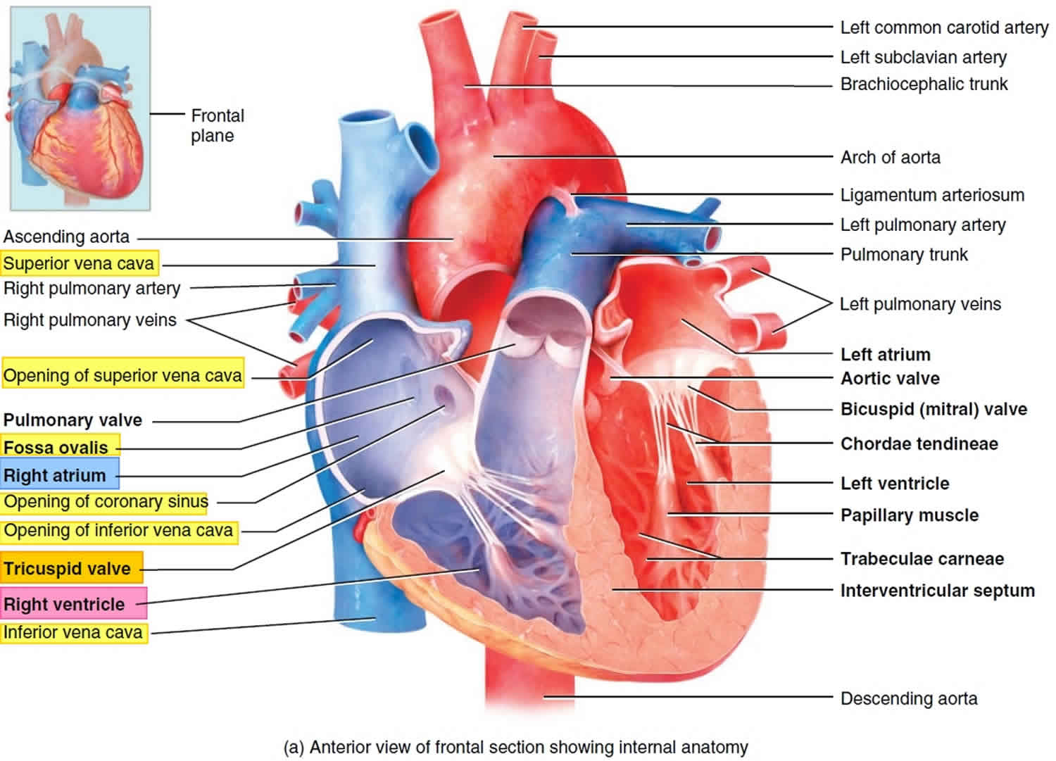
The right atrium is about 2–3 mm (0.08–0.12 in.) in average thickness. The interior of the right atrium is divided into two continuous spaces: a smooth-walled posterior part and an anterior part lined by horizontal muscular ridges called the pectinate muscles (pectin = comb), which also extend into the auricle (Figures 2). Externally, this separation is indicated by a shallow, vertical groove (the sulcus terminalis cordis), which extends from the right side of the opening of the superior vena cava to the right side of the opening of the inferior vena cava. Internally, these two anterior and posterior walls of the right atrium are separated by a large, C-shaped ridge called the crista terminalis (“terminal crest”), which is a smooth, muscular ridge that begins on the roof of the atrium just in front of the opening of the superior vena cava and extends down the lateral wall to the anterior lip of the inferior vena cava. The crista terminalis is an important landmark in locating the sites where veins enter the right atrium: The superior vena cava opens into the atrium just posterior to the superior bend of the crista; the inferior vena cava opens into the right atrium just posterior to the inferior bend of the crista; and the coronary sinus opens into the atrium just anterior to the inferior end of the crista. Additionally, just posterior to this end of the crista is the fossa ovalis (the remnant of the foramen ovale), a depression in the interatrial septum (inter- = between; septum = a dividing wall or partition) that marks the spot where an opening existed in the fetal heart: the foramen ovale, that normally closes soon after birth.
An additional structure in the right atrium is the opening of the coronary sinus, which receives blood from most of the cardiac veins and opens medially to the opening of the inferior vena cava. Associated with these openings are small folds of tissue derived from the valve of the embryonic sinus venosus (the valve of the coronary sinus and the valve of inferior vena cava, respectively) . During development, the valve of the inferior vena cava helps direct incoming oxygenated blood through the foramen ovale and into the left atrium.
Numerous small openings-the openings of the smallest cardiac veins (the foramina of the venae cordis minimae)-are scattered along the walls of the right atrium. These are small veins that drain the myocardium directly into the right atrium.
Blood passes from the right atrium into the right ventricle through a valve that is called the tricuspid valve (tri- = three; -cuspid = point) because it consists of three cusps or leaflets (Figure 2). The tricuspid valve is also called the right atrioventricular valve. The valves of the heart are composed of dense connective tissue covered by endocardium.
Externally the right atrium is a small flap shaped like a dog’s ear (auricle = little ear) called the right auricle, projects anteriorly from the superior corner of the atrium (Figure 1).
Figure 1. Right atrium
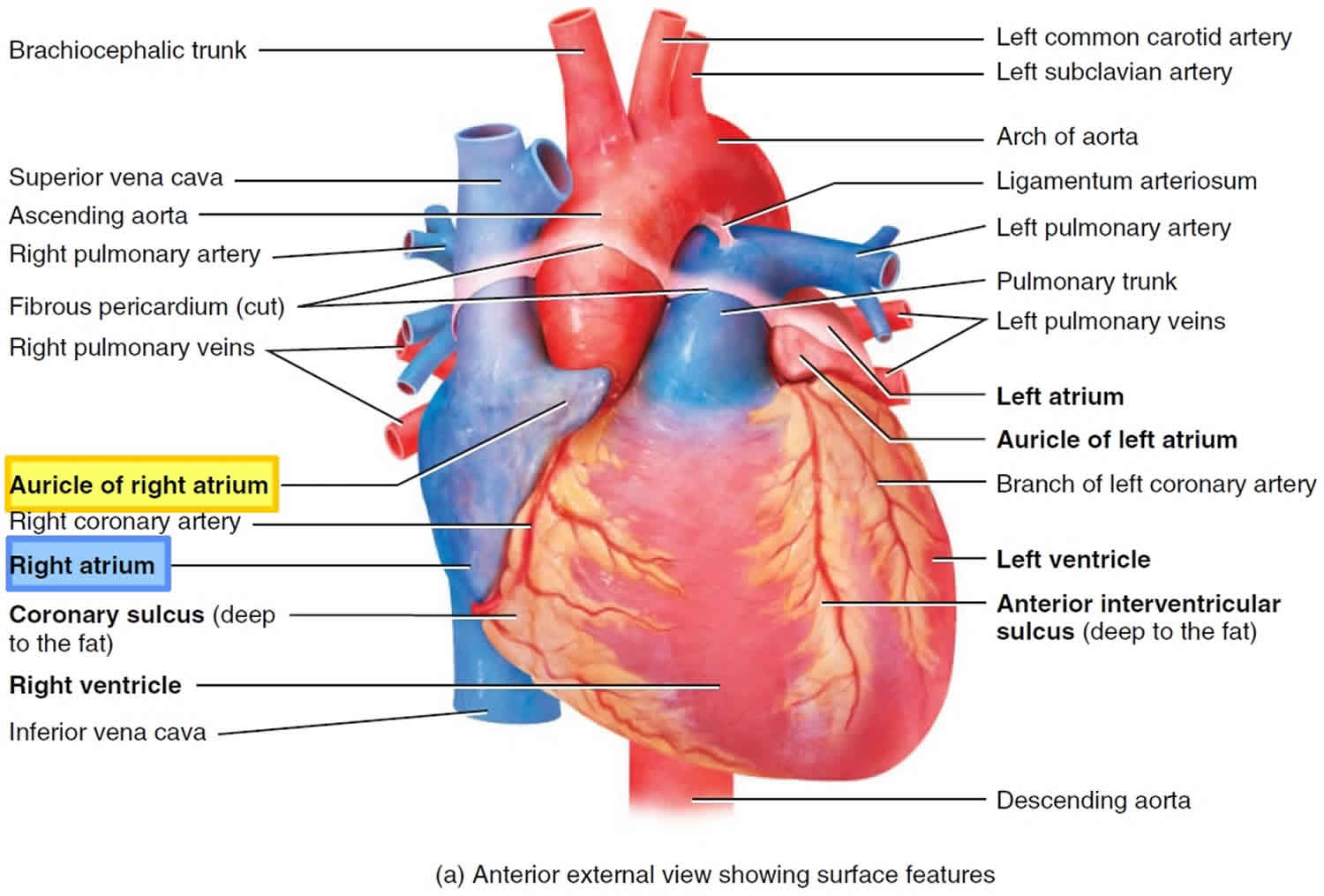
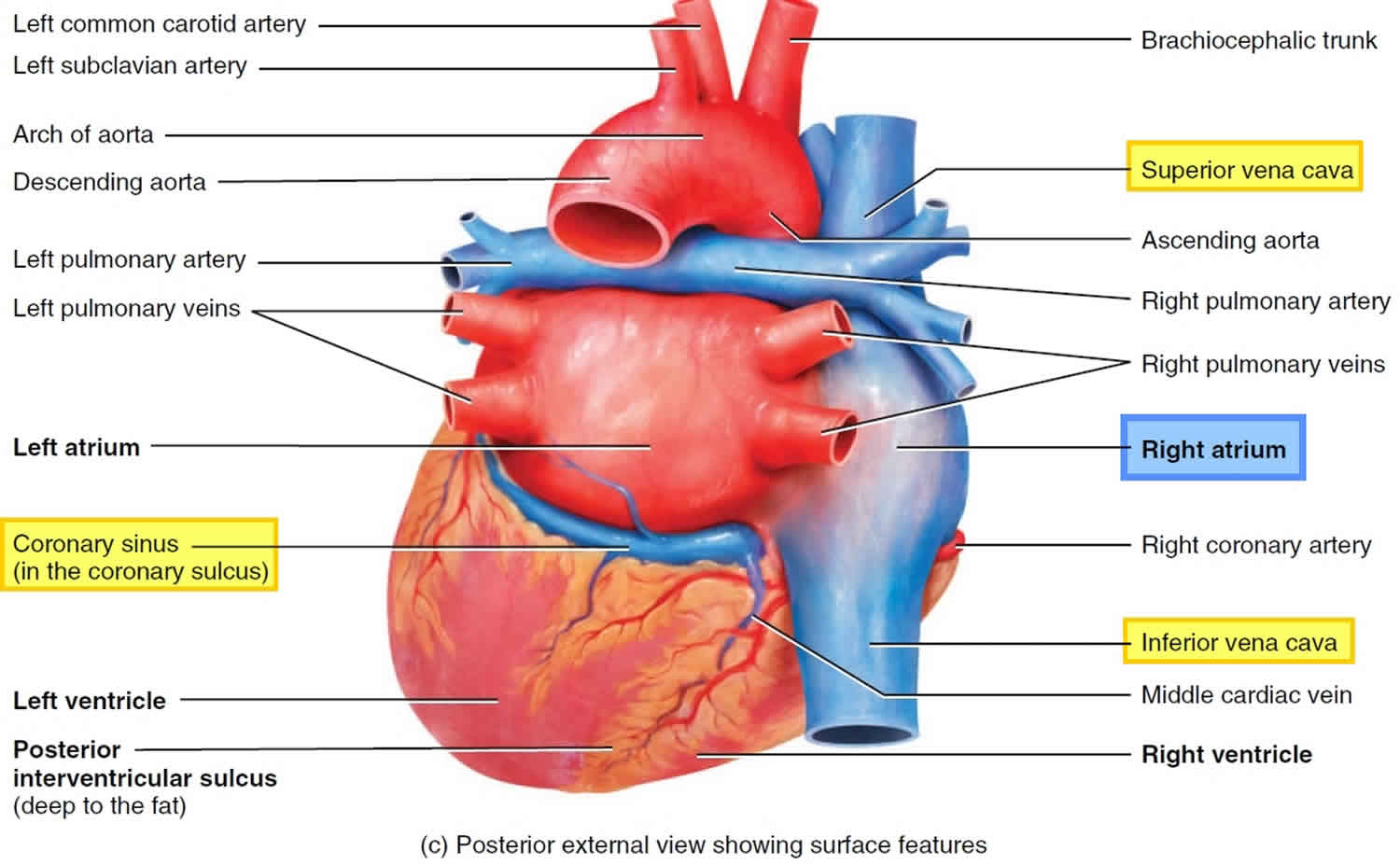
Figure 2. Right atrium anatomy
What is the function of the right atrium?
The heart provides the body’s organs and tissues with a constant supply of blood – and with it vital oxygen and nutrients. You can think of the heart as a central pump that keeps the blood circulating around the body. Your heart is about the same size as your fist and weighs around 300 g (about 0.7 pounds). In people who do endurance sports, it can weigh up to 500 g (about 1.1 pounds). The heart is located more or less in the middle of the chest, slightly to the left, behind the breastbone (sternum). You can normally feel someone’s heart beat if you put your hand on their chest.
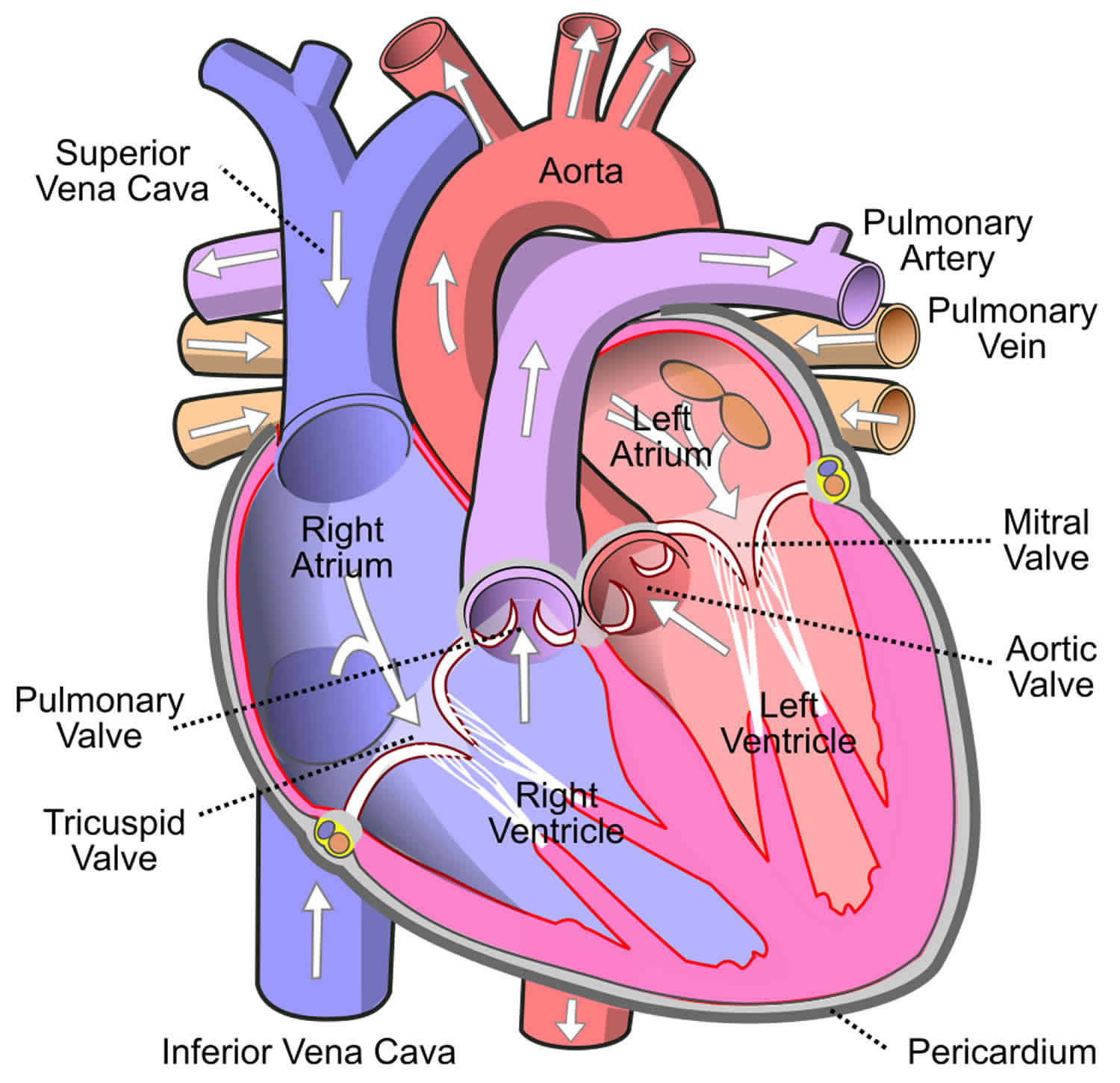
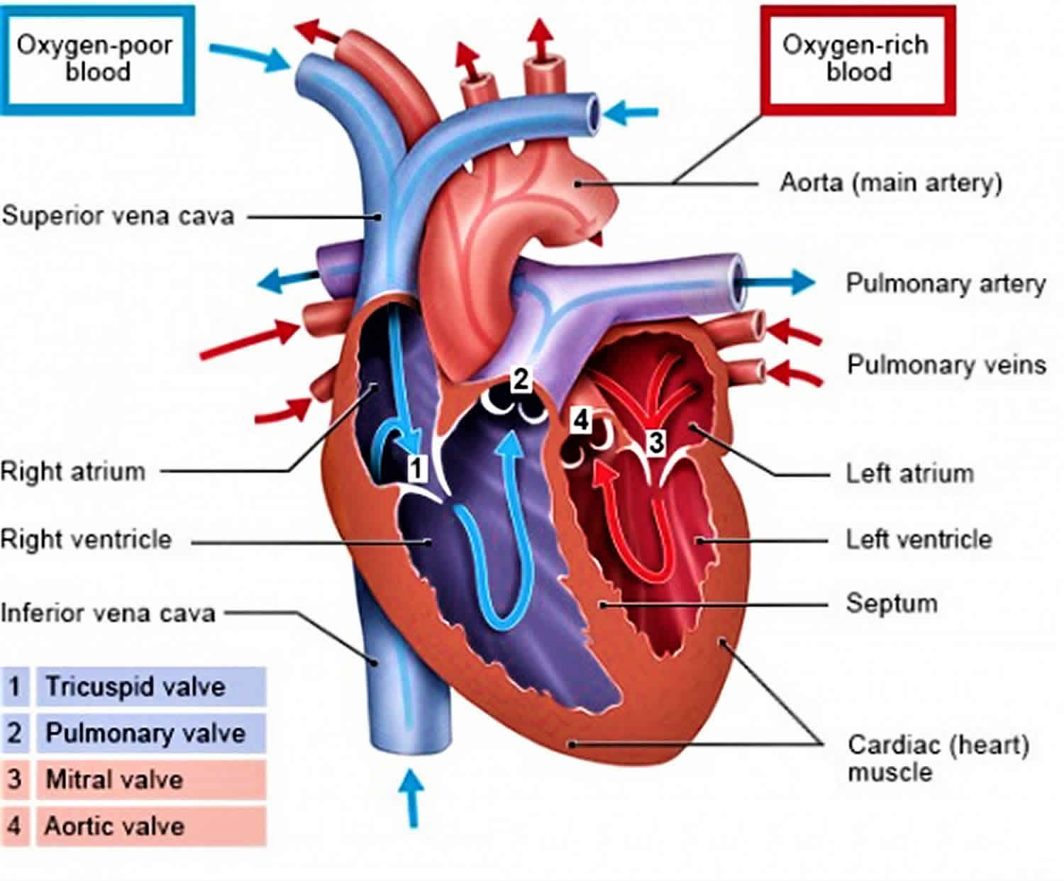
Blood enters the right atrium of the heart through the superior vena cava and inferior vena cava. The right atrium contracts and pushes the blood cells through the tricuspid valve into the right ventricle. The right ventricle then contracts and pushes the blood through the pulmonary valve into the pulmonary artery, which takes it to the lungs. In the lungs, the blood cells exchange carbon dioxide for oxygen. The oxygenated blood returns to the heart via the pulmonary veins and enters the left atrium. The left atrium contracts and pumps the blood through the mitral valve into the left ventricle. Finally, the left ventricle contracts and pushes the blood into the aorta. The aorta branches off into several different arteries that pump the oxygenated blood to various parts of the body.
Your heart’s pumping action is controlled by tiny electrical impulses produced by a part of the right atrium called the sinus node. The sinus node is sometimes called your heart’s ‘natural pacemaker’. The sinus node which is the main pacemaker of the heart is situated at the junction of the superior vena cava and the right atrium. These impulses make the atria (right and left atrium) contract and push blood into the ventricles. The impulses travel to the ventricles through the AV node (atrio-ventricular node). This acts like a junction box and is sometimes called the AV junction. When the impulse reaches the ventricles, the ventricles both contract, pushing the blood out of your heart to your lungs, your brain, and the rest of your body. In a normal heart rhythm, each impulse from the sinus node makes the atria and the ventricles contract regularly.
Figure 3. Right atrium function


