Contents
- What is telangiectasia
- Telangiectasia signs and symptoms
- Classification of telangiectasia
- Hereditary telangiectasia
- Hereditary hemorrhagic telangiectasia causes
- Hereditary hemorrhagic telangiectasia prevention
- Hereditary hemorrhagic telangiectasia symptoms
- Hereditary hemorrhagic telangiectasia possible complications
- Hereditary hemorrhagic telangiectasia diagnosis
- Hereditary hemorrhagic telangiectasia treatment
- Hereditary hemorrhagic telangiectasia prognosis
- Spider telangiectasia
- Are spider telangiectasia hereditary?
- Can a spider telangiectasia be cured?
- What do spider telangiectasia look like?
- Who gets spider telangiectasia
- What causes a spider telangiectasia?
- What are the symptoms of a spider telangiectasia?
- How is a spider telangiectasia diagnosed?
- Spider telangiectasia treatment
- Spider telangiectasia prognosis
- Telangiectasia macularis eruptiva perstans
- Retinal telangiectasia
- Telangiectasia treatment
What is telangiectasia
Telangiectasia is a condition in which there are visible small linear widely open (dilated) blood vessels (broken capillaries) in the outer layer of the skin. Visible small blood vessels that are blue in color (venulectasia) are called spider veins, because venules are involved.
Telangiectasias are very common in healthy people and are usually caused by sun damage or aging. When seen on the legs, they do not necessarily indicate a vein disorder, such as varicose veins or underlying deep vein problems. However, they are seen with a number of diseases, including acne rosacea, birthmarks (e.g., port-wine stains), scleroderma, several types of inherited disorders (ataxia-telangiectasia, hereditary hemorrhagic telangiectasia, xeroderma pigmentosum, and others), or with prolonged use of oral or topical corticosteroids.
If you are concerned about a potential disease causing the telangiectasias, blood tests and evaluations of family members may be done.
If you have acne rosacea, oral or topical antibiotics may be prescribed.
The appearance of telangiectasias may be improved by laser treatments and burning (electrocautery).
Laser treatment of telangiectasias on the leg is more difficult and may leave marks. Injection of chemicals to cause scarring in the blood vessel, thereby closing it (sclerotherapy), is often preferred for treating spider veins.
Self-Care
No treatment is necessary for telangiectasia unless the appearance is bothersome.
You might try the following:
- Use cover-up makeup or self-tanning lotion to cover them up. Waterproof leg makeup is also available.
- If in a sun-exposed area, use sun-protection measures, such as a hat or sunscreen.
- If you have been using topical corticosteroid cream, stop its use on the affected area.
See your doctor if you have a family history of similar marks, any personal or family history of bleeding from the gastrointestinal tract, lesions on the mouth or eye lining, or repeated bleeding from a lesion.
If the lesions are cosmetically displeasing, you may want to seek medical advice for treatment, although treatment for telangiectasias is usually not covered by insurance.
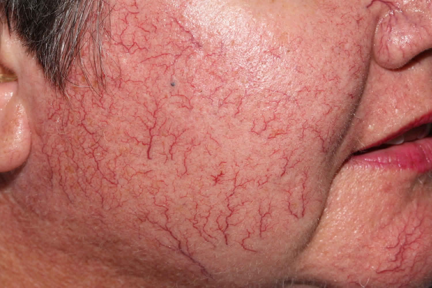
Telangiectasia signs and symptoms
Telangiectasias can be seen anywhere on the body. They are common on the face (nose, cheeks, and chin) and legs (particularly the thighs, just below the knees and the ankles).
Telangiectasias are red, blue, or purple linear marks measuring less than 1–3 mm in width and several millimeters to centimeters in length, and they can disappear temporarily if you press on them with your finger.
Classification of telangiectasia
Inherited telangiectasia
- Hereditary hemorrhagic telangiectasia
- Benign hereditary telangiectasia
- Generalized essential telangiectasia
- Unilateral nevoid telangiectasia
- Angioma serpiginosum
- Rothmund Thompson syndrome
- Ataxia-telangiectasia
- Bloom syndrome (congenial telangiectatic erythema)
- Cockayne syndrome
- Cutis marmorata telangiectatica congenita
- Focal dermal hypoplasia (Goltz syndrome)
- Kindler syndrome
Note that telangiectasia may be noted as a normal feature of facial skin in some families.
Acquired telangiectasia
- Rosacea
- Sun damaged and aged skin especially in those who smoke
- Spider telangiectasis (also called spider naevus or spider angioma)
- Poikiloderma of Civatte (sun damage affecting the sides of the neck)
- Pregnancy
- Liver disease particularly when associated with alcohol or viral infection
- Cushing syndrome
- Systemic sclerosis especiallly CRST syndrome (which forms telangiectatic mats)
- Cutaneous lupus erythematosus
- Mixed connective tissue disease
- Dermatomyositis
- Mastocytosis, specifically the variant, telangiectasia macularis eruptiva perstans
- Carcinoid syndrome
- Necrobiosis lipoidica
- Male genital dysaesthesia
Telangiectasia may follow cutaneous injury. For example:
- Scarring, including hypertrophic and keloid scars
- Livedoid vasculopathy and atrophie blanche
- Radiation damage
- Erythema ab igne
Some tumors are characterized by telangiectasia, such as:
- Sebaceous hyperplasia
- Basal cell carcinoma
- Merkel cell carcinoma
- Kaposi sarcoma
- Cutaneous T-cell lymphoma (CTCL), particularly poikiloderma vasculare variant
- Intravascular B-cell lymphoma.
Certain medications may give rise to telangiectasia:
- Vasodilators especially calcium channel blockers; sun exposed sites are mainly affected
- Long-term systemic corticosteroids
- Long-term topical corticosteroids including steroid rosacea
- Intralesional triamcinolone injections
Figure 1. Telangiectasia face
Hereditary telangiectasia
Hereditary hemorrhagic telangiectasia is known as Osler-Weber-Rendu syndrome, is an inherited disorder of the blood vessels that can cause excessive bleeding. People with hereditary hemorrhagic telangiectasia can develop abnormal blood vessels called arteriovenous malformations (AVMs) in several areas of the body 1. In hereditary hemorrhagic telangiectasia, some arterial vessels flow directly into veins rather than into the capillaries. These abnormalities are called arteriovenous malformations. When they occur in vessels near the surface of the skin, where they are visible as red markings, they are called telangiectasias. Arteriovenous malformations can also develop in other parts of the body, including the brain, lungs, liver, or intestines 2.
Without the normal buffer of the capillaries, the blood moves from the arteries at high pressure into the thinner walled, less elastic veins. The extra pressure tends to strain and enlarge these blood vessels, and may result in compression or irritation of adjacent tissues and frequent episodes of severe bleeding (hemorrhage). Nosebleeds are very common in people with hereditary hemorrhagic telangiectasia, and more serious problems may arise from hemorrhages in the brain, liver, lungs, or other organs.
There are several forms of hereditary hemorrhagic telangiectasia, distinguished mainly by their genetic cause but with some differences in patterns of signs and symptoms. People with type 1 tend to develop symptoms earlier than those with type 2, and are more likely to have blood vessel malformations in the lungs and brain. Type 2 and type 3 may be associated with a higher risk of liver involvement. Women are more likely than men to develop blood vessel malformations in the lungs with type 1, and are also at higher risk of liver involvement with both type 1 and type 2. Individuals with any form of hereditary hemorrhagic telangiectasia, however, can have any of these problems.
Juvenile polyposis/hereditary hemorrhagic telangiectasia syndrome is a condition that involves both arteriovenous malformations and a tendency to develop growths (polyps) in the gastrointestinal tract. Hereditary hemorrhagic telangiectasia types 1, 2 and 3 do not appear to increase the likelihood of such polyps.
Hereditary hemorrhagic telangiectasia is caused by a mutation in one of several genes, including ACVRL1, ENG, SMAD4, and GDF2. Changes in at least other two unknown genes are also suspected of causing hereditary hemorrhagic telangiectasia in some people 3. Hereditary hemorrhagic telangiectasia is inherited in an autosomal dominant pattern, which means a person only needs to inherit one copy of the changed gene to have hereditary hemorrhagic telangiectasia 4.
The incidence of hereditary hemorrhagic telangiectasia is difficult to determine because the severity of symptoms can vary widely and some symptoms, such as frequent nosebleeds, are common in the general population. In addition, arteriovenous malformations may be associated with other medical conditions. Hereditary hemorrhagic telangiectasia is widely distributed, occurring in many ethnic groups around the world. It is believed to affect between 1 in 5,000 and 1 in 10,000 people.
While there is no cure for hereditary hemorrhagic telangiectasia, treatment is symptomatic and supportive, with a focus on controlling bleeding and preventing medical complications, either through surgery or medication 2. People with severe nosebleeds may need emergency nasal packing, where the nose is packed with ribbon gauze or a special nasal sponge. Some people may need to see an ear, nose and throat specialist for treatment. Laser therapy may help. More severe cases may be treated with skin grafting or other surgery. If a lot of blood has been lost from bleeding inside the body or after nosebleeds, a blood transfusion may be needed.
If you have regular nosebleeds you will probably lose a lot of iron through this loss of blood, especially if you also bleed from telangiectasia in the gut. It’s important to replace the lost iron with iron supplements. Dietary changes alone may not be enough.
Figure 2. Hereditary hemorrhagic telangiectasia

Footnote: Clinical manifestations of telangiectasias. (A) Small red telangiectasias are often seen on the skin of hereditary hemorrhagic telangiectasia patients. (B) Similar lesions may be present on the tongue, lips, or palate.
[Source 5 ]Hereditary hemorrhagic telangiectasia causes
Mutations in several genes, including the ACVRL1, ENG, and SMAD4 genes, cause hereditary hemorrhagic telangiectasia.
Hereditary hemorrhagic telangiectasia type 1 is caused by mutations in the ENG gene. Type 2 is caused by mutations in the ACVRL1 gene. Juvenile polyposis/hereditary hemorrhagic telangiectasia syndrome is caused by mutations in the SMAD4 gene. All these genes provide instructions for making proteins that are found in the lining of the blood vessels. These proteins interact with growth factors that control blood vessel development. Mutations in other genes, some of which have not been identified, account for other forms of hereditary hemorrhagic telangiectasia.
Mutations in these genes generally prevent the production of the associated protein or result in the production of a defective protein that cannot fulfill its function. An individual with a mutated gene will therefore have a reduced amount of the functional protein available in the tissue lining the blood vessels. This shortage is believed to result in the signs and symptoms of hereditary hemorrhagic telangiectasia.
Hereditary hemorrhagic telangiectasia inheritance pattern
Hereditary hemorrhagic telangiectasia is inherited in an autosomal dominant pattern, which means one copy of the altered gene in each cell is sufficient to cause the disorder.
Hereditary hemorrhagic telangiectasia prevention
Genetic counseling is recommended for couples who want to have children and who have a family history of hereditary hemorrhagic telangiectasia. If you have this condition, medical treatments can prevent certain types of strokes and heart failure.
Hereditary hemorrhagic telangiectasia symptoms
People with hereditary hemorrhagic telangiectasia can develop abnormal blood vessels in several areas of the body. These vessels are called arteriovenous malformations (AVMs).
If they are on the skin, they are called telangiectasias. The most common sites include the lips, tongue, ears, and fingers. The abnormal blood vessels can also develop in the brain, lungs, liver, intestines, or other areas.
Symptoms of hereditary hemorrhagic telangiectasia include:
- Frequent nosebleeds in children
- Bleeding in the gastrointestinal tract (GI), including loss of blood in the stool, or dark or black stools
- Seizures or unexplained, small strokes (from bleeding into the brain)
- Shortness of breath
- Enlarged liver
- Heart failure
- Anemia caused by low iron
Hereditary hemorrhagic telangiectasia possible complications
These complications can occur:
- Heart failure
- High blood pressure in the lungs (pulmonary hypertension)
- Internal bleeding
- Shortness of breath
Hereditary hemorrhagic telangiectasia diagnosis
The health care provider will perform a physical examination and ask about your symptoms. An experienced provider can detect telangiectases during a physical examination. There is often a family history of this condition.
Tests include:
- Blood gas tests
- Blood tests
- Imaging test of the heart called an echocardiogram
- Endoscopy, which uses a tiny camera attached to a thin tube to look inside your body
- MRI to detect AVMs in the brain
- CT or ultrasound scans to detect AVMs in the liver
Genetic testing is available to look for changes in genes associated with this syndrome.
Hereditary hemorrhagic telangiectasia treatment
Although current treatment cannot stop telangiectasias or arteriovenous malformations from forming, many of the symptoms and complications associated with hereditary hemorrhagic telangiectasia can be treated or prevented 6. Management of hereditary hemorrhagic telangiectasia includes checking for new or worsening arteriovenous malformations (AVMs) and the treatment of complications such as nosebleeds, bleeding from the intestines or stomach, and anemia. Treatment of arteriovenous malformations (AVMs) of the lung (pulmonary), brain (cerebral) and liver (hepatic) may also be recommended 3.
Treatments may include:
- Surgery to treat bleeding in some areas
- Electrocautery (heating tissue with electricity) or laser surgery to treat frequent or heavy nosebleeds
- Endovascular embolization (injecting a substance through a thin tube) to treat abnormal blood vessels in the brain and other parts of the body
Reducing the number and severity of nosebleeds can help prevent anemia. Treatment of nosebleeds may include using a vaporizer to increase the moisture of room air and keeping the inside of nose moist using nasal lubricants or sprays. Laser therapy may be used to remove the abnormal blood vessels (laser ablation). Other treatment may include medication or hormone therapy, but the effectiveness and safety of these treatments has not been established. Some people respond to estrogen therapy, which can reduce bleeding episodes. Iron may also be given if there is a lot of blood loss, leading to anemia. Avoid taking blood-thinning medicines. Some drugs that affect blood vessel development are being studied as possible future treatments.
Some people may need to take antibiotics before having dental work or surgery.
If the nosebleeds continue despite other treatment, skin from a different part of the body may be grafted to replace the thin lining of the nose (septal dermoplasty) in an effort to cover and protect the fragile telangiectases 3.
Bleeding in the intestine or stomach is usually only treated if oral iron supplements cannot keep iron levels high enough to avoid anemia. Treatment may include surgical removal of arteriovenous malformations (AVMs) or laser therapy to destroy and close the arteriovenous malformations (AVMs). If severe bleeding with uncontrolled anemia develops, treatment of arteriovenous malformations (AVMs) in the stomach or intestine may include medication or hormone therapy, but the effectiveness and safety of these treatments has not been established. In addition to oral iron supplements, anemia may be treated by intravenous (IV) iron therapy or, in more severe cases, red blood cell transfusions 3.
People with lung arteriovenous malformations (AVMs) should avoid scuba diving to prevent decompression sickness (the bends). Ask your provider what other precautions you should take.
Treatment of arteriovenous malformations (AVMs) of the lungs (pulmonary arteriovenous malformations) is recommended if the person with hereditary hemorrhagic telangiectasia is having a hard time breathing (dyspnea), is unable to exercise without extreme fatigue (exercise intolerance), or has low blood oxygen levels (hypoxemia). Treatment of pulmonary arteriovenous malformations (AVMs) may also be performed to prevent lung hemorrhage and the neurologic complications of brain abscesses and/or stroke. Treatment may include inserting a small inflated balloon or small metal coil in the artery that leads into the arteriovenous malformation in order to stop the blood flow through the arteriovenous malformation (embolotherapy) or surgical removal of the arteriovenous malformation 3. People with pulmonary arteriovenous malformations (AVMs) are advised to take extra precautions to avoid serious complications. These recommendations include taking antibiotics before dental or surgical procedures, using special filters in IV lines to prevent even tiny air bubbles from entering the blood stream, avoiding blood thinners and non-steroidal anti-inflammatory drugs (such as aspirin, ibuprofin, and naproxen), and regular monitoring by a doctor familiar with hereditary hemorrhagic telangiectasia 6.
Arteriovenous malformations (AVMs) in the brain (cerebral arteriovenous malformations) greater than 1.0 cm in diameter may be surgically removed. Alternative treatment includes inserting a small inflated balloon or glue-like substance to stop the blood flow through the artery involved in the arteriovenous malformation (embolotherapy) and/or using a narrow, focused beam of radiation to destroy the arteriovenous malformation (stereotactic radiosurgery) 6.
Arteriovenous malformations (AVMs) in the liver (hepatic arteriovenous malformations) are currently treated only if a person shows signs of heart failure or significant health problems related to the liver not working properly. Treatment might include standard heart failure medications, liver transplantation, or medications like bevacizumab 6.
In addition, guidelines for people with hereditary hemorrhagic telangiectasia recommend annual evaluations for anemia and neurologic conditions and re-evaluation for pulmonary arteriovenous malformations (AVMs) every one to two years during childhood and every five years thereafter. Blood tests to check for anemia may be recommended more often depending on the frequency and severity of nose bleeds or if an intestinal or stomach arteriovenous malformation is bleeding. Women with hereditary hemorrhagic telangiectasia considering pregnancy are screened and treated for pulmonary arteriovenous malformations (AVMs); if pulmonary arteriovenous malformations (AVMs) are discovered during pregnancy, they are treated during the second trimester 3.
Hereditary hemorrhagic telangiectasia prognosis
People with hereditary hemorrhagic telangiectasia can live a completely normal lifespan, depending on where in the body the arteriovenous malformations (AVMs) are located.
Spider telangiectasia
A spider telangiectasia is also called spider angioma (a misnomer), spider nevus, arterial spider, vascular spider and naevus araneus, is composed of dilated blood vessels, and is clinically characterized by its spider-like appearance. Spider telangiectasia is given that name because it has a central red papule (the body of the ‘spider’) from which fine red lines (smaller blood vessels) resembling the spider’s legs extend radially. An alternative explanation for the name explains that the red lines form a spider-like network.
Some of these names are Latin: ‘araneus’ for ‘spider’ and ‘angioma’ for ‘blood vessel’. ‘Naevus’ means an increase in normal or healthy tissue within the skin.
Spider telangiectasia can develop at any age, but are more common in children. The vast majority affect healthy people, and most people have only one spider telangiectasia or a just a few.
The majority of spider telangiectases are on the face, upper chest, back and upper arms. In children, the back of the hands are also often affected.
Spider telangiectasia are of cosmetic concern only and so are not usually treated. In cosmetic clinics, the central artery can be treated with an electric current (‘electrodessication’), causing it to dry up. A vascular laser such as the pulsed dye laser or KTP (potassium titanyl phosphate) laser can target the blood in the central small artery, causing it to shrink.
These treatments may hurt but do not usually need any local anesthetic. They may leave a small permanent scar like a dent in the skin, which is less common after laser treatment than after electrodessication.
About a third of spider telangiectasia come back after treatment.
Cosmetic camouflage can be useful if there are many spider telangiectasia which are causing cosmetic concern. Camouflage is a type of special make-up, which is matched to the color of the person’s skin and which is water resistant.
Are spider telangiectasia hereditary?
Spider telangiectases are very common and affect at least one in ten of healthy adults and are even more common in children. Spider telangiectases do not run in families.
A spider telangiectasia is not contagious or cancerous.
Can a spider telangiectasia be cured?
In children and some adults, spider telangiectasia may go away on their own, which can take several years. Treatment is usually not necessary.
If spider telangiectases are related to increased estrogen hormones and the levels then go back to normal (after a pregnancy or on stopping an oral contraceptive pill), the spider telangiectasia may go away within about nine months.
A spider telangiectasia can also completely disappear after treatment, but sometimes repeated treatments may be required. The problem may come back a few months later after treatment.
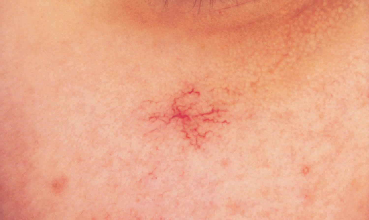
What do spider telangiectasia look like?
Spider telangiectases are often located on the face, neck, and upper chest (this has been postulated to relate to the distribution of a large vein draining the heart, the superior vena cava). Spider telangiectases may also occur on the hands, arms, or other sites. Spider telangiectases vary in size and number, tending to be larger and more numerous in people with severe liver disease when other cutaneous signs of liver disease may be present such as palmar erythema, leukonychia, and jaundice. A central dilated arteriole may be present without radial capillaries, and the capillaries may vary in diameter, length, and number. They can also be star-shaped.
Spider telangiectasia lesions may briefly bleed on trauma but otherwise do not cause any symptoms or complications.
Figure 3. Spider telangiectasia
Who gets spider telangiectasia
A solitary spider telangiectasia is common in children and adults, affecting 10–15% of the population. Although spider telangiectases can affect people of any race, spider telangiectases are more easily seen in fair skin compared to skin of color. Multiple spider telangiectases arise most frequently in pregnancy, in women taking a combined oral contraceptive pill, in patients with liver disease (particularly, in cirrhosis due to alcohol abuse), and in those with thyrotoxicosis.
What causes a spider telangiectasia?
The cause of spider telangiectasia is not known. Spider telangiectasia is an acquired vascular malformation. It occurs because of the failure of a tiny smooth muscle restricting the size of an arteriole. Increased pulsating flow through the vessel (the central papule) results in the dilatation of distal vessels (the red lines).
Spider telangiectasia may arise spontaneously or may be induced by circulating estrogen, which is increased in pregnancy, women taking combined oral contraceptive pill and in those with liver or thyroid disease. Various vascular endothelial growth factors may be involved.
What are the symptoms of a spider telangiectasia?
Apart from its appearance, a spider telangiectasia does not usually cause any symptoms. Bleeding from a spider telangiectasia is unusual but may occur if picked or scratched.
How is a spider telangiectasia diagnosed?
A spider telangiectasia is diagnosed by its typical appearance. Compression of the central arteriole results in the disappearance of the radial capillaries, which rapidly refill when the compression is relieved. This is best seen through a transparent object, such as a glass slide or the lens of a contact dermatoscope.
Spider telangiectasia treatment
A spider telangiectasia is harmless and does not require treatment. A lesion that is unsightly can be removed by destroying the feeding central arteriole, but this may result in a small scar. Treatments to remove spider telangiectasia include:
- Cryotherapy
- Electrocautery
- Intense pulsed light
- Vascular laser.
Surgical excision is rarely necessary and inevitably leaves a scar.
Spider telangiectasia prognosis
Spider telangiectases can persist or disappear. In women, estrogen-induced telangiectases often disappear within 6 months after having a baby or of stopping the combined contraceptive pill. Spider telangiectasia can also reappear after initially successful treatment.
Telangiectasia macularis eruptiva perstans
Telangiectasia macularis eruptiva perstans, described by Parkes Weber in 1930, represents a rare form of cutaneous mastocytosis 7 and accounts for less than 1 percent of cases of mastocytosis 8. The typical lesions are telangiectatic macules with background color ranging from light to dark brown. Telangiectasia macularis eruptiva perstans develops more frequently in adults, although there are reports of cases affecting children 9. Telangiectasia macularis eruptiva perstans presents most often in adulthood and clinically is comprised of tan macules and small patches with telangiectases that most often involve the trunk 10. On dermatoscopic examination, the telangiectases are aligned in a reticular pattern 11. Most patients have only cutaneous findings with variable degrees of pruritus; the lesions typically do not urticate 12.
Mastocytosis is characterized by an increase in the number of mast cells in one or more organs. In the skin, more than 20 mast cells per high power field establishes a diagnosis. However, if lesional skin is shown to have more mast cells when compared to the normal skin, the diagnsosis of cutaneous mastocytosis also may be made. A mutation in the c-kit oncogene results in mast cell hyperplasia, but the role of c-kit mutations in telangiectasia macularis eruptiva perstans are limited to case reports 13.
Despite most cases being limited to the skin, telangiectasia macularis eruptiva perstans lesions on the chest and extremities, systemic involvement may occur. Bone marrow, gastrointestinal tract, liver, spleen, and lymph node involvement are not uncommon. Hematological abnormalities may also be present and the association with multiple myeloma and polycythemia vera has been reported 14. Histologically, the lesions are characterized by an infiltrate consisting of mast cells around venules and dilated capillaries of the superficial venous plexus of the dermis 15. The treatment is based on the symptoms presented by the patient, and there is no gold standard treatment to date 16.
In the past five years, there have been case reports of telangiectasia macularis eruptiva perstans that present in association with an underlying illness, such as myelodysplasia, Sjögren syndrome, and renal cancer 17. It is unclear if these patients demonstrated the rare systemic symptoms that are associated with telangiectasia macularis eruptiva perstans, such as flushing, diarrhea, headaches, and abnormal skeletal surveys. Although telangiectasia macularis eruptiva perstans is a cutaneous manifestation of mastocytosis, systemic nvolvement may occur. Tryptase levels may serve as a guide to establish this involvement 18. In the absence of systemic involvement, there is no need for the treatment of telangiectasia macularis eruptiva perstans. Patients who experience burning and pruritus of their lesions should be counseled about avoiding agents that are known to be mast cell degranulators. Nonetheless, the literature highlights several successful modalities that are used to treat telangiectasia macularis eruptiva perstans successfully. A recent publication reports the successful use of PUVA photochemotherapy in treating the stinging sensation associated with telangiectasia macularis eruptiva perstans 19. Montelukast also has been used 20. For cosmetic improvement of lesions, a variety of lasers have been used, which include the 585 nm flash-pumped dye laser in two patients and total beam radiation in one patient 21.
Figure 4. Telangiectasia macularis eruptiva perstans
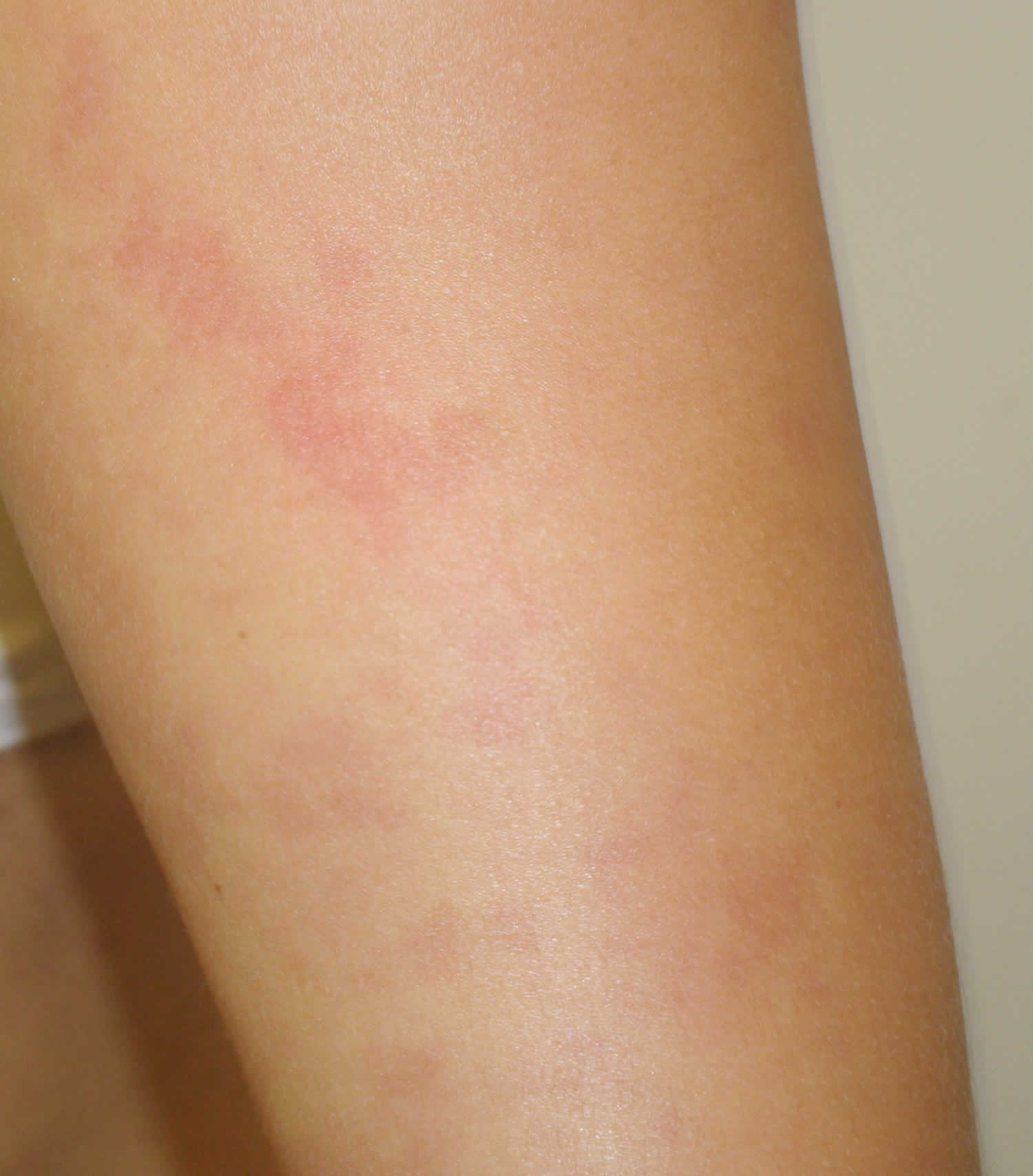
Footnote: A 31-year-old woman presented to Bellevue Hospital Center with a ten-year history of blemishes that were distributed mostly on her limbs. The eruption started on the arms and began to involve her legs over the course of years. Recently, new lesions appeared on her back. The eruption is mildly pruritic and is fixed in place. She denied headache, irritability, flushing, rhinorrhea, bone pain, nasuea, vomiting, diarrhea, and abdominal pain. She denied symptoms when taking non-steroidal anti-inflammatory agents or while drinking alcohol. She did not recall having a reaction to any type of antibiotics and had never taken morphine. The patient had no family history of any skin disease. A biopsy was consistent with telangiectasia macularis eruptiva perstans. There were no signs and symptoms of systemic involvement despite the large body surface area of involvement.
[Source 22 ]Retinal telangiectasia
Macular telangiectasia leads to abnormalities of capillaries of the fovea or perifoveal region associated with loss outer nuclear layers and ellipsoid zone that can progress to cystic cavitation-like changes in all retinal layers, or development of full-thickness macular hole or subretinal neovascularization in advanced stages 23. First described by Gass in 1968 as a different entity from Coats disease.
Macular telangiectasia is usually divided into 3 main groups:
- Type 1: congenital and unilateral. Thought to be similar to and/or possibly a variant of Coats disease. Uncommon.
- Type 2: acquired and bilateral. The most common form of the three types. Usually found in middle-aged or older patients. According to the Beaver Dam Eye Study which graded only color fundus images, macular telangiectasia has a prevalence of 0.1% with an average age of 63 years. Although not initially described by Gass, Yannuzzi found a slight female preponderance of 58%. Of note, on common usage, the term ‘Mac Tel’ is often used to refer to macular telangiectasia type 2. This entry will largely focus on type 2 as it has the most clinical relevance.
- Type 3: an poorly understood primarily occlusive phenomena which is quite rare.
What causes macular telangiectasia
The Macular Telangiectasia Project found a prevalence of diabetes mellitus of 28% and hypertension of 52% in macular telangiectasia type 2. Such high prevalence of diabetes mellitus, high blood pressure and the age of onset of the disease can point toward a long-term vascular stress as the etiology for this entity. These vascular changes are associated with disruption of the outer nuclear layer, ellipsoid zone and lead to pseudolamellar macular holes. Retinal pigment epithelium (RPE) hyperplasia and migration into the inner retinal layers is also seen with time.
Gass postulated that Muller cells and parafoveal neural abnormalities or degeneration may be the primary sites initially affected. Histopathology has shown disruption of pericytes, ectatic vessels, with dilated venules diving at a right angle into deeper retinal layers with evidence of retinal pigment epithelium (RPE) hyperplasia along them. Vascular occlusion, inflammatory changes and other neurodegenerative processes lead to loss of the outer nuclear layer, ellipsoid zone with later formation of cysts that can encompass all retinal layers.
Macular telangiectasia pathophysiology
Chronic neurodegenerative processes, vascular inflammation, occlusion and ectatic capillary changes lead to loss of outer retinal layers and formation of pseudolamellar macular holes with cysts or cavitations that can also be in inner retinal layers. Patients can develop subretinal neovascularization temporal to the fovea and can lead to exudates, hemorrhages and in late stages a disciform scar.
Risk factors for macular telangiectasia
Risk factors include age as it is mainly encountered in middle-aged and older patients. Higher prevalence of hypertension and diabetes mellitus have been found and could play a part in association with neurodegenerative processes.
Macular telangiectasia prevention
No prevention has been shown to be beneficial, however most clinicians would recommend control of hypertension and diabetes mellitus if present as these two diseases have been shown to be more prevalent in macular telangiectasia.
Macular telangiectasia signs and symptoms
If asked, most patients with macular telangiectasia type 2 will complain of metamorphopsia or scotoma. With progression of the disease, an increased scotoma can be perceived with decreased visual acuity as this entity affects the fovea and perifoveal area. However visual acuity rarely reaches legal blindness, with 50% keeping 20/32 or better vision according to the Macular Telangiectasia Project. Vision can decrease if a macular hole develops. Vision can be severely diminished in macular telangiectasia type 1 and 3.
Macular telangiectasia diagnosis
The diagnosis of macular telangiectasia is made by ophthalmic clinical exam and ophthalmic imaging. Symptoms and history can help in diagnosis. No labratory test are helpful in diagnosis.
Physical examination
In macular telangiectasia type 2, fundoscopic exam is initially significant for a decrease of foveal pit, with subsequent foveal or perifoveal ectatic vessels with possible presence of venules diving at a right angle into deeper retinal layers. Grayish discoloration of temporal perifoveal area is the earliest sign present. Ectatic vessels with evidence of venules diving at a right angle into deeper retinal layers. Of note, these findings may be subtle on routine retinal exam. Subretinal neovascularization temporal to fovea may be seen in this disease and can be associated with exudates or hemorrhage. RPE hyperplasia can also be seen at the fovea or temporal perifovea. Also, in later stages, pseudolamellar holes can develop into full-thickness macular holes.
Imaging
Fluorescein angiography (FA) shows temporal foveal telangiectatic vessels in macular telangiectasia type 2 that leak in later stages. Evidence of venules diving at a right angle into deeper retinal layers can also be seen. Subretinal neovascularization is evidenced to be retinal in origin as FA shows a feeding arteriole and a draining venule.
Optical coherence tomography (OCT) shows temporal foveal pit enlargement secondary to loss of outer nuclear layer and ellipsoid zone that can progress into large cysts (often called ‘cavitation’) that can encompass all retinal layers. Often only the internal limiting membrane is left in place over these areas, lending to the term of an “ILM drape.” Hyperreflective areas on OCT correlate to areas of RPE hyperplasia and migration.
Fundus autofluorescence (FAF) shows decreased foveal hypofluorescence secondary to loss of foveal luteopigment. Areas of retinal pigment epithelium (RPE) hyperplasia will appear as hypofluorescent spots on Fundus autofluorescence.
Macular telangiectasia treatment
There has been no randomized clinical study for this entity. In non-neovascular cases, laser, intravitreal anti-vascular endothelial growth factor (VEGF) or steroid has not been shown to be effective in controlling the disease. For neovascular cases, intravitreal anti-VEGF are the mainstay of treatment, although transpupillary therapy and photodynamic therapy have been used in the past. Neuroprotective agents are currently being investigated. A Phase II trial studying intraocular delivery of ciliary neurotrophic factor (CNTF) in eyes with macular telangiectasia found decreased ellipsoid zone loss, increased macular thickness, and stable reading speed when compared to controls. Phase 3 trial is currently underway. Associated full thickness macular holes may be treated with pars plana vitrectomy surgery, membrane peeling and gas tamponade, but are likely to have a lower-than-average closure rate.
Macular telangiectasia prognosis
According to the macular telangiectasia Project, 50% of type 2 patients with have a visual acuity of 20/32 or better, with a mean visual acuity of 20/40. macular telangiectasia can rarely lead to 20/200 or worse vision when associated with full-thickness macular hole or subretinal neaovascularization that can lead to a disciform scar in very advanced stages.
Telangiectasia treatment
Telangiectases are generally harmless. Treatment may be sought because of bleeding or unsightly appearance. Facial red vein treatment methods include:
- Electrosurgery
- Intense pulsed light (IPL)
- Vascular laser treatment
- Sclerotherapy.
Laser treatment for telangiectasia
Telangiectasia on the skin or in the lining of the nose can sometimes be improved with vascular laser or intense pulsed light (IPL) treatment:
- for the skin, you’ll usually need a referral to a dermatologist – this may be expensive as two to four treatments a year may be necessary
- for the nose, you’ll need a referral to an ear, nose and throat (ENT) specialist
Laser and intense pulsed light machines produce narrow beams of light aimed at the visible blood vessels in the skin. The heat from the lasers makes the veins shrink so they’re no longer visible. This should leave minimal scarring or damage to the surrounding area.
Laser treatment can be uncomfortable, but most people don’t need local anesthetic.
Treating arteriovenous malformations inside the body
Arteriovenous malformations (AVMs) in the body may require specialist treatment. For example, arteriovenous malformations (AVMs) in the lung are usually treated, even if they’re not causing any apparent problems. They’re treated by embolisation, a procedure that blocks the blood supply to the arteriovenous malformation. A tiny plug is inserted inside the artery supplying the abnormal blood vessel.
Embolization is preferred to open surgery and is normally carried out under sedation (which helps relax you during the procedure).
For brain arteriovenous malformations (AVMs), embolisation, surgery and stereotactic radiotherapy (precisely delivering radiation to the blood vessel) are possible treatment options. Liver arteriovenous malformations (AVMs) may require different types of specialist treatments.
However, these treatments aren’t always appropriate. Your specialist will explain why many arteriovenous malformations (AVMs), such as those in the liver and in the brain, may be better left alone. The risks of treatments may outweigh the potential benefits.
- Hereditary hemorrhagic telangiectasia. https://rarediseases.info.nih.gov/diseases/6626/hereditary-hemorrhagic-telangiectasia[↩]
- Hereditary hemorrhagic telangiectasia. https://medlineplus.gov/ency/article/000837.htm[↩][↩]
- McDonald J, Pyeritz RE. Hereditary Hemorrhagic Telangiectasia. 2000 Jun 26 [Updated 2017 Feb 2]. In: Adam MP, Ardinger HH, Pagon RA, et al., editors. GeneReviews® [Internet]. Seattle (WA): University of Washington, Seattle; 1993-2019. Available from: https://www.ncbi.nlm.nih.gov/books/NBK1351[↩][↩][↩][↩][↩][↩]
- Hereditary hemorrhagic telangiectasia. https://ghr.nlm.nih.gov/condition/hereditary-hemorrhagic-telangiectasia[↩]
- Hereditary hemorrhagic telangiectasia: diagnosis and management from the hematologist’s perspective. Athena Kritharis, Hanny Al-Samkari, David J Kuter. Haematologica Sep 2018, 103 (9) 1433-1443; DOI: 10.3324/haematol.2018.193003 http://www.haematologica.org/content/103/9/1433[↩]
- Diagnosis & Treatment. Cure HHT. https://curehht.org/understanding-hht/diagnosis-treatment[↩][↩][↩][↩]
- Costa DL, Moura HH, Rodrigues R, Pineiro-Maceira J, Ramos-E-Silva M. Telangiectasia macularis eruptiva perstans: a rare form of adult mastocytosis. J Clin Aesthet Dermatol. 2011;4(10):52-4. https://www.ncbi.nlm.nih.gov/pmc/articles/PMC3196299/[↩]
- Cohn MS, Mahon MJ. Telangiectasia macularis eruptiva perstans. J Am Osteopathic Assoc 1994;94:246[↩]
- Rishpon A, Matz H, Gat A, Brenner S. Telangiectasia macularis eruptiva perstans: unusual presentation and treatment. Skinmed. 2006;5(6):300–302.[↩]
- Tharp, M.D. Mastocytosis. In: Bolognia JJ, et al. Mastocytosis. Dermatology. London: Mosby Elsevier 2008:1848[↩]
- Akay BN, et al. Dermatoscopic findings of cutaneous mastocytosis. Dermatology 2009;218:226[↩]
- Soter NA. The skin in mastocytosis. J Invest Dermatol 1991;96:32[↩]
- Lee HW, et al. Two cases of telangiectasia macularis eruptiva perstans demonstrated by immunohistochemistry for c-kit (CD 117). J Dermatol 2005;32:817[↩]
- Sotiriou E, Apalla Z, Ioannides D. Telangiectasia macularis eruptive perstans successfully treated with PUVA therapy. Photodermatol Photoimmunol Photomed. 2010;26(1):46–47.[↩]
- Soter NA. The skin in mastocytosis. J Invest Dermatol. 1991;96(3 Suppl):32S–39S.[↩]
- Nguyen NQ. Telangiectasia macularis eruptiva perstans. Dermatol Online J. 10(3):1.[↩]
- Turchin I, Irani AM. Unusual cutaneous findings of urticaria pigmentosa and telangiectasia macularis eruptiva perstans associated with marked myelofibrosis. Int J Dermatol 2006;45:1215[↩]
- Schwartz LB, Irani AM. Serum tryptase and the laboratory diagnosis of systemic mastocytosis. Hematol Oncol Clin North Am 2000;14:641[↩]
- Sotiriou E, et al. Telangiectasia macularis eruptive perstans successfully treated with PUVA therapy. Photodermatol Photoimmunol Photomed. 2010;26:46[↩]
- Cengizlier R, et al. Treatment of telangiectasia macularis eruptive perstans with montelukast. Allergol Immunopathol (Madr). 2009;37:334[↩]
- Monahan TP, Petropolis AA. Treatment of telangiectasia macularis eruptiva perstans with total skin electron beam radiation. Cutis 2003; 71:357.[↩]
- Altiner, A., Tzu, J., Patel, R., Meehan, S., & Sanchez, M. (2011). Telangiectasia macularis eruptiva perstans. Dermatology Online Journal, 17(10). Retrieved from https://escholarship.org/uc/item/7pc4764x[↩]
- Macular telangiectasia. http://eyewiki.org/Macular_telangiectasia[↩]


