What are tendons
Tendons are connective tissues that attach muscles to bones and and transfer muscular tension to bones.
Ligaments are structurally similar to tendons that connect bones to other bones and tightly bind bones together and resist stress.
Both tendons and ligaments are dense regular connective tissue, because of its two properties: (1) The collagen fibers are closely packed (dense) and leave relatively little open space, and (2) the fibers are parallel to each other (regular). The parallel arrangement of fibers is an adaptation to the fact that musculoskeletal stresses pull tendons and ligaments in predictable directions. With minor exceptions such as blood vessels and sensory nerve fibers, the only cells in this tissue are fibroblasts, visible by their slender, violet-staining nuclei squeezed between bundles of collagen. This type of tissue has few blood vessels, so injured tendons and ligaments are slow to heal.
Muscles have two forms of attachment to bones—direct and indirect.
In a direct (fleshy) attachment, such as in the brachialis and the lateral head of the triceps brachii, there is so little separation between muscle and bone that to the naked eye, the red muscular tissue seems to emerge directly from the bone.
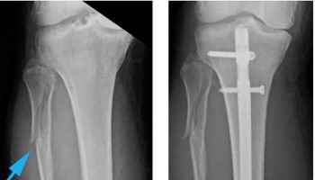
In an indirect attachment, the muscle ends conspicuously short of its bony destination, and the gap is bridged by a fibrous band or sheet called a tendon. See, for example, the two ends of the biceps brachii and the photographs of tendons in figures. You can easily palpate tendons and feel their texture just above your heel (your calcaneal or Achilles tendon) and on the anterior side of your wrist (tendons of the palmaris longus and flexor carpi radialis muscles). Collagen fibers of the muscle continue into the tendon and from there into the periosteum and matrix of the bone, creating very strong structural continuity from muscle to bone.
In some cases, the tendon is a broad sheet called an aponeurosis. This term originally referred to the tendon located beneath the scalp, but now it also refers to similar tendons associated with certain abdominal, lumbar, hand, and foot muscles. For example, the palmaris longus tendon passes through the wrist and then expands into a fanlike palmar aponeurosis beneath the skin of the palm.
In some places, groups of tendons from separate muscles pass under a band of connective tissue called a retinaculum. One of these covers each surface of the wrist like a bracelet, for example. The tendons of several forearm muscles pass under them on their way to the hand.
Figure 1. Tendons of the wrist and hand (flexors)

Note: Anterior views of the forearm. (a) Superficial flexors. (b) The flexor digitorum superficialis, deep to the muscles in part (a). (c) Deep flexors. Flexor muscles of each compartment are labeled in boldface.
Figure 2. Tendons of the wrist and hand (extensors)
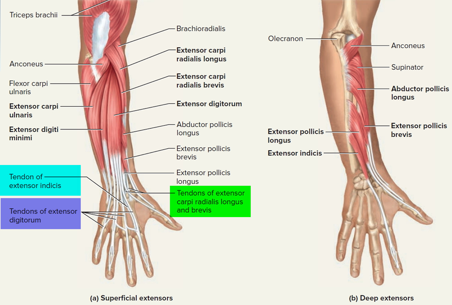
Figure 3. Finger and hand tendons
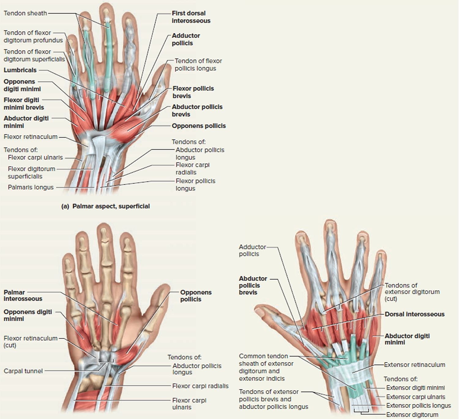
Figure 4. Tendons of the leg and foot
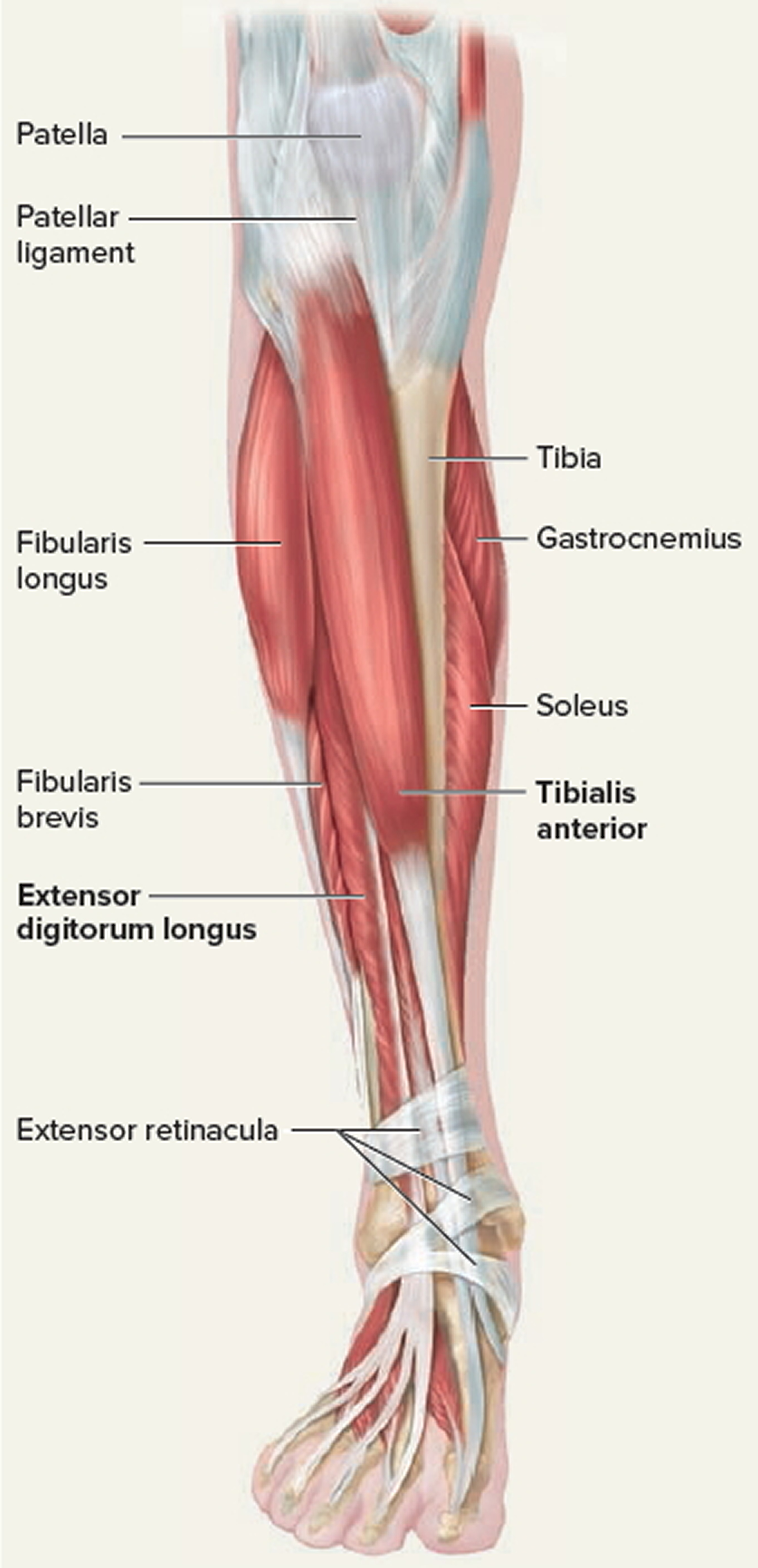
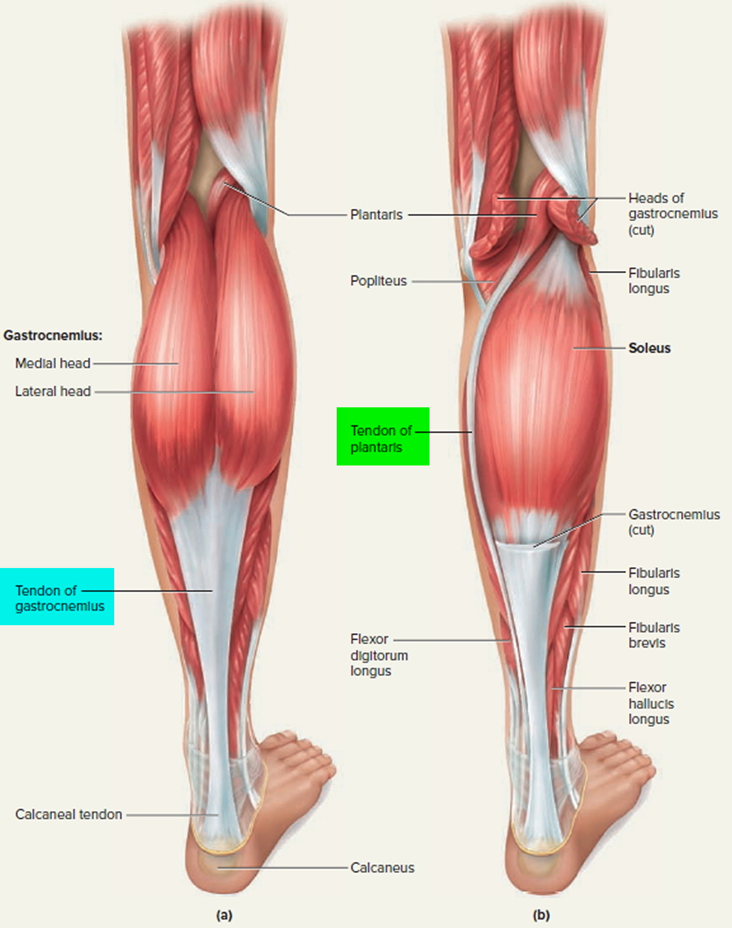
Figure 5. Tendons of the calf and foot (back of leg)