Contents
What is urinalysis
A urinalysis is a test of your urine to screen for, help diagnose and/or monitor a wide range of disorders and diseases, such as urinary tract infections (UTIs), kidney disorders, diabetes, liver problems and other metabolic conditions, to name a few. A urinalysis is a group of physical, chemical, visual examinations and microscopic tests that involves checking the appearance, concentration and content of urine. The urinalysis tests detect and/or measure several substances in the urine, such as byproducts of normal and abnormal metabolism, cells, cellular fragments, crystals, casts and bacteria. Abnormal urinalysis results may point to a disease or illness. For example, a urinary tract infection can make urine look cloudy instead of clear. Increased levels of protein in urine can be a sign of kidney disease. Unusual urinalysis results often require follow-up investigation and additional testing to uncover the source of the problem. Often, substances such as protein or glucose will begin to appear in the urine before people are aware that they may have a problem.
In people diagnosed with diseases or conditions, such as kidney disease or diabetes, the urinalysis may be used in conjunction with other tests, such as urine albumin, to follow treatment.
There are many factors that can affect or interfere with the tests that comprise a urinalysis. If instructed to do so, it is important to follow the directions carefully for a “clean-catch” urine sample. Give a complete history to your healthcare practitioner, including any prescribed or over-the-counter medications or supplements you may be taking. If you are a women, be sure to tell your healthcare practitioner whether you are menstruating.
Urinalysis test preparation
None, if your urine is being tested only for a urinalysis, you can eat and drink normally before the urinalysis test. If you’re having other tests at the same time, you may need to fast for a certain amount of time before the test. Your doctor will give you specific instructions.
Many drugs, including nonprescription medications and supplements, can affect the results of a urinalysis. Before a urinalysis, tell your doctor about any medications, vitamins or other supplements you’re taking.
Is the time of day a factor when collecting a urine sample?
Because this is a general screening test, time of collection is usually not important, although a first morning void may be preferred because it is more concentrated. However, if your healthcare provider is looking for a specific finding, you may be asked to collect a sample at a specific time.
Sample required for a urinalysis
Depending on your situation, you may collect a urine sample at home or at your doctor’s office. Your doctor will provide a container for the urine sample. You may be asked to collect the sample first thing in the morning because at that time your urine is more concentrated, and abnormal results may be more obvious.
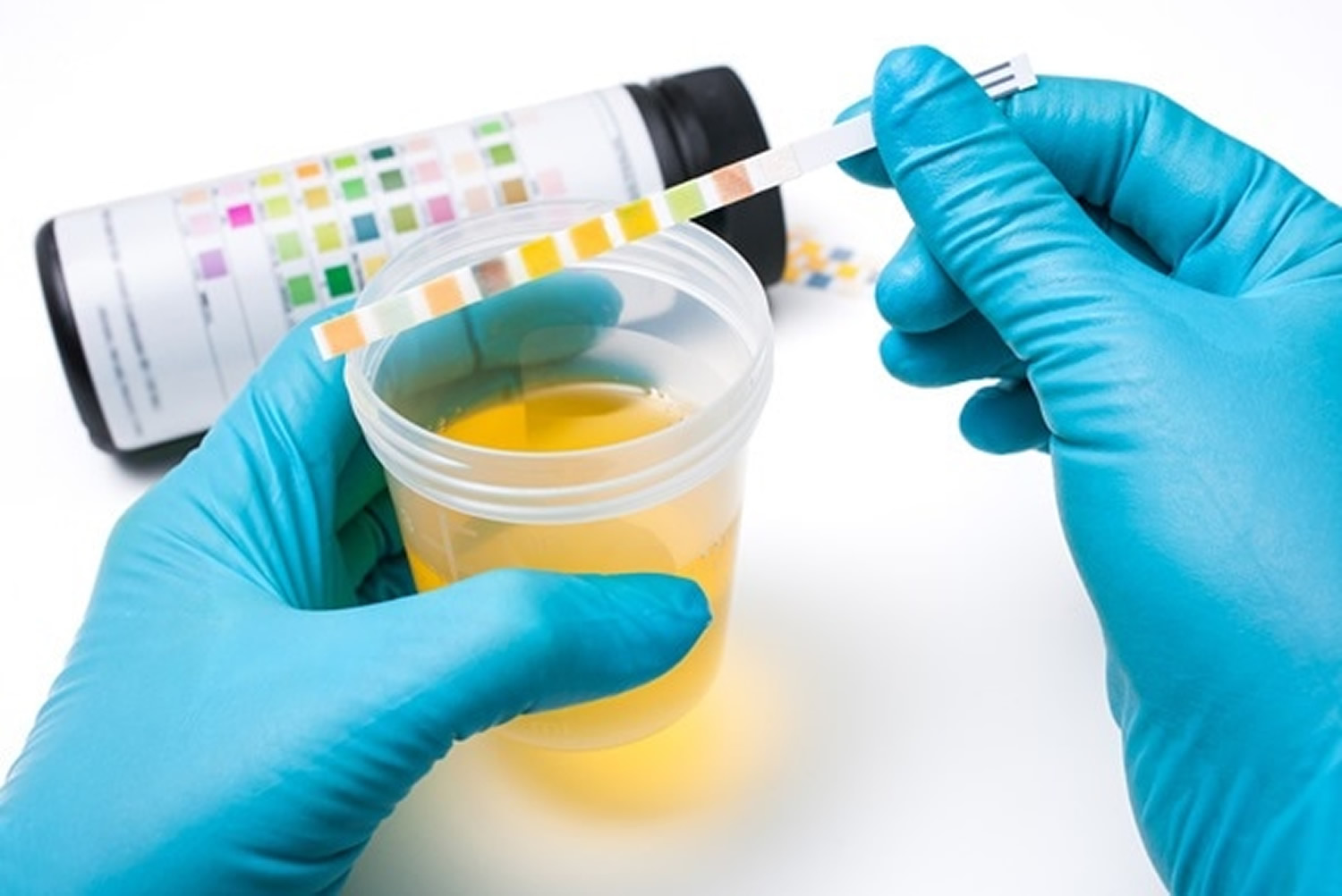
One to two ounces (30 to 59 milliliters) of urine—a sufficient sample is required for accurate results; sometimes you may be directed to collect a sample using a “clean-catch” technique. For this, it is important to clean the genital area before collecting the urine. Bacteria and cells from the surrounding skin can contaminate the sample and interfere with the interpretation of test results. With women, menstrual blood and vaginal secretions can also be a source of contamination.
To get the most accurate results, the sample may need to be collected midstream, using a “clean-catch method”. This method involves the following steps:
- Cleanse the urinary opening. Women should spread their labia and clean from front to back. Men should wipe the tip of the penis.
- Begin to urinate into the toilet.
- Pass the collection container into your urine stream.
- Urinate at least 1 to 2 ounces (30 to 59 milliliters) into the collection container.
- Finish urinating into the toilet.
- Deliver the sample as directed by your doctor.
- If you can’t deliver the sample to the designated area within 60 minutes of collection, refrigerate the sample, unless you’ve been instructed otherwise by your doctor.
In some cases, your doctor may insert a thin, flexible tube (catheter) through the urinary tract opening and into the bladder to collect the urine sample.
- A urine sample will only be useful for a urinalysis if taken to the healthcare provider’s office or laboratory for processing within a short period of time. If it will be longer than an hour between collection and transport time, then the urine should be refrigerated or a preservative may be added.
The urine sample is sent to a lab for analysis. You can return to your usual activities immediately.
Can a urinalysis be done in my healthcare practitioner’s office?
Many healthcare providers’ offices and clinics can perform the visual and chemical examinations of urine. Some may also be able to provide microscopic examinations. Sometimes, if abnormal results are found on the visual or chemical exams, your urine sample may be sent to a laboratory for the microscopic exam. Alternatively, your sample may be sent to a laboratory for a full urinalysis.
Are there home test kits available to test my urine?
Kits to perform a full urinalysis are not available because the test requires special equipment and technical skills. However, some commercial testing strips can be purchased at a pharmacy to perform part of the chemical examination, such as urine pH, urine glucose, and urine ketones.
Why urinalysis is done?
A urinalysis is a common test that’s done for several reasons:
- To check your overall health. Your doctor may recommend a urinalysis as part of a routine medical exam, pregnancy checkup, pre-surgery preparation, or on hospital admission to screen for a variety of disorders, such as diabetes, kidney disease and liver disease.
- To diagnose a medical condition. Your doctor may suggest a urinalysis if you’re experiencing abdominal pain, back pain, frequent or painful urination, blood in your urine, or other urinary problems. A urinalysis may help diagnose the cause of these symptoms.
- To monitor a medical condition. If you’ve been diagnosed with a medical condition, such as kidney disease or a urinary tract disease, your doctor may recommend a urinalysis on a regular basis to monitor your condition and treatment.
Other tests, such as pregnancy testing and drug screenings, also may rely on a urine sample, but these tests look for substances that aren’t included in a typical urinalysis. For example, pregnancy testing measures a hormone called human chorionic gonadotropin (hCG). Drug screenings detect specific drugs or their metabolic products, depending on the purpose of the testing.
When to get tested?
When you have symptoms, such as abdominal pain, back pain, frequent or painful urination; sometimes as part of a routine wellness examination, pregnancy check-up, hospital admission, or pre-surgical work-up.
A urinalysis will likely be ordered when a person sees a healthcare practitioner complaining of symptoms of a urinary tract infection or other urinary system problem, such as kidney disease. Some signs and symptoms may include:
- Abdominal pain
- Back pain
- Painful or frequent urination
- Blood in the urine
Testing may also be ordered at regular intervals when monitoring certain conditions over time.
Urinalysis interpretation
Urinalysis results can have many interpretations. Abnormal findings are a warning that something may be wrong and should be evaluated further. A healthcare practitioner must correlate the urinalysis results with a person’s symptoms and clinical findings and search for the causes of abnormal findings with other targeted tests, such as a comprehensive metabolic panel (CMP), complete blood count (CBC), renal panel, liver panel, or urine culture (for urinary tract infection).
Generally, the greater the concentration of the atypical substance, such as greatly increased amounts of glucose, protein, or red blood cells, the more likely it is that there is a problem that needs to be addressed. However, the results do not tell the healthcare practitioner exactly what the cause of the finding is or whether it is a temporary or chronic condition.
A normal urinalysis does not guarantee that there is no illness. Some people will not release elevated amounts of a substance early in a disease process, and some will release them sporadically during the day, which means that they may be missed by a single urine sample. In very dilute urine, small quantities of chemicals may be undetectable.
For additional details on what specific results may mean, see the sections below on:
- Visual examination
- Chemical examination
- Microscopic examination
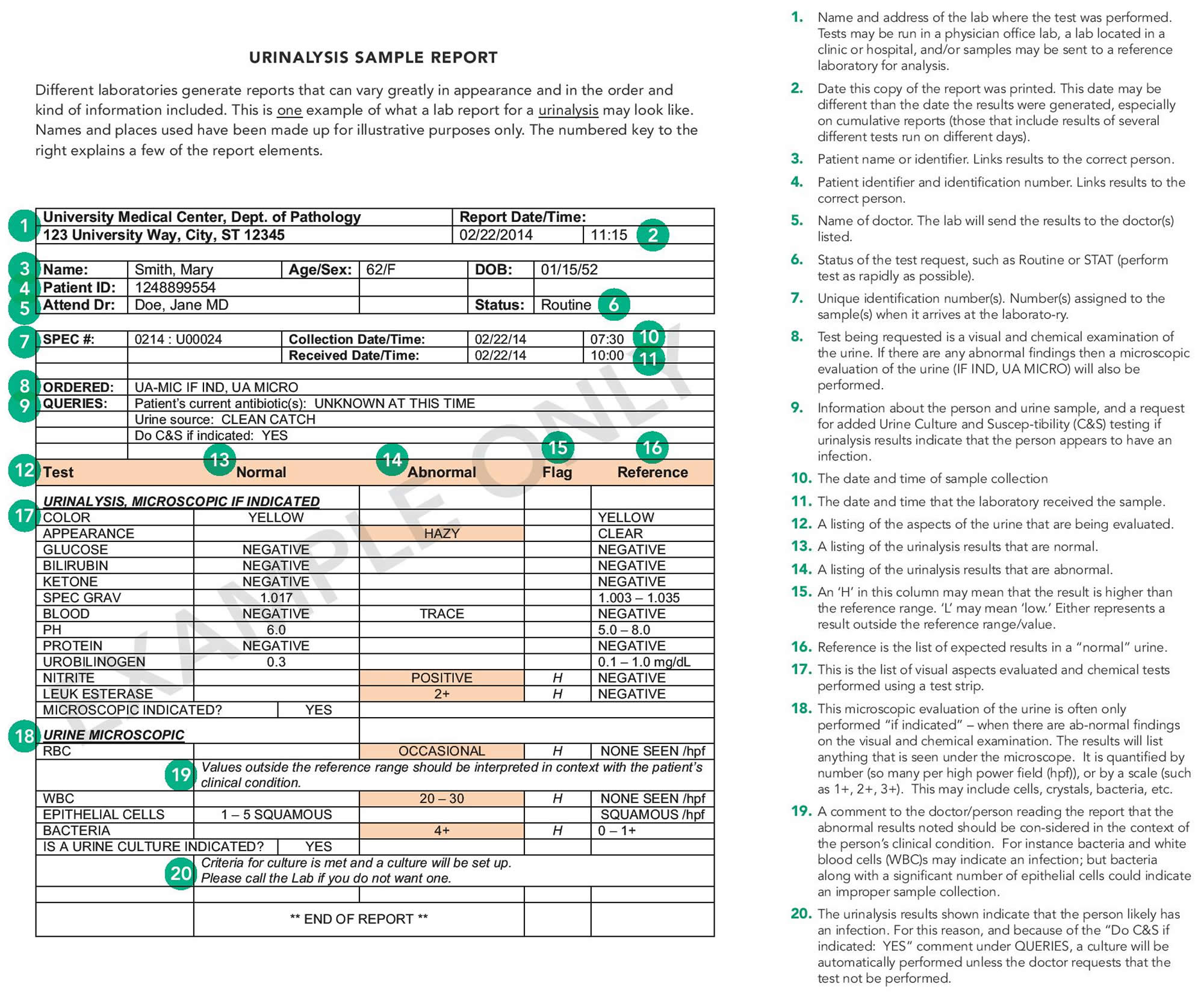
To see an example of a urinalysis lab report, see Figure 1 Sample urinalysis report.
Figure 1. Sample urinalysis report
What does a urinalysis test for?
A urinalysis is a group of physical, chemical, and microscopic tests. The tests detect and/or measure several substances in the urine, such as byproducts of normal and abnormal metabolism, cells, cellular fragments, and bacteria.
Urine is produced by the kidneys, two fist-sized organs located on either side of the spine at the bottom of the ribcage. The kidneys filter wastes out of the blood, help regulate the amount of water in the body, and conserve proteins, electrolytes, and other compounds that the body can reuse. Anything that is not needed is eliminated in the urine, traveling from the kidneys through ureters to the bladder and then through the urethra and out of the body. Urine is generally yellow and relatively clear, but each time a person urinates, the color, quantity, concentration, and content of the urine will be slightly different because of varying constituents.
Many disorders may be detected in their early stages by identifying substances that are not normally present in the urine and/or by measuring abnormal levels of certain substances. Some examples include glucose, protein, bilirubin, red blood cells, white blood cells, crystals, and bacteria. They may be present because:
- There is an elevated level of the substance in the blood and the body responds by trying to eliminate the excess in the urine.
- Kidney disease is present.
- There is a urinary tract infection present, as in the case of bacteria and white blood cells.
A complete urinalysis consists of three distinct testing phases:
- Visual examination, which evaluates the urine’s color and clarity
- Chemical examination, which tests chemically for about 9 substances that provide valuable information about health and disease and determines the concentration of the urine
- Microscopic examination, which identifies and counts the type of cells, casts, crystals, and other components such as bacteria and mucus that can be present in urine
See below for details on each of these examinations.
Visual exam urinalysis
During the visual examination of the urine, a lab technician examines the urine’s appearance – urine’s color and clarity. Urine is typically clear. Cloudiness or an unusual odor may indicate a problem, such as an infection. These can be signs of what substances may be present in the urine. They are interpreted in conjunction with results obtained during the chemical and microscopic examinations to confirm what substances are present.
Blood in the urine may make it look red or brown. Urine color can be influenced by what you’ve just eaten. For example, beets or rhubarb may add a red tint to your urine.
Urine color
Urine can be a variety of colors, most often shades of yellow, from very pale or colorless to very dark or amber. Unusual or abnormal urine colors can be the result of a disease process, several medications (e.g., multivitamins can turn urine bright yellow), or the result of eating certain foods. For example, some people can have red-colored urine after eating beets; the color is from the natural pigment of beets and is not a cause for worry. However, red-colored urine can also occur when blood is present in the urine and can be an indicator of disease or damage to some part of the urinary system. Another example is yellow-brown or greenish-brown urine that may be a sign of bilirubin in the urine (see the Chemical Examination section below).
Urine clarity
Urine clarity refers to how clear the urine is. Usually, lab technicians report the clarity of the urine using one of the following terms: clear, slightly cloudy, cloudy, or turbid. “Normal” urine can be clear or cloudy. Substances that cause cloudiness but that are not considered unhealthy include mucus, sperm and prostatic fluid, cells from the skin, normal urine crystals, and contaminants such as body lotions and powders. Other substances that can make urine cloudy, like red blood cells, white blood cells, or bacteria, indicate a condition that requires attention.
Chemical examination or Dipstick urinalysis test
To perform the chemical examination, most clinical laboratories use commercially prepared test strips (a dipstick) —a thin, plastic stick with strips of chemicals on it. The lab technician dips the strip into urine, chemical reactions change the colors of the pads within seconds to minutes, and the lab technician determines the result for each test. The chemical strips change color if certain substances are present or if their levels are above normal. To reduce timing errors and eliminate variations in color interpretation, automated instruments are frequently used to “read” the results of the test strip.
The degree of color change on a test pad can give an estimate of the amount of substance present. For example, a slight color change in the test pad for protein may indicate a small amount of protein present in the urine whereas a deep color change may indicate a large amount.
The chemical examination is often done in conjunction with or may be followed by a microscopic examination of the urine if there are any abnormal results. Results from both sets of tests are then considered together for interpretation. Abnormal findings may be followed by additional urine and/or blood tests.
The most frequently performed chemical tests using reagent test strips are described below.
Figure 2. Dipstick urinalysis test (Chemical urinalysis test)
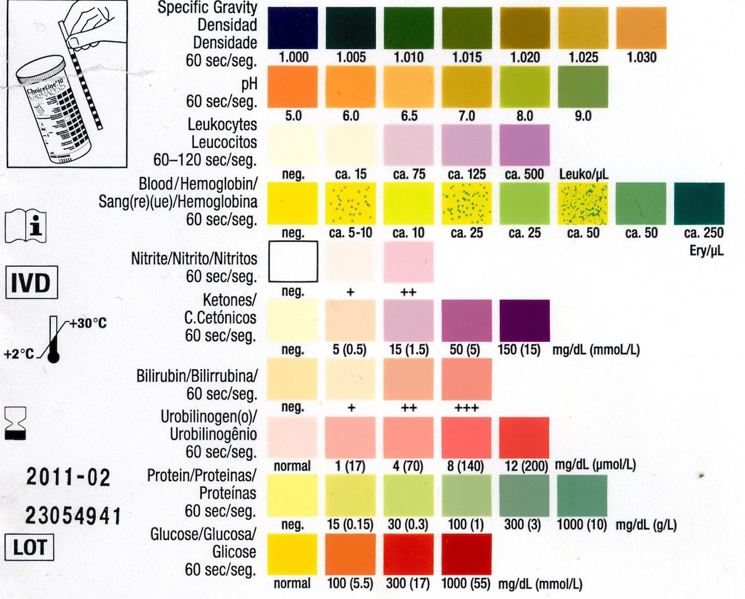
A dipstick urinalysis test checks for:
Acidity (pH)
The pH level indicates the amount of acid in urine. The urine is usually slightly acidic, about pH 6, but can range from 4.5-8. The kidneys play an important role in maintaining the acid-base balance of the body. Therefore, any condition that produces acids or bases in the body, such as acidosis or alkalosis, or the ingestion of acidic or basic foods can directly affect urine pH. Some of the substances dissolved in urine will precipitate out to form crystals when the urine is acidic; others will form crystals when the urine is basic. If crystals form while the urine is being produced in the kidneys, a kidney stone or “calculus” can develop. By modifying urine pH through diet or medications, the formation of these crystals can be reduced or eliminated. Abnormal pH levels may indicate a kidney or urinary tract disorder.
Concentration or specific gravity
A measure of concentration, or specific gravity, shows how concentrated particles are in your urine. A higher than normal concentration often is a result of not drinking enough fluids. Specific gravity measurements are a comparison of the amount of substances dissolved in urine as compared to pure water. If there were no substances present, the specific gravity of the urine would be 1.000 (the same as pure water). Since all urine has some substances in it, a urine specific gravity of 1.000 is not possible. If a person drinks excessive quantities of water in a short period of time or gets an intravenous (IV) infusion of large volumes of fluid, then the urine specific gravity may be very close to that of water. The upper limit of the test pad, a specific gravity of 1.035, indicates concentrated urine, one with many substances in a limited amount of water. Knowing the urine concentration helps healthcare practitioners understand whether a urine specimen they are evaluating is the best one to detect a particular substance. For example, if they are looking for very small amounts of protein, a concentrated morning urine specimen would be the best sample.
Protein
The protein test pad provides a rough estimate of the amount of albumin in the urine. Albumin makes up about 60% of the total protein in the blood. Low levels of protein in urine are normal. Small increases in protein in urine usually aren’t a cause for concern, but larger amounts may indicate a kidney problem.
When urine protein is elevated, a person has a condition called proteinuria. Proteinuria may occasionally be seen in healthy individuals. Healthy people can have temporary or persistent proteinuria due to stress, exercise, fever, aspirin therapy, or exposure to cold, for example. Repeat testing may be done once these conditions have resolved to determine whether the proteinuria is persistent.
If trace amounts of protein are detected, and depending on the person’s signs, symptoms and medical history, a repeat urinalysis and dipstick protein may be performed at a later time to see if there is still protein in the urine or if it has dropped back to undetectable levels.
If a large amount of protein is detected on a urinalysis and/or if the protein persists in repeated tests, a 24-hour urine protein test may be used as a follow-up test. Since the dipstick primarily measures albumin, the 24-hour urine protein test also may be ordered if a healthcare practitioner suspects that proteins other than albumin are being released into the urine.
Protein in the urine may be a sign of kidney disease. Small amounts of albumin may be found in the urine when kidney dysfunction begins to develop. A different test called a urine albumin test detects and measures small amounts of albumin in the urine. The urine albumin test is more sensitive than a dipstick urinalysis and is routinely used to screen people with chronic conditions that put them at risk for kidney disease, such as diabetes and high blood pressure.
Proteinuria may also be associated with many other diseases and conditions. A healthcare practitioner may order other types of follow-up tests to help determine the cause of protein in the urine.
Glucose
Glucose (sugar) is normally not present in urine. When glucose is present, the condition is called glucosuria. It results from either:
- An excessively high glucose level in the blood, such as may be seen with people who have uncontrolled diabetes. Any detection of sugar on this test usually calls for follow-up testing for diabetes.
- A reduction in the “renal threshold;” when blood glucose levels reach a certain concentration, the kidneys begin to eliminate glucose into the urine to decrease blood concentrations. Sometimes the threshold concentration is reduced and glucose enters the urine sooner, at a lower blood glucose concentration.
Some other conditions that can cause glucosuria include hormonal disorders, liver disease, medications, and pregnancy. When glucosuria occurs, other tests such as a fasting blood glucose are usually performed to further identify the specific cause.
Ketones
Ketones are not normally found in the urine. They are intermediate products of fat metabolism. They are produced when glucose is not available to the body’s cells as an energy source. They can form when a person does not eat enough carbohydrates (for example, in cases of fasting, starvation, or high-protein diets) or when a person’s body cannot use carbohydrates properly. When carbohydrates are not available, the body metabolizes fat instead to get the energy it needs to keep functioning. Strenuous exercise, exposure to cold, frequent, prolonged vomiting, and several digestive system diseases can also increase fat metabolism, resulting in ketonuria.
As with sugar, any amount of ketones detected in your urine could be a sign of diabetes and requires follow-up testing.
In a person who has diabetes, ketones in urine may also be an early indication of insufficient insulin. With insufficient insulin, a diabetic cannot process glucose and instead metabolizes fat. This can cause ketones to build up in the blood, resulting first in ketosis and then progressing to ketoacidosis, a form of metabolic acidosis. Excess ketones and glucose are dumped into the urine by the kidneys in an effort to flush them from the body. This condition, called diabetic ketoacidosis (DKA), is most frequently seen with uncontrolled type 1 diabetes and can be a medical emergency.
Bilirubin
Bilirubin is a product from the hemoglobin of red blood cells that are broken down and removed from circulation. Normally, bilirubin is carried in the blood and passes into your liver, where it’s removed and becomes part of bile, a fluid that is released into the intestines to aid in food digestion. Bilirubin is not present in the urine of normal, healthy individuals. Bilirubin in your urine may indicate liver damage or disease. In certain liver diseases, such as biliary obstruction or hepatitis, excess bilirubin can build up in the blood and is eliminated in urine. The presence of bilirubin in urine is an early indicator of liver disease and can occur before clinical symptoms such as jaundice develop. The results of this test will be considered along with the result of urobilinogen (see below). If positive, the healthcare practitioner will likely follow up with other laboratory tests, such as a liver panel (liver function test), to help establish a diagnosis.
Urobilinogen
This test screens for urobilinogen in the urine. The results are considered along with those for urine bilirubin (above). Urobilinogen is normally present in urine in low concentrations. It is formed in the intestine from bilirubin, and a portion of it is absorbed back into the blood. Positive test results may indicate liver diseases such as viral hepatitis, cirrhosis, liver damage due to drugs or toxic substances, or conditions associated with increased red blood cells (RBCs) destruction (hemolytic anemia). When urine urobilinogen is low or absent in a person with urine bilirubin and/or signs of liver dysfunction, it can indicate the presence of hepatic or biliary obstruction.
Leukocyte esterase
Leukocyte esterase is an enzyme present in most white blood cells (WBCs). A few white blood cells are normally present in urine and usually give a negative chemical test result. When the number of white blood cells (WBCs) in urine increases significantly, this screening test will become positive. Results of this test will be considered along with a microscopic examination for white blood cells (WBCs) in the urine.
When this test is positive and/or the WBC count in urine is high, it may indicate that there is inflammation in the urinary tract or kidneys. The most common cause for WBCs in urine (leukocyturia) is a bacterial urinary tract infection (UTI), such as a bladder or kidney infection. In addition to white blood cells (WBCs), bacteria and red blood cells (RBCs) may also be seen in the microscopic examination. If bacteria are present, the chemical test for nitrite may also be positive (see below).
If either nitrites or leukocyte esterase — a product of white blood cells — is detected in your urine, it may be a sign of a urinary tract infection.
Nitrite
This test detects nitrite and is based upon the fact that many bacteria can convert nitrate (a normal substance in urine) to nitrite. Normally, the urinary tract and urine are free of bacteria and nitrite. When bacteria enter the urinary tract, they can cause a urinary tract infection. A positive nitrite test result can indicate a urinary tract infection (UTI). However, since not all bacteria are capable of converting nitrate to nitrite, someone can still have a urinary tract infection (UTI) despite a negative nitrite test. The results of this test will be considered along with the leukocyte esterase (above) and a microscopic examination.
Blood (Hemoglobin) and Myoglobin
This test is used to detect hemoglobin in the urine (hemoglobinuria). Hemoglobin is an oxygen-transporting protein found inside red blood cells (RBCs). Its presence in the urine indicates blood in the urine (known as hematuria).
A small number of red blood cells (RBCs) are normally present in urine and usually result in a “negative” chemical test. An increased amount of hemoglobin and/or increased number of red blood cells (RBCs) are detected as a “positive” chemical test result. Results of this test are typically interpreted along with those from the microscopic examination of the urine to determine whether red blood cells (RBCs) are present in the urine. A positive result on this test with no red blood cells (RBCs) present may indicate the presence of hemoglobin in the urine (which can occur when red blood cells have broken apart) or myoglobin from muscle injury.
Blood in the urine is not a normal finding, but it is not uncommon and not necessarily a cause for alarm. Your healthcare practitioner will investigate further to try to determine the source and underlying cause of the blood and may request repeat testing to determine whether the blood is persistent.
Blood in your urine requires additional testing — it may be a sign of kidney damage, infection, kidney or bladder stones, kidney or bladder cancer, or blood disorders.
Ascorbic Acid (Vitamin C)
Occasionally, people taking vitamin C or multivitamins may have large amounts of ascorbic acid in their urine. When this is suspected to be the case, a laboratorian may test the sample for ascorbic acid (vitamin C) because it has been known to interfere with the accuracy of some of the results of the chemical test strip, causing them to be falsely low or falsely negative. Examples of tests that may be affected include the urine dipstick tests for glucose, blood, bilirubin, nitrite, and leukocyte esterase.
Microscopic examination
A microscopic examination may or may not be performed as part of a routine urinalysis. A microscopic examination is typically performed when there is an abnormal finding on the visual or chemical examination, or if a healthcare practitioner specifically orders it.
A microscopic examination will typically be done when there are abnormal findings on the physical or chemical examination and the results from all will be taken into account for interpretation.
The microscopic exam is performed on urine sediment – urine that has been centrifuged to concentrate the substances in it at the bottom of a tube. The fluid at the top of the tube is then discarded and the drops of fluid remaining are examined under a microscope. Cells, crystals, and other substances are counted and reported either as the number observed “per low power field” (LPF) or “per high power field” (HPF). In addition, some entities, if present, are estimated as “few,” “moderate,” or “many,” such as epithelial cells, bacteria, and crystals. Cells and other substances that may be seen are listed below.
During this exam, several drops of urine are viewed with a microscope. If any of the following are observed in above-average levels, additional testing may be necessary:
- White blood cells (leukocytes) may be a sign of an infection.
- Red blood cells (erythrocytes) may be a sign of kidney disease, a blood disorder or another underlying medical condition, such as bladder cancer.
- Bacteria or yeasts may indicate an infection.
- Casts — tube-shaped proteins — may form as a result of kidney disorders.
- Crystals that form from chemicals in urine may be a sign of kidney stones.
A urinalysis alone usually doesn’t provide a definite diagnosis. Depending on the reason your doctor recommended this test, abnormal results may or may not require follow-up.
Abnormal findings on a urinalysis may prompt repeat testing to see if the results are still abnormal and/or may be followed by additional urine and blood tests to help establish a diagnosis.
Your doctor may evaluate the urinalysis results along with those of other tests — or order additional tests — to determine next steps. For specifics about what your urinalysis results mean, talk with your doctor.
For example, if you are otherwise healthy and have no signs or symptoms of illness, results slightly above normal on a urinalysis may not be a cause for concern and follow-up may not be needed. However, if you’ve been diagnosed with a kidney or urinary tract disease, elevated levels may indicate a need to change your treatment plan.
Red blood cells (RBCs)
Normally, a few red blood cells (RBCs) are present in urine sediment (0-5 RBCs per high power field, HPF). A positive chemical test for hemoglobin and an increase in the number of red blood cells (RBCs) seen under the microscope indicates that there is blood in the urine. However, this test cannot be used to identify where the blood is coming from. For instance, contamination of urine with blood from hemorrhoids or vaginal bleeding cannot be distinguished from a bleed in the urinary tract. This is why it is important to collect a urine specimen correctly and for women to tell their healthcare provider that they are menstruating when asked to collect a urine specimen.
Blood in the urine is not a normal finding, but it is not uncommon and is not necessarily a cause for alarm. Hematuria (blood in urine) is a sign or an indicator that prompts a healthcare practitioner to investigate further to try to determine the underlying cause of the blood. As part of the investigation, a healthcare practitioner will evaluate an individual’s medical history, physical examination, and accompanying signs and symptoms. Additional urine and blood tests may be done to help determine the source.
Some of the underlying causes of hematuria are benign, temporary states that do no lasting harm and resolve with little or no specific treatment. If there is blood in the urine along with white blood cells and bacteria, it may be caused by a urinary tract infection that can be easily treated with antibiotics. Some causes of hematuria, however, may be critical conditions or represent a chronic condition that requires treatment and monitoring.
White blood cells (WBCs)
The number of white blood cells (WBCs) in urine sediment is normally low (0-5 WBCs per high power field, HPF). White blood cells (WBCs) can be a contaminant, such as those from vaginal secretions.
An increased number of white blood cells (WBCs) seen in the urine under a microscope and/or positive test for leukocyte esterase may indicate an infection or inflammation somewhere in the urinary tract. If also seen with bacteria (see below), they indicate a likely urinary tract infection.
Bacteria, yeast and parasites
In healthy people, the urinary tract is sterile and, if the urine sample is collected as a “clean-catch” sample, there will be no microbes seen in the urine sediment under the microscope. Special care must be taken during specimen collection, particularly in women, to prevent bacteria that normally live on the skin or in vaginal secretions from contaminating the urine sample.
If microbes are seen, they are usually reported as “few,” “moderate,” or “many” present per high power field (HPF).
- Bacteria from the surrounding skin can enter the urinary tract at the urethra and move up to the bladder, causing a urinary tract infection (UTI). If the infection is not treated, it can eventually move to the kidneys and cause kidney infection (pyelonephritis). If a person has an uncomplicated lower urinary tract infection, then the person may be treated without need for a urine culture. However, if the person has had recurrent urinary tract infections (UTIs), suspected complicated infection, or is hospitalized, a urine culture and susceptibility testing may be performed to help guide treatment.
- In women (and rarely in men), yeast can also be present in urine. They are most often present in women who have a vaginal yeast infection because the urine has been contaminated with vaginal secretions during collection. If yeast are observed in urine, then the person may be treated for a yeast infection.
- Trichomonas vaginalis is a parasite that may be found in the urine of women, or rarely, men. As with yeast, Trichomonas vaginalis infects the vaginal canal and their presence in urine is due to contamination during sample collection. If these are found during a urinalysis, then Trichomonas testing may be performed to look for a vaginal infection.
Casts
Casts are cylindrical particles sometimes found in urine that are formed from coagulated protein released by kidney cells. They are formed in the long, thin, hollow tubes of the kidneys known as tubules and usually take the shape of the tubule (hence the name). Under the microscope, they often look like the shape of a “hot dog” and in healthy people they appear nearly clear. This type of cast is called a “hyaline” cast. Normally, healthy people may have a few (0–5) hyaline casts per low power field (LPF). After strenuous exercise, more hyaline casts may be detected.
Other types of casts are associated with different kidney diseases, and the type of casts found in the urine may give clues as to which disorder is affecting the kidney. Cellular casts, such as red blood cell and white blood cell casts, indicate a kidney disorder. Some other examples of types of casts include granular casts, fatty casts, and waxy casts. When a disease process is present in the kidney, cells or other substances can become trapped in the protein as the cast is formed. When this happens, the cast is identified by the substances inside it, for example, as a red blood cell cast or white blood cell cast.
Crystals
Urine contains many dissolved substances (solutes) – waste chemicals that the body needs to eliminate. These solutes can form crystals, solid forms of a particular substance, in the urine if:
- The urine pH is increasingly acidic or basic;
- The concentration of dissolved substances is increased; and
- The urine temperature promotes their formation.
Crystals are identified by their shape, color, and by the urine pH. They may be small, sand-like particles with no specific shape (amorphous) or have specific shapes, such as needle-like. Crystals are considered “normal” if they are from solutes that are typically found in the urine; these usually form as urine cools after collection and were not present in the body.
Some examples of crystals that can be found in the urine of healthy individuals include:
- Amorphous urates
- Crystalline uric acid
- Calcium oxalates
- Amorphous phosphates
If the crystals are from substances that are not normally in the urine, they are considered “abnormal.”
Abnormal crystals may indicate an abnormal metabolic process. Some of these include:
- Calcium carbonate
- Cystine
- Tyrosine
- Leucine
Normal or abnormal crystals can form within the kidneys as urine is being made and may group together to form kidney “stones” or calculi. These stones can become lodged in the kidney itself or in the ureters, tubes that pass the urine from kidney to the bladder, causing extreme pain.
Medications, drugs, and x-ray dye can also crystallize in urine. Therefore, the lab technician must be familiar with and trained in the identification of urine crystals.
Epithelial cells
Epithelial cells are usually reported as “few,” “moderate,” or “many” present per low power field (LPF). Normally, in men and women, a few epithelial cells can be found in the urine sediment. In urinary tract conditions such as infections, inflammation, and malignancies, an increased number of epithelial cells are present. Determining the kinds of cells present may sometimes help to identify certain conditions. For example, epithelial cells containing large amounts of broken-down hemoglobin (called hemosiderin) may indicate that there were red blood cells or hemoglobin in the urine recently, even if there are none now.


