Contents
- Newborn conjunctivitis
Newborn conjunctivitis
Newborn babies can develop conjunctivitis or an infection of the eye surface that affects newborn babies within the first 30 days of life also called neonatal conjunctivitis or ophthalmia neonatorum, caused by bacterial or viral infection, irritation or a blocked tear duct 1, 2, 3. If your baby has a discharge from their eye, seek advice from your doctor or pediatrician. Newborn conjunctivitis if left untreated can cause blindness and serious health problems for newborns if it’s not treated. Newborns should be seen by a doctor right away if they have conjunctivitis symptoms. Newborns with conjunctivitis can develop watery eyes within a few days to several weeks after birth. Their eyelids become puffy, red, and tender.
Conjunctivitis in a newborn may be caused by:
- A blocked tear duct. Babies are often born with blocked tear ducts. This can cause a watery or sticky discharge in their eyes. It usually goes away without treatment. Occasionally it can lead to an infection that needs antibiotics. If your baby has a discharge from their eye, seek advice from your doctor, maternal health nurse or paediatrician.
- Irritation produced by the topical antibiotics given at birth
- An infection passed from the mother to baby during childbirth
Even mothers without symptoms at the time of delivery can carry and pass bacteria or viruses to babies during birth.
To prevent newborn conjunctivitis, most states have laws requiring doctors to put drops or ointment in a newborn’s eyes. This typically happens within 2 to 3 hours of birth. In the past, hospitals used silver nitrate 1% solution (Crede’s method) 4. Now, hospitals mostly use antibiotic eye drops or ointment, such as tetracycline hydrochloride 1% eye ointment, erythromycin 0.5% eye ointment, chloramphenicol 1% eye ointment or povidone‐iodine 2.5% solution (water‐based) 5. Erythromycin is the only ophthalmic ointment recommended for use among neonates in the United States 6. The recommended regimen to prevent newborn conjunctivitis caused by Neisseria gonorrhoeae bacteria is erythromycin 0.5% ophthalmic ointment in each eye in a single application at birth 6. Silver nitrate and tetracycline ophthalmic ointments are no longer manufactured in the United States, bacitracin is ineffective, and povidone iodine has not been studied adequately 7, 8. Gentamicin ophthalmic ointment has been associated with severe ocular reactions 9, 10. If erythromycin ointment is unavailable, infants at risk for exposure to gonococcal infection, especially those born to a mother at risk for gonococcal infection or with no prenatal care, can be administered ceftriaxone 25–50 mg/kg body weight IV or IM, not to exceed 250 mg in a single dose 6.
Prevalence of newborn conjunctivitis has decreased significantly in developed countries since the abandonment of silver nitrate as topical prophylaxis. Current estimates of prevalence of newborn conjunctivitis in developed countries are typically < 0.5%. However, a higher incidence of newborn conjunctivitis is still found certain regions of the world, particularly in developing countries. A recent study found an estimated prevalence of 17% among nearly 1000 newborn infants in Pakistan 11. Incidence of neonatal conjunctivitis remains high in Africa 12. Furthermore, studies from sub-Saharan Africa suggest that Chlamydia trachomatis accounted for up to 33% of neonatal conjunctivitis 13.
Aseptic neonatal conjunctivitis most often is a chemical conjunctivitis that is induced by silver nitrate solution, which is used at birth for prophylaxis of infectious conjunctivitis. Chemical conjunctivitis is becoming less common owing to the use of erythromycin ointment or povidone iodide in place of silver nitrate solution for the prophylaxis of infectious conjunctivitis.
Bacterial and viral infections are major causes of septic newborn conjunctivitis, with Chlamydia being the most common infectious agent. Infants may acquire these infective agents as they pass through the birth canal during the birth process 14.
The incidence of infectious ophthalmia neonatorum or newborn conjunctivitis ranges from 1-2%, depending on the socioeconomic character of the area. Chlamydia is the most common infectious agent that causes ophthalmia neonatorum in the United States, where 2%-40% of neonatal conjunctivitis cases are caused by Chlamydia 15.
In contrast, the incidence of gonococcal newborn conjunctivitis has been reduced dramatically and causes less than 1% of cases of neonatal conjunctivitis 16.
The pathology of newborn conjunctivitis is influenced by the anatomy of the conjunctival tissues in the newborn. The inflammation of the conjunctiva may cause blood vessel dilation, potentially dramatic swelling of the conjunctiva (chemosis) and excessive secretion. This infection tends to be more serious in neonates owing to their lack of immunity, lack of lymphoid tissue in the conjunctiva, and absence of tears at birth.
Prompt diagnosis is key in establishing proper treatment and minimizing potential serious complications of neonatal conjunctivitis.
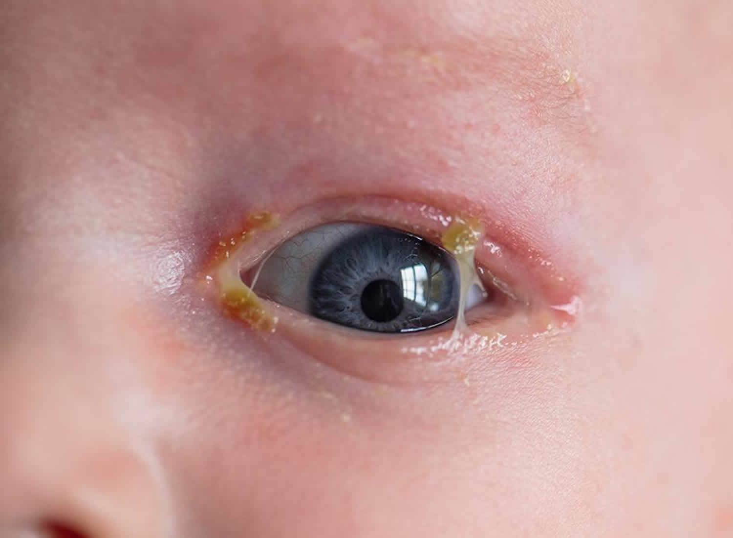
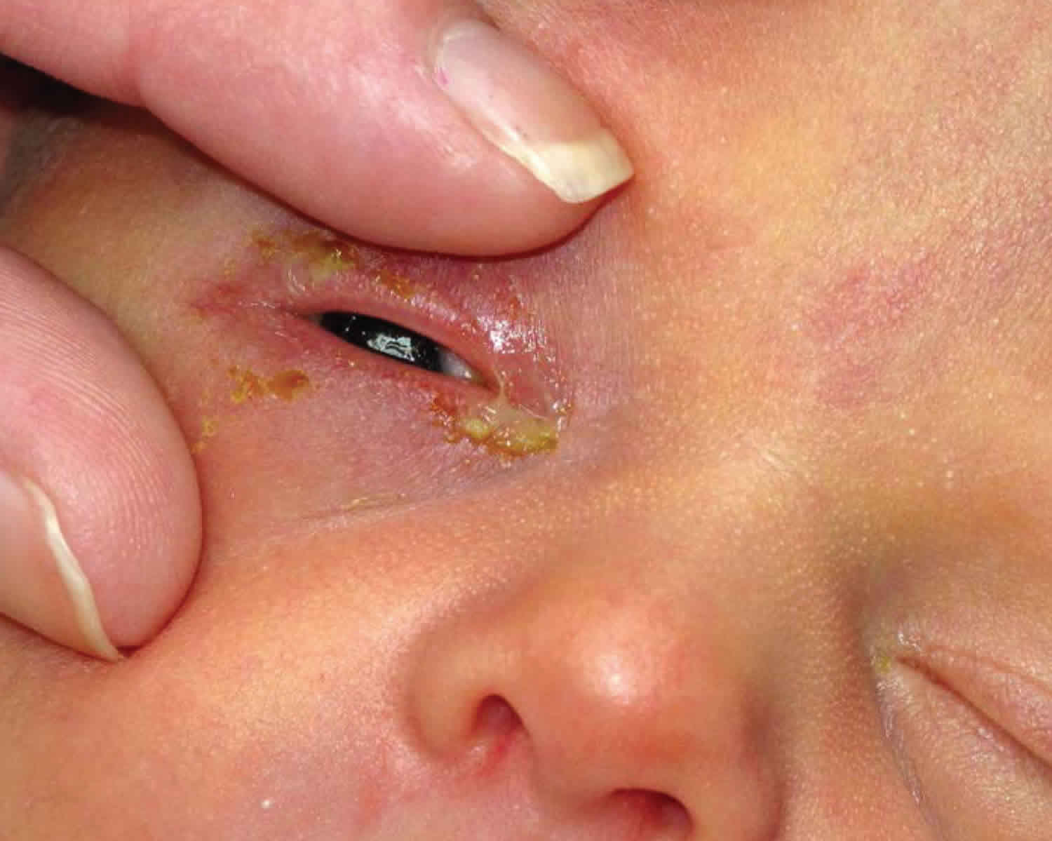
Figure 1. Newborn conjunctivitis
Newborn conjunctivitis causes
The cause of newborn conjunctivitis is often difficult to determine because symptoms are often very similar for each cause.
The most common causes of newborn conjunctivitis include 3:
- Chemical: Classically, the most common cause of neonatal conjunctivitis due to use of post-delivery use of ophthalmic silver nitrate used in the prevention of gonococcal eye infections. However, the incidence of chemical conjunctivitis in the United States has significantly decreased since replacement of silver nitrate with erythromycin ointment
- Bacterial (Chlamydia trachomatis trachomatis most common). Bacterial cause of neonatal conjunctivitis include:
- Chlamydia trachomatis (most common). Newborn chlamydial conjunctivitis is much more prevalent than newborn gonococcal conjunctivitis and has historically been underdiagnosed due to lack of accurate diagnostic techniques 17, 18.
- Neisseria gonorrhoeae. Neisseria gonorrhoeae is one of the most severe and feared causes of neonatal conjunctivitis, requiring prompt diagnosis and treatment.
- Staphylococcus aureus 11, 19, 20, 21, 22.
- Pseudomonas aeruginosa. Pseudomonas, although rare, may lead to potentially blinding complications such as rapid corneal ulceration and perforation.
- Streptococcus spp. (including Streptococcu haemolyticus, Streptococcu pneumonia)
- Other bacteria include Klebsiella, Proteus, Enterobacter, Serratia, and Eikenella corrodens 23
- Viral. Herpes simplex virus (HSV).
Often, no causative pathogen can be found in newborns with conjunctivitis due to methods for obtaining and culturing for bacteria, or due to causes other than bacteria such as chemical conjunctivitis or nasolacrimal duct obstruction 24.
Chlamydial conjunctivitis
- Cause: A mother with untreated chlamydia can pass the Chlamydia trachomatis bacteria to the baby during childbirth (chlamydia-infected cervix during the birth process). Chlamydial infections occur in 4–10% of pregnant women in the United States. Infants whose mothers have untreated chlamydial infections have a 30–40% chance of developing conjunctivitis (incidence of 6.2 per 1000 live births). The risk of chlamydial transmission from an infected mother to newborns is 15% on average (range = 8% to 44%) 25. Furthermore, an increased prevalence of chlamydial infection in some high‐income countries is associated with a commensurate rise in risk of chlamydial conjunctivitis 26. Pooled mean prevalence of Chlamydia trachomatis bacteria was higher at 6.9% in pregnant women in Eastern and Southern Africa, and 6.1% in pregnant women in West and Central Africa 27.
- Symptoms: Redness of the eyes, swelling of the eyelids, and discharge of pus. Some newborns with chlamydial conjunctivitis can have the infection in other parts of their bodies. The bacteria can infect the lungs and nasopharynx (the area at the back of the nose to the mouth).
Newborn chlamydial conjunctivitis or chlamydial ophthalmia neonatorum symptoms are likely to appear 5 to 12 days after birth and is associated with a high risk of corneal and conjunctival scarring, hemorrhagic conjunctivitis, and rarely, loss of vision if left untreated 28, 17, 29. Symptoms can develop earlier if the amniotic sac is ruptured during delivery.
The presentation of chlamydial conjunctivitis may range from mild hyperemia with scant mucoid discharge to eyelid swelling, swelling of the conjunctiva (chemosis) and pseudomembrane formation. Patients typically present with unilateral or bilateral watery discharge, which may become more copious and purulent later.
Although most cases are mild and self-limited, chlamydial conjunctivitis occasionally may be severe. Pseudomembranes, thickened palpebral conjunctiva, significant peripheral pannus, and corneal opacification may be present.
Blindness, although rare and much slower to develop than in gonococcal conjunctivitis, is generally not due to corneal involvement as in gonococcal conjunctivitis. Instead, eyelid scarring and corneal pannus can gradually progress to central corneal opacification by mechanisms reminiscent of trachoma.
A follicular reaction does not occur, because newborns have no requisite lymphoid tissue present in the conjunctiva.
Like gonococcal conjunctivitis, chlamydial conjunctivitis also may be associated with extraocular involvement, including pneumonitis, otitis, and pharyngeal and rectal colonization.
Gonococcal conjunctivitis
- Cause: A mother with untreated gonorrhea can pass the Neisseria gonorrhoeae bacteria to the baby during childbirth. Newborn gonococcal conjunctivitis is mainly contracted from the mother’s infected birth canal during delivery, but can also be contracted in utero by ascending infections. Neonates born to gonorrhoea‐infected mothers have a 30% to 50% risk of developing gonococcal conjunctivitis 30. Furthermore, there is increasing incidence of drug‐resistant strains of Neisseria gonorrhoeae bacteria globally 31, 32, 33. The pooled mean prevalence of Neisseria gonorrhoeae bacteria was estimated at 3.7% in pregnant women in Eastern and Southern Africa, and 2.7% in pregnant women in West and Central Africa 27.
- Symptoms: Red eyes, thick pus in the eyes, and swelling of the eyelids. It can also progress to serious infections of the bloodstream (bacteremia) and lining of the brain and spinal cord (meningitis) in newborns.
Newborn gonococcal conjunctivitis or gonococcal ophthalmia neonatorum tends to be the most serious form of neonatal conjunctivitis. Untreated or inappropriately treated gonococcal conjunctivitis can result in corneal perforation and vision loss in 24 hours 34, 35. In one case series, the mean duration of corneal perforation from untreated gonococcal conjunctivitis was 11 days 36. In areas with low incidence of gonococcal neonatal conjunctivitis or limited access to appropriate health care, appropriate clinical diagnosis and appropriate therapy may be delayed, which can lead to loss of vision 37, 38, 39, 40.
Newborn gonococcal conjunctivitis symptoms are likely to appear 2 to 5 days after birth. Gonococcal conjunctivitis presents with the most rapid onset, usually occurring 24-48 hours following birth. Typically, patients develop a hyperacute conjunctivitis, associated with marked lid edema, swelling of the conjunctiva (chemosis) and purulent discharge (pus). The classic presentation is severe bilateral purulent conjunctivitis.
Corneal ulceration may occur, particularly in the periphery, where massive limbal conjunctival chemosis traps inflammatory mediators and organisms, with rapid progression to perforation the cornea if treatment is delayed and endophthalmitis.
Patients also may have systemic manifestations, including rhinitis, stomatitis, arthritis, meningitis, anorectal infection, and septicemia.
Chemical conjunctivitis
- Cause: When eye drops are given to newborns to help prevent a bacterial infection, the newborn’s eye may become irritated.
- Symptoms: Mildly red eye and some swelling of the eyelids. Symptoms are likely to last for only 24 to 36 hours.
Chemical conjunctivitis secondary to silver nitrate solution application usually occurs in the first day of life, disappearing spontaneously within 2-4 days.
Neonatal conjunctivitis due to other microbial agents is usually milder.
Other neonatal conjunctivitis
- Causes: Viruses that cause genital or oral herpes (herpes simplex virus 1 and 2), and bacteria other than chlamydia and gonorrhea, can cause conjunctivitis and severe eye damage in newborns. This includes bacteria that normally live in a mother’s vagina and are not sexually transmitted. They may pass such viruses to the baby during childbirth. However, herpes conjunctivitis is less common than conjunctivitis caused by gonorrhea and chlamydia.
- Symptoms: Red eye and swollen eyelids with some pus.
Herpes simplex virus (HSV) is a rare cause of neonatal keratoconjunctivitis, found in less than 1% of cases 41 and can be associated with a generalized herpes simplex infection.
Herpes simplex keratoconjunctivitis often presents in infants with generalized herpes simplex infections, characterized by corneal epithelial involvement or vesicles on the periocular skin. Serious systemic complications, such as encephalitis, may also occur in these neonates owing to their poor immunologic response.
Most infants with such an infection acquire the infection during the birth process. Caesarean delivery is strongly considered when active maternal genital disease is recognized at term since the risk of transmitting HSV (herpes simplex virus) to the neonate during vaginal delivery is 25 to 60% 42.
During pregnancy and prior to giving birth, mothers with genital herpes should consult with their doctor about ways to minimize the chances of spread to their newborn baby.
Risk factors for developing newborn conjunctivitis
Risk factors of neonatal conjunctivitis may include:
- Maternal infections harbored in the mother’s birth canal
- HIV-infected mothers 43
- Exposure of the infant to infectious organisms
- Increased birth weight 43
- Inadequacy of ocular prophylaxis immediately after birth
- Premature rupture of membranes (PROM) 44
- Ocular trauma during delivery
- Mechanical ventilation
- Prematurity
- Poor prenatal care
- Poor hygienic delivery conditions
- Post-delivery infection due to direct contact with health care workers or by aerosolization
- Silver nitrate exposure.
Newborn conjunctivitis prevention
Newborn conjunctivitis prevention through good prenatal care and treatment of chlamydial, gonococcal, or herpetic infections during pregnancy remains the best preventative method 3.
Topical prophylaxis
Use of topical silver nitrate to prevent neonatal gonococcal conjunctivitis was first introduced by Credé in 1880 and has been classically been cited as the most common cause of neonatal chemical conjunctivitis 4. However, the incidence of chemical conjunctivitis has declined as the use of silver nitrate as prophylaxis has been abandoned in many modern countries in favor of topical medications with a more favorable side effect profile such as erythromycin. However, countries often vary in their accessibility to these more favorable topical antibiotics. One study evaluating the current methods of treatment in maternity hospitals in Croatia showed that 75% of all maternity hospitals in Croatia routinely administered preventive topical treatment, including topical tobramycin (83.3%), povidone-iodine (8.3%), erythromycin (4.2%), and silver nitrate (4.2%), partially due to the lack of availability of erythromycin and tetracycline in Croatia 45. Data supporting the superiority of one topical antibiotic over another seem to remain clear cut. A recent systematic review including 30 trials found little evidence that any prophylaxis involving tetracycline 1%, erythromycin 0.5%, povidone-iodine 2.5%, or silver nitrate 1% provided better prevention from serious outcomes such as blindness or any adverse visual outcomes 5.
Systemic prophylaxis
Infants with possible infectious exposure in utero or during birth process should receive appropriate prophylaxis following birth in attempt to prevent ocular and systemic complications 3. Gonococcal prophylaxis includes single injection of ceftriaxone 50 mg/kg IM or IV in those neonates born to mothers with untreated or suspected gonococcal infection 3. Other preventative measures include proper hand-washing techniques by peripartum and nursery staff.
Maternal screening
The United States Preventive Services Task (USPSTF) currently recommends routine screening for chlamydial and gonorrheal infection in all sexually active or pregnant women if they are 24 years or younger, or at least 25 years and at increased risk for infection 3. These preventative screening measures have significantly decreased perinatal chlamydial and gonorrheal infections in the United States. One study done at a medical center in the United States reports a decrease from 15.6% positivity rate of Chlamydia trachomatis eye cultures before the implementation of universal prenatal screening (1986-1993), to 1.8% during the screening period (1994-2002) 46.
Newborn conjunctivitis signs and symptoms
Non-specific signs of neonatal conjunctivitis include conjunctival injection, tearing, mucopurulent or non-purulent discharge, chemosis, and eyelid swelling.
Chemical conjunctivitis
Typically results in mild conjunctival injection accompanied by tearing, spontaneously resolving within 2-4 days
Chlamydia trachomatis conjunctivitis
Presentation may range from mild hyperemia with scant mucoid discharge to eyelid swelling, chemosis, and pseudomembrane formation
Neisseria gonorrhoeae conjunctivitis
- Typically, patients present with acute conjunctivitis, associated with chemosis, severe lid edema, and mucopurulent discharge
- Corneal involvement is the most serious complication, involving diffuse epithelial edema and ulceration that may progress to perforation of the cornea and endophthalmitis
- Initially, superficial keratitis gives the corneal surface a lackluster appearance followed by marginal and central infiltrates appear, which then ulcerate
HSV conjunctivitis
- Typically, patients present with unilateral or bilateral lid edema, moderate amount of conjunctival injection, and non-purulent, serosanguinous discharge
- Other signs include vesicles on the skin surrounding the eye and corneal epithelial involvement with microdendrites or geographic ulcers (in contrast to typical dendrites as seen in adults)
Newborn conjunctivitis complications
Eye complications of neonatal conjunctivitis include pseudomembrane formation, corneal edema, thickened palpebral conjunctivia, peripheral pannus formation, corneal opacification, staphyloma, corneal perforation, endophthalmitis, loss of eye, and blindness. Risk of complications can minimized with prompt diagnosis and appropriate antibiotic therapy.
If untreated, peripheral corneal ulceration may occur in Neisseria gonorrhoeae infection and rapidly progress to corneal perforation. Complications of gonococcal conjunctivitis and subsquent systemic involvement include arthritis, meningitis, anorectal infection, septicemia, and death.
When unrecognized and not immediately treated, Pseudomonas infection may lead to endophthalmitis and subsequent death.
Systemic complications of chlamydia conjunctivitis include pneumonitis, otitis, and pharyngeal and rectal colonization. Pneumonia has been reported in 10-20% of infants with chlamydial conjunctivitis 47.
HSV keratoconjunctivitis can cause corneal scarring and ulceration. Additionally, disseminated HSV infection often includes central nervous system involvement 42.
Newborn conjunctivitis diagnosis
History
Time frame of signs/symptoms following birth play an important role in determining the most likely cause and subsequent proper diagnosis and treatment 3:
- Chemical conjunctivitis (typically presents within first 24 hours following birth)
- Neisseria gonorrhea (3-5 days after birth)
- Chlamydia trachomatis (5-14 days)
- HSV or herpes simplex virus (1-2 weeks)
Physical examination
Your baby’s doctor will perform an eye exam on the baby. A thorough examination of the globe and periocular structures of a neonate suspected to have neonatal conjunctivitis is crucial. Corneal involvement should be investigated closely with and without fluorescein and blue cobalt light. A complete systemic examination should be performed by trained physician familiar with the physical exam of a neonate.
If the eye does not appear normal, the following tests may be done:
- Culture of the drainage from the eye to look for bacteria or viruses
- Slit-lamp exam to look for damage to the surface of the eyeball
Laboratory studies for neonatal conjunctivitis should include the following:
- Conjunctival scraping for Gram stain or Giemsa stain
- Conjunctival scraping for polymerase chain reaction (PCR) assay to detect chlamydia and gonorrhea
- Culture on chocolate agar and/or Thayer-Martin for Neisseria gonorrhoeae bacteria
- Culture on blood agar for other bacteria
- Culture of corneal epithelial cells for HSV if cornea is involved; PCR should also be considered in cases of possible HSV conjunctivitis
Culture and histology
Bacterial cultures on blood and chocolate agar are indicated in every case of neonatal conjunctivitis and remain the criterion standard despite newer diagnostic methods.
Since Chlamydia bacteria are obligate intracellular organisms, the culture specimens need to contain epithelial cells and not just exudative material. PCR is generally accepted as the most useful test for chlamydial conjunctivitis owing to its high sensitivity 47.
In cases in which gonorrhea is suspected, the agar should be inoculated immediately since Ngonorrhoeae is very sensitive to moisture and temperature changes.
Laboratory evaluation for the presence of HSV infection is indicated if a corneal epithelial defect is present, if vesicles are present on the eyelids or other parts of the body, and if the diagnosis cannot be made on ocular examination. The presence of HSV in tissue culture remains the criterion standard in the diagnosis of HSV, despite a high false-negative rate. HSV infections may be more rapidly diagnosed with PCR, and PCR testing for HSV is more sensitive than viral culture 47. Laboratory evaluation for suspected HSV becomes more important in neonatal disease because the clinical presentation may be highly atypical in an immunologically immature newborn.
Cytologic findings for various forms of conjunctivitis are as follows:
- Chemical conjunctivitis – Neutrophils, occasional lymphocytes on Gram stain
- Bacterial conjunctivitis – Bacteria, neutrophils on Gram stain
- Gonococcal conjunctivitis – Neutrophils, Gram-negative intracellular diplococci on Gram stain
- Chlamydial conjunctivitis – Neutrophils, lymphocytes, plasma cells on Gram stain; basophilic intracytoplasmic inclusions in epithelial cells on Giemsa stain
- Herpetic conjunctivitis – Lymphocytes, plasma cells, multinucleate giant cells on Gram stain; eosinophilic intranuclear inclusions in epithelial cells on Papanicolaou smear, but with low sensitivity
Newer diagnostic techniques
Nucleic acid amplification tests such as polymerase chain reaction (PCR) and transcription-mediated amplification are more sensitive than culture in detecting chlamydial and gonorrheal organisms 16.
PCR assays may have a higher sensitivity and similar specificity in diagnosing neonatal chlamydial conjunctivitis, compared with conventional methods 48.
PCR for HSV from conjunctival scrapings has high sensitivity and specificity, but it is expensive, not always readily available, and is usually reserved for the diagnosis of encephalitis. Direct florescent antibody (DFA) studies are useful for rapid detection, have high sensitivity and specificity, and can be used to type the virus 42.
Newborn conjunctivitis treatment
Doctors may treat conjunctivitis in infants caused by a bacterial infection with antibiotics. It will depend on the severity of the infection and the bacteria that caused it. Rinsing the newborn’s infected eye with a saline solution will remove any debris that may develop in response to the infection.
How antibiotics are applied:
- As an eye drop or ointment in the eye (topical)
- By mouth (orally)
- Through a vein (intravenous or IV)
- As a shot (intramuscular)
- Doctors may combine eye antibiotic eye drop or ointment with one of the other methods
If your baby has a blocked tear duct that causes conjunctivitis, a gentle, warm massage between the eye and nasal area may help. If the blocked tear duct does not clear by 1 year of age, the newborn may require surgery.
The treatment prior to laboratory results should include topical erythromycin ointment and an IV or IM third-generation cephalosporin. Prompt treatment of gonococcal conjunctivitis is important, since this organism can penetrate an intact corneal epithelium and rapidly cause corneal ulceration. Because of the rapid progression of gonococcal conjunctivitis, patients with acute neonatal conjunctivitis should be treated for gonococcal conjunctivitis until culture results are available; the treatment is altered according to the laboratory results.
In cases of chlamydial conjunctivitis, systemic treatment is necessary because of the significant risk for life-threatening pneumonia.
Infants with a potentially sexually transmitted disease, such as gonorrhea or chlamydia, should undergo evaluation for other sexually transmitted diseases, such as syphilis and HIV 49, as should the mother and her sexual partner(s).
Newborns with conjunctivitis are at risk for secondary infections, such as pneumonia, meningitis, and septicemia, which can lead to sepsis and death and thus should be admitted for full workup and treatment.
Bacterial conjunctivitis rarely fails to respond to treatment.
A consultation can be made with a pediatrician or pediatric infectious specialist in neonatal conjunctivitis, and the patient should be seen daily until response to treatment is confirmed.
Discharged patients should continue the treatment, according to clinical presentations and available culture results. Treatment may be modified later per culture results.
Avoid eye patching
Treatment of neonatal chemical conjunctivitis is not necessary. Lubrication with artificial tear preparations may ease mild discomfort.
Chemical conjunctivitis
No treatment required; supportive care only (may use artificial tears four times daily). Lubrication with artificial tear preparations may ease mild discomfort.
Typically disappears spontaneously within 2-4 days.
Neonatal Chlamydial conjunctivitis
Treatment should be initiated upon a positive diagnostic test. Neonatal Chlamydial conjunctivitis is treated with erythromycin drops four times daily plus oral erythromycin (50 mg/kg/day divided four times daily) for 14 days 3. Alternatively, oral azithromycin may be used for both chlamydial conjunctivitis and pneumonia, 20 mg/kg once daily for three days 3.
While outpatient treatment is an option, hospitalization may be required. Evaluation of systemic involvement needed.
Topical therapy for chlamydial conjunctivitis has proven to be relatively ineffective compared to oral therapy, particularly in treating nasopharyngeal infection 50. Topical erythromycin ointment may be beneficial as an adjunctive therapy. Doctors usually use oral antibiotics to treat inclusion conjunctivitis.
Since the efficacy of systemic erythromycin therapy is approximately 80%, a second course sometimes is required.
Systemic treatment is important in cases of chlamydial conjunctivitis since topical therapy is ineffective in eradicating the bacteria in the nasopharynx of the infant, which could cause a life-threatening pneumonia if left untreated.
The use of azithromycin and erythromycin are associated with increased risk of infantile hypertrophic pyloric stenosis (IHPS) in infants within 2 weeks of life. Infants should be closely monitored for potential signs of infantile hypertrophic pyloric stenosis when started on azithromycin or erythromycin 3.
Neonatal Gonococcal conjunctivitis
Newborns who have gonococcal conjunctivitis should be managed in consultation with an infectious disease specialist. Topical irrigation with normal saline should be used to remove mucopurulent discharge, as Gonococcal infection may produce more mucopurulent discharge than other pathogens. Ceftriaxone in a single dose (Ceftriaxone 25–50 mg/kg body weight IV or IM in a single dose, not to exceed 250 mg) 6. One dose of ceftriaxone is adequate therapy for gonococcal conjunctivitis 6. Ceftriaxone should be administered cautiously to neonates with hyperbilirubinemia, especially those born prematurely 6. Cefotaxime 100 mg/kg body weight IV or IM as a single dose can be administered for those neonates unable to receive ceftriaxone because of simultaneous administration of IV calcium 6. Topical antibiotic therapy alone is inadequate and unnecessary if systemic treatment is administered 6.
If there is systemic disease, treatment is required for 7 to 14 days, depending on the nature of the invasive infection. Bacitracin or erythromycin ointment every 2 to 4 hours in addition to systemic treatment of ceftriaxone 3. Hospitalization and evaluation for disseminated Neisseria gonorrhoeae infection should be initiated. Topical saline drops to remove discharge. Topical atropine should be considered for pain relief if there is corneal involvement.
- Note: Chlamydial testing should be performed simultaneously from the inverted eyelid specimen. Mothers of newborns with gonococcal conjunctivitis caused by Neisseria gonorrhoeae should be evaluated, tested, and presumptively treated for gonorrhea, along with their sex partners. All neonates with gonococcal conjunctivitis should also be treated for chalmydia. Mother and sexual partner should be treated as well.
Other bacterial neonatal conjunctivitis
- Gram positive: Bacitracin ointment four times daily for 2 weeks
- Gram negative: Gentamicin, tobramycin or ciprofloxacin four times daily for 2 weeks
Neonatal herpetic conjunctivitis
Neonates with a suspected herpes simplex infection should be treated with systemic acyclovir to reduce the risk of a systemic infection. An effective dose is Acyclovir 45mg/kg/day IV plus vidarabine 3% ointment 5x/day 3. The recommended minimal duration is 14 days, but a course as long as 21 days may be required depending on presence of central nervous system involvement.
Infants with neonatal HSV keratitis should also receive a topical ophthalmic drug, most commonly 1% trifluridine drops or 3% vidarabine ointment 5x/day for 14-21 days depending on presence or absence of central nervous system involvement. Topical ganciclovir 0.15% gel is now also available, although none of these topical agents is specifically approved for neonatal use 51.
Topical antibiotics can also be considered to prevent secondary bacterial infections in cases with significant epithelial defects.
Medical follow up
- Patients with neonatal conjunctivitis should be followed daily for signs of improvement or worsening, especially acutely due to concerns of rapidly progressing infectious complications.
- Patient should be followed closely by pediatrician for evaluation and treatment of potential systemic infection.
Newborn conjunctivitis prognosis
The prognosis of neonatal conjunctivitis is generally considered to be good as long as early diagnosis is made and prompt medical therapy is initiated 3. Most cases of infectious conjunctivitis respond to appropriate treatment. However, morbidity and mortality increases in cases of systemic involvement requiring hospitalization and intensive monitoring.
- World Health Organization. Conjunctivitis of the newborn: prevention and treatment at the primary health care level. apps.who.int/iris/bitstream/10665/39482/1/9241560886_eng.pdf[↩]
- Pink Eye in Newborns. https://www.cdc.gov/conjunctivitis/newborns/index.html[↩]
- Neonatal Conjunctivitis. https://eyewiki.org/Neonatal_Conjunctivitis[↩][↩][↩][↩][↩][↩][↩][↩][↩][↩][↩][↩][↩]
- Crede CS. Ophthalmia neonatorum in newborn children [Die verhürtung der augenentzündung der neugeborenen]. Archiv fur Gynaekologie 1881;17:50-3.[↩][↩]
- Kapoor VS, Evans JR, Vedula SS. Interventions for preventing ophthalmia neonatorum. Cochrane Database Syst Rev. 2020 Sep 21;9(9):CD001862. doi: 10.1002/14651858.CD001862.pub4[↩][↩]
- Gonococcal Infections Among Neonates. https://www.cdc.gov/std/treatment-guidelines/gonorrhea-neonates.htm[↩][↩][↩][↩][↩][↩][↩][↩]
- Scott WJ, Eck CD. Povidone-iodine and ophthalmia neonatorum. Ophthalmology. 2012 Mar;119(3):653-4; author reply 654. doi: 10.1016/j.ophtha.2011.11.037[↩]
- David M, Rumelt S, Weintraub Z. Efficacy comparison between povidone iodine 2.5% and tetracycline 1% in prevention of ophthalmia neonatorum. Ophthalmology. 2011 Jul;118(7):1454-8. doi: 10.1016/j.ophtha.2010.12.003[↩]
- Binenbaum G, Bruno CJ, Forbes BJ, Snyder M, Mollen TJ, Schmidt B, Peterside I. Periocular ulcerative dermatitis associated with gentamicin ointment prophylaxis in newborns. J Pediatr. 2010 Feb;156(2):320-1. doi: 10.1016/j.jpeds.2009.11.060[↩]
- Nathawad R, Mendez H, Ahmad A, Laungani S, Hoa BT, Garlick J, Hammerschlag MR. Severe ocular reactions after neonatal ocular prophylaxis with gentamicin ophthalmic ointment. Pediatr Infect Dis J. 2011 Feb;30(2):175-6. doi: 10.1097/INF.0b013e3181f6c2e5[↩]
- Gul SS, Jamal M, Khan N. Ophthalmia neonatorum. J Coll Physicians Surg Pak. 2010 Sep;20(9):595-8.[↩][↩]
- Isenberg SJ et al. A double application approach to ophthalmia neonatorum prophylaxis. Br J Ophthalmol. 2003 Dec;87(12):1449-52.[↩]
- Adachi KN, Nielsen-Saines K, Klausner JD. Chlamydia trachomatis Screening and Treatment in Pregnancy to Reduce Adverse Pregnancy and Neonatal Outcomes: A Review. Front Public Health. 2021 Jun 10;9:531073. doi: 10.3389/fpubh.2021.531073[↩]
- Neonatal Conjunctivitis (Ophthalmia Neonatorum). https://emedicine.medscape.com/article/1192190-overview[↩]
- American Academy of Pediatrics. Chlamydia Trachomatis. Pickering LK, Baker CJ, Kimberlin DW, Long SS, eds. Red Book: Report of the Committee on Infectious Diseases. 28th ed. Elk Grove Village, Ill: American Academy of Pediatrics; 2009. 255-9.[↩]
- American Academy of Pediatrics. Gonococcal Infections. Pickering LK, Baker CJ, Kimberlin DW, Long SS eds. Red Book 2009 Report of the Committee on Infectious Diseases. 28th ed. Elk Grove Village, Ill: American Academy of Pediatrics; 2009. 305-13.[↩][↩]
- Darville T. Chlamydia trachomatis infections in neonates and young children. Semin Pediatr Infect Dis. 2005 Oct;16(4):235-44. doi: 10.1053/j.spid.2005.06.004[↩][↩]
- Yip PP, Chan WH, Yip KT, Que TL, Kwong NS, Ho CK. The use of polymerase chain reaction assay versus conventional methods in detecting neonatal chlamydial conjunctivitis. J Pediatr Ophthalmol Strabismus. 2008 Jul-Aug;45(4):234-9. doi: 10.3928/01913913-20080701-17[↩]
- Olatunji FO, Fadeyi A, Ayanniyi AA, Akanbi AA 2nd. Non-gonococcal bacterial agents of conjunctivitis and their antibiotic susceptibility patterns in Ilorin, Nigeria. Afr J Med Med Sci. 2007 Sep;36(3):243-7.[↩]
- Amini E, Ghasemi M, Daneshjou K. A five-year study in Iran of ophthalmia neonatorum: prevalence and etiology. Med Sci Monit. 2008 Feb;14(2):CR90-96.[↩]
- Nsanze H, Dawodu A, Usmani A, Sabarinathan K, Varady E. Ophthalmia neonatorum in the United Arab Emirates. Ann Trop Paediatr. 1996 Mar;16(1):27-32. doi: 10.1080/02724936.1996.11747800[↩]
- Verma M, Chhatwal J, Varughese PV. Neonatal conjunctivitis: a profile. Indian Pediatr. 1994 Nov;31(11):1357-61.[↩]
- Chhabra MS, Motley WW 3rd, Mortensen JE. Eikenella corrodens as a causative agent for neonatal conjunctivitis.J AAPOS. 2008 Oct;12(5):524-5.[↩]
- Sandström I. Etiology and diagnosis of neonatal conjunctivitis. Acta Paediatr Scand. 1987 Mar;76(2):221-7. doi: 10.1111/j.1651-2227.1987.tb10451.x[↩]
- Rosenman MB, Mahon BE, Downs SM, Kleiman MB. Oral erythromycin prophylaxis vs watchful waiting in caring for newborns exposed to Chlamydia trachomatis. Arch Pediatr Adolesc Med. 2003 Jun;157(6):565-71. doi: 10.1001/archpedi.157.6.565[↩]
- Quirke M, Cullinane A. Recent trends in chlamydial and gonococcal conjunctivitis among neonates and adults in an Irish hospital. Int J Infect Dis. 2008 Jul;12(4):371-3. doi: 10.1016/j.ijid.2007.09.013[↩]
- Chico RM, Mayaud P, Ariti C, Mabey D, Ronsmans C, Chandramohan D. Prevalence of malaria and sexually transmitted and reproductive tract infections in pregnancy in sub-Saharan Africa: a systematic review. JAMA. 2012 May 16;307(19):2079-86. doi: 10.1001/jama.2012.3428[↩][↩]
- Chang K, Cheng VY, Kwong NS. Neonatal haemorrhagic conjunctivitis: a specific sign of chlamydial infection. Hong Kong Med J. 2006 Feb;12(1):27-32. https://www.hkmj.org/system/files/hkm0602p27.pdf[↩]
- Whitcher JP, Srinivasan M, Upadhyay MP. Corneal blindness: a global perspective. Bull World Health Organ. 2001;79(3):214-21. https://pmc.ncbi.nlm.nih.gov/articles/instance/2566379/pdf/11285665.pdf[↩]
- Laga M, Meheus A, Piot P. Epidemiology and control of gonococcal ophthalmia neonatorum. Bull World Health Organ. 1989;67(5):471-7. Erratum in: Bull World Health Organ 1990;68(5):690. https://pmc.ncbi.nlm.nih.gov/articles/instance/2491298/pdf/bullwho00063-0002.pdf[↩]
- World Health Organization. Baseline report on global sexually transmitted infection surveillance. apps.who.int/iris/bitstream/10665/85376/1/9789241505895_eng.pdf[↩]
- Martin I, Sawatzky P, Liu G, Mulvey MR. Antimicrobial resistance to Neisseria gonorrhoeae in Canada: 2009-2013. Can Commun Dis Rep. 2015 Feb 5;41(2):35-41. doi: 10.14745/ccdr.v41i02a04[↩]
- Van de Laar M, Spiteri G. Increasing trends of gonorrhoea and syphilis and the threat of drug-resistant gonorrhoea in Europe. Euro Surveill. 2012 Jul 19;17(29):20225. https://www.eurosurveillance.org/content/10.2807/ese.17.29.20225-en[↩]
- Duke-Elder S. Diseases of the outer eye. In: System of Ophthalmology. Vol. 8 (Part 1). St Louis: CV Mosby Co, 1965:167-74.[↩]
- Donham BP, Gibler WB. Images in emergency medicine. Gonococcal conjunctivitis. Ann Emerg Med. 2008 Jul;52(1):11, 16. doi: 10.1016/j.annemergmed.2007.10.003[↩]
- Kawashima M, Kawakita T, Den S, Tomita M, Shimazaki J. Surgical management of corneal perforation secondary to gonococcal keratoconjunctivitis. Eye (Lond). 2009 Feb;23(2):339-44. doi: 10.1038/sj.eye.6703051[↩]
- Bastion ML, Prakash K, Siow YC, Loh SS. Bilateral corneal perforation in a sexually active adult male with gonococcal conjunctivitis. Med J Malaysia. 2006 Aug;61(3):366-8. https://www.e-mjm.org/2006/v61n3/Gonococcal_Coniunctivitis.pdf[↩]
- McElnea E, Stapleton P, Khan S, Stokes J, Higgins G. Challenges in the management of Neisseria gonorrhoeae keratitis. Int Ophthalmol. 2015 Feb;35(1):135-40. doi: 10.1007/s10792-014-0032-8[↩]
- Schwab L, Tizazu T. Destructive epidemic Neisseria gonorrheae keratoconjunctivitis in African adults. Br J Ophthalmol. 1985 Jul;69(7):525-8. doi: 10.1136/bjo.69.7.525 https://pmc.ncbi.nlm.nih.gov/articles/instance/1040661/pdf/brjopthal00139-0059.pdf[↩]
- Wan WL, Farkas GC, May WN, Robin JB. The clinical characteristics and course of adult gonococcal conjunctivitis. Am J Ophthalmol. 1986 Nov 15;102(5):575-83. doi: 10.1016/0002-9394(86)90527-1[↩]
- American Academy of Pediatrics. Prevention of Neonatal Ophthalmia. Pickering LK, Baker CJ, Kimberlin DW, Long SS eds. Red Book 2009 Report of the Committee on Infectious Diseases. 28th ed. Elk Grove Village, Ill: American Academy of Pediatrics; 2009. 827-9.[↩]
- American Academy of Pediatrics. Herpes Simplex. Pickering LK, Baker CJ, Kimberlin DW, Long SS eds. Red Book 2009 Report of the Committee on Infectious Diseases. 28th ed. Elk Grove Village, Ill: American Academy of Pediatrics; 2009. 363-73.[↩][↩][↩]
- Gichuhi S et al. Risk factors for neonatal conjunctivitis in babies of HIV-1 infected mothers. Ophthalmic Epidemiol. 2009 Nov-Dec;16(6):337-45.[↩][↩]
- Wu J et al. Influence of premature rupture of membranes on neonatal health. Zhonghua Er Ke Za Zhi. 2009 Jun;47(6):452-6.[↩]
- Kaštelan S, Anić Jurica S, Orešković S, Župić T, Herman M, Gverović Antunica A, Marković I, Bakija I. A Survey of Current Prophylactic Treatment for Ophthalmia Neonatorum in Croatia and a Review of International Preventive Practices. Med Sci Monit. 2018 Nov 10;24:8042-8047. doi: 10.12659/MSM.910705[↩]
- Kohlhoff S, Roblin PM, Clement S, Banniettis N, Hammerschlag MR. Universal Prenatal Screening and Testing and Chlamydia trachomatis Conjunctivitis in Infants. Sex Transm Dis. 2021 Sep 1;48(9):e122-e123. doi: 10.1097/OLQ.0000000000001344[↩]
- Zuppa AA, D’Andrea V, Catenazzi P, Scorrano A, Romagnoli C. Ophthalmia neonatorum: what kind of prophylaxis?. J Matern Fetal Neonatal Med. 2011 Jun. 24(6):769-73.[↩][↩][↩]
- Yip PP, Chan WH, Yip KT, Que TL, Kwong NS, Ho CK. The use of polymerase chain reaction assay versus conventional methods in detecting neonatal chlamydial conjunctivitis. J Pediatr Ophthalmol Strabismus. 2008 Jul-Aug. 45(4):234-9.[↩]
- Gichuhi S, Bosire R, Mbori-Ngacha D, Gichuhi C, Wamalwa D, Maleche-Obimbo E, et al. Risk factors for neonatal conjunctivitis in babies of HIV-1 infected mothers. Ophthalmic Epidemiol. 2009 Nov-Dec. 16(6):337-45.[↩]
- Heggie AD, Jaffe AC, Stuart LA, Thombre PS, Sorensen RU. Topical sulfacetamide vs oral erythromycin for neonatal chlamydial conjunctivitis. Am J Dis Child. 1985 Jun;139(6):564-6. doi: 10.1001/archpedi.1985.02140080034027[↩]
- McDonald M, Hardten D, Mah F, O’Brien T, Rapuano C, Schanzlin D, et al. Management of Epithelial Herpetic Keratitis: An Evidence-Based Algorithm. Optometric Management. https://www.optometricmanagement.com/content/bl/2/b-l_treament-finalnb.pdf[↩]

