Contents
What is adenopathy
Adenopathy more commonly known lymphadenopathy, which means swollen lymph nodes. Lymph nodes are small, bean-shaped glands throughout the body. Lymph nodes are are part of your body’s lymphatic system, which carries fluid (lymph fluid), nutrients, and waste material between the body tissues and the bloodstream. The lymph system is an important part of your immune system, the body’s defence system against disease.
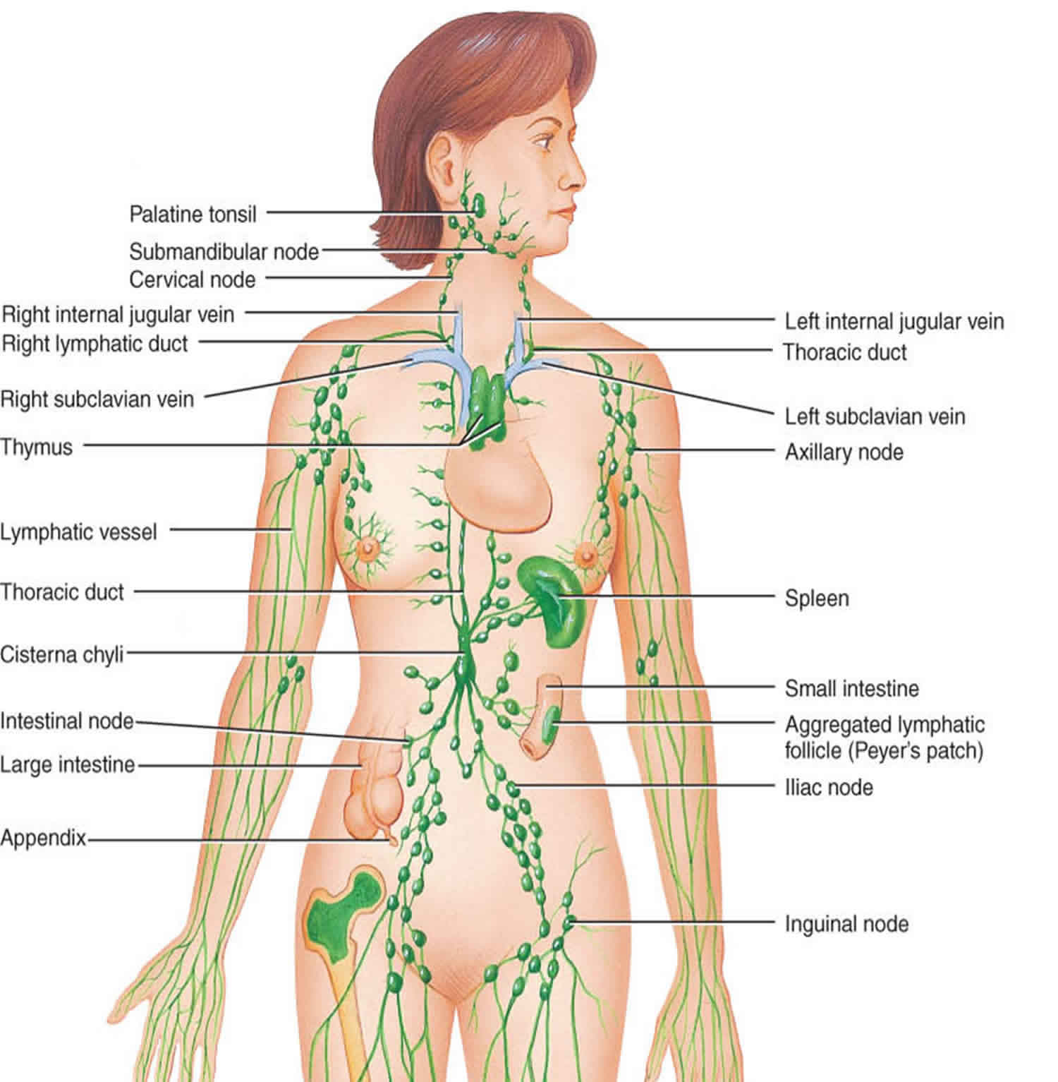
Lymph nodes are found widely throughout the body and are connected to one another by lymph vessels. Groups of lymph nodes are located in the neck, underarms, chest, abdomen, and groin. A clear fluid called lymph flows through lymph vessels and lymph nodes.
Lymph nodes contain B lymphocytes, T lymphocytes, and other types of immune system cells. These cells monitor lymph for the presence of “foreign” substances, such as bacteria and viruses. If a foreign substance is detected, some of the cells will become activated and an immune response will be triggered.
Lymph nodes are also important in helping to determine whether cancer cells have developed the ability to spread to other parts of the body. Many types of cancer spread through the lymphatic system, and one of the earliest sites of spread for these cancers is nearby lymph nodes.
The lymph nodes filter lymph fluid as it flows through them, trapping bacteria, viruses, and other foreign substances, which are then destroyed by special white blood cells called lymphocytes.
Lymph nodes may be found singly or in groups. And they may be as small as the head of a pin or as large as an olive. Groups of lymph nodes can be felt in the neck, groin, and underarms. Lymph nodes generally are not tender or painful. Most lymph nodes in the body cannot be felt.
The three most common sites of swollen lymph nodes are the neck, armpit, and groin.
Lymph glands become swollen for different reasons. If you have adenopathy because of a virus, they usually get better without treatment. If you’re not sure why you have swollen glands, if they are painful or getting bigger, or if you are feeling unwell, it’s a good idea to see your doctor.
Physical examination can be quite revealing especially with the location of the adenopathy and consideration of the lymphatic drainage of the related areas. Once the determination has been made that the lymphadenopathy is either localized or general, strict attention to the localized area must be paid. For example:
- Submandibular nodes typically drain the tongue the lips and the mouth and the conjunctiva
- Submental nodes typically drain the lower lip portions of the oropharynx and the cheek
- Jugular lymphadenopathy typically drains the tongue, the tonsils, the pinna, and the parotid gland
- Posterior cervical adenopathy typically is indicative of scalp, neck, skin of the arms and legs
- Pectoral thoracic cervical and axillary drainage
- Suboccipital nodes reflect drainage of the scalp in the head, and preauricular nodes reflect drainage the eyelids, conjunctiva temporal region, and pinna.
- Postauricular nodes reflect drainage at the scalp in the external auditory meatus.
- The right supraclavicular node represents drainage of the mediastinum the lungs in the esophagus
- Axillary nodes typically creating the arm at the thoracic wall and the breast.
- The epitrochlear nerve roots typically drain the ulnar aspect of the forearm and the hand.
- Inguinal nodes drain the penis, the scrotum, the vulva, vagina, the perineum, the gluteal region, and the lower abdominal wall and portions of the lower anal canal
Characterization of the lymph node morphology itself:
- Tenderness-pain may result from an inflammatory process or perforation and also may result from hemorrhage into the necrotic center of a malignant node. (Presence or absence of pain not a reliable differentiating factor for malignant nodes though.)
- Consistently firm rubbery nodes may suggest lymphoma; softer nodes are usually the result of infection or inflammatory conditions; hard stonelike nodes are typically a sign of cancer more commonly metastatic than primary.
- “Shotty” nodes refers to very small, scattered nodes that feel like shotgun pellets under the skin. This configuration is typically is found in cervical nodes of children with viral illnesses
- The designation of a “matting” configuration of nodes describes the pattern of clustered, seemingly conjoined lymph nodes. This is indicative of, but not pathognomonic, for malignancy.
What causes adenopathy?
Lymph nodes often swell in one location when a problem such as an injury, infection, or tumor develops in or near the lymph node. Which lymph nodes are swollen can help identify the problem.
- The glands on either side of the neck, under the jaw, or behind the ears commonly swell when you have a cold or sore throat. Glands can also swell following an injury, such as a cut or bite, near the gland or when a tumour or infection occurs in the mouth, head, or neck.
- Glands in the armpit (axillary lymph nodes) may swell from an injury or infection to the arm or hand. A rare cause of axillary swelling may be breast cancer or lymphoma.
- The lymph nodes in the groin (femoral or inguinal lymph nodes) may swell from an injury or infection in the foot, leg, groin, or genitals. In rare cases, testicular cancer, lymphoma, or melanoma may cause a lump in this area.
- Glands above the collarbone (supraclavicular lymph nodes) may swell from an infection or tumour in the areas of the lungs, breasts, neck, or abdomen.
Common sites for swollen lymph nodes include the neck, groin, and underarms.
The causes of adenopathy includes the following:
- Infectious disease
- Cancer
- Inflammatory disease
- Autoimmune disease
- Inborn metabolic storage disorder
- Exposure to toxic/medication
Infectious disease can be of viral, bacterial, mycobacterial, fungal or parasitic causes:
- Viral causes of adenopathy include HIV, mononucleosis caused by EBV (Ebstein-Barr Virus) or CMV (cytomegalovirus), roseola, HSV (herpes simplex virus), varicella, and adenovirus.
- Bacterial causes of adenopathy include Staphylococcus, Streptococcus, Salmonella, Syphilis, and Yersinia
- Mycobacterial cause of adenopathy include tuberculosis (TB) and Mycobacterium avium intracellulare (MAI)
- Fungal cause of adenopathy include coccidiomycosis and Candida
- Parasitic cause of adenopathy include toxoplasmosis, histoplasmosis, Chagas, and many of the ectoparasites
- Cancer causes of adenopathy include both primary malignancies and metastatic malignancies: Acute lymphoblastic leukemia (ALL), Hodgkin lymphoma, non-Hodgkin lymphoma, neuroblastoma, pediatric acute myelocytic leukemia, rhabdomyosarcoma, metastatic carcinoma of the lung, metastatic carcinoma of the viscera of the gastrointestinal (GI) tract, metastatic breast cancer, and metastatic thyroid cancer and metastatic renal cancer.
- Autoimmune disease these causes of adenopathy include sarcoidosis, juvenile rheumatoid arthritis (JRA), serum sickness, systemic lupus erythematosus (SLE)
- Exposures to toxins and medications that are common causes of adenopathy include the medications allopurinol, atenolol, captopril, carbamazepine, many of the cephalosporins, gold, hydralazine, penicillin, phenytoin, primidone, para methylamine, quinidine, the sulfonamides, and sulindac. The lifestyle exposures to alcohol, ultraviolet (UV) radiation, and tobacco can cause cancers with secondary adenopathy.
- Inborn metabolic storage disorders (including Niemann-Pick disease and Gaucher disease) are possible additional causes of adenopathy
The patient’s location and circumstance are very revealing for adenopathy. For example, in the developing world (sub-Saharan Africa, Southeast Asia, Indian subcontinent), exposure to parasites, HIV, and miliary tuberculosis (TB) are far more likely to be causes of generalized adenopathy then in the United States and Europe 1. Whereas, Epstein-Barr virus (EBV), streptococcal pharyngitis, and some cancer processes are more likely candidates to cause adenopathy in the United States and the remainder of the localized industrial world. An exposure history is very important for diagnosis.
- Exposure to blood and blood-borne products either through transfusion, unsafe sexual practices, intravenous drug abuse, or vocation
- Exposure to infectious disease whether it be travel, in the workplace, or the home
- Medication exposure-prescription, nonprescription, or supplements
- Exposure to animal-borne illness either via pets or the workplace
- Exposure to arthropod bites
Generalized adenopathy
Common Infective causes:
- Mononucleosis
- HIV
- Tuberculosis
- Typhoid fever
- Syphilis
- Plague
Cancers:
- Acute leukemia
- Hodgkin’s lymphoma
- Non-Hodgkin’s lymphoma
Metabolic Storage Disorders:
- Gaucher disease
- Niemann-Pick disease
Medication Reactions:
- Allopurinol
- Atenolol
- Captopril
- Carbamazepine
- Cephalosporin(s)
- Gold
- Hydralazine
- Penicillin
- Phenytoin
- Primidone
- Pyrimethamine
- Quinidine
- Sulfonamides
- Sulidac
Autoimmune Disease:
- Sjogren syndrome
- Sarcoidosis
- Rheumatoid arthritis
- Systemic lupus erythematosus
Localized Peripheral adenopathy
Head and Neck Lymph Nodes
Viral infection:
- Viral upper respiratory infection
- Mononucleosis
- Herpes virus
- Coxsackievirus
- Cytomegalovirus
- HIV
Bacterial infection:
- Staphylococcal aureus
- Group A Streptococcus pyogenes
- Mycobacterium
- Dental abscess
- Cat scratch disease
Cancer
- Hodgkin disease
- Non-Hodgkin lymphoma
- Thyroid cancer
- Squamous cell carcinomas of the head and neck
Inguinal Peripheral adenopathy
Infection
- Sexually transmitted infections (STIs)
- Cellulitis
Cancer
- Lymphoma
- Squamous cell carcinoma of genitalia
- Malignant melanoma
Axillary adenopathy
Infection
- Localized Staphylococcal aureus
- Cat-scratch disease
- Brucellosis
Cancer
- Lymphoma
- Breast cancer
- Melanoma
Reaction to breast implants
Supraclavicular Adenopathy
Infections
- Mycobacteria
- Fungi
Cancer
- Thoracic and abdominal neoplasms
- Hodgkin disease
- Non-Hodgkin lymphoma
What does it mean when lymph nodes swell in two or more areas of the body?
When lymph nodes swell in two or more areas of the body, it is called generalized adenopathy. This may be caused by:
- A viral illness, such as measles, rubella, chickenpox (varicella), or mumps.
- Mononucleosis (Epstein-Barr virus), which results in fever, sore throat, and fatigue, or cytomegalovirus (CMV), a viral infection that causes symptoms similar to those of mononucleosis.
- A bacterial illness, such as strep throat (caused by the streptococcus bacterium) or Lyme disease (a bacterial infection spread by certain types of ticks).
- Side effects of phenytoin (Dilantin), a medicine used to prevent seizures.
- Side effects of measles-mumps-rubella (MMR) vaccination.
- Cancer, such as leukemia, Hodgkin’s disease, and non-Hodgkin’s lymphoma.
- Acquired immunodeficiency syndrome (AIDS) , which develops after a person contracts HIV (human immunodeficiency virus). This virus attacks the immune system, making it difficult for the body to fight off infection and some disease.
- Syphilis , a sexually transmitted infection.
Adenopathy diagnosis
Laboratory evaluation of adenopathy
- Complete blood count (CBC) with manual differential: This is a foundational test in the diagnosis of both generalized and regional adenopathy. The number and differential of the white blood cells can indicate bacterial, viral, or fungal pathology. In addition, characteristic white blood cell (WBC) patterns are observed with several of the hematological neoplasms producing adenopathy
- EBV serology: Epstein-Barr viral mono is present causing regionalized adenopathy
- Sedimentation rate (ESR): A measure of inflammation though not diagnostic, it can contribute to diagnostic reasoning
- Cytomegalovirus titers: This viral serology is indicative of possible of CMV mononucleosis
- HIV serology: This serology can be used to diagnose acute HIV syndrome related adenopathy or to infer the diagnosis of secondary HIV-elated pathologies causing adenopathy.
- Bartonella henselae serology: Serology that may be indicative of the diagnosis of cat scratch adenopathy
- Fluorescent treponemal antibody absorption (FTA) \rapid plasma reagin (RPR) tests: These tests can establish if syphilis is a cause of adenopathy
- Herpes simplex serology: Serological testing to discern if herpes-related, mononucleosis-like syndrome is present or if regionalized inguinal adenopathy is secondary to herpes simplex exposure
- Toxoplasmosis serology: These serological tests can lead to a diagnosis of acute toxoplasmosis as a cause of adenopathy
- Hepatitis B serology: Serological tests for hepatitis B to establish it as a contributing factor for adenopathy
- Antinuclear antibody (ANA) test: A serological screening test for SLE that can help establish it as a cause for generalized adenopathy
Diagnostic imaging testing
- Chest x-ray: This radiological imaging modality can reveal tuberculosis, pulmonary sarcoidosis, and pulmonary neoplasm.
- Chest CT scan: This modality of radiological imaging can define the above processes and revealing of hilar adenopathy.
- Abdominal and pelvic CT scan: These images, in combination with chest CAT scan, can be revealing in cases of supraclavicular adenopathy and the diagnosis of secondary neoplasm.
- Ultrasonography: This imaging modality can be used in the assessment of number, size, site, shape, the marginal definition, and internal structures in patients with adenopathy. Of note, color Doppler ultrasonography is of use in distinguishing the vascular pattern between older pre-existing adenopathy and recent (newly active) adenopathy. Studies have indicated that a low long axis to short axis ratio of adenopathy as measured by ultrasound can be a significant indicator of lymphoma and metastatic cancer as a cause of adenopathy.
- MRI scanning: As with CAT scanning, this modality of diagnostic imaging has great utility in the evaluation of thoracic, abdominal, and pelvic masses.
Purified protein derivative (PPD) skin test
Tuberculosis is among the leading cause of both regional and generalized adenopathy in the non-industrialized world
How are adenopathy treated?
Treatment for swollen glands focuses on treating the cause. For example, a bacterial infection may be treated with antibiotics, while a viral infection often goes away on its own. If cancer is suspected, a biopsy may be done to confirm the diagnosis.
- Adenopathy caused by a primary cancer: Treatment of the cancer
- Adenopathy caused by metastatic cancer: Treatment of the metastasis and primary cancer
- Adenopathy caused by bacterial disease: Supportive care, antibiotics, and elimination of nidus of infection if applicable
- Adenopathy caused by viral disease: Observation and supportive care or treatment of the virus if particular antiviral medications exist
- Adenopathy caused by a toxin or medication exposure: Removal of offending medication if possible or avoidance of toxin
Any swollen lymph nodes that don’t go away or return to normal size within about a month should be checked by your doctor.
How long will lymph nodes remain swollen?
Lymph nodes may remain swollen or firm long after an initial infection is gone. This is especially true in children, whose glands may decrease in size while remaining firm and visible for many weeks.
- Freeman AM, Matto P. Adenopathy. [Updated 2018 Dec 13]. In: StatPearls [Internet]. Treasure Island (FL): StatPearls Publishing; 2019 Jan-. Available from: https://www.ncbi.nlm.nih.gov/books/NBK513250[↩]


