Contents
What is anasarca
Anasarca means massive generalized edema, which is is a serious condition caused an accumulation of fluid in the body that occurs as the capillary filtration exceeds the amount of fluid take out by lymphatic drainage 1. Edema is defined as a palpable swelling on the body produced by expansion of the interstitial fluid volume. Anasarca is caused by a variety of clinical conditions like heart failure, renal failure, liver failure, or problems with the lymphatic system. Edema usually becomes clinically apparent as the interstitial volume exceeds 2.5 -3 liters 2.
Because there are many causes of anasarca, it is best managed by a multidisciplinary team of healthcare workers. A team approach for management is necessary with support of nurses and pharmacists. The outcomes of patients with anasarca is guarded, for those with failing organs, the mortality rates are very high. Even benign causes can require long-term admission and recovery is a very slow process.
Anasarca complications
If anasarca is left untreated, common complications include, but not limited to skin ulcerations, skin infections, dyspnea, congestive heart failure, and death.
Anasarca causes
The most common causes of anasarca seen by clinicians are heart failure, cirrhosis, renal disease, and pregnancy. Anasarca may form as a response to an elevation in capillary hydraulic pressure or increased capillary permeability, a lower plasma oncotic pressure, or a combination of these changes. Edema also can be induced by lymphatic obstruction since the fluid that is usually filtered is not returned to the systemic circulation. The following are the clinical conditions for the various mechanisms described 3.
- An elevation in capillary hydraulic pressure
- Heart failure, kidney disease, early cirrhosis, pregnancy, drugs
- Venous obstruction or insufficiency states like DVT, hepatic venous congestion
- Increased capillary permeability
- Burns, trauma, sepsis, allergic reactions, malignant ascites
- Lymphatic obstruction
- Malignancy, Post lymph node dissection
- Hypoalbuminemia
- Nephrotic syndrome, liver disease, malnutrition
As a first step, when the fluid moves from the vascular space to the interstitium, it reduces the plasma volume. This reduces tissue perfusion. Poor tissue perfusion causes retention of sodium and water by the kidneys. Some of the excess fluid gained will be retained in the intravascular compartment. However, the alteration in capillary hemodynamics results in most of the retained fluid entering the interstitium and, eventually, becoming apparent as edema.
Anasarca diagnosis
Medical history should include the timing of the edema, and position changes, and whether it is unilateral or bilateral, and medication history as well as an assessment for systemic diseases. Chronic accumulation of more generalized edema is caused by the onset or exacerbation of systemic conditions such as congestive heart failure, renal disease, or hepatic disease. Dependent edema caused by venous insufficiency is more likely to improve with elevation and deteriorate with dependency. Edema associated with decreased plasma oncotic pressure (e.g., malabsorption, liver failure, nephrotic syndrome) does not change with dependency.
The physical examination can aid in establishing the diagnosis. The exam should be focused on identifying the pattern of edema – peripheral vs. pulmonary edema, pitting vs. non-pitting edema, and presence of jugular venous distension. Below are the exam findings of common clinical conditions that can cause anasarca.
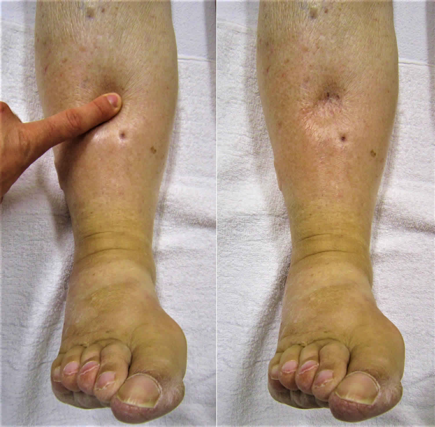
Patients with pulmonary edema complain primarily of dyspnea on exertion and orthopnea. Physical examination usually reveals wet rales, possible diastolic gallop (S3), and heart murmurs. Cardiac and renal disease are common causes of pulmonary edema. Peripheral edema is usually detected by the presence of pitting after pressure is applied to the edematous area for at least 5 seconds. Pitting reflects the movement of the excess interstitial water in response to pressure. It is usually seen in dependent areas like lower extremities in ambulatory patients and over the sacrum in patients who are bed-bound. Scrotal edema is also common in males. Nonpitting edema suggests lymphatic obstruction or hypothyroidism. Acute onset of unexplained unilateral leg edema should raise the possibility of deep vein thrombosis (DVT). Cirrhotic patients can develop ascites and then edema in the lower extremities because of an increase in venous pressure below the diseased liver. The presence of other signs of portal hypertension, such as distended abdominal wall veins and splenomegaly, also is suggestive of a primary hepatic disease.
Cause of edema can be determined by noting changes in skin temperature, color, and texture. Acute DVT and cellulitis may produce increased warmth over the affected area. The deposition of hemosiderin or chronic venous insufficiency often causes the skin to have a brawny, reddish hue and commonly involves the medial malleolus. As venous insufficiency progresses, it can result in lipodermatosclerosis which is associated with marked sclerotic and hyperpigmented tissue and characterized by fibrosis and hemosiderin deposition that can lead to venous ulcers over the medial malleolus. The ulcers may progress to deep, weeping erosions. Myxedema from hypothyroidism presents with a generalized, dry, thick skin with nonpitting periorbital edema and yellow to orange skin discoloration over the knees, elbows, palms, and soles. Pretibial myxedema occurs in patients with thyroid disease and is characterized by bilateral, asymmetric, nonpitting, scaly thickening and induration of the skin. These ulcers may be violaceous or slightly pigmented (yellow-brown) and often have an orange-peel appearance. The most frequent location of pretibial myxedema is over the lower legs, especially the pretibial areas or the dorsum of the foot.
Laboratory tests
Routine labs tests like a comprehensive metabolic panel can help assess renal function, albumin, and liver function tests. Urinalysis must be obtained in all children with edema. Dipstick testing principally detects albumin and need additional protein sulfosalicylic acid precipitation test (SAS) to detect globulins and Bence-Jones proteins. Urine protein/creatinine ratio or 24-hr urine for protein can obviate the need for sulfosalicylic acid precipitation testing. The finding of a markedly positive dipstick for protein in combination with hypoalbuminemia and clinical edema is virtually diagnostic of a nephrotic syndrome. Brain natriuretic peptide measurement can give clues to underlying congestive heart failure. Albumin and liver function tests can aid in diagnosing liver cirrhosis. Chest radiography is helpful in detecting cardiac failure, pulmonary edema, and pleural effusions. Because of safety, ease of use, and the information provided, the most commonly used radiographic technique in patients presenting with renal disease is renal ultrasonography. Ultrasonography allows the clinician to characterize kidney size and assess for cystic renal disease and hydronephrosis. Cardiac echocardiography can evaluate ventricular function, assess for the presence of a pericardial effusion, and aid in the diagnosis of cardiac disease 4.
Imaging tests
Echocardiography to evaluate pulmonary arterial pressures is recommended for patients with obstructive sleep apnea and edema. Venous ultrasonography is the imaging of choice in the evaluation of suspected DVT. Duplex ultrasonography also can be used to confirm a chronic venous insufficiency. Magnetic resonance angiography with venography of the lower extremity and pelvis can be used to evaluate for intrinsic or extrinsic pelvic or thigh DVT. Left iliac vein compression by the right iliac artery (May-Thurner syndrome) should be suspected in women who are 18 and 30 years of age and present with edema of the left lower extremity. Lymphoscintigraphy is the method of choice for evaluating lymphedema when the diagnosis cannot be made clinically. MRI may aid in the diagnosis of musculoskeletal etiologies like a gastrocnemius tear or popliteal cyst. T1-weighted magnetic resonance lymphangiography can visualize the lymphatic channels when lymphedema is suspected.
Anasarca treatment
Management of anasarca is guided by the underlying cause. This commonly includes chronic venous insufficiency, lymphedema, DVT, and medication-induced edema. Pulmonary edema is the only form of generalized edema that is life-threatening and requires immediate therapy. In all other edematous states, removal of the excess fluid can proceed more slowly as it is not acutely life-threatening to the patient. In patients with generalized edema due to heart failure, the nephrotic syndrome, or primary sodium retention, the edema fluid can be mobilized rapidly. In patients with anasarca, removing 2 to 3 liters or more of edema fluid in 24 hours can ordinarily be accomplished without a clinically significant reduction in plasma volume. In patients with localized edema due to venous or lymphatic obstruction or malignant ascites, diuretic therapy can lead to volume depletion. Diuretic therapy in generalized edematous states is usually begun with a loop diuretic, such as furosemide. In patients with cirrhosis, the combination of spironolactone and a loop diuretic is the preferred initial diuretic regimen to prevent hypokalemia. As hypokalemia predisposes to increased production of ammonia. Higher doses of diuretics may be required in patients with nephrotic syndrome. Some cases of idiopathic edema are diuretic-induced, and the initial approach in patients with idiopathic edema who are already on diuretics is to stop the diuretics for at least 2 to 3 weeks and encourage sodium restriction in diet. The mainstays of therapy of lower-extremity edema due to venous insufficiency are mechanical therapies, including leg elevation and compression stockings with 20 to 30 mmHg for mild edema and 30 to 40 mmHg for severe edema complicated by ulceration. Compression therapy is contraindicated in patients with a peripheral arterial disease. Local skin and wound care of venous ulcers is essential in preventing secondary cellulitis and dermatitis. Eczematous (stasis) dermatitis, characterized by dry, inflamed, scaling skin overlying superficial varicose veins, often occurs in patients with chronic venous insufficiency. Treatment includes daily hydration with emollients and short courses of topical steroid creams for the severely inflamed skin. Primary lymphedema treatment involves complex decongestive physiotherapy, including manual lymphatic massage and multilayer bandages. The first goal is the improvement of fluid resorption and continue until achieving the maximum therapeutic response. The maintenance phase of treatment includes compression stockings at 30 to 40 mmHg. Pneumatic compression devices can be used to augment standard therapies. Surgical debulking or bypass procedures are limited to severe refractory cases. Diuretics are not demonstrated to be effective in the treatment of lymphedema.
Anasarca prognosis
Prognosis of anasarca depends on the underlying cause. Reversible causes may carry favorable outcomes, whereas irreversible etiologies and malignancies have a poor prognosis. However in most of the situations, by the time anasarca has developed, the underlying problem has progressed beyond cure.
- Kattula SRST, Baradhi KM. Anasarca. [Updated 2018 Dec 20]. In: StatPearls [Internet]. Treasure Island (FL): StatPearls Publishing; 2019 Jan-. Available from: https://www.ncbi.nlm.nih.gov/books/NBK519013[↩]
- Wang CS, Greenbaum LA. Nephrotic Syndrome. Pediatr. Clin. North Am. 2019 Feb;66(1):73-85[↩]
- Klanderman RB, Bosboom JJ, Migdady Y, Veelo DP, Geerts BF, Murphy MF, Vlaar APJ. Transfusion-associated circulatory overload-a systematic review of diagnostic biomarkers. Transfusion. 2019 Feb;59(2):795-805.[↩]
- Sharma R, Sharma S. StatPearls [Internet]. StatPearls Publishing; Treasure Island (FL): Oct 27, 2018. Physiology, Blood Volume.[↩]


