
What is dermis
The dermis, the second major region of the skin, is a strong, flexible connective tissue. The cells of the dermis are typical of any connective tissue proper: fibroblasts, macrophages, mast cells, and scattered white blood cells. The fiber types—collagen, elastic, and reticular—also are typical.
The function of the dermis is to bind the entire body together like a body stocking. It is your “hide” and corresponds to animal hides used to make leather products.
The dermis is also the site where all the accessory structures of the skin – your hair, nails, and a variety of multicellular exocrine glands originate. These structures are located in the dermis and protrude through the epidermis to the surface.
The dermis has two regions:
- the Papillary Dermis and
- the Reticular Dermis.
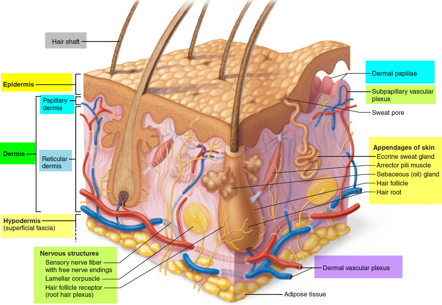
Figure 1. Dermis layers
Papillary layer of Dermis
The papillary dermis, the superficial 20% of the dermis, is areolar connective tissue containing very thin collagen and elastic fibers. It includes the dermal papillae (“nipples”), fingerlike projections that extend into the overlying epidermis. These projections of the dermal papillae into the epidermis increase the surface area for exchange of gases, nutrients, and waste products between these layers. Recall that the epidermis is avascular and depends on the diffusion of these materials from the underlying dermis.
The inter-digitation of these layers also strengthens the dermal-epidermal junction and thus reduces blister formation.
On the palms of the hands and the soles of the feet, the dermal papillae lie atop larger mounds called dermal ridges. These elevate the overlying epidermis into epidermal ridges or friction ridges, which create fingerprints, palm-prints, and footprints. Epidermal ridges increase friction and enhance the gripping ability of the hands and feet.
Patterns of these ridges are genetically determined and unique to each person. Because sweat pores open along the crests of the friction ridges, they leave distinct fingerprints on almost anything they touch. Thus, fingerprints are “sweat films.”
Reticular layer of Dermis
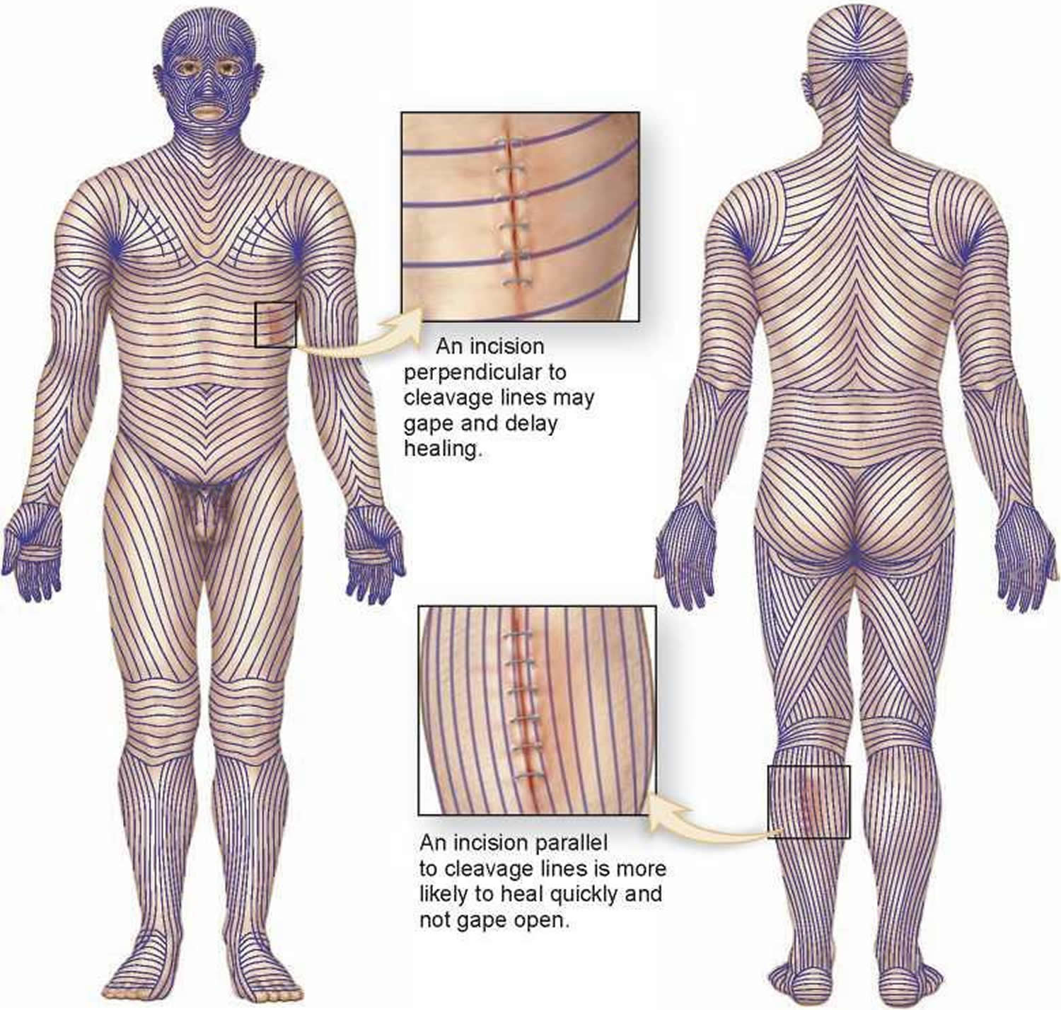
The deeper reticular dermis, which accounts for about 80% of the thickness of the dermis, is dense irregular connective tissue. Its extracellular matrix contains thick bundles of interlacing collagen and elastic fibers that run in many different planes. However, most run parallel to the skin surface. The reticular layer is named for its networks of collagen fibers (reticulum = network); the name does not imply any special abundance of reticular fibers. Separations or less dense regions between the collagen bundles form the cleavage lines or tension lines of the skin.
These invisible lines occur over the entire body: They run longitudinally in the skin of the limbs and head and in circular patterns around the neck and trunk. A knowledge of cleavage lines is important to surgeons. Incisions made parallel to these lines tend to gape less and heal more readily than incisions made across cleavage lines.
Figure 2. Cleavage or tension lines of the skin
The collagen fibers of the dermis give skin its strength and resilience. Thus, many jabs and scrapes do not penetrate this tough layer. Furthermore, elastic fibers in the dermis provide the skin with stretch-recoil properties. Extreme stretching of the skin, as occurs in obesity and pregnancy, can tear the collagen in the dermis. Such dermal tearing results in silvery white scars called striae (“streaks”), which is commonly known as “stretch marks.” The dermis is also the receptive site for the pigments used in tattoos.
From the deep part of the dermis arise the skin surface markings called flexure lines. Observe, for example, the deep skin creases on your palm. These result from
a continual folding of the skin, often over joints, where the dermis attaches tightly to underlying structures. Flexure lines are also visible on the wrists, soles, fingers, and toes.
The dermis is richly supplied with nerve fiber and blood vessels. The dermal blood vessels consist of two vascular plexuses (a plexus is a network of converging and diverging vessels). The deep dermal plexus is located between the hypodermis and the dermis. It nourishes the hypodermis and the structures located within the deeper portions of the dermis. The more superficial subpapillary plexus, located just below the dermal papillae, supplies the more superficial dermal structures, the dermal papillae, and the epidermis.
Dermal blood vessels do more than just nourish the dermis and overlying epidermis; they also perform a critical role in temperature regulation. These vessels are so extensive that they can hold 5% of all blood in the body. When internal organs need more blood or more heat, nerves stimulate the dermal vessels to constrict, shunting more blood into the general circulation and making it available to the internal organs. By contrast, on hot days the dermal vessels engorge with warm blood, cooling the body by radiating heat away from it.
Below the dermis is another connective tissue layer, the hypodermis, which is not part of the skin but is customarily studied in conjunction with it. Most of the skin is 1 to 2 mm thick, but it ranges from less than 0.5 mm on the eyelids to 6 mm between the shoulder blades. The difference is due mainly to variation in thickness of the dermis, although skin is classified as thick or thin based on the relative thickness of the epidermis alone.
The Epidermis
The skin consists of two main parts, the most superficial part of the skin is the epidermis. The epidermis is a thinner portion of the skin, which is composed of
epithelial tissue. While the epidermis is avascular, the dermis is vascular. For this reason, if you cut the epidermis there is no bleeding, but if the cut penetrates to the dermis there is bleeding.
The epidermis is composed of keratinized stratified squamous epithelium. It contains five principal types of cells: stem cells, keratinocytes, melanocytes, Merkel cells (Tactile cells) and Dendritic cells (Langerhans cells). About 90% of epidermal cells are keratinocytes, which are arranged in four or five layers and produce the protein keratin. Keratin is a tough, fibrous protein that helps protect the skin and underlying tissues from abrasions, heat, microbes, and chemicals.
Keratinocytes also produce lamellar granules, which release a water-repellent sealant that decreases water entry and loss and inhibits the entry of foreign materials.
About 8% of the epidermal cells are melanocytes, which produce the pigment melanin. Their long, slender projections extend between the keratinocytes and transfer melanin granules to them. Melanin is a yellowred or brown-black pigment that contributes to skin color and absorbs damaging ultraviolet (UV) light. Once inside keratinocytes, the melanin granules cluster to form a protective veil over the nucleus, on the side toward the skin surface. In this way, they shield the nuclear DNA from damage by UV light. Although their melanin granules effectively protect keratinocytes, melanocytes themselves are particularly susceptible to damage by UV light.
Intraepidermal macrophages or Langerhans cells (Dendritic cells) arise from red bone marrow and migrate to the epidermis, where they constitute a small fraction of the epidermal cells. They participate in immune responses mounted against microbes that invade the skin, and are easily damaged by UV light. Their role in the immune response is to help other cells of the immune system recognize an invading microbe and destroy it.
Tactile epithelial cells, or Merkel cells, are the least numerous of the epidermal cells. They are located in the deepest layer of the epidermis, where they contact the flattened process of a sensory neuron (nerve cell), a structure called a tactile disc or Merkel disc. Tactile epithelial cells and their associated tactile discs detect touch sensations.
Several distinct layers of keratinocytes in various stages of development form the epidermis. In most regions of the body the epidermis has four strata or layers —stratum basale, stratum spinosum, stratum granulosum, and a thin stratum corneum. This is called thin skin. Where exposure to friction is greatest, such as in the fingertips, palms, and soles, the epidermis has five layers—stratum basale, stratum spinosum, stratum granulosum, stratum lucidum, and a thick stratum corneum. This is called thick skin.

