Contents
What is an epidermoid cyst
Epidermoid cysts, sometimes known as epidermoid inclusion cyst, epidermal inclusion cyst, infundibular cyst, epidermal cyst or sebaceous cysts (a misnomer), is a sphere of skin within the skin containing a soft “cheesy” material composed of keratin, a protein component of skin, hair, and nails. In the past, epidermoid cysts were wrongly known as ‘sebaceous’ cysts but this term should be used only for a quite different and much less common type of cyst that is filled with a clear oily liquid made by sebaceous (grease) glands. The epidermoid cyst wall constantly flakes into the center of the cyst causing it to enlarge over time. Multiple epidermoid cysts may indicate Gardner’s syndrome. Gardner’s syndrome is a rare autosomal dominant disorder. It is associated with multiple epidermoid cysts (usually irregularly distributed on the face, scalp and extremities), polyposis of the colon, osteomas (mainly of the skull) and fibromas.
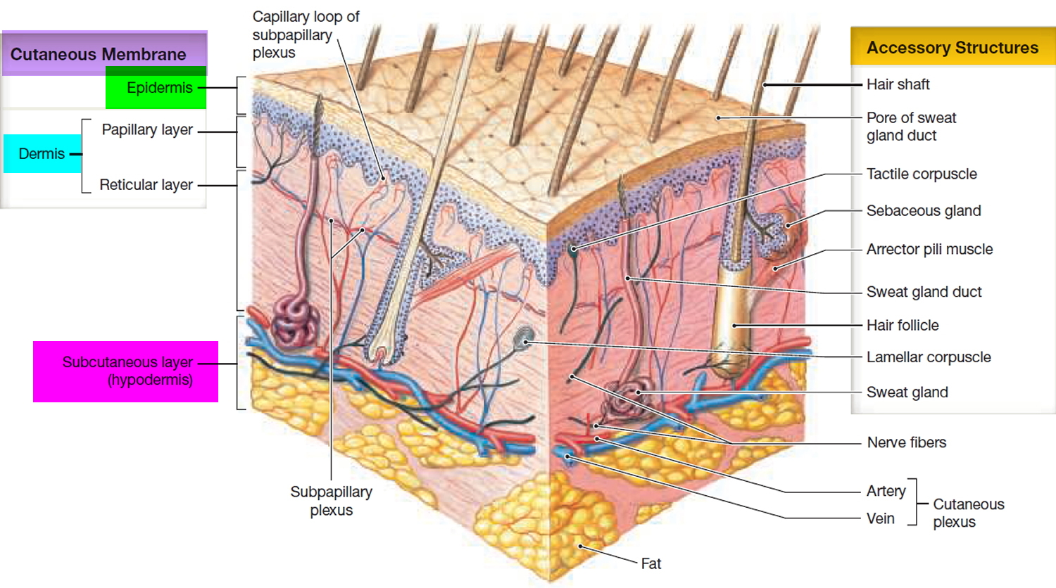
Epidermoid cysts form when the top layer of skin (epidermis) grows into the middle layer of the skin (dermis). Their origin is the follicular infundibulum. This may occur due to injury causing dermal implantation of epidermis (epidermal inclusion cyst) or blocked hair follicles. The lesion may be asymptomatic, but rupture of the epidermoid cyst can result in significant discomfort.
Although an epidermoid cyst is a benign walled-off cavity filled with keratin which originates from the hair follicle unit. In one review of 1904 epidermoid cysts, three cases of squamous cell carcinoma arose from an epidermoid cyst and one case of squamous cell carcinoma developed from a pilar cyst 1.
Epidermoid cysts are common, not cancerous, and not contagious. Age of presentation – most frequently affected are young and middle-aged adults, they are rare in childhood.
Epidermoid cysts tend to be asymptomatic unless they become infected (see infected epidermoid cyst in Figure 6 below). Infected epidermoid cysts enlarge, becoming red and tender, and eventually discharge pus.
Can epidermoid cyst be cured?
Yes. There are several simple and effective ways of removing them under local anesthetic. However, it is fairly common for new cysts to grow at a later date, especially on the scalp or genital skin.
Most epidermoid cysts don’t cause problems or need treatment. See your doctor if you have one or more that:
- Grows rapidly
- Ruptures or becomes painful or infected
- Occurs in a spot that’s constantly irritated
- Bothers you for cosmetic reasons
- Is in an unusual location, such as a finger and toe
Furthermore, if you find any sort of lump in your skin, you should consult your doctor. Epidermoid cysts are not dangerous, but your doctor should be asked to look at them to make sure that the diagnosis is right.
Figure 1. Skin structure
Figure 2. Epidermoid inclusion cyst
Figure 3. Epidermoid cyst
Epidermoid cyst brain
Epidermoid brain cysts (also called intracranial epidermoid cysts or tumors) usually form in the very early stages of the development of a baby (embryo). The cysts develop when cells that are meant to become skin, hair, and nails (epithelial cells) are trapped among the cells that form the brain 2. Less commonly, the cysts may develop later in life (acquired) when an injury or surgery causes epithelial cells to be trapped in brain tissue 3. An epidermoid brain cyst has a thin outer layer of epithelial cells surrounding fluid, keratin, and cholesterol. Epidermoid brain cysts tend to be located in the area where the top part of the brain meets the brain stem.
Although epidermoid brain cysts are usually benign (not cancerous) and slow growing, the cysts may grow around and encase cranial nerves and arteries. Thus, epidermoid brain cysts are most often diagnosed in middle-aged adults when the cysts have grown large enough to cause symptoms. Symptoms may include hearing loss, ringing in the ears (tinnitus), headaches, involuntary twitching of the face, or chronic, severe face pain (trigeminal neuralgia). Rarely, an epidermoid cyst may leak into surrounding tissue and cause the lining of the brain (meninges) to become inflamed (aseptic meningitis, meaning the meningitis is not caused by a virus or bacteria).
Epidermoid brain cysts may be diagnosed by MRI and CT scans. Treatment usually involves surgery. Complete removal may be difficult if the cysts have surrounded or are very close to cranial nerves, arteries, or brain tissue. Regrowth of the cysts may occur, but in most cases, due to slow growth, symptoms may not return for years. If aseptic meningitis develops due to leakage of the cyst, steroids may be used to control the inflammation. There have been some reports of existing cysts or remnants of cysts left behind after surgery developing into cancer (since the cyst is made of skin cells, the cancer is usually a form of skin cancer, most commonly squamous cell carcinoma).
Epidermoid cyst causes
The surface of your skin (epidermis) is made up of a thin, protective layer of cells that your body continuously sheds. Most epidermoid cysts form when these cells move deeper into your skin and multiply rather than slough off. Sometimes the cysts form due to irritation or injury of the skin or the most superficial portion of a hair follicle.
The epidermal cells form the walls of the cyst and then secrete the protein keratin into the interior. The keratin is the thick, yellow substance that sometimes drains from the cyst. This abnormal growth of cells may be due to a damaged hair follicle or oil gland in your skin.
Epidermoid cysts are the most common type of cyst. Epidermoid cysts affect young and middle aged adults. They may be primary or they may arise from disrupted follicular structures due to trauma (epidermal inclusion cyst) or comedone formation (blackheads), so they are common in acne. Many result from inflammation around a pilosebaceous hair follicle and can, therefore, follow on from the more severe lesions of acne vulgaris (common acne).
Multiple cysts may occur in the conjunction with Gardner syndrome and in nevoid basal cell carcinoma syndrome.
Tiny superficial epidermoid cysts are known as milia.
Risk factors for epidermoid cyst
Nearly anyone can develop one or more epidermoid cysts, but these factors make you more susceptible:
- Being past puberty
- Having certain rare genetic disorders
- Injuring the skin
Epidermoid cyst symptoms
Epidermoid cysts appear as flesh colored to yellowish, firm, round nodules of variable size – 0.5-2.0 cm in diameter is typical. A central pore or punctum may be present. Patient often relates being able to squeeze out a foul-smelling material.
They are usually symptomless but sometimes discharge a foul smelling, “cheese-like” material. Less frequently, the cysts can be painful due to inflammation or infection.
- Epidermoid cysts occur on face, neck, trunk or anywhere where there is little hair.
- Epidermoid cysts are most common on the face, neck, genital skin and upper trunk.
- Most epidermoid cysts arise in adult life.
- They are more than twice as common in men as in women.
- They present as one or more flesh–colored to yellowish, adherent, firm, round nodules of variable size.
- A central pore or punctum may be present.
- Keratinous contents are soft, cheese-like and malodorous.
- Scrotal and labial cysts are frequently multiple and may calcify.
Epidermoid cyst is also called follicular infundibular cyst, epidermal cyst, keratin cyst.
Figure 4. Epidermoid cyst (note the central pore)
Figure 5. Epidermoid cyst – foul-smelling soft “cheesy” material composed of keratin coming out from the center of the cyst
Figure 6. Infected epidermoid cyst
Epidermoid cyst complications
Potential complications of epidermoid cysts include:
- Inflammation. An epidermoid cyst can become tender and swollen, even if it’s not infected. An inflamed cyst is difficult to remove. Your doctor is likely to postpone removing it until the inflammation subsides.
- Rupture. A ruptured cyst often leads to a boil-like infection that requires prompt treatment.
- Infection. Cysts can become infected and painful (abscessed).
- Skin cancer. In very rare cases, epidermoid cysts can lead to skin cancer.
Epidermoid cyst diagnosis
Doctors can usually make a diagnosis by looking at the cyst. Your doctor may also scrape off skin cells and examine them under a microscope or take a skin sample (biopsy) for detailed analysis in the laboratory.
Epidermoid cysts look like sebaceous cysts, but they’re different. True epidermoid cysts result from damage to hair follicles or the outer layer of skin (epidermis).
Epidermoid cyst treatment
Epidermoid cysts are harmless, and small ones that give no trouble can safely be left alone.
Your doctor may give you an antibiotic if your cyst becomes infected.
Talk with your doctor about these options:
- Injection. This treatment involves injecting the cyst with a medicine that reduces swelling and inflammation.
- Incision and drainage. With this method, your doctor makes a small cut in the cyst and gently squeezes out the contents. This is a fairly quick and easy method, but cysts often recur after this treatment.
- Minor surgery. Your doctor can remove the entire cyst under a local anesthetic, but this does leave a scar. You may need to return to the doctor’s office to have stitches removed. Minor surgery is safe and effective and usually prevents cysts from recurring. If your cyst is inflamed, your doctor may delay the surgery.
Epidermoid cyst removal
Reasons for epidermoid cyst removal may include the following:
- If the epidermoid cyst is unsightly and easily seen by others.
- If it interferes with everyday life, for example by catching on your comb.
- If the epidermoid cyst becomes infected.
It is important that the doctor removes the whole of the lining during the operation (and doesn’t just cut into it to remove the contents), as doing so cuts down the chance of the cyst growing back.
Figure 7. Epidermoid cyst removal – note the sac that must be removed or else the epidermoid cyst will recur.
Epidermoid cyst home treatment
You can’t stop epidermoid cysts from forming. But you can help prevent scarring and infection by:
- Not squeezing a cyst yourself
- Placing a warm, moist cloth over the area to help the cyst drain and heal.
- J Dermatolog Treat. 2016;27:95[↩]
- Cysts. http://www.abta.org/brain-tumor-information/types-of-tumors/brain-cysts.html[↩]
- Ravindran K, Rogers TW, Yuen T, Gaillard F. Intracranial white epidermoid cyst with dystrophic calcification – A case report and literature review. J Clin Neurosc. August 2017; 42:43-47. https://www.ncbi.nlm.nih.gov/pubmed/28342703[↩]


