Contents
What are causes of dark inner thighs
Intertrigo is inflammation and irritation of touching skin surfaces in body fold regions (armpits, under the breasts, belly folds, buttocks, inner thighs and groin folds and sometimes between fingers or toes) 1. Redness and breaks in the skin (erosions) of opposing skin surfaces may be noted. The area may ooze or be sore or itchy. Intertrigo can be worsened by any conditions causing increased heat, wetness, and friction. Intertrigo may be complicated by superficial skin infection with yeast or bacteria.
Figure 1. Intertrigo of the thigh area

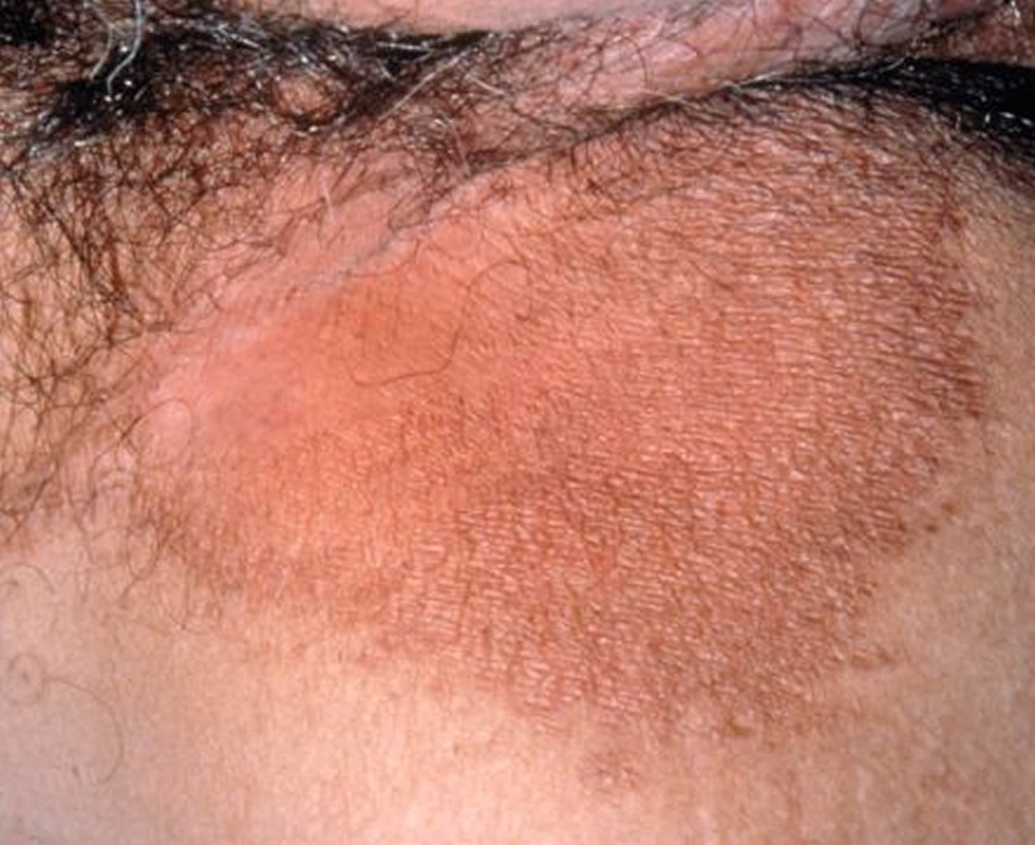
Figure 2. Inner thigh Candida (yeast) skin infection
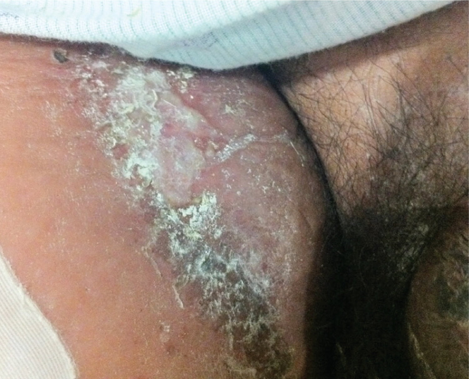
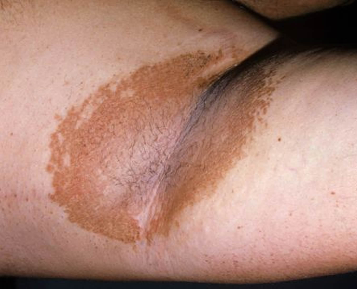
Figure 3. Erythrasma (bacterial infection causing dark inner thighs)
Symptoms of intertrigo (of dark inner thighs)
Symptoms of intertrigo include a red or reddish-brown rash that can appear anywhere skin rubs together or traps wetness. This rash may burn or itch. The most common areas include:
- between toes
- in the armpits
- in the groin area
- on the underside of the belly or breasts
- in the crease of the neck.
Intertrigo can also affect the skin between the buttocks. The affected skin will often be very raw and may itch or ooze. In severe cases, intertrigo may cause a foul odor, and the skin may crack and bleed.
Intertrigo associated with a fungal superinfection may produce satellite papules and pustules. Candidal intertrigo (Figure 2) is often associated with a foul-smelling odor. In the presence of a bacterial superinfection, plaques and abscesses / blackheads may form 2.
Causes of intertrigo (dark inner thighs)
Intertrigo affects the top layers of skin. It is caused by moisture, bacteria, or fungus in the folds of the skin. The affected areas of skin are usually pink to brown. If the skin is very moist, it may begin to break down. In severe cases, there may be a bad odor.
Intertrigo most often occurs in patients with obesity (body mass index more than 30 kg per m2), diabetes mellitus, or human immunodeficiency virus infection, and in those who are bedridden. It also occurs in patients with large skin folds and those who wear diapers or other items that trap moisture against the skin. It may also occur in people who must stay in bed or who wear medical devices such as artificial limbs, splints, and braces. These devices may trap moisture against the skin. There is a linear increase in the severity of obesity and the presence of intertrigo 3. Patients who are obese sweat more profusely because of their thick layers of subcutaneous brown fat, generating more heat than persons with normal body mass 4. This increases thermal, frictional, and moisture components of the skin.
As the stratum corneum becomes macerated because of hyperhydration (excess moisture), the friction intensifies and further weakens and damages the epidermal tissue. The condition can progress to severe inflammation and skin breakdown. This erosion of the epidermal barrier may create an entry point for micro-organisms that cause secondary infections 5.
The diagnosis of secondary fungal infections is commonly made clinically, based on the characteristic appearance and distribution of satellite papules and pustules 6. The diagnosis may, however, be confirmed with a potassium hydroxide preparation positive for pseudohyphae and spores. Additionally, a potassium hydroxide preparation, Wood lamp examination, or culture of skin scrapings can diagnose conditions such as Candida or dermatophyte infections. The presence of hyphae on potassium hydroxide examination confirms dermatophytic lesions, including tinea versicolor and tinea corporis, and the presence of pseudohyphae confirms Candida infection. A video showing a potassium hydroxide examination of a fungal infection is available at http://www.youtube.com/watch?v=ugeMsyEDJaw. The Wood lamp examination fluoresces green with Pseudomonas infection and coral-red with erythrasma (Figure 4), a bacterial infection caused by Corynebacterium minutissimum 7.
The condition is most common in warm, moist climates.
Table 1. Differential Diagnosis of Cutaneous Diseases Resembling Intertrigo
| Disease | Characteristics |
|---|---|
Infectious diseases | |
Candidiasis (moniliasis) | Superficial erythematous infection, commonly affecting moist, cutaneous areas of the skin; satellite pustules |
Dermatophytosis (tinea corporis, tinea versicolor) | Pruritic infections of nonviable keratinized tissues, such as nails and hair; contains a leading scale |
Erythrasma | Small, red-brown macules that may coalesce into larger patches with sharp borders; may be asymptomatic or pruritic; fluoresces coral-red on Wood lamp examination |
Pyoderma | Aggressive infection with boggy, blue-red bullae that progress to deep ulcers with hemorrhagic bases |
Scabies | Infection with intense pruritus and minimal cutaneous manifestations, including intertriginous burrows and papules; the head is spared in all age groups except infants |
Seborrheic dermatitis | Yellow, greasy, scaly plaques with overlying erythema; most often affects the face, postauricular region, and chest |
Noninfectious inflammatory diseases | |
Atopic dermatitis | Red or brownish patches with intense pruritus; personal and/or family history of seasonal allergies and asthma is common |
Pemphigus vulgaris | Serious, often fatal, autoimmune disease; flaccid bullae, Nikolsky sign (i.e., disruption of the epidermal layer with lateral pressure, leading to ulceration) |
Psoriasis | Chronic scaling papules and plaques; often affects extensor surfaces of the elbows and knees; associated with silvery scale |
Noninflammatory diseases | |
Acanthosis nigricans | Hyperpigmentation with velvety, thickened skin, predominantly on posterior neck and body folds |
Hidradenitis suppurativa | Chronic condition of apocrine gland–bearing skin in the axillae or anogenital region; deep abscesses and old scars |
Intertrigo | Pruritic regions of erythema and peripheral scaling between skin folds |
Lichen sclerosus | Well-demarcated, hypopigmented, atrophic plaques on the genitalia, trunk, and axillae |
Neoplasms | |
Bowen disease | Solitary, enlarging, erythematous, well-defined plaque |
Paget disease | Erythematous plaques with scaling, crusting, and/or exudation; affects the breast, axillae, or anogenital region; may represent a neoplastic process |
Superficial basal cell carcinoma | Most common skin cancer; pink or flesh-colored papule often containing a telangiectatic vessel |
What is Erythrasma
Dark inner thighs is a long-term top skin infection involving layers of the skin caused by bacteria Corynebacterium minutissimum, also known as erythrasma 8.
Erythrasma affects mostly adults, especially those with diabetes and those living in the tropics. Erythrasma is most common in the foot, where it causes scaling, cracking, and breakdown of the skin between the 4th and 5th toes. This infection is also common in the groin, where it causes irregularly shaped pink or brown patches and fine scaling especially where the thighs touch the scrotum (in men). The armpits, skinfolds under the breasts or on the abdomen, and the area between the vaginal opening and the anus (perineum) are prone to this infection, particularly among people with diabetes and among obese middle-aged women. In some people, the infection spreads to the torso and anal area.
Although erythrasma may be confused with a fungal infection, doctors can easily diagnose erythrasma because skin infected with Corynebacterium glows coral-red under an ultraviolet light (Wood’s lamp).
An antibiotic given by mouth, such as erythromycin or tetracycline, can eliminate the infection. Antibacterial soaps, such as chlorhexidine, may also help. Drugs applied directly to the affected area (topically), such as erythromycin and clindamycin, are also effective. Antifungal creams such as miconazole may be helpful if yeast or fungus is present in the affected areas as well. Erythrasma may recur, necessitating a second treatment.
Causes of Erythrasma
Erythrasma is caused by the bacteria Corynebacterium minutissimum.
Erythrasma is more common in warm climates. You are more likely to develop this condition if you are overweight, older, or have diabetes.
Symptoms of Erythrasma
The main symptoms are reddish-brown slightly scaly patches with sharp borders (see Figures 3, 4, 5 and 6). They may itch slightly. The patches occur in moist areas such as the groin, armpit, and skin folds.
The patches often look similar to other fungal infections, such as ringworm.
Exams and Tests
The health care provider will check your skin and ask about the symptoms.
These tests can help diagnose erythrasma:
- Lab tests of scrapings from the skin patch
- Examination under a special UV lamp called a Wood’s lamp
- A skin biopsy.
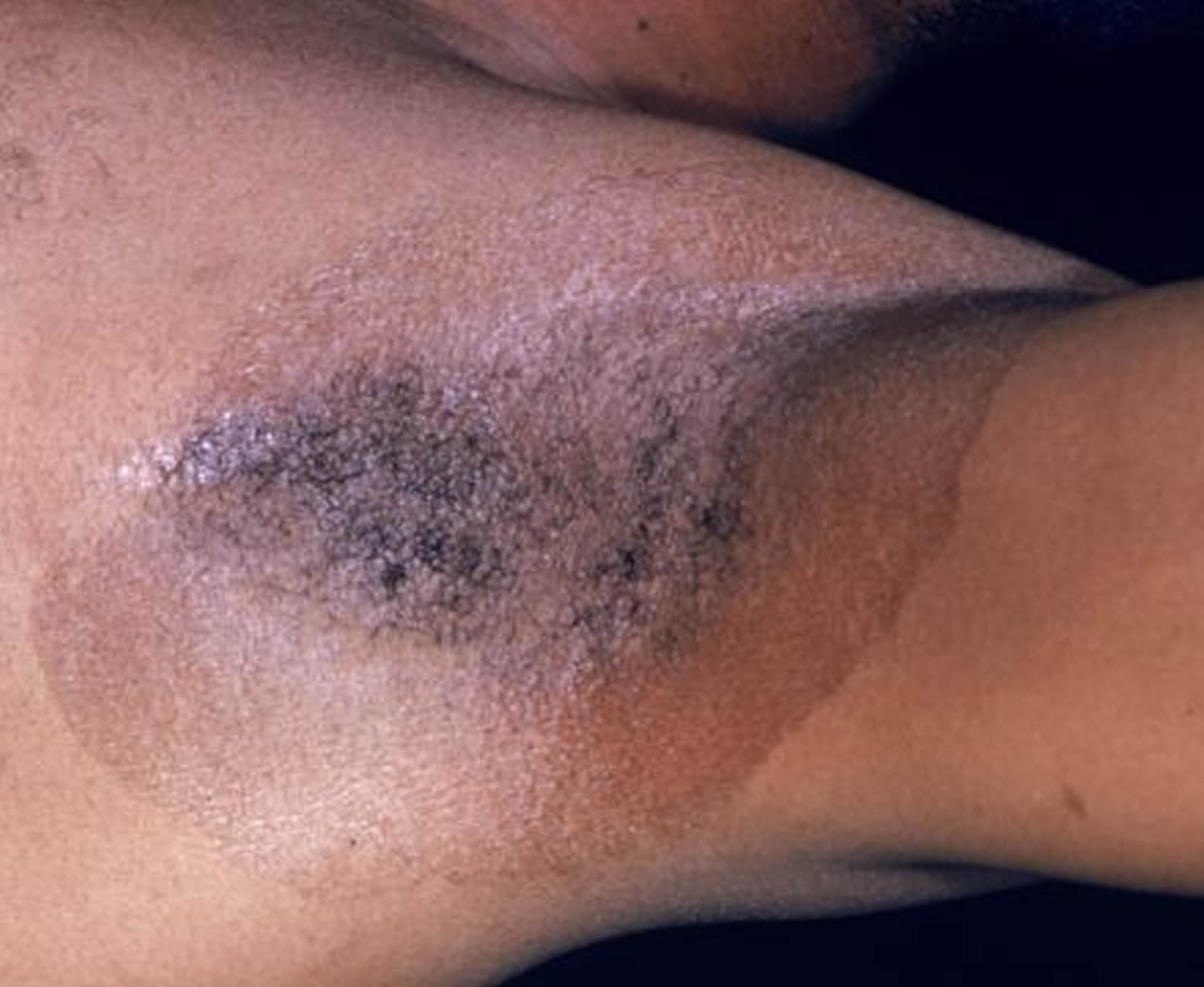
Figure 4. Underarm erythrasma
Note: Erythrasma is displayed as slowly enlarging brown or pink, rough areas in body folds.
Figure 5. Underarm erythrasma
Note: Erythrasma often has a very sharp border
Figure 6. Underarm erythrasma under Wood’s lamp
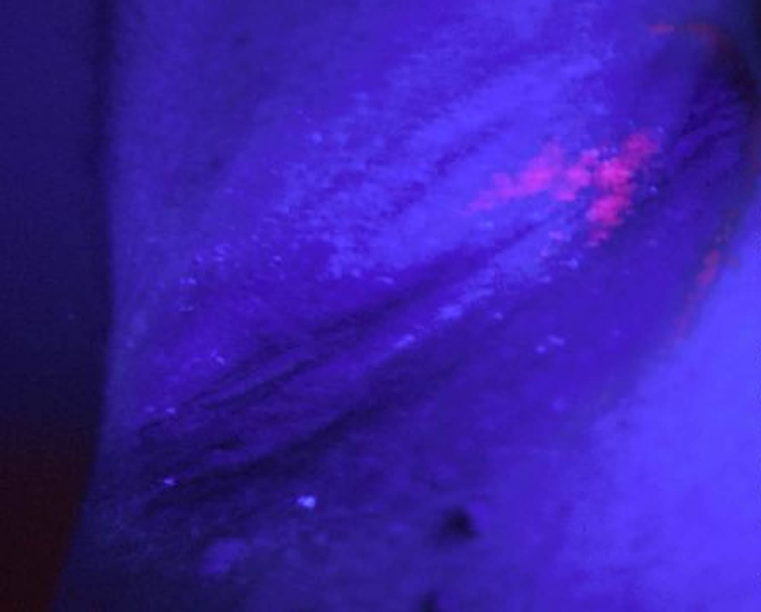
Note: This image shows the pink-red fluorescence of corynebacteria, an infection of the body folds known as erythrasma, shown under a “Woods lamp” in a dark room.
Treatment of Erythrasma
Once the diagnosis of erythrasma is established, the doctor may try one of the following treatments:
- Topical antibiotic lotions such as erythromycin or clindamycin
- Whitfield’s ointment (a mixture of benzoic acid and salicylic acid)
- Aluminum chloride solution to inhibit sweating and moisture
- Oral antibiotics such as erythromycin or clarithromycin.
Treatment is a single dose of clarithromycin 1 gram orally. One to two treatments (80 J/cm2) of broadband red light (635 nm) have been successful in a small case series. Topical erythromycin or clindamycin is also effective. Recurrence is common 9.
Outlook (Prognosis) of Erythrasma
The condition should go away after treatment.
Who’s at risk of intertrigo (dark inner thighs) ?
Intertrigo can affect people of all ages.
Intertrigo is most frequently seen in:
- Overweight or obese people,
- Diabetics,
- People spending a lot of time in bed,
- Diaper users, or
- Anyone with incontinence problems.
It can also occur in individuals wearing or using anything that causes friction or holds moisture against the skin surface.
When to Contact a Medical Professional
Call your health care provider if:
- The condition does not go away, even with good home care.
- The area of affected skin spreads beyond a skin fold.
Intertrigo treatment
For mild cases, your doctor will tell you to keep the affected area of your skin dry and exposed to air. Your doctor may also want to prescribe a topical steroid cream. For more severe cases, your doctor may prescribe an antibiotic or antifungal cream. There are also antifungal powders that may help dry the skin. Talk to your doctor about which treatment is right for you.
Treatments Your Physician May Prescribe
- Low-potency topical steroid creams.
- Calcineurin inhibitors (pimecrolimus cream and tacrolimus ointment), which are non-steroid topical agents, can be helpful in difficult cases.
- Coexisting infection should be treated if present. In cases with limited yeast involvement, topical miconazole, clotrimazole, or ciclopirox olamine may be used. If more severe yeast involvement is noted, oral antifungal agents, such as fluconazole, may be used. Topical or oral antibiotics may be needed for bacterial infections.
- A biopsy may be recommended if the diagnosis is uncertain.
Table 2. Treatment Options for Inflammatory and Infectious Intertrigo
| Condition | Treatments | |
|---|---|---|
Intertrigo | Topical: zinc oxide ointment, petrolatum, talcum powder, aluminum sulfate, calcium acetate solution | |
Intertrigo complicated by secondary bacterial infections | ||
Erythrasma | Topical: erythromycin, clindamycin, Whitfield ointment, chlorhexidine | |
Oral: erythromycin | ||
Group A beta-hemolytic streptococcus | Topical: mupirocin (Bactroban), erythromycin, low-potency steroids | |
Oral: penicillin, cephalexin (Keflex), ceftriaxone (Rocephin), cefazolin, clindamycin | ||
Intertrigo complicated by secondary fungal infections | ||
Candida | Topical: nystatin, clotrimazole, ketoconazole, oxiconazole (Oxistat), econazole | |
Oral: fluconazole (Diflucan; used for resistant cases) | ||
Dermatophytes | Topical: clotrimazole, ketoconazole, oxiconazole, econazole | |
Home remedies for dark inner thighs
If you are overweight, it may help to lose weight and often change your body position.
Other things you can do are:
- Separate skin folds with dry towels.
- Blow a fan on moist areas.
- Wear loose clothing and moisture wicking fabrics.
- Keep skin cool and dry.
- Do not wear tight shoes or clothing. Wear a bra that has good support.
- Wear clothes made with absorbent fabrics, such as cotton. Avoid nylon or other synthetic (manmade) fibers.
- After exercising, shower and dry off completely. Use a hair dryer with a cool setting to dry areas that can trap wetness, such as under your arms or breasts.
- Gently cleanse the affected areas daily with mild soap substitutes.
- Barrier creams, such as zinc oxide paste, may be helpful for individuals wearing diapers or having incontinence problems.
- Intertrigo. Medline Plus. https://medlineplus.gov/ency/article/003223.htm[↩]
- Fitzpatrick TB, ed. Color Atlas and Synopsis of Clinical Dermatology: Common and Serious Diseases. 3rd ed. New York, NY: McGraw-Hill; 1997.[↩][↩]
- Yosipovitch G, DeVore A, Dawn A. Obesity and the skin: skin physiology and skin manifestations of obesity. J Am Acad Dermatol. 2007;56(6):901–916.[↩]
- Seale P, Lazar MA. Brown fat in humans: turning up the heat on obesity. Diabetes. 2009;58(7):1482–1484.[↩]
- Del Rosso JQ, Draelos ZD, Jorizzo JL, Joseph WS, Ribotsky BM, Rich P. Modern methods to treat superficial fungal disease. Cutis. 2007;79(2 suppl):6–29.[↩]
- Guitart J, Woodley DT. Intertrigo: a practical approach. Compr Ther. 1994;20(7):402–409.[↩]
- Janniger CK, Schwartz RA, Szepietowski JC, Reich A. Intertrigo and common secondary skin infections. Am Fam Physician. 2005;72(5):833–838.[↩]
- Erythrasma. Medline Plus. https://medlineplus.gov/ency/article/001470.htm[↩]
- Erythrasma. Merck Manual. https://www.merckmanuals.com/professional/dermatologic-disorders/bacterial-skin-infections/erythrasma[↩]
- Intertrigo and Secondary Skin Infections. Am Fam Physician. 2014 Apr 1;89(7):569-573. http://www.aafp.org/afp/2014/0401/p569.html[↩]


