Contents
What is hyaluronic acid
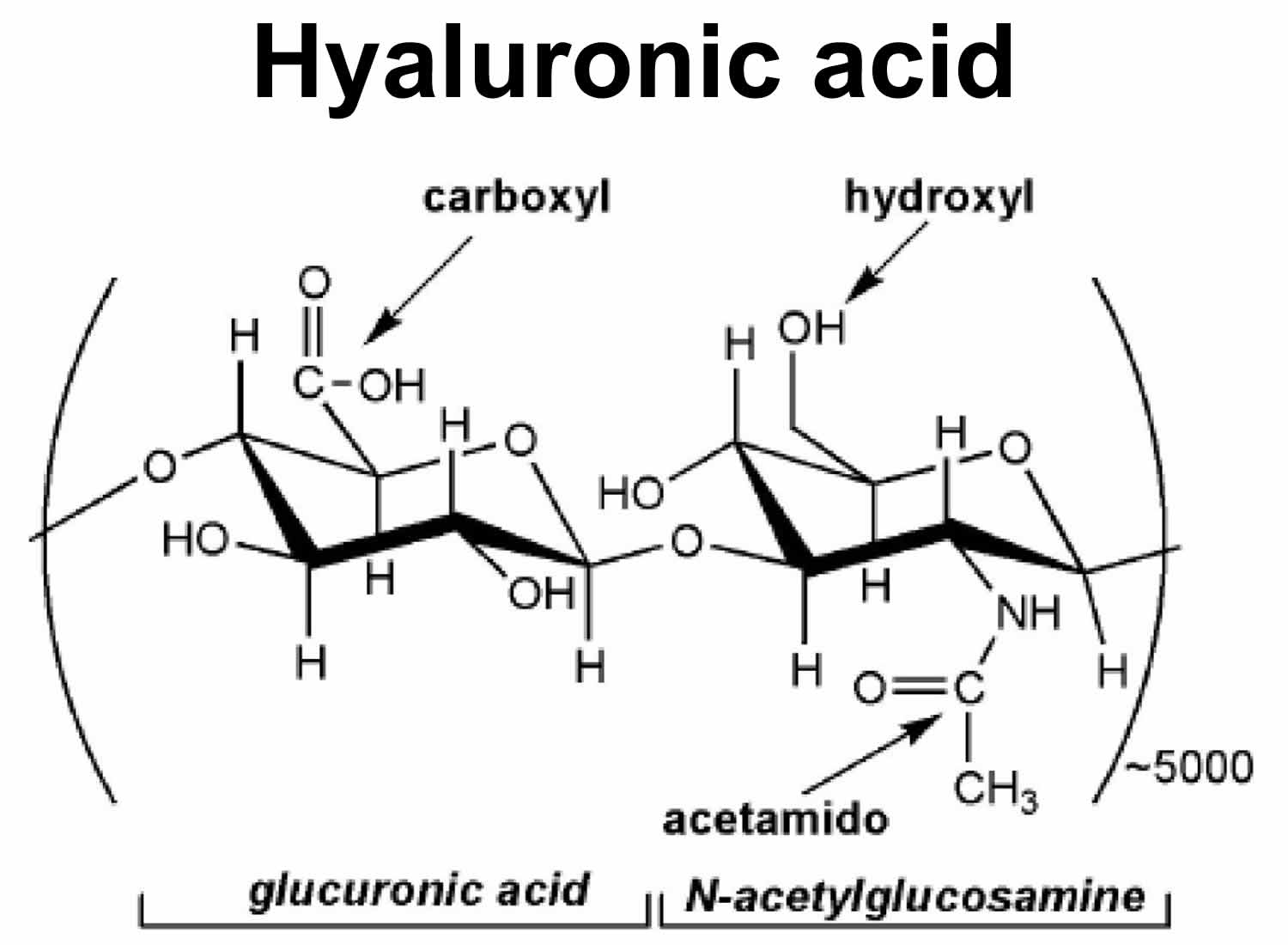
Hyaluronic acid is a non-sulphated glycosaminoglycan and is composed of repeating polymeric disaccharide repeats of D-glucuronic acid (GlcUA) and N-acetylglucosamine (GlcNAc) joined alternately by β-1, 3 and β-1, 4 glycosidic bonds (Figure 1) with a molecular weight up to 6 million Daltons 1). Traditionally hyaluronic acid was extracted from rooster combs, and now it is mainly produced via streptococcal fermentation with lower production costs and less environmental pollution 2). Hyaluronic acid has been successfully produced on an industrial scale with Streptococcus species bacteria as the main producer. Nevertheless, the production of hyaluronic acid from Streptococcus species bacteria is facing a growing concern due to the fact that streptococci are pathogenic 3). In this background, the recombinant hyaluronic acid production has attracted an increasing interest, and Novozymes has produced hyaluronic acid with recombinant Bacillus subtilis on an industrial scale 4).
Hyaluronic acid is also synthesized by your body by specific enzymes called hyaluronic acid synthases (HAS). These are membrane bound enzymes that synthesize hyaluronic acid on the inner surface of the plasma membrane 5) and then hyaluronic acid is extruded through pore-like structures into the extracellular space 6). There are three mammalian enzymes HAS -1, -2 and -3, which exhibit distinct enzymatic properties and synthesize hyaluronic acid chains of various length 7). In aqueous solutions hyaluronic acid forms specific stable tertiary structures 8). Hyaluronic acid is traditionally regarded as a biological ‘goo’ that participates in lubricating joints or holding together gel-like connective tissues. Although these are common physiological roles of hyaluronic acid in adult organisms, hyaluronic acid also functions as a microenvironmental cue that co-regulates cell behavior during embryonic development, healing processes, inflammation and tumor development 9). Hyaluronic acid polymers occur in a vast number of configurations and shapes, depending on their size, salt concentration, pH, and associated cations 10). Hyaluronic acid encompasses a large volume of water giving solutions high viscosity, even at low concentrations 11).
Molecular weight is an important quality parameter for a commercial hyaluronic acid product, as it determines the hyaluronic acid’s rheological properties, affects physiological response, and defines suitable applications 12). Hyaluronic acid with a high molecular weight (greater than 10 kDa) has good viscoelasticity, moisture retention, and mucoadhesion, — qualities desirable in the areas of ophthalmology, orthopedics, wound healing, and cosmetics. Whereas, hyaluronic acid with a relatively low molecular weight (2-3.5 kDa) or hyaluronic acid oligosaccharides (10-20 sugars in length) have shown to promote angiogenesis, induce expression of inflammatory mediators, and inhibit tumor growth 13).
Figure 1. Hyaluronic acid
In humans, hyaluronic acid is most abundant in the skin 14), accounting for 50% of the total body hyaluronic acid 15), the vitreous of the eye 16), the umbilical cord 17), and synovial fluid 18), but it is also present in all tissues and fluids of the body, such as skeletal tissues 19), heart valves 20), the lung 21), the aorta 22), the prostate 23), tunica albuginea, corpora cavernosa and corpus spongiosum of the penis 24). Hyaluronic acid is produced primarily by mesenchymal cells but also by other cell types 25).
What does hyaluronic acid do
Functions of hyaluronic acid include the following: hydration, lubrication of joints, a space filling capacity, and the framework through which cells migrate 26). The synthesis of hyaluronic acid increases during tissue injury and wound healing 27) and hyaluronic acid regulates several aspects of tissue repair, including activation of inflammatory cells to enhance immune response 28) and the response to injury of fibroblasts 29) and epithelial cells 30). Hyaluronic acid also provides the framework for blood vessel formation 31) and fibroblast migration 32), that may be involved in tumor progression 33). The correlation of hyaluronic acid levels on the cell surface of cancer cells with the aggressiveness of tumors has also been reported 34).
The size of hyaluronic acid appears to be of critical importance for its various functions described above 35). Hyaluronic acid of high molecular size, usually in excess of 1,000 kDa, is present in intact tissues and is antiangiogenic and immunosuppressive, whereas smaller polymers of hyaluronic acid are distress signals and potent inducers of inflammation and angiogenesis 36).
Hyaluronic acid receptors
There is a variety of proteins that bind hyaluronic acid, called hyaladherins, which are widely distributed in the extracellular matrix, the cell surface, the cytoplasm and the nucleus 37). Those that attach hyaluronic acid to the cell surface constitute hyaluronic acid receptors. The most prominent among these receptors is the transmembrane glycoprotein “cluster of differentiation 44” (CD44) that occurs in many isoforms, which are the products of a single gene with variable exon expression 38). CD44 is found on virtually all cells, except red blood cells, and regulates cell adhesion, migration, lymphocyte activation and homing, and cancer metastasis.
The receptor for hyaluronic acid-mediated motility (RHAMM) is another major receptor for hyaluronic acid, and it is expressed in various isoforms 39). RHAMM is a functional receptor in many cell types, including endothelial cells 40) and in smooth muscle cells from human pulmonary arteries 41) and airways 42). The interactions of hyaluronic acid with RHAMM control cell growth and migration by a complex network of signal transduction events and interactions with the cytoskeleton 43). Transforming growth factor (TGF)-β1, which is a potent stimulator of cell motility, elicits the synthesis and expression of RHAMM and hyaluronic acid, and thus initiates locomotion 44).
Degradation of hyaluronic acid
Hyaluronic acid has a dynamic turnover rate. Hyaluronic acid has a half-life of 3 to 5 min in the blood, less than a day in the skin and 1 to 3 weeks in the cartilage 45). Hyaluronic acid is degraded into fragments of varying size by hyaluronidases (HYAL) by hydrolyzing the hexosaminidic β (1–4) linkages between N-acetyl-D-glucosamine and D-glucuronic acid residues in hyaluronic acid. In humans, six hyaluronidases (HYAL) have been identified so far: HYAL-1, -2, -3, -4, PH-20 and HYALP1 46). The family of hyaluronidase (HYAL) enzymes received little attention until recently 47), because they are found at extremely low concentrations and they are difficult to purify, characterize and measure their activity, which is high but unstable 48). New procedures have now enabled the isolation and characterization of HYAL. HYAL-1 is the major HYAL in serum 49). Mutations in the HYAL-1 gene are associated with HYAL deficiency and mucopolysaccharidosis type IX 50). HYAL-2 has very low activity in comparison to plasma HYAL-1 and it hydrolyzes specifically hyaluronic acid of high molecular weight, yielding hyaluronic acid fragments of approximately 20 kDa, which are further degraded to small oligosaccharides by PH-20 51). HYAL-3 is mainly expressed in bone marrow and testis 52), but also in other organs, such as the human lung 53). The role of HYAL-3 in the catabolism of hyaluronic acid is not clear and it is suggested that it may contribute to hyaluronic acid degradation by enhancing the activity of HYAL-1 54).
Hyaluronic acid can also be degraded non-enzymatically by a free-radical mechanism 55) in the presence of reducing agents such as ascorbic acid, thiols, ferrous, or cuprous ions, a process that requires the presence of molecular oxygen. Thus, agents that could delay the free-radical-catalyzed degradation of hyaluronic acid may be useful in maintaining the integrity of dermal hyaluronic acid and its moisturizing properties 56).
Hyaluronic acid for skin
Human skin aging is a complex biological process, not yet fully understood. It is the result of two biologically independent processes. The first is intrinsic or innate aging, an unpreventable process, which affects the skin in the same pattern as it affects all internal organs. The second is extrinsic aging, which is the result of exposure to external factors, mainly ultraviolet (UV) irradiation, that is also referred to as photoaging 57). Intrinsic skin aging is influenced by hormonal changes that occur with age 58), such as the gradual decreased production of sex hormones from the mid-twenties and the diminution of estrogens and progesterone associated with menopause. It is well established that the deficiency in estrogens and androgens results in collagen degradation, dryness, loss of elasticity, epidermal atrophy and wrinkling of the skin 59).
Even though intrinsic and extrinsic skin aging are distinctive processes, they share similarities in molecular mechanisms. For example, reactive oxygen species (ROS), arising from oxidative cell metabolism, play a major role in both processes 60). Reactive oxygen species (ROS) in extrinsic or intrinsic skin aging induce the transcription factor c-Jun via mitogen-activated protein kinases (MAPK), leading to overexpression of matrix metalloproteinase (MMP)-1, MMP-3 and MMP-9 and prevention of the expression of procollagen-1 61). Therefore, elevated levels of degraded collagen and reduced collagen synthesis are pathologies occurring in intrinsically aged as well as photoaged skin.
Skin aging is also associated with loss of skin moisture. The key molecule involved in skin moisture is hyaluronic acid (hyaluronic acid), a glycosaminoglycan with a unique capacity to bind and retain water molecules 62). Hyaluronic acid belongs to the extracellular matrix (ECM) molecules. During the past decades the constituents of the skin have been well characterized. In the beginning, most of the studies focused on the cells that comprise the skin layers, such as the epidermis, the dermis and the underlying subcutis. Recently, it is appreciated that extracellular matrix (ECM) molecules that lie between cells, in addition to providing a constructive framework, they exert major effects on cellular function. These extracellular matrix (ECM) molecules, although they appear amorphous by light microscopy, they form a highly organized structure, comprising mainly of glycosaminoglycan, proteoglycans, growth factors and structural proteins such as collagens. Yet, the predominant component of the skin extracellular matrix (ECM) is hyaluronic acid.
As mentioned above, skin hyaluronic acid accounts for most of 50% of total body hyaluronic acid 63). The hyaluronic acid content of the dermis is significantly higher than that of the epidermis, while papillary dermis has much greater levels of hyaluronic acid than reticular dermis 64). The hyaluronic acid of the dermis is in continuity with the lymphatic and vascular systems. Hyaluronic acid in the dermis regulates water balance, osmotic pressure and ion flow and functions as a sieve, excluding certain molecules, enhancing the extracellular domain of cell surfaces and stabilizes skin structures by electrostatic interactions 65). Elevated levels of hyaluronic acid are synthesized during scar-free fetal tissue repair and the prolonged presence of hyaluronic acid assures such scar-free tissue repair 66). Dermal fibroblasts provide the synthetic machinery for dermal hyaluronic acid and should be the target for pharmacologic attempts to enhance skin hydration. Unfortunately, exogenous hyaluronic acid is cleared from the dermis and is rapidly degraded 67).
Hyaluronic acid and skin aging
The most dramatic histochemical change observed in aged skin is the marked disappearance of epidermal hyaluronic acid, while hyaluronic acid is still present in the dermis 68). The reasons for this change in hyaluronic acid homeostasis with aging is unknown. As mentioned above, the synthesis of epidermal hyaluronic acid is influenced by the underlying dermis and is under separate controls from the synthesis of dermal hyaluronic acid 69). Progressive reduction of the size of the hyaluronic acid polymers in skin as a result of aging has also been reported 70). Thus, the epidermis loses the principle molecule responsible for binding and retaining water molecules, resulting in loss of skin moisture. In the dermis, the major age-related change is the increasing avidity of hyaluronic acid with tissue structures with the concomitant loss of hyaluronic acid extractability. This parallels the progressive cross-linking of collagen and the steady loss of collagen extractability with age 71). All of the above age related phenomena contribute to the apparent dehydration, atrophy and loss of elasticity that characterizes aged skin.
Premature aging of skin is the result of repeated and extended exposure to UV (ultraviolet) radiation 72). Approximately 80% of facial skin aging is attributed to UV-exposure 73). UV radiation damage causes initially a mild form of wound healing and is associated at first with an increase of dermal hyaluronic acid. As little as 5 minute of UV exposure in nude mice caused enhanced deposition of hyaluronic acid, indicating that UV radiation induced skin damage is an extremely rapid event 74). The initial redness of the skin following exposure to UV (ultraviolet) radiation may be due to a mild edematous reaction induced by the enhanced hyaluronic acid deposition and histamine release. Repeated and extensive exposures to UV ultimately simulate a typical wound healing response with deposition of scarlike type I collagen, rather than the usual types I and III collagen mixture that gives skin resilience and pliability 75).
In the skin, photoaging results in abnormal glycosaminoglycan content and distribution compared with that found in scars, or in the wound healing response, with diminished hyaluronic acid and increased levels of chondroitin sulfate proteoglycans 76). In dermal fibroblasts this reduction in hyaluronic acid synthesis was attributed to collagen fragments, which activate αvβ3-integrins and in turn inhibit Rho kinase signaling and nuclear translocation of phosphoERK, resulting in reduced hyaluronic acid synthase-2 (HAS-2) expression 77). We have recently unraveled some of the biochemical changes that may distinguish photoaging and natural aging. Using photoexposed and photoprotected human skin tissue specimens, obtained from the same patient, we have shown a significant increase in the expression of hyaluronic acid of lower molecular mass in photoexposed skin, as compared with photoprotected skin. This increase of degraded hyaluronic acid was associated with a significant decrease in the expression of HAS-1 (hyaluronic acid synthase-1) and an increased expression of hyaluronidase-1, -2 and -3. Furthermore, the expression of hyaluronic acid receptors CD44 (cluster of differentiation 44) and RHAMM (receptor for hyaluronic acid-mediated motility) was significantly downregulated in photoexposed, as compared with photoprotected skin. These findings indicate that photoexposed skin, and therefore extrinsic skin aging, is characterized by distinct homeostasis of hyaluronic acid 78). Scientists have also assessed photoprotected skin tissue specimens from adults and juvenile patients and observed that intrinsic skin aging was associated with a significant reduction in the content of hyaluronic acid and downregulation of HAS-1, HAS -2, CD44 and RHAMM 79). Similar results for photoprotected skin have also been reported for both genders for hyaluronic acid, HAS-2 and CD44 80).
Conclusion
The available data suggest that hyaluronic acid homeostasis exhibits a distinct profile in intrinsic skin aging (innate or biological aging), which is totally different of that in extrinsic skin aging (photoaging due to exposure to external factors, mainly ultraviolet (UV) irradiation). Additional insight needs to be gained in understanding the metabolism of hyaluronic acid in skin layers and the interactions of hyaluronic acid with other skin components. Such information will facilitate the ability to modulate skin moisture in a rational manner and may contribute to the refinement of current drugs and the development of novel treatments for skin aging 81).
Hyaluronic acid fillers for facial volume restoration and contouring
Loss of appropriate facial fullness and youthful appearance occurs in most areas of the face to varying degrees, including the periorbital, malar, forehead, temporal, glabellar, mandibular, mental and perioral zones 82). A recent study determined that facial subcutaneous fat is partitioned into multiple, independent anatomical compartments, which may age independently, resulting in abrupt contour changes between them 83).
Re-establishing the integrity of these foundation structures through appropriate volume restoration and contouring has proven to be a highly effective means of harmonizing features, contours, proportions and balance associated with the youthful face 84).
The products chosen most frequently by some experts for volume restoration and contouring were a high-viscosity, low-molecular-weight hyaluronic acid (LMWHA) for structural support and high molecular weight hyaluronic acids for lines, grooves and furrows 85).
That the recent addition of a high-viscosity, low molecular weight hyaluronic acid (LMWHA) to the volumizing product has resulted in significantly improved patient outcomes. This is a smooth consistency gel that is uniquely composed of a mix of low and high molecular weight hyaluronic acid. Compared to hyaluronic acid fillers with 100% high molecular weight, the LMWHA (low molecular weight hyaluronic acid) allows a combination of high cohesivity and viscosity. This property enables the LMWHA (low molecular weight hyaluronic acid) to retain its structure following a deep injection and makes it particularly well suited for facial volumizing and contouring 86).
As a point of interest, while the term ‘dermal filler’ is still widely used, it is arguably not anatomically accurate. Facial fillers are frequently used at multiple levels beneath the dermis – sub-muscularly above the periosteum and in the subcutaneous plane 87).
For the purposes of treatment discussion, the face is divided into three fundamental, but overlapping sections: Mid, upper and lower. However, the global, whole-face and three-dimensional approach must always be the physician’s primary consideration. Treatment is always carried out in the context of how it will integrate with the rest of the face, both at the moment of implementation and over time, taking changing facial dynamics into consideration.
It should be noted that these are recently developed procedures representing the collective experience and best practices of the authors at the time of writing and should not be construed as absolute, validated directives. It is important that each individual be treated uniquely while keeping these general principles in mind.
Table 1. Five categories of facial filler
| Category | Agent | Durationa |
|---|---|---|
| High-viscosity, low molecular weight hyaluronic acid (LMWHA) | Voluma (Allergan, Inc) | Long duration (up to 18 months) |
| Hyaluronic acid (HA) | Juvéderm (Allergan, Inc) | Intermediate duration (~12 mo) |
| Restylane (Medicis Aesthetics) | Intermediate duration (~6 mo) | |
| Perlane (Medicis Aesthetics) | ||
| Teosyal (Clarion Medical Technologies) | ||
| Esthelis (Anteis) | ||
| Prevelle (Mentor) | ||
| Revanesse (Prollenium Medical) | ||
| Calcium hydroxylapatite | Radiesse (Merz Aesthetics) | Intermediate duration (~1 yr) |
| Poly-L-lactic acid (PLLA) | Sculptra (Valeant) | Long duration (up to 18 months) |
| Polymethyl methacrylate (PMMA) | Artefill (Artes Medical/suneva) | Permanent (~10 years) |
| Platelet-Rich Fibrin Matrix (PRFM) | Selphyl (Canderm) | Long duration (up to 18 months) |
Footnote:
a) Some authors have seen duration of action which is longer than the amounts selphyl (Canderm) listed above.
[Source 88)]Table 2. Strategies for volume restoration and contouring in the upper face with hyaluronic acid
| Treated area | Preferred product | Technique | Dosinga (per degree of severity of deficit) | Needles/cannulae (gauge and length) |
|---|---|---|---|---|
| Temporal hollow | Hyaluronic acid |
| ||
| Hyaluronic acid Mild: 0.5 cc/side Moderate: 1–2 cc/side Severe: 3+ cc/side | Hyaluronic acid 27 gauge (deep) 30 gauge (superficial) 22 or 25 g cannula | ||
| TIPS/PEARLS |
| |||
| Brow | Hyaluronic acid |
| 0.2–0.5 cc/side | 27 gauge 30 gauge |
| TIPS/PEARLS |
| |||
| Upper eyelid hollow | Hyaluronic acid |
| Use small aliquots (<0.25 cc) Rarely need more than 1 cc/side | 30–31 gauge ½”–1” 30 g microcannula |
| TIPS/PEARLS |
| |||
Footnote:
a) General dosing range based on consensus group experience.
Abbreviations: HA = hyaluronic acid; LMWHA = low-molecular-weight hyaluronic acid; Poly-L = lactic acid.
[Source 89)]Table 3. Strategies for volume restoration and contouring in the mid-face with hyaluronic acid
| Treated area | Preferred product | Technique | Dosinga (per degree of severity of deficit) | Needles/cannulae (gauge and length) |
|---|---|---|---|---|
| Cheek | HA LMWHA |
| Mild: 0.5–1 cc/side Medium: 1–2 cc/side Severe: 3+ cc/side | Needle: 25–27–29 UTW gauge ½” Cannula: 25 g 30 mm/22 g 40 mm |
| TIPS/PEARLS |
| |||
| Submalar cheek | Hyaluronic acid |
| Hyaluronic acid Mild: 1 cc/side Medium: 2 cc/side Severe: 3 cc/side | 27–30 gauge needle 25 g or 27 g microcannula |
| TIPS/PEARLS |
| |||
| Tear Trough/Infra-orbital hollows | Hyaluronic acid |
| Use small aliquots (<0.25 cc) | 30–31 gauge ½”–1” or BD tuberculin syringe (backloaded) or 30 g microcannula |
| Rarely need more than 1 cc/side | |||
| TIPS/PEARLS |
| |||
| Nasolabial folds (NLF) | Hyaluronic acid |
| Mild: <0.5 cc/side Medium: 0.5–1 cc/side Severe: 1+ cc/side | 27–31 gauge 3/8”–½” 25 g cannula, 30 mm 22 g cannula, 40 mm |
| TIPS/PEARLS |
| |||
Footnote:
a) General dosing range based on consensus group experience.
Abbreviations: HA = hyaluronic acid; LMWHA = low-molecular-weight hyaluronic acid; NLF = nasolabial fold.
[Source 90)]Table 4. Strategies for volume restoration and contouring in the lower face with hyaluronic acid
| Treated area | Preferred product | Technique | Dosinga (per degree of severity of deficit) | Needles/cannulae (gauge and length) |
|---|---|---|---|---|
| Chin | LMWHA/HA |
| Hyaluronic acid Mild: 1 cc Moderate: 2 cc Severe: 3 cc | 27–30 gauge ½” 22 or 25 g cannula |
| TIPS/PEARLS |
| |||
| Pre-jowl sulcus/marionette | LMWHA/HA |
| Hyaluronic acid Mild: 0.5 cc/side Moderate: 1 cc/side Severe: 2+ cc/side | 27–30 gauge ½”–1” 22 or 25 g cannula |
| TIPS/PEARLS |
| |||
| Post jowl sulcus, mandibular angle and preauricular | LMWHA/HA |
| Hyaluronic acid Mild: 1–2 cc/side Moderate: 2–3 cc/side Severe: 3–4 cc/side | 27 gauge ½”–1½” |
| TIPS/PEARLS |
| |||
Footnote:
a) General dosing range based on consensus group experience.
Abbreviations: HA = hyaluronic acid; LMWHA = low-molecular-weight hyaluronic acid.
[Source 91)]Summary
Low-molecular-weight hyaluronic acid (LMWHA) has made it possible for physicians to achieve new levels of excellence in facial aesthetics. In clinical practice, physicians have demonstrated that high- viscosity LMWHA has advantages when used for volume restoration and contouring, including versatility of use (different facial planes), high malleability, minimal swelling, limited downtime, and immediate patient satisfaction 92).
Combination treatment of fillers, neuromodulators and energy devices to create optimal contours is forecast to remain the mainstay of non-surgical facial aesthetics. Further advances are expected to standardize techniques, increase reproducibility and consistency of results, and augment patient experiences by minimizing bruising and downtime.
Serum hyaluronic acid
Serum hyaluronic acid (sHA) is a serum biomarker for knee osteoarthritis (OA). Serum hyaluronic acid concentration is elevated in patients with knee osteoarthritis and the concentration of serum hyaluronic acid was positively correlated with the severity of radiographic knee osteoarthritis 93). Several previous cross-sectional studies have reported that measuring serum hyaluronic acid concentration may be useful for not only diagnosing knee osteoarthritis, but also identifying disease duration, severity, and the extent of osteoarthritis-related knee pain 94). Furthermore, over 5 years, higher serum hyaluronic acid concentrations were positively correlated with the progression of Kellgren–Lawrence grade in those with knee osteoarthritis and of joint space narrowing in those both with and without knee osteoarthritis. These results suggest that elevated serum hyaluronic acid concentration can be used as a predictor of knee osteoarthritis progression. Thus, serum hyaluronic acid concentration may be useful as a prognostic marker for predicting joint space narrowing, though not for predicting osteophyte formation (Sasaki E, Tsuda E, Yamamoto Y, et al. Serum hyaluronic acid concentration predicts the progression of joint space narrowing in normal knees and established knee osteoarthritis – a five-year prospective cohort study. Arthritis Research & Therapy. 2015;17:283. doi:10.1186/s13075-015-0793-0. https://www.ncbi.nlm.nih.gov/pmc/articles/PMC4600294/)). However, serum hyaluronic acid concentration increases with age because of an impaired ability to metabolize hyaluronic acid in the elderly 95). Hyaluronic acid is actively secreted into joint fluid by the surrounding synovial lining cells 96). When intra-articular fluid pressure is increased, hyaluronic acid is also distributed to the liver (90 %), kidney (9 %), and spleen (1 %) via the lymphatic and capillary systems 97). The increase in serum hyaluronic acid concentration with age may even be a consequence of progressive age-related hepatic and renal impairment 98). serum hyaluronic acid concentration is reported to be influenced by other factors, including renal failure 99), liver failure 100), rheumatoid arthritis 101), malignancy 102), and physical activity 103).
The mechanisms underpinning the phenomenon of elevated serum hyaluronic acid concentration in progressive knee osteoarthritis are thought to be mechanical stress and synovitis with cartilage degeneration 104). Osteoarthritis is characterized by focal damage to the articular cartilage centered on the load-bearing areas, osteophyte formation at the joint margins, subchondral bone changes, and synovitis 105). Synovitis is present at the onset of osteoarthritis 106) and results in the production of hyaluronic acid (HA) and pro-inflammatory cytokines 107). In addition, synovitis activates fibroblasts, which promote the production of other pro-inflammatory cytokines such as tumor necrosis factor-α and interleukin-1β 108). These cytokines promote matrix metalloproteinase production by fibroblasts, which degrade the articular cartilage matrix 109). Thus, the presence of synovitis is thought to contribute to the progression of knee osteoarthritis. Ayral et al. 110) suggested that synovitis was also predictive of subsequent chondropathy.
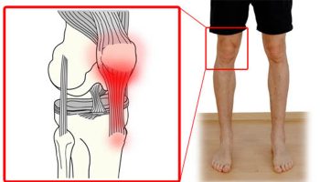
Hyaluronic acid for joints
Hyaluronic acid is a substance already in the fluid of your knee. It helps lubricate the joint. When you have arthritis, the hyaluronic acid in your joint becomes thinner and less effective. Your doctor can inject a form of hyaluronic acid into your joint to help lubricate and protect it. This is sometimes called artificial joint fluid, or viscosupplementation. Hyaluronic acid injections are one treatment option doctors may offer when a patient is no longer able to control osteoarthritis pain with ibuprofen or other nonsteroidal anti-inflammatory drugs (NSAIDs), or the patient can’t tolerate these drugs (which can cause side effects such as stomach bleeding and kidney problems). The treatment regimen for hyaluronic acid usually involves receiving one injection in the affected joint per week for three to five weeks. Many patients appear to get at least some relief – eventually.
Hyaluronic acid injections are approved by the U.S. Food and Drug Administration (FDA) for treating osteoarthritis of the knee, though some doctors have used the therapy on other joints, such as the hip and ankle. While studies of hyaluronic acid injections have occasionally yielded disappointing results, many doctors who treat osteoarthritis say that the weight of scientific evidence and their own clinical experience – suggests that hyaluronic acid injection in the knee can produce significant relief for some patients. Furthermore, lab and clinical research hints that hyaluronic acid may do much more than simply re-grease a creaky joint.
In 2006, a team led by Nicholas Bellamy, MD, of the University of Queensland in Brisbane, Australia, reviewed 76 studies examining the use of hyaluronic acid for treating knee osteoarthritis. The review, the largest and most comprehensive of its kind, found that pain levels in the average patient who receives these injections are reduced by 28 to 54 percent. That’s roughly what a patient might expect from taking NSAIDs, the authors concluded. Meanwhile, hyaluronic acid improved the ability to move about and perform daily activities by 9 to 32 percent.
As a patient soon learns, though, hyaluronic acid is no quick fix. According to Bellamy’s review (which was conducted on behalf of the Cochrane Collaboration, an international consortium that reviews scientific evidence for medical treatments), it takes about five weeks, on average, before a patient experiences the full benefits of hyaluronic acid. By contrast, corticosteroid injections – the other primary treatment choice when NSAIDs aren’t an option – provide significant relief within a few days. However, pain relief from corticosteroids diminishes markedly within a month or so. What’s more, overuse of corticosteroids can have a catabolic effect – that is, it could cause cartilage to break down and deteriorate further. Meanwhile, the Cochrane review found that pain-relieving benefits of hyaluronic acid persist at peak levels for about three months, on average. Some doctors sometimes give patients a double shot in the knee – one injection each of hyaluronic acid and corticosteroids – for quick-acting, long-lasting relief.
About 30 percent of people who undergo hyaluronic acid injections become virtually pain free, and symptom relief may last up to two years. Yet, another 20 percent of patients experience no benefit. Unfortunately, experts don’t know how to pick out those people who are going to have an outstanding response versus a modest response versus no response at all. Researchers have tried to identify ways to predict how a patient will respond to hyaluronic acid, but so far have come up empty.
However, hyaluronic acid isn’t universally approved. In June 2013, the American Academy of Orthopedic Surgeons (AAOS) issued a new set of recommendations for the treatment of knee osteoarthritis. Based on a review of 14 studies, the organization determined hyaluronic acid did not meet the minimum clinically important improvement measures.
With five brands available in the United States, it’s natural to ask which is most effective. There have been relatively few head-to-head comparisons of the various products in clinical trials. Experts believe that they all work about the same. Likewise, the risk of side effects is similar among the different products, the most common being pain and swelling at the injection site that fades within a few days.
Beyond the question of how well hyaluronic acid injections (viscosupplements) work lies another intriguing area of inquiry: How do they work? Hyaluronic acid may act as a lubricant and shock absorber. Hyaluronic acid has a lot of other activities in the joint. For example, research suggests that hyaluronic acid interfere with prostaglandins and cytokines, naturally occurring compounds that promote inflammation.
What’s more, studies indicate that injecting supplemental hyaluronic acid may coax the joint into increasing its own production of this important substance, which may in turn help to preserve cartilage. There’s a lot of data to suggest that it can slow the disease down. However, as pointed out above, hyaluronic acid is not a magic pill, but it has a definite role in the armamentarium for treating osteoarthritis of the knee.
Hyaluronic acid injection
Hyaluronic acid injection is used to treat knee pain caused by osteoarthritis (OA) in patients who have already been treated with pain relievers (e.g., acetaminophen) and other treatments that did not work well.
Hyaluronic acid is similar to a substance that occurs naturally in the joints. It works by acting like a lubricant and shock absorber in the joints and helps the joints to work properly.
Hyaluronic acid injection is to be administered only by or under the immediate supervision of your doctor. How often you have the hyaluronic acid injections depends on the type of hyaluronan (hyaluronic acid) preparation used. You’ll probably be given a course of 3–5 injections, each separated by 1–3 weeks. Some doctors give the first two injections close together and then extend the period between those remaining. You probably won’t need to rest the joint after the hyaluronic acid injections. However, some doctors recommend you avoid activities such as jogging, soccer, tennis, heavy lifting, or standing on your feet for a long time for two days after the injection.
Hyaluronic acid injection is available in the following dosage forms:
- Solution
- Gel/Jelly
Before having hyaluronic acid injection
In deciding to use hyaluronic acid injection, the risks of taking the medicine must be weighed against the good it will do. This is a decision you and your doctor will make. For this medicine, the following should be considered:
- Allergies: Tell your doctor if you have ever had any unusual or allergic reaction to this medicine or any other medicines. Also tell your health care professional if you have any other types of allergies, such as to foods, dyes, preservatives, or animals. For non-prescription products, read the label or package ingredients carefully.
- Pediatric: Appropriate studies have not been performed on the relationship of age to the effects of hyaluronic acid injection in the pediatric population. Safety and efficacy have not been established.
- Geriatric: No information is available on the relationship of age to the effects of hyaluronic acid injection in geriatric patients.
- Breastfeeding: There are no adequate studies in women for determining infant risk when using this medication during breastfeeding. Weigh the potential benefits against the potential risks before taking this medication while breastfeeding.
Drug Interactions
Although certain medicines should not be used together at all, in other cases two different medicines may be used together even if an interaction might occur. In these cases, your doctor may want to change the dose, or other precautions may be necessary. Tell your healthcare professional if you are taking any other prescription or nonprescription (over-the-counter [OTC]) medicine.
Other Interactions
Certain medicines should not be used at or around the time of eating food or eating certain types of food since interactions may occur. Using alcohol or tobacco with certain medicines may also cause interactions to occur. Discuss with your healthcare professional the use of your medicine with food, alcohol, or tobacco.
Other Medical Problems
The presence of other medical problems may affect the use of hyaluronic acid injection. Make sure you tell your doctor if you have any other medical problems, especially:
- Allergy to bacterial proteins, gram positive or
- Allergy to hyaluronate preparations or
- Skin or knee joint infections or other problems at the place where the injection is to be given—Should not be given in patients with these conditions.
- Joint effusion (too much fluid in the knees)—Patients with this condition should be treated first before receiving hyaluronic acid injection.
How long do they take to work?
The hyaluronic acid injections should work within days if they’re effective and they’ll probably last for a few months. Sometimes your doctor may recommend you have repeat courses.
The particles in hyaluronan are quite large, so they’ll stay in your joint for some time before they’re absorbed into your blood stream. Some of the hyaluronic acid preparations available contain significantly larger particles than others and this can influence your doctor’s choice.
Side-effects and risks of hyaluronic acid injection
Hyaluronan injections have very few side-effects, although some people have a slight allergic reaction, which causes temporary pain and swelling in their joint after the injection. There’s also a small risk of infection. If the injection is done in slightly the wrong place, it won’t damage your skin or muscle.
Temporary pain or swelling in the knee joint may occur after receiving hyaluronic acid injection. See your doctor if the pain or swelling in the knee persists or becomes worse after receiving hyaluronic acid injection.
Do not use hyaluronic acid injection with disinfectants containing quaternary ammonium salts (e.g., benzalkonium chloride). This may prevent hyaluronic acid injection from working properly.
More common side effects
- Difficulty with moving
- Muscle pain or stiffness
- Pain in the joints
Less common
- Swelling or redness in the joints
Some side effects may occur that usually do not need medical attention. These side effects may go away during treatment as your body adjusts to the medicine. Also, your health care professional may be able to tell you about ways to prevent or reduce some of these side effects. Check with your health care professional if any of the following side effects continue or are bothersome or if you have any questions about them:
Less common
- Bleeding, blistering, burning, coldness, discoloration of the skin, feeling of pressure, hives, infection, inflammation, itching, lumps, numbness, pain, rash, redness, scarring, soreness, stinging, swelling, tenderness, tingling, ulceration, or warmth at the injection site
Other side effects not listed may also occur in some patients. If you notice any other effects, check with your healthcare professional.
References [ + ]


