Contents
What is Ludwig’s angina
Ludwig’s angina is a type of bacterial infection that occurs in the floor of the mouth, under the tongue. Ludwig angina often develops after an infection of the roots of the teeth (such as tooth abscess) or a mouth injury. Ludwig’s angina can be life threatening. Prior to the development of antibiotics, mortality exceeded 50% 1. With antibiotic therapy, along with improved imaging modalities and surgical techniques, mortality is approximately 8% 1.
Ludwig angina can be cured with getting treatment to keep the airways open and taking antibiotic medicine.
Ludwig’s angina was named after a German physician, Wilhelm Friedrich von Ludwig who first described the condition in 1836 1. Ludwig’s angina involves two compartments on the floor of the mouth namely sublingual and submaxillary space. Ludwig’s angina usually does not involve lymphatic system nor it forms abscess. Infection of the lower molars is the most common cause of Ludwig’s angina. The infection is rapidly progressive leading to aspiration pneumonia and airway obstruction.
Ludwig angina is uncommon in children.
If the swelling blocks your airway, you need to get emergency medical help right away. A breathing tube may needed to be placed through your mouth or nose and into the lungs to restore breathing. You may need to have surgery called a tracheostomy that creates an opening through the neck into the windpipe. Airway compromise is always synonymous with the term Ludwig’s angina, and it is the leading cause of death 2.
Antibiotics are given to fight the infection. They are most often given through a vein until symptoms go away. Antibiotics taken by mouth may be continued until tests show that the bacteria have gone away.
Dental treatment may be needed for tooth infections that cause Ludwig’s angina.
Surgery may be needed to drain fluids that are causing the swelling.
Breathing difficulty is an emergency situation. Go to the emergency room or call your local emergency number right away.
Call your doctor if you have symptoms of Ludwig angina or if your symptoms do not get better after treatment.
Figure 1. Ludwig angina
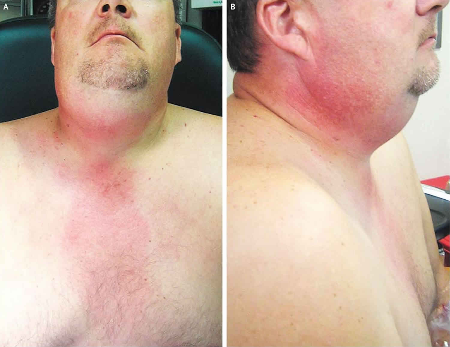
Footnote: A 58-year-old healthy man was evaluated for a toothache of 2 days’ duration. He was treated with oral penicillin and an opiate analgesic and advised to have the affected teeth pulled. He returned less than 24 hours later reporting severe swelling in the neck, sore throat, chills, and chest pain. On examination, the floor of his oral cavity was indurated and woody, and he had marked tenderness and adenopathy throughout his neck. He had erythema spreading from his neck down over his anterior chest wall where his chest pain was localized (Panels A and B). He did not have appreciable stridor or trismus, and he was able to swallow his own secretions. Ludwig’s angina is an infectious process involving the submental, sublingual, and submandibular spaces. It can rapidly progress to hemodynamic instability and airway obstruction; in rare cases, it spreads into the mediastinum. Compromise of the airway can progress rapidly, with lethal consequences; therefore, immediate consideration should be given to surgical débridement of the infected areas and antimicrobial therapy. In addition, options for surgical management of the airway should be available. The patient was admitted and underwent extensive surgical débridement. He recovered after a stay in the intensive care unit.
[Source 3 ]Is Ludwig angina contagious
No. Ludwig’s angina is a submandibular space or sublingual space infection that often develops after an infection of the roots of the teeth (such as tooth abscess) or a mouth injury.
Ludwig’s angina causes
The most common cause of Ludwig’s angina is dental disease in the lower molars mainly second and third which accounts for over 90% of cases 1. Any recent infection or injury to the area may predispose the patient to develop Ludwig’s angina. Some common causes include injury or laceration to the floor of the mouth, mandible fracture, tongue injury, oral piercing, osteomyelitis, traumatic intubation, peritonsillar abscess, submandibular sialadenitis and infected thyroglossal cysts. Predisposing factors include diabetes, oral malignancy, dental caries, alcoholism, malnutrition, and immunocompromised status.
Ludwig’s angina usually starts as a cellulitis of the submandibular space 1. The infection usually starts as a dental infection of the second or third mandibular molar teeth 1. Other sources of infection include local spread from a peritonsillar abscess or suppurative parotitis. The infection spreads medially rather than laterally because the medial side of the periodontal bones is thin. The infection initially spreads to the sublingual space and progresses to the submandibular space. Since the infection does not spread via the lymphatic system, the infection is bilateral. The infection is usually polymicrobial involving the oral flora. The most common organisms are Staphylococcus, Streptococcus, Peptostreptococcus, Fusobacterium, Bacteroides and Actinomyces. Immunocompromised patients are at higher risk of Ludwig’s angina 1.
Ludwig angina prevention
Visit the dentist for regular checkups.
Treat symptoms of mouth or tooth infection right away.
Ludwig’s angina differential diagnosis
Differential diagnosis includes peritonsillar abscess, retropharyngeal abscess, submandibular abscess, epiglottitis, oral carcinoma, angioedema, submandibular hematoma, and diphtheria. Although Ludwig’s angina is a clinical diagnosis, it may be difficult to differentiate from other diseases initially. Imaging may be helpful in this situation for Ludwig’s angina and also to rule out other causes of the patient’s symptoms. Typically, it does not result in abscess formation and it does not involve the lymphatic system.
Ludwig’s angina symptoms
In Ludwig angina the infected area swells quickly. This may block the airway or prevent you from swallowing saliva.
The most common presenting symptoms of Ludwig angina are fever and chills with neck swelling, neck pain, odynophagia (painful swallowing) and dysphagia (difficulty swallowing). People often describe the appearance as a “bull neck.” Less common symptoms include mouth pain, hoarse voice, drooling, tongue swelling, stiff neck and sore throat. Stridor may indicate impending airway obstruction. Patients will not have trismus unless the infection has spread into the parapharyngeal space. On physical exam, patients will have a fever with bilateral induration due to submandibular swelling and tenderness, swelling to the floor of the mouth, tenderness to the involved teeth, stiff neck, edema in the upper part of the neck, and crepitus. The patient will not typically have lymphadenopathy.
Ludwig angina symptoms include:
- Breathing difficulty
- Difficulty swallowing
- Drooling
- Unusual speech (sounds like the person has a “hot potato” in the mouth)
- Tongue swelling or protrusion of the tongue out of the mouth
- Fever
- Neck pain
- Neck swelling
- Redness of the neck
Other symptoms that may occur with Ludwig angina:
- Weakness, fatigue, excess tiredness
- Confusion or other mental changes
- Earache
Ludwig angina possible complications
Ludwig’s angina is a rapidly progressive cellulitis which can cause airway obstruction requiring immediate intervention. Close monitoring is required to prevent extension of the cellulitis to the adjacent areas. Ludwig’s angina can cause mediastinitis or necrotizing cellulitis of the neck. Ludwig’s angina can also cause aspiration pneumonia.
Ludwig angina complications may include:
- Airway blockage
- Generalized infection (sepsis)
- Septic shock
Ludwig angina diagnosis
A clinical diagnosis should be made based on presentation. Your health care provider will do an exam of your neck and head to look for redness and swelling of the upper neck, under the chin.
The swelling may reach to the floor of the mouth. Your tongue may be swollen or pushed up to top of your mouth.
You may need a CT scan. CT scan of the soft tissue neck with intravenous (IV) contrast is used to evaluate the severity of the infection and airway obstruction. CT is also useful to determine which patients will require surgical intervention for the formation of an abscess. Ultrasound may also be useful to identify the formation of an abscess. However, Ludwig’s angina usually does not result in an abscess formation. Therefore, it is often difficult to obtain cultures to determine what bacteria is causing the infection.
A sample of the fluid from the tissue may be sent to the lab to test for bacteria. Laboratory testing, although common in clinical practice, may be of little value as this is a clinical diagnosis. Blood cultures should be obtained to determine if there is the hematogenous spread of the infection.
Ludwig angina treatment
Early airway management is critical to the treatment of Ludwig’s angina as the most common cause of death is sudden asphyxiation from airway obstruction. Flexible fiberoptic nasal intubation is clinician’s favored method of intubation. The provider with the most experience should manage the airway as it will often be very challenging. Video laryngoscopy may be an option although there are no studies to date on this issue. Standard direct laryngoscope may be very challenging because of the swelling of the upper airway. It is important to manage the airway before the presence of stridor or cyanosis as these are late findings. If the patient is not able to be intubated, the next step would be an emergency tracheotomy. Cricothyrotomy is very challenging because of the edema in the neck which can obscure the anatomy.
Early broad-spectrum IV antibiotics have been shown to be helpful. For patients who are immunocompetent, a reasonable first choice would be ampicillin-sulbactam or clindamycin. Antibiotics should cover gram-positive bacteria, gram-negative bacteria, and anaerobes. For patients who are immunocompromised, the coverage should be broadened to cover for pseudomonas. Some options include cefepime, meropenem, or piperacillin-tazobactam. MRSA coverage should be considered for patients who are immunocompromised, increased risk of methicillin-resistant Staphylococcus aureus (MRSA) or prior MRSA infection. IV steroids are controversial. Several case reports have shown the decrease in the need for airway management with the use of steroids. However, more studies are needed before it becomes standard of care. Duration of the antibiotics are usually two to three weeks. White blood cell count and fever needs to be monitored closely.
Ludwig angina surgery
Dental extraction is recommended if the source of the infection is odontogenic. For patients who do not respond to initial antibiotics or develop a fluid collection on imaging, needle aspiration or surgical incision and drainage may be performed. Surgery is usually reserved for patients who fail medical therapy as early surgical decompression has not been shown to improve outcomes.
Ludwig angina prognosis
Due to the life-threatening complication of airway obstruction from Ludwig’s angina, mortality exceeded 50% prior to the development of antibiotics. With antibiotic therapy, along with improved imaging modalities and surgical techniques, mortality is approximately 8%.
- An J, Singhal M. Ludwig Angina. [Updated 2018 Dec 6]. In: StatPearls [Internet]. Treasure Island (FL): StatPearls Publishing; 2018 Jan-. Available from: https://www.ncbi.nlm.nih.gov/books/NBK482354[↩][↩][↩][↩][↩][↩][↩]
- [↩]
- Ludwig’s Angina. N Engl J Med 2008; 359:1501 DOI: 10.1056/NEJMicm065036 https://www.nejm.org/doi/full/10.1056/NEJMicm065036[↩]


