Contents
What is pericarditis
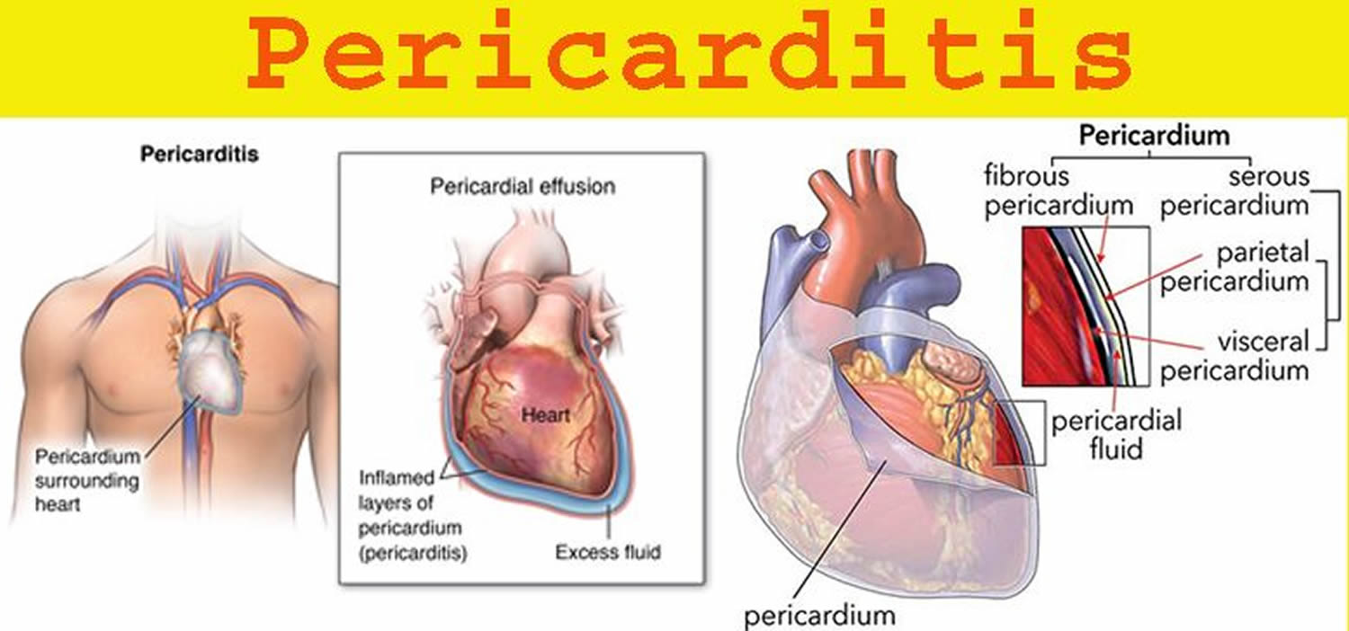
Pericarditis is inflammation of the pericardium, the thin protective sac that surrounds your heart. The pericardium has an inner and outer layer and can become inflamed if blood or fluid leaks between these two layers (see Figures 1 and 2). In pericarditis, the pericardium gets inflamed, and blood or fluid can leak into it. Pericarditis is often associated with pericardial effusion (build-up of fluid within the structures). Pericarditis causes chest pain and a high temperature (fever).
Symptoms of pericarditis include:
- Sharp chest pain that’s similar to a stabbing sensation, which may feel worse when swallowing or piercing chest pain in the center or left side of your chest. However, some people with acute pericarditis describe their chest pain as dull, achy or pressure-like instead, and of varying intensity.
- Pain in the neck that may extend across the shoulders and/or arms
- An intermittent fever
- Nausea
- Light headedness
- In some cases, a sudden onset shortness of breath (if this occurs seek urgent medical attention).
The pain can resolve itself if sitting forward, allowing the heart to relax within the chest cavity.
Usually the cause of pericarditis can’t be found. A viral infection is often suspected, but is difficult to confirm. Pericarditis can also develop after a heart attack. Other less common causes include autoimmune disorders, complication of a bacterial infection, and heart or chest injury.
- Pericarditis is not usually serious, but it can cause complications. Get medical advice if you have chest pain.
Most people recover from pericarditis quickly, but for some it can take several months or never fully resolve, making long term sufferers vulnerable to physical and psychological issues. This can impact on quality of life for the sufferer and their families.
Unfortunately, pericarditis can come back despite medical or surgical intervention, so patients may feel uncertain about the future of their health. As this rare condition is not visible or associated with an unhealthy lifestyle, there is often a lack of understanding about the effects of living with pericarditis, meaning sufferers can often feel isolated, causing them to experience other effects such as anxiety, palpitations and panic.
Most people with pericarditis need monitoring and treatment to reduce the pain and swelling.
If complications develop, surgery may be needed.
Seek immediate medical care if you develop new symptoms of chest pain.
Many of the symptoms of pericarditis are similar to those of other heart and lung conditions. The sooner you are evaluated, the sooner you can receive proper diagnosis and treatment. For example, although the cause of acute chest pain may be pericarditis, the original cause could have been a heart attack or a blood clot of the lungs (pulmonary embolus).
The pericardium
The serous membrane is a thin double-layered membrane that covers, lines, partitions, or connects structures. The serous membrane lining the heart cavity and covering the heart is the pericardium. The visceral pericardium covers the surface of the heart; the parietal pericardium lines the chest wall. Between them is the pericardial cavity, filled with a small amount of lubricating serous fluid (see Figure 1).
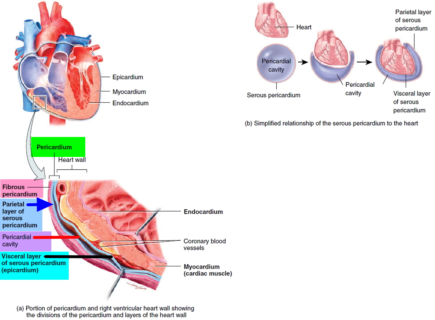
The pericardium confines the heart to its position in the mediastinum, while allowing sufficient freedom of movement for vigorous and rapid contraction. The pericardium consists of two main parts: (1) the fibrous pericardium and (2) the serous pericardium (Figures 1 and 2). The superficial fibrous pericardium is composed of tough, inelastic, dense irregular connective tissue. It resembles a bag that rests on and attaches to the diaphragm; its open end is fused to the connective tissues of the blood vessels entering and leaving the heart. The fibrous pericardium prevents overstretching of the heart, provides protection, and anchors the heart in the mediastinum. The fibrous pericardium near the apex of the heart is partially fused to the central tendon of the diaphragm and therefore movement of the diaphragm, as in deep breathing, facilitates the movement of blood by the heart.
The deeper serous pericardium is a thinner, more delicate membrane that forms a double layer around the heart (Figure 2). The outer parietal layer of the serous pericardium is fused to the fibrous pericardium. The inner visceral layer of the serous pericardium, which is also called the epicardium, is one of the layers of the heart wall and adheres tightly to the surface of the heart. Between the parietal and visceral layers of the serous pericardium is a thin film of lubricating serous fluid. This slippery secretion of the pericardial cells, known as pericardial fluid, reduces friction between the layers of the serous pericardium as the heart moves. The space that contains the few milliliters of pericardial fluid is called the pericardial cavity.
Figure 1. Pericardium
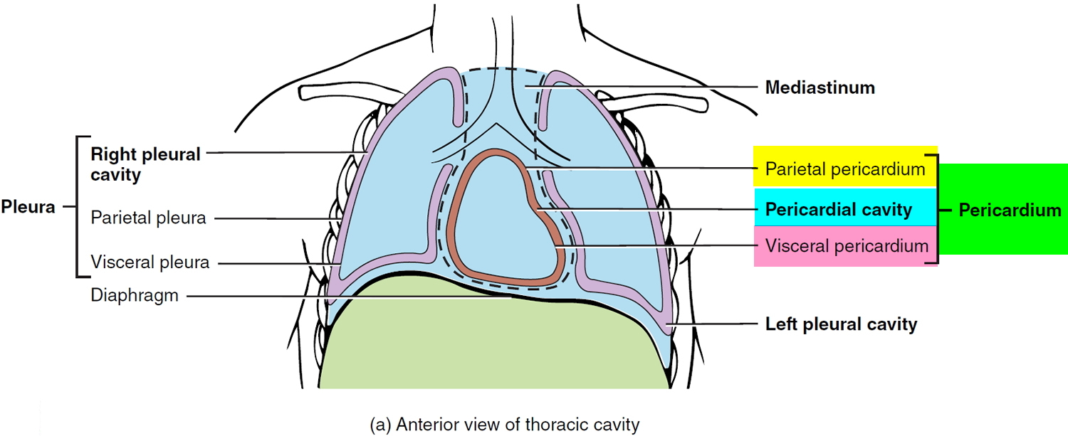
Figure 2. Pericardium, pericardial cavity and heart wall
Pericarditis types
The main types of pericarditis are:
- Acute pericarditis – symptoms begin suddenly and usually lasts less than three weeks.
- Incessant pericarditis lasts about four to six weeks but less than three months and is continuous.
- Chronic pericarditis – symptoms develop gradually and persist, or may persist after an acute attack but last longer than three months.
- Recurring pericarditis – repeated attacks of acute pericarditis with a symptom-free interval in between and it occurs about four to six weeks after an episode of acute pericarditis.
Acute episodes of pericarditis typically last a few weeks, but future episodes can occur. Some people with pericarditis have a recurrence within months after the original episode.
Pericarditis complications
Complications of pericarditis can be:
- Constrictive pericarditis – Although uncommon, some people with pericarditis, particularly those with long-term inflammation and chronic recurrences, can develop permanent thickening, scarring and contraction of the pericardium. In these people, the pericardium loses much of its elasticity and resembles a rigid case that’s tight around the heart, which keeps the heart from working properly. This condition is called constrictive pericarditis and often leads to severe swelling of the legs and abdomen, as well as shortness of breath.
- Cardiac tamponade – a dangerous condition, where too much fluid collects in the pericardium, which puts pressure on the heart and causes blood pressure to drop dramatically. This is life-threatening and requires emergency treatment. Excess fluid puts pressure on the heart and doesn’t allow it to fill properly. That means less blood leaves the heart, which causes a dramatic drop in blood pressure. Cardiac tamponade can be fatal if it isn’t promptly treated.
Pericarditis causes
Under normal circumstances, the two-layered pericardial sac that surrounds your heart contains a small amount of lubricating fluid. In pericarditis, the sac becomes inflamed and the resulting friction from the inflamed sac leads to chest pain.
The cause of pericarditis is often hard to determine. In most cases, doctors either are unable to determine a cause (idiopathic) or suspect a viral infection.
Pericarditis can also develop shortly after a major heart attack, due to the irritation of the underlying damaged heart muscle. In addition, a delayed form of pericarditis may occur weeks after a heart attack or heart surgery.
This delayed pericarditis is known as Dressler’s syndrome. Dressler’s syndrome may also be called postpericardiotomy syndrome, post-myocardial infarction syndrome and post-cardiac injury syndrome.
What causes pericarditis
Pericarditis is a complex condition with many variations and causes. A patient may need a physical examination and a doctor will consider their medical history in order for them to be diagnosed with the condition.
Pericarditis can be caused by:
- Cardiovascular disorders, such as acute myocardial infarction (heart attack), Dressler syndrome and aortic dissection
- Heart surgery
- Infectious conditions, such as viral (such as the flu), bacterial, HIV/AIDS and tuberculous infections
- Inflammatory disorders, such as rheumatoid arthritis, SLE (Systemic Lupus Erythematosus), scleroderma, and rheumatic fever
- Metabolic disorders, such as renal failure and hypothyroidism
- in rare cases, some form of cancer
- inflammation of the myocardium (the heart muscle) rubbing against the pericardium
- Miscellaneous causes, such as iatrogenic, neoplasms, drugs, irradiation, sarcoidosis, cardiovascular procedures, and trauma
- Trauma. Injury to your heart or chest may occur as a result of a motor vehicle or other accident
- Certain medications. Some medications can cause pericarditis, although this is unusual.
Sometimes the cause will be unknown.
Pericarditis symptoms
If you have acute pericarditis, the most common symptom of pericarditis is sharp, piercing or stabbing chest pain in the center or left side of your chest. However, some people with acute pericarditis describe their chest pain as dull, achy or pressure-like instead, and of varying intensity.
The pain of acute pericarditis may travel into your left shoulder and neck. It often intensifies when you cough, lie down or inhale deeply. Sitting up and leaning forward can often ease the pain. At times, it may be difficult to distinguish pericardial pain from the pain that occurs with a heart attack.
Depending on the cause of pericarditis, symptoms may also include some or all of the following:
- Sharp, piercing chest pain over the center or left side of the chest, which is generally more intense when breathing in
- Shortness of breath when reclining
- Heart palpitations
- Low-grade fever
- An overall sense of weakness, fatigue or feeling sick
- Cough
- Abdominal or leg swelling
- Nausea
- Dry cough
Chronic pericarditis is usually associated with chronic inflammation and may result in fluid around the heart (pericardial effusion). The most common symptom of chronic pericarditis is chest pain.
Pericarditis diagnosis
Your doctor likely will start by taking your medical history and asking questions about your chest pain and other symptoms. As part of your initial evaluation, your doctor will also perform a physical exam and check your heart sounds.
While listening to your heart, your doctor will place a stethoscope on your chest to check for the sounds characteristic of pericarditis, which are made when the pericardial layers rub against each other. This characteristic noise is called a pericardial rub.
Your doctor may have you undergo tests that can help determine whether you’ve had a heart attack, whether fluid has collected in the pericardial sac or whether there are signs of inflammation. Your doctor may use blood tests to determine if a bacterial or other type of infection is present.
Tests for pericarditis include:
- Electrocardiogram (ECG). In this test, patches with wires (electrodes) are attached to your skin to measure the electrical impulses given off by your heart. Impulses are recorded as waves displayed on a monitor or printed on paper. Certain ECG results may indicate pericarditis, while others could indicate a heart attack.
- Chest X-ray. With an X-ray of your chest, your doctor can study the size and shape of your heart. Images of your heart may show an enlarged heart if excess fluid has accumulated in the pericardium.
- Echocardiogram. This test uses high-frequency sound waves to create an image of your heart and its structures, including fluid accumulation in the pericardium. Your doctor can view and analyze this image on a monitor.
- Computerized tomography (CT). This X-ray technique can produce more-detailed images of your heart and the pericardium than can conventional X-ray studies. CT scanning may be done to exclude other causes of acute chest pain, such as a blood clot in a lung artery (pulmonary embolus) or a tear in your aorta (aortic dissection). CT scanning can also be used to look for thickening of the pericardium that might indicate constrictive pericarditis.
- Cardiac magnetic resonance imaging (MRI). This technique uses a magnetic field and radio waves to create cross-sectional images of your heart that can reveal thickening, inflammation or other changes in the pericardium.
Pericarditis treatment
Treatment will depend on type, the cause as well as the severity of pericarditis you have. Mild cases of pericarditis may get better on their own without treatment.
Pericarditis treatment may include:
- Non-steroidal anti-inflammatory drugs (NSAIDs), such as Ibuprofen to reduce swelling. You should feel better within 1 to 2 weeks.
- Antibiotics. When a bacterial infection is the underlying cause of pericarditis, you’ll be treated with antibiotics and drainage if necessary.
- Colchicine (Colcrys, Mitigare). This drug, which reduces inflammation in the body, may be prescribed for acute pericarditis or as a treatment for recurrent symptoms. Colchicine can reduce the length of pericarditis symptoms and decrease the risk that the condition will recur. However, the drug is not safe for people with certain pre-existing health problems, such as liver or kidney disease, and for those taking certain medications. Your doctor will carefully check your health history before prescribing colchicine.
- Pain killers. Most pain associated with pericarditis responds well to treatment with pain relievers available without a prescription, such as aspirin or ibuprofen (Advil, Motrin IB, others). These medications also help lessen inflammation. Prescription-strength pain relievers also may be used.
- Corticosteroids. If you don’t respond to pain killers or colchicine or if you have recurrent symptoms of pericarditis, your doctor may prescribe a steroid medication, such as prednisone.
- If symptoms don’t resolve surgery may be necessary – pericardial window (a procedure to drain the sac surrounding the heart) is normally performed to treat and permanently prevent symptoms from persisting or coming back.
If an underlying cause is found, it will be treated where possible. You may also be monitored for potential complications.
For most mild cases of pericarditis, rest and over-the-counter pain medications — taken under your doctor’s direction — may be all that’s needed. While you recuperate, avoid rigorous physical activity. Strenuous activity can trigger pericarditis symptoms. Ask your doctor how long you need to take it easy.
Hospitalization and procedures
You’ll likely need hospitalization if your doctor suspects cardiac tamponade, a dangerous complication of pericarditis due to fluid buildup around the heart.
If cardiac tamponade is present, your doctor may recommend a procedure to relieve fluid buildup, such as:
- Pericardiocentesis. In this procedure, a doctor uses a sterile needle or a small tube (catheter) to remove and drain the excess fluid from the pericardial cavity. You’ll receive a local anesthetic before undergoing pericardiocentesis, which is often done with echocardiogram monitoring and ultrasound guidance. This drainage may continue for several days during the course of your hospitalization.
- Pericardiectomy. If you’re diagnosed with constrictive pericarditis, you may need to undergo a surgical procedure (pericardiectomy) to remove the entire pericardium that has become rigid and is making it hard for your heart to pump.
Coping and support for pericarditis
Living with pericarditis can be emotionally challenging for you and your family. It is important to manage anxiety and stress and there are many outlets of support to help you. Talk to your doctor about being referred for counseling or Cognitive Behavioral Therapy (CBT).


