Contents
What is the peripheral nervous system
The peripheral nervous system consists of all nervous tissue outside the central nervous system (brain and spinal cord). Components of the peripheral nervous system include nerves and sensory receptors. A nerve is a bundle of hundreds to thousands of axons plus associated connective tissue and blood vessels that lies outside the brain and spinal cord. Twelve pairs of cranial nerves emerge from the brain and thirty-one pairs of spinal nerves emerge from the spinal cord. Each nerve follows a defined path and serves a specific region of the body. The term sensory receptor refers to a structure of the nervous system that monitors changes in the external or internal environment. Examples of sensory receptors include touch receptors in the skin, photoreceptors in the eye, and olfactory (smell) receptors in the nose.
The peripheral nervous system is functionally divided into sensory and motor divisions, and each of these is further divided into somatic and visceral subdivisions.
The sensory (afferent) division carries signals from various receptors (sense organs and simple sensory nerve endings) to the central nervous system (CNS). This pathway informs the central nervous system (brain and spinal cord) of stimuli within and around the body.
- The somatic sensory division carries signals from receptors in the skin, muscles, bones, and joints.
- The visceral sensory division carries signals mainly from the viscera of the thoracic and abdominal cavities, such as the heart, lungs, stomach, and urinary bladder.
The motor (efferent) division carries signals from the central nervous system (the brain and the spinal cord) mainly to gland and muscle cells that carry out the body’s responses. Cells and organs that respond to these signals are called effectors.
- The somatic motor division carries signals to the skeletal muscles. This produces voluntary muscle contractions as well as involuntary somatic reflexes.
- The visceral motor division (autonomic nervous system) carries signals to glands, cardiac muscle, and smooth muscle. You usually have no voluntary control over these effectors, and the autonomic nervous system operates at an unconscious level. The responses of the autonomic nervous system and its effectors are visceral reflexes. The autonomic nervous system has two further divisions:
- The sympathetic division tends to arouse the body for action—for example, by accelerating the heartbeat and increasing respiratory airflow—but it inhibits digestion.
- The parasympathetic division tends to have a calming effect—slowing the heartbeat, for example—but it stimulates digestion.
Figure 1. Nervous system and its parts
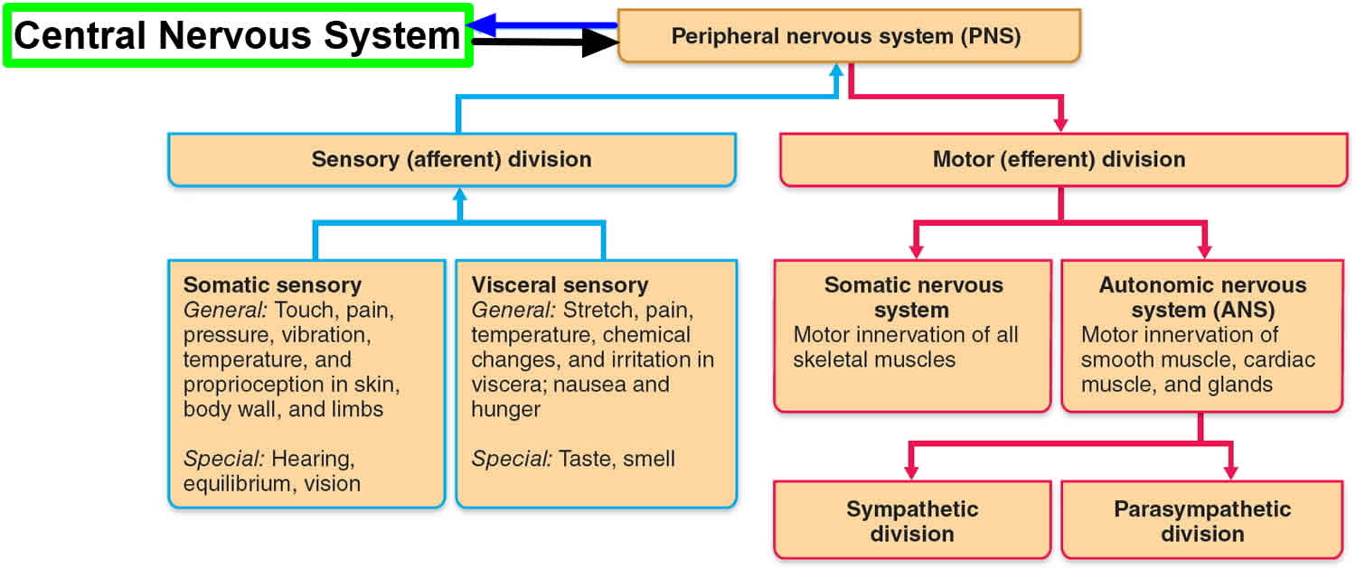
Figure 2. Peripheral nervous system
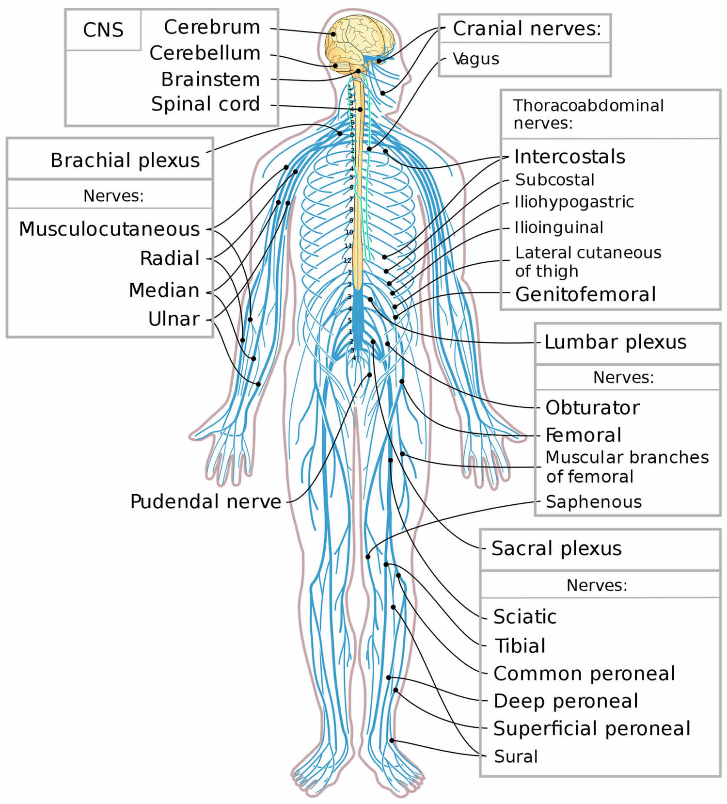 Figure 3. Autonomic nervous system (part of peripheral nervous system)
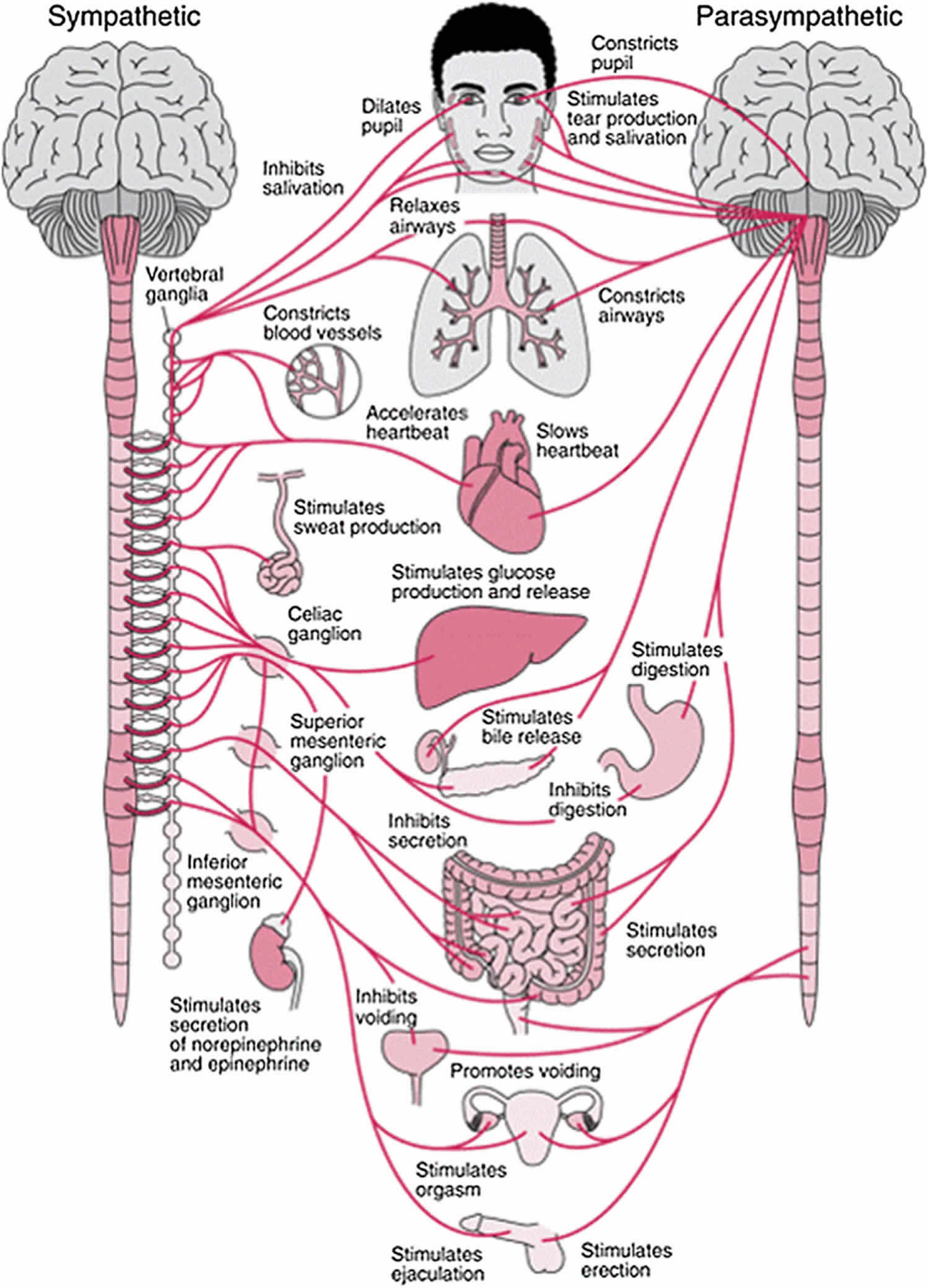
Central nervous system vs Peripheral nervous system
The central nervous system (CNS) consists of the brain and spinal cord. The brain is the part of the central nervous system that is located in the skull and contains about 85 billion neurons. The spinal cord is connected to the brain through the foramen magnum of the occipital bone and is encircled by the bones of the vertebral column. The spinal cord contains about 100 million neurons. The central nervous system (CNS) processes many different kinds of incoming sensory information. It is also the source of thoughts, emotions, and memories. Most signals that stimulate muscles to contract and glands to secrete originate in the central nervous system (brain and spinal cord).
The brain is divided into the cerebrum, diencephalons, brain stem, and cerebellum.
Cerebrum
The largest and most obvious portion of the brain is the cerebrum, which is divided by a deep longitudinal fissure into two cerebral hemispheres. The two hemispheres are two separate entities but are connected by an arching band of white fibers, called the [glossary term:] corpus callosum that provides a communication pathway between the two halves.
Each cerebral hemisphere is divided into five lobes, four of which have the same name as the bone over them: the fontal lobe, the parietal lobe, the occipital lobe, and the temporal lobe. A fifth lobe, the insula or Island of Reil, lies deep within the lateral sulcus.
Diencephalon
The diencephalons is centrally located and is nearly surrounded by the cerebral hemispheres. It includes the thalamus, hypothalamus, and epithalamus. The thalamus, about 80 percent of the diencephalons, consists of two oval masses of gray matter that serve as relay stations for sensory impulses, except for the sense of smell, going to the cerebral cortex. The hypothalamus is a small region below the thalamus, which plays a key role in maintaining homeostasis because it regulates many visceral activities. The epithalamus is the most dorsal portion of the diencephalons. This small gland is involved with the onset of puberty and rhythmic cycles in the body. It is like a biological clock.
Brain Stem
The brain stem is the region between the diencephalons and the spinal cord. It consists of three parts: midbrain, pons, and medulla oblongata. The midbrain is the most superior portion of the brain stem. The pons is the bulging middle portion of the brain stem. This region primarily consists of nerve fibers that form conduction tracts between the higher brain centers and spinal cord. The medulla oblongata, or simply medulla, extends inferiorly from the pons. It is continuous with the spinal cord at the foramen magnum. All the ascending (sensory) and descending (motor) nerve fibers connecting the brain and spinal cord pass through the medulla.
Cerebellum
The cerebellum, the second largest portion of the brain, is located below the occipital lobes of the cerebrum. Three paired bundles of myelinated nerve fibers, called cerebellar peduncles, form communication pathways between the cerebellum and other parts of the central nervous system.
Ventricles and Cerebrospinal Fluid
A series of interconnected, fluid-filled cavities are found within the brain. These cavities are the ventricles of the brain, and the fluid is cerebrospinal fluid (CSF).
Spinal Cord
The spinal cord extends from the foramen magnum at the base of the skull to the level of the first lumbar vertebra. The cord is continuous with the medulla oblongata at the foramen magnum. Like the brain, the spinal cord is surrounded by bone, meninges, and cerebrospinal fluid.
The spinal cord is divided into 31 segments with each segment giving rise to a pair of spinal nerves. At the distal end of the cord, many spinal nerves extend beyond the conus medullaris to form a collection that resembles a horse’s tail. This is the cauda equina. In cross section, the spinal cord appears oval in shape.
The spinal cord has two main functions:
- Serving as a conduction pathway for impulses going to and from the brain. Sensory impulses travel to the brain on ascending tracts in the cord. Motor impulses travel on descending tracts.
- Serving as a reflex center. The reflex arc is the functional unit of the nervous system. Reflexes are responses to stimuli that do not require conscious thought and consequently, they occur more quickly than reactions that require thought processes. For example, with the withdrawal reflex, the reflex action withdraws the affected part before you are aware of the pain. Many reflexes are mediated in the spinal cord without going to the higher brain centers.
Peripheral nervous system divisions
The peripheral nervous system is functionally divided into sensory (afferent) and motor (efferent) divisions (Figure 1). The sensory inputs (afferent division) and motor outputs (efferent division) of the peripheral nervous system are subcategorized as either somatic (innervating the outer tube) or visceral (innervating
the visceral organs or inner tube).
The sensory or afferent division of the peripheral nervous system conveys input into the central nervous system (brain and spinal cord) from sensory receptors in the body. Within the sensory division, sensory inputs are differentiated as general (widespread) or special (localized, i.e., the special senses). The sensory or afferent division of the peripheral nervous system provides the central nervous system (brain and spinal cord) with sensory information about the somatic senses (tactile, thermal, pain, and proprioceptive sensations) and special senses (smell, taste, vision, hearing, and equilibrium).
The motor or efferent division of the peripheral nervous system conveys output from the central nervous system (brain and spinal cord) to effectors (muscles and glands). The motor division is further subdivided into a somatic nervous system and an autonomic nervous system. The somatic nervous system conveys output from the central nervous system (brain and spinal cord) to skeletal muscles only. Because its motor responses can be consciously controlled, the action of this part of the peripheral nervous system is voluntary. The visceral motor component of the peripheral nervous system is the autonomic nervous system. The autonomic nervous system has parasympathetic and sympathetic divisions. The autonomic nervous system conveys output from the central nervous system (brain and spinal cord) to smooth muscle, cardiac muscle, and glands. Because its motor responses are not normally under conscious control, the action of the autonomic nervous system is involuntary.
The autonomic nervous system is comprised of two main branches, the sympathetic nervous system and the parasympathetic nervous system. With a few exceptions, effectors (muscles and glands) receive innervation from both of these branches, and usually the two branches have opposing actions. For example, neurons of the sympathetic nervous system increase heart rate, and neurons of the parasympathetic nervous system slow it down. In general, the parasympathetic nervous system takes care of “rest-and-digest” activities, and the sympathetic nervous system helps support exercise or emergency actions—the so-called “fight-or-flight” responses. A third branch of the autonomic nervous system is the enteric nervous system (intestines nervous system), an extensive network of over 100 million neurons confined to the wall of the gastrointestinal (GI) tract. The enteric nervous system (intestines nervous system) helps regulate the activity of the smooth muscle and glands of the gastrointestinal tract. Although the enteric nervous system can function independently, it communicates with and is regulated by the other branches of the autonomic nervous system.
Peripheral nervous system parts
The peripheral nervous system is further subdivided into the somatic nervous system and the autonomic nervous system. The somatic nervous system consists of nerves that go to the skin and muscles and is involved in conscious activities. The autonomic nervous system consists of nerves that connect the central nervous system (brain and spinal cord) to the visceral organs such as the heart, stomach, and intestines. It mediates unconscious activities.
It is important to recognize that the structures of the peripheral nervous system reflect this functional organization—nerve impulses from each modality (somatic sensory, visceral sensory, somatic motor, visceral motor) are carried on distinct neurons. The following are the structures of the peripheral nervous system:
- The sensory receptors. Sensory receptors pick up stimuli (environmental changes) from inside and outside the body and then initiate impulses in sensory axons, which carry the impulse to the CNS. The receptors for the special senses are specialized receptor cells.
- The nerves and ganglia. Nerves are bundles of peripheral axons or dendrites surrounded by connective tissue and ganglia are clusters of peripheral cell bodies, such as the cell bodies of the sensory neurons. Most nerves contain both sensory and motor axons and are called mixed nerves. Certain cranial nerves (the nerves attached to the brain) contain only sensory axons and thus are purely sensory in function; certain others contain primarily motor axons and are motor in function. The cranial nerves and the spinal nerves (the nerves attached to the spinal cord) are described later, focusing on their somatic functions. Visceral innervation is discussed with the autonomic nervous system. A connective tissue sheath called the epineurium surrounds each nerve. Each bundle of nerve fibers is called a fasciculus and is surrounded by a layer of connective tissue called the perineurium. Within the fasciculus, each individual nerve fiber, with its myelin and neurilemma, is surrounded by connective tissue called the endoneurium. A nerve may also have blood vessels enclosed in its connective tissue wrappings.
- The motor endings. The motor endings are the axon terminals of motor neurons that innervate the effector organs, muscles, and glands. Motor endings to the muscles and glands of the body: skeletal muscle innervation; cardiac muscle innervation; and the innervation of smooth muscle and glands.
Cranial Nerves
Twelve pairs of cranial nerves emerge from the inferior surface of the brain. All of these nerves, except the [glossary term:] vagus nerve, pass through foramina of the skull to innervate structures in the head, neck, and facial region.
The cranial nerves are designated both by name and by Roman numerals, according to the order in which they appear on the inferior surface of the brain. Most of the nerves have both sensory and motor components. Three of the nerves are associated with the special senses of smell, vision, hearing, and equilibrium and have only sensory fibers. Five other nerves are primarily motor in function but do have some sensory fibers for proprioception. The remaining four nerves consist of significant amounts of both sensory and motor fibers.
Acoustic neuromas are benign fibrous growths that arise from the balance nerve, also called the eighth cranial nerve or vestibulocochlear nerve. These tumors are non-malignant, meaning that they do not spread or metastasize to other parts of the body. The location of these tumors is deep inside the skull, adjacent to vital brain centers in the brain stem. As the tumors enlarge, they involve surrounding structures which have to do with vital functions. In the majority of cases, these tumors grow slowly over a period of years. In other cases, the growth rate is more rapid and patients develop symptoms at a faster pace. Usually, the symptoms are mild and many patients are not diagnosed until some time after their tumor has developed. Many patients also exhibit no tumor growth over a number of years when followed by yearly MRI scans.
Spinal Nerves
Thirty-one pairs of spinal nerves emerge laterally from the spinal cord. Each pair of nerves corresponds to a segment of the cord and they are named accordingly. This means there are 8 cervical nerves, 12 thoracic nerves, 5 lumbar nerves, 5 sacral nerves, and 1 coccygeal nerve.
Each spinal nerve is connected to the spinal cord by a dorsal root and a ventral root. The cell bodies of the sensory neurons are in the dorsal root ganglion, but the motor neuron cell bodies are in the gray matter. The two roots join to form the spinal nerve just before the nerve leaves the vertebral column. Because all spinal nerves have both sensory and motor components, they are all mixed nerves.
Autonomic Nervous System
The autonomic nervous system is a visceral efferent system, which means it sends motor impulses to the visceral organs. It functions automatically and continuously, without conscious effort, to innervate smooth muscle, cardiac muscle, and glands. It is concerned with heart rate, breathing rate, blood pressure, body temperature, and other visceral activities that work together to maintain homeostasis.
The autonomic nervous system is unique in that it requires a sequential two-neuron efferent pathway; the preganglionic neuron must first synapse onto a postganglionic neuron before innervating the target organ. The preganglionic, or first, neuron will begin at the “outflow” and will synapse at the postganglionic, or second, neuron’s cell body. The postganglionic neuron will then synapse at the target organ.
The autonomic nervous system has two parts, the sympathetic division and the parasympathetic division. Many visceral organs are supplied with fibers from both divisions. In this case, one stimulates and the other inhibits. This antagonistic functional relationship serves as a balance to help maintain homeostasis.
Sympathetic nervous system
The sympathetic division emerges from the spinal cord in the thoracic and lumbar areas, terminating around L2-3.
The sympathetic system is activated during a “fight or flight” situation in which great mental stress or physical danger is encountered. Neurotransmitters such as norepinephrine, and epinephrine are released, which increases heart rate and blood flow in certain areas like muscle, while simultaneously decreasing activities of non-critical functions for survival, like digestion. The systems are independent to each other, which allows activation of certain parts of the body, while others remain rested.
Parasympathetic nervous system
The parasympathetic division has craniosacral “outflow”, meaning that the neurons begin at the cranial nerves (specifically the oculomotor nerve, facial nerve, glossopharyngeal nerve and vagus nerve) and sacral (S2, S3, S4) spinal cord.
The parasympathetic nervous system consists of cells with bodies in one of two locations: the brainstem (Cranial Nerves III, VII, IX, X) or the sacral spinal cord (S2, S3, S4). These are the preganglionic neurons, which synapse with postganglionic neurons in these locations:
- Parasympathetic ganglia of the head: Ciliary (Cranial nerve III), Submandibular (Cranial nerve VII), Pterygopalatine (Cranial nerve VII), and Otic (Cranial nerve IX)
- In or near the wall of an organ innervated by the Vagus (Cranial nerve X) or Sacral nerves (S2, S3, S4)
These ganglia provide the postganglionic neurons from which innervations of target organs follows. Examples are:
- The postganglionic parasympathetic splanchnic (visceral) nerves
- The vagus nerve, which passes through the thorax and abdominal regions innervating, among other organs, the heart, lungs, liver and stomach
Primarily using the neurotransmitter acetylcholine (ACh) as a mediator, the parasympathetic system allows the body to function in a “rest and digest” state. Consequently, when the parasympathetic system dominates the body, there are increases in salivation and activities in digestion, while heart rate and other sympathetic response decrease. Unlike the sympathetic system, humans have some voluntary controls in the parasympathetic system. The most prominent examples of this control are urination and defecation.
Peripheral nervous system function
The peripheral nervous system carries out a complex array of tasks. It allows you to sense various smells and produce speech; in addition, it provides signals that control body movements and regulates the operation of internal organs. These diverse activities can be grouped into two basic functions: sensory (input) and motor (output).
- Sensory function. Sensory receptors detect internal stimuli, such as an increase in blood pressure, or external stimuli (for example, a raindrop landing on your arm). This sensory information is then carried into the brain and spinal cord through cranial and spinal nerves.
- Motor function. Once sensory information is integrated in the central nervous system, the central nervous system may elicit an appropriate motor response by activating effectors (muscles and glands) through cranial and spinal nerves. Stimulation of the effectors causes muscles to contract and glands
to secrete.
The two basic functions of the peripheral nervous system occur, for example, when you answer your cell phone after hearing it ring. The sound of the ringing cell phone stimulates sensory receptors in your ears (sensory function). This auditory information is subsequently relayed into your brain where it is processed and the decision to answer the phone is made (integrative function). The brain then stimulates the contraction of specific muscles that will allow you to grab the phone and press the appropriate button to answer it (motor function).
Autonomic nervous system function
Sympathetic and parasympathetic divisions of the peripheral nervous system typically function in opposition to each other. But this opposition is better termed complementary in nature rather than antagonistic. For an analogy, one may think of the sympathetic division as the accelerator and the parasympathetic division as the brake. The sympathetic division typically functions in actions requiring quick responses. The parasympathetic division functions with actions that do not require immediate reaction. The sympathetic system is often considered the “fight or flight” system, while the parasympathetic system is often considered the “rest and digest” or “feed and breed” system.
However, many instances of sympathetic and parasympathetic activity cannot be ascribed to “fight” or “rest” situations. For example, standing up from a reclining or sitting position would entail an unsustainable drop in blood pressure if not for a compensatory increase in the arterial sympathetic tonus. Another example is the constant, second-to-second, modulation of heart rate by sympathetic and parasympathetic influences, as a function of the respiratory cycles. In general, these two systems should be seen as permanently modulating vital functions, in usually antagonistic fashion, to achieve homeostasis. Higher organisms maintain their integrity via homeostasis which relies on negative feedback regulation which, in turn, typically depends on the autonomic nervous system. Some typical actions of the sympathetic and parasympathetic nervous systems are listed below.
Sympathetic nervous system
Promotes a fight-or-flight response, corresponds with arousal and energy generation, and inhibits digestion (see Figure 3):
- Diverts blood flow away from the gastro-intestinal (GI) tract and skin via vasoconstriction
- Blood flow to skeletal muscles and the lungs is enhanced (by as much as 1200% in the case of skeletal muscles)
- Dilates bronchioles of the lung through circulating epinephrine, which allows for greater alveolar oxygen exchange
- Increases heart rate and the contractility of cardiac cells (myocytes), thereby providing a mechanism for enhanced blood flow to skeletal muscles
- Dilates pupils and relaxes the ciliary muscle to the lens, allowing more light to enter the eye and enhances far vision
- Provides vasodilation for the coronary vessels of the heart
- Constricts all the intestinal sphincters and the urinary sphincter
- Inhibits peristalsis
- Stimulates orgasm
Parasympathetic nervous system
The parasympathetic nervous system has been said to promote a “rest and digest” response, promotes calming of the nerves return to regular function, and enhancing digestion. Functions of nerves within the parasympathetic nervous system include:
- Dilating blood vessels leading to the GI tract, increasing the blood flow.
- Constricting the bronchiolar diameter when the need for oxygen has diminished
- Dedicated cardiac branches of the vagus and thoracic spinal accessory nerves impart parasympathetic control of the heart (myocardium)
- Constriction of the pupil and contraction of the ciliary muscles, facilitating accommodation and allowing for closer vision
- Stimulating salivary gland secretion, and accelerates peristalsis, mediating digestion of food and, indirectly, the absorption of nutrients
- Sexual. Nerves of the peripheral nervous system are involved in the erection of genital tissues via the pelvic splanchnic nerves 2–4. They are also responsible for stimulating sexual arousal.
Enteric nervous system
The enteric nervous system is the intrinsic nervous system of the gastrointestinal system. Its functions include:
- Sensing chemical and mechanical changes in the gut
- Regulating secretions in the gut
- Controlling peristalsis and some other movements


