Contents
- Pernicious anemia
- What is vitamin B12?
- Pernicious anemia causes
- Risk Factors for pernicious anemia
- Pernicious anemia prevention
- Pernicious anemia signs and symptoms
- Pernicious anemia complications
- Pernicious anemia diagnosis
- Pernicious anemia treatment
- Pernicious anemia prognosis
Pernicious anemia
Pernicious anemia is a condition in which your body can’t make enough healthy red blood cells because of severe vitamin B12 deficiency due to an auto-immune inflammation of the stomach (autoimmune gastritis), resulting in destruction of stomach parietal cells by your own antibodies resulting in intrinsic factor (IF) deficiency and malabsorption of dietary vitamin B12, recycled biliary vitamin B12, and free vitamin B12 1, 2, 3, 4, 5. Pernicious anemia is caused by autoimmune metaplastic atrophic gastritis, which predominantly manifests in the stomach body and fundus. In pernicious anemia, anti-parietal cell antibodies (PCA) are detected specifically against the hydrogen potassium adenosine triphosphatase (H+/K+‐ATPase) proton pump 6. Helicobacter pylori (H. pylori) are not generally considered to be associated with autoimmune metaplastic atrophic gastritis. However, Hershko et al. 7 have reported that Helicobacter pylori (H. pylori) might serve as a trigger of autoimmune metaplastic atrophic gastritis and pernicious anemia, based on their observation that the prevalence of H. pylori infection was 87.5% in patients under 20 years of age. In addition, one theory regarding the initiating event of autoimmune metaplastic atrophic gastritis is molecular mimicry between H. pylori antigens and gastric H+/K+−ATPase proton pump 8.
Vitamin B12 is a nutrient found in some foods. The body needs vitamin B12 to make healthy red blood cells and to keep its nervous system working properly. People who have pernicious anemia can’t absorb enough vitamin B12 from food. This is because they lack intrinsic factor (IF), a protein made in the stomach. A lack of this protein leads to vitamin B12 deficiency.
Other conditions and factors also can cause vitamin B12 deficiency. Examples include infections, surgery, medicines, and diet. Technically, the term “pernicious anemia” refers to vitamin B12 deficiency due to a lack of intrinsic factor. Often though, vitamin B12 deficiency due to other causes also is called pernicious anemia. Therefore, pernicious anemia must be differentiated from other disorders that interfere with the absorption and metabolism of vitamin B12.
Pernicious anemia has often been confused with vitamin B12 deficiency 9. Pernicious anemia denotes only vitamin B12 deficiency due to gastric atrophy and/or intrinsic factor deficiency or autoimmune gastritis 9. Pernicious anemia is considered a late stage of autoimmune gastritis.
These circulating anti-intrinsic factor antibodies (IFA) and anti-parietal cell antibodies (PCA) cause autoimmune chronic atrophic gastritis with parietal cell loss and eventual vitamin B12 deficiency 10. However, autoimmune gastritis-associated gastric corpus atrophy may progress without developing pernicious anemia 9. Progressive destruction of the parietal cells that line your stomach (autoimmune gastritis) cause decreased secretion of stomach acid and enzymes required to release food bound vitamin B12. Antibodies to intrinsic factor (IFA) bind to intrinsic factor (IF), a protein made in your stomach, preventing formation of the IF-B12 complex, further inhibiting vitamin B12 absorption which leads to vitamin B12 deficiency. People who have pernicious anemia can’t absorb enough vitamin B12 from food. Without enough vitamin B12, your red blood cells don’t divide normally and are too large (megaloblasts). These changes occur because vitamin B12 is necessary for DNA synthesis 11. Without treatment, pernicious anemia causes vitamin B12 deficiency, even in the presence of adequate vitamin B12 intakes. Generally, it takes about 10–12 years to clinically develop symptomatic pernicious anemia, so pernicious anemia may onset with subclinical vitamin B12 deficiency 12.
Rarely, children are born with an inherited disorder that prevents their bodies from making intrinsic factor. This disorder is called congenital pernicious anemia 9. This condition is quite rare and distinguishable from the usual form of pernicious anemia due to the early age of onset and the absence of gastric corpus atrophy.
Pernicious anemia is one of two major types of “macrocystic” or “megaloblastic” anemia. These terms refer to anemia in which the red blood cells are larger than normal. The other major type of macrocystic anemia is caused by folic acid deficiency. In addition to megaloblasts, hypersegmented neutrophils are also present. The large red blood cells may have trouble getting out of the bone marrow—a sponge-like tissue inside the bones where blood cells are made 11. Megaloblastic anemia is characterized by large nucleated red blood cell precursors called megaloblasts in the bone marrow 11. Without enough red blood cells to carry oxygen to your body, you may feel tired and weak. Severe or long-lasting pernicious anemia can damage your heart, brain, and other organs in your body. Note that the causes of megaloblastic anemia other than vitamin B12 deficiency caused by impaired intrinsic factor (IF) production can include folic acid deficiency, altered pH in the small intestine, and lack of absorption of vitamin B12 complexes in the terminal ileum.
Pernicious anemia is the most common cause of clinically evident vitamin B12 deficiency around the world 4, 1. Pernicious anemia accounts for 20%‐50% of the vitamin B12 deficiency in adults 13 and is associated with autoimmune gastritis, resulting in the destruction of gastric parietal cells and the associated lack of intrinsic factor 3. The incidence of pernicious anemia in the United States is an estimated 151 per 100,000, and this condition is more common in women and in people of European ancestry (North Europeans and Caucasian Americans) 1. The prevalence of pernicious anemia in Japan is rare, 1 to 5 per 100,000 persons compared with the West 14.
Pernicious anemia can also cause other problems, such as nerve damage, neurological problems (such as memory loss), and digestive tract problems. People who have pernicious anemia also may be at higher risk for weakened bone strength (osteoporosis) and stomach cancer. Pernicious anemia is frequently presenting with other autoimmune conditions such as autoimmune thyroid disease, type 1 diabetes, and vitiligo 15.
An important point is that pernicious anemia may lead to potentially serious long-term complications that may be related to micronutrient deficiencies and the development of gastric neoplasms, in particular, gastric cancer and type 1 gastric neuroendocrine tumors 9. When not recognized in a timely manner or when pernicious anemia is diagnosed with delay, these complications may be potentially life-threatening and sometimes irreversible.
Vitamin B12 therapy resolves the anemia of pernicious anemia, but does not cure the atrophic gastritis, which can progress to stomach cancer 16. The incidence of gastric adenocarcinoma is 2- to 3-fold greater in patients with pernicious anemia than in the general population of the same age 17. Presently, periodic gastroscopy and/or barium studies are not advocated in patients with treated pernicious anemia who are asymptomatic, because such screening has not been demonstrated to prolong lifespan 17.
A population-based, case-control study using the Surveillance, Epidemiology, and End Results (SEER)–Medicare database found that elderly persons with pernicious anemia were not only at significantly increased risk for noncardia gastric adenocarcinoma and gastric carcinoid tumors, they were also at increased risk for the following 16:
- Tonsillar cancer
- Hypopharyngeal cancer
- Esophageal squamous cell carcinoma
- Small intestinal cancer
- Liver cancer
- Myeloma
- Acute myeloid leukemia
- Myelodysplastic syndrome
In a longitudinal study of 199 intrinsic factor antibody (IFA)–positive and 168 IFA-negative Chinese patients, Chan et al 18 found that despite a good hematologic response to therapy, both groups had an unsatisfactory neurologic response, and newly diagnosed hypothyroidism was found during follow-up. In addition, newly diagnosed cancers were also found (24 in IFA-positive patients, seven in IFA-negative patients), of which 20% were stomach cancer 18.
For the intrinsic factor antibody (IFA)-positive patients with a cancer, mean survival was 64 months; for those without a cancer, it was 129 months. Mortality was 31% in this group, in which cancer-related deaths represented 37% of the total 18. For the intrinsic factor antibody (IFA)-negative patients with a cancer, mean survival was 36 months. For those without a cancer, it was 126 months. Mortality was 21% in this group, in which cancer-related deaths represented 14% of the total.
Chan et al 18 concluded that although Chinese patients treated for pernicious anemia demonstrated a good survival period, they remained at increased risk for gastric carcinoma, and IFA-positive patients had a higher risk of developing all types of cancers and cancer-related deaths than did IFA-negative patients.
Pernicious anemia has been estimated to be present in approximately 2% of individuals over 60 years of age 19. Although anemia is often a symptom, the condition is actually the end stage of an autoimmune inflammation of the stomach known as autoimmune atrophic gastritis, resulting in destruction of stomach cells by one’s own antibodies (autoantibodies). Progressive destruction of the cells that line the stomach causes decreased secretion of acid and enzymes required to release food-bound vitamin B12. Antibodies to intrinsic factor (IF) bind to IF preventing formation of the IF-B12 complex, further inhibiting vitamin B12 absorption. About 20% of the relatives of pernicious anemia patients also have the condition, suggesting a genetic predisposition. It is also thought that H. pylori infection could be involved in initiating the autoimmune response in a subset of individuals 20. Further, co-occurrence of autoimmune atrophic gastritis with other autoimmune conditions, especially autoimmune thyroiditis and type 1 diabetes mellitus, has been reported 21.
Pernicious anemia signs and symptoms are similar to other vitamin B12 deficiencies, but pernicious anemia is sometimes associated with other autoimmune diseases such as type 1 diabetes, autoimmune thyroiditis, and Addison’s disease.
When pernicious anemia is suspected, the first step is usually a full blood panel to test for anemia and/or macrocytosis, together with testing for cobalamin deficiency and increased levels of homocysteine and/or methylmalonic acid (MMA) 9. Next, the positivity of gastric autoantibodies towards parietal cells and/or intrinsic factor (IF) is commonly assessed 9. Sensitivity and specificity of the anti‐intrinsic factor antibody test were 50%‐70%, and greater than 95%, respectively 22. Sensitivity and specificity of the antigastric parietal cell antibody test were more than 90% and 50%, respectively 23. In any case, the hematological and/or serological suspicion of pernicious anemia always needs to be confirmed by histological assessment of gastric antral and corpus biopsies obtained during gastroscopy to ascertain the presence of autoimmune gastritis 9.
The treatment for pernicious anemia is lifelong administration of vitamin B12. Treatment of pernicious anemia generally requires injections of vitamin B12 to bypass intestinal absorption. High-dose oral supplementation is another treatment option, because consuming 1,000 mcg (1 mg)/day of vitamin B12 orally should result in the absorption of about 10 mcg/day (1% of dose) by passive diffusion. In fact, high-dose oral therapy is considered to be as effective as intramuscular injection 24.
Patients with pernicious anemia are at high risk of developing stomach cancer such as gastric adenocarcinoma and carcinoid tumors 25. Significant risk factors for the development of gastric carcinoma in autoimmune metaplastic atrophic gastritis include the presence of pernicious anemia, severity of mucosal atrophy, intestinal metaplasia, disease duration, and over 50 years of age 26. Periodic stomach examinations are recommended for patients with pernicious anemia.
What is vitamin B12?
Vitamin B12 is also known as cobalamin or cyanocobalamin (man-made form of vitamin B12), is a nutrient that helps keep your body’s nerve and blood cells healthy and helps make DNA, the genetic material in all cells. Vitamin B-12 is a water-soluble vitamin that is naturally present in some foods, added to others, and available as a dietary supplement and a prescription medication. Vitamin B12 has the largest and most complex chemical structure of all the vitamins. Vitamin B12 is unique among vitamins in that it contains a metal ion, cobalt 27, 28, 29, 30, 31. For this reason cobalamin is the term used to refer to compounds having vitamin B12 activity 27. Methylcobalamin and adenosylcobalamin (5-deoxyadenosylcobalamin) are the two forms of “active” vitamin B12 used by your body 32, 33, 34. The form of cobalamin used in most nutritional supplements and fortified foods, cyanocobalamin (man-made form of vitamin B12), is readily converted to adenosylcobalamin (5-deoxyadenosylcobalamin) and methylcobalamin in your body. In mammals, vitamin B-12 is a cofactor for only two enzymes, methionine synthase and L-methylmalonyl-coenzyme A mutase 35, 36. Methionine synthase catalyzes the conversion of homocysteine to methionine 34, 37. Methionine is required for the formation of S-adenosylmethionine, a universal methyl donor for almost 100 different substrates, including DNA, RNA, hormones, proteins, and lipids. L-methylmalonyl-CoA mutase converts L-methylmalonyl-CoA to succinyl-CoA in the degradation of propionate 30, 34, 37, an essential biochemical reaction in fat and protein metabolism. Succinyl-CoA is also required for hemoglobin synthesis.
Vitamin B12 is required for the development, myelination, and function of the central nervous system; healthy red blood cell formation; and DNA synthesis 28, 29, 30, 31, 34.
Large amounts of Vitamin B-12 seem to be nontoxic but are not recommended for regular use (ie, as a general tonic). The Recommended Dietary Allowance (RDA) for vitamin B12 is 2.4 micrograms per day (μg/day) for adolescents and adults. It is slightly higher for women who are pregnant (2.6 mcg/day) or breastfeeding (2.8 mcg/day) 27. The Food and Nutrition Board at the National Academies of Sciences, Engineering, and Medicine did not establish a Tolerable Upper Intake Level (maximum daily intake unlikely to cause adverse health effects) for vitamin B12 because of its low potential for toxicity 34. Even at large doses, vitamin B12 is generally considered to be safe because your body does not store excess amounts 38.
Vitamin B-12 also helps prevent a type of anemia called megaloblastic anemia that makes people tired and weak. Your body cannot make vitamin B12. Vitamin B-12 is synthesized only by bacteria. While present in animal products, including meats, fish, shellfish, dairy products, and eggs, it is absent in plant-based foods. People most at risk for vitamin B12 deficiency are vegans, as diets devoid of animal products will result in B12 deficiency. However, vitamin B12 issues can be caused by taking some types of stomach acid blockers. Also, some people have an autoimmune or inflammatory condition of the stomach wall that degrade the proteins that aid vitamin B12 absorption.
Two steps are required for your body to absorb Vitamin B-12 from food.
- First, food-bound Vitamin B-12 is released in the stomach’s acid environment (hydrochloric acid and and gastric protease in the stomach separate Vitamin B-12 from the protein to which Vitamin B-12 is attached in food) and is bound to R protein (haptocorrin) 34. Approximately 1.2% of vitamin B12 is absorbed passively without the help of intrinsic factor (IF) 39. When synthetic Vitamin B-12 is added to fortified foods and dietary supplements, it is already in free form and thus, does not require this separation step. If a patient receives the oral formulation of cobalamin at high doses, this passive absorption is sufficient to replenish vitamin B12 deficiency (a lack of vitamin B12). If intrinsic factor (IF) is present in an adequate amount, then oral cobalamin is absorbed with the help of intrinsic factor (IF). When administering cobalamin parenterally, it bypasses the intestinal barrier, absorbs quickly by diffusion, and enters into the systemic circulation 40.
- Second, pancreatic enzymes cleave this B12 complex (B12-R protein) in the small intestine. After cleavage, intrinsic factor (IF), a protein secreted by parietal cells situated in the mucosa of your stomach, binds with the free Vitamin B-12. Intrinsic factor is required for absorption of Vitamin B-12, which takes place in the terminal ileum 34, 41. Intrinsic factor (IF) binds to vitamin B12 and the complex is transported across the cell membrane bound to another glycoprotein called transcobalamin 40. Approximately 56% of a 1 mcg oral dose of Vitamin B-12 is absorbed, but absorption decreases drastically when the capacity of intrinsic factor is exceeded (at 1–2 mcg of Vitamin B-12) 24. Some people have pernicious anemia, a condition where they cannot make intrinsic factor (IF). As a result, they have trouble absorbing Vitamin B-12 from all foods and dietary supplements.
Pernicious anemia is an autoimmune disease that affects the gastric mucosa and results in gastric atrophy. This leads to the destruction of parietal cells, achlorhydria, and failure to produce intrinsic factor, resulting in Vitamin B-12 malabsorption 30, 34, 42, 43, 44. If pernicious anemia is left untreated, it causes vitamin B-12 deficiency (a lack of vitamin B12), leading to megaloblastic anemia and neurological disorders, even in the presence of adequate dietary intake of vitamin B-12. Pernicious anemia can cause fatigue, weakness, constipation, loss of appetite, and weight loss. Numbness and tingling in the hands and feet, depression, confusion, or poor memory can also occur. Symptoms of vitamin B12 deficiency can take decades to develop, and can usually only be diagnosed by a medical professional. For more details see below – Groups at Risk of Vitamin B12 deficiency.
In the blood plasma, Vitamin B-12 is bound to transcobalamins 1 and 2 45. Transcobalamin 2 is responsible for delivering Vitamin B-12 to tissues. The liver stores large amounts of Vitamin B-12. Enterohepatic reabsorption helps retain Vitamin B-12. Liver Vitamin B-12 stores can normally sustain physiologic needs for 3 to 5 years if B12 intake stops (eg, in people who become vegans) and for months to 1 year if enterohepatic reabsorption capacity is absent.
In healthy adults, vitamin B12 deficiency is uncommon, mainly because total body stores can exceed 2,500 mcg, daily turnover is slow, and dietary intake of only 2.4 mcg/day is sufficient to maintain adequate vitamin B12 status 24. In elderly individuals, vitamin B12 deficiency is more common mainly because of impaired intestinal absorption that can result in marginal to severe vitamin B12 deficiency in this population.
Vitamin B12 status is typically assessed by measurements of serum or plasma vitamin B12 levels 38. The cutoff between normal vitamin B12 levels and vitamin B12 deficiency varies by method and laboratory, but most laboratories define subnormal serum or plasma values as those lower than 200 or 250 pg/mL (148 or 185 pmol/L) 46. Levels of serum methylmalonic acid (MMA), a vitamin B12-associated metabolite, are the most sensitive markers of vitamin B12 status, and an methylmalonic acid (MMA) level greater than 0.271 micromol/L suggests vitamin B12 deficiency 5, 47, 48. However, MMA levels also rise with kidney failure and tend to be higher in older adults 5, 49, 50. Another marker is total plasma homocysteine levels, which rise quickly as vitamin B12 status declines; a serum homocysteine level higher than 15 micromol/L, for example, suggests vitamin B12 deficiency 51. However, this indicator has poor specificity because it is influenced by other factors, such as low folate levels and, especially, by declines in kidney function 5. Experts suggest that if a patient’s serum vitamin B12 level is less than 150 pg/ml (111 pmol/L), the patient’s serum methylmalonic acid (MMA) levels should be checked to confirm a diagnosis of vitamin B12 deficiency 47, 49.
Vitamin B12 key points
- Vitamin B12 or cobalamin plays essential roles in folate metabolism and in the synthesis of the citric acid cycle intermediate, succinyl-CoA.
- Vitamin B12 deficiency is commonly associated with chronic stomach inflammation, which may contribute to an autoimmune vitamin B12 malabsorption syndrome called pernicious anemia and to a food-bound vitamin B12 malabsorption syndrome. Impairment of vitamin B12 absorption can cause megaloblastic anemia and neurologic disorders in deficient subjects.
- Normal function of the digestive system required for food-bound vitamin B12 absorption is commonly impaired in individuals over 60 years of age, placing them at risk for vitamin B12 deficiency.
- Vitamin B12 and folate are important for homocysteine metabolism. Elevated homocysteine levels in blood are a risk factor for cardiovascular disease. Although B vitamin supplementation has been proven effective to control homocysteine levels, current data from intervention trials have not shown that lowering homocysteine levels decreases cardiovascular disease risk.
- The preservation of DNA integrity is dependent on folate and vitamin B12 availability. Poor vitamin B12 status has been linked to increased risk of breast cancer in some, but not all, observational studies. There is a need to evaluate whether supplemental vitamin B12, along with folic acid, could help reduce breast cancer incidence.
- Low maternal vitamin B12 status has been associated with an increased risk of neural tube defects, but it is not known whether vitamin B12 supplementation could help reduce the risk of neural tube defects.
- Vitamin B12 is essential for the preservation of the myelin sheath around neurons and for the synthesis of neurotransmitters. While hyperhomocysteinemia may increase the risk of cognitive impairment, it is not clear whether vitamin B12 deficiency contributes to the risk of dementia in the elderly. Although B-vitamin supplementation lowers homocysteine levels in older subjects, the long-term benefit is not yet known.
- Both depression and osteoporosis have been linked to diminished vitamin B12 status and high homocysteine levels.
- Products of animal origin constitute the primary source of vitamin B12. Older individuals and vegans are advised to use vitamin B12 fortified foods and supplements to meet their needs.
- The long-term use of certain medications, such as inhibitors of stomach acid secretion, can adversely affect vitamin B12 absorption.
Figure 1. Vitamin B12 absorption and transport
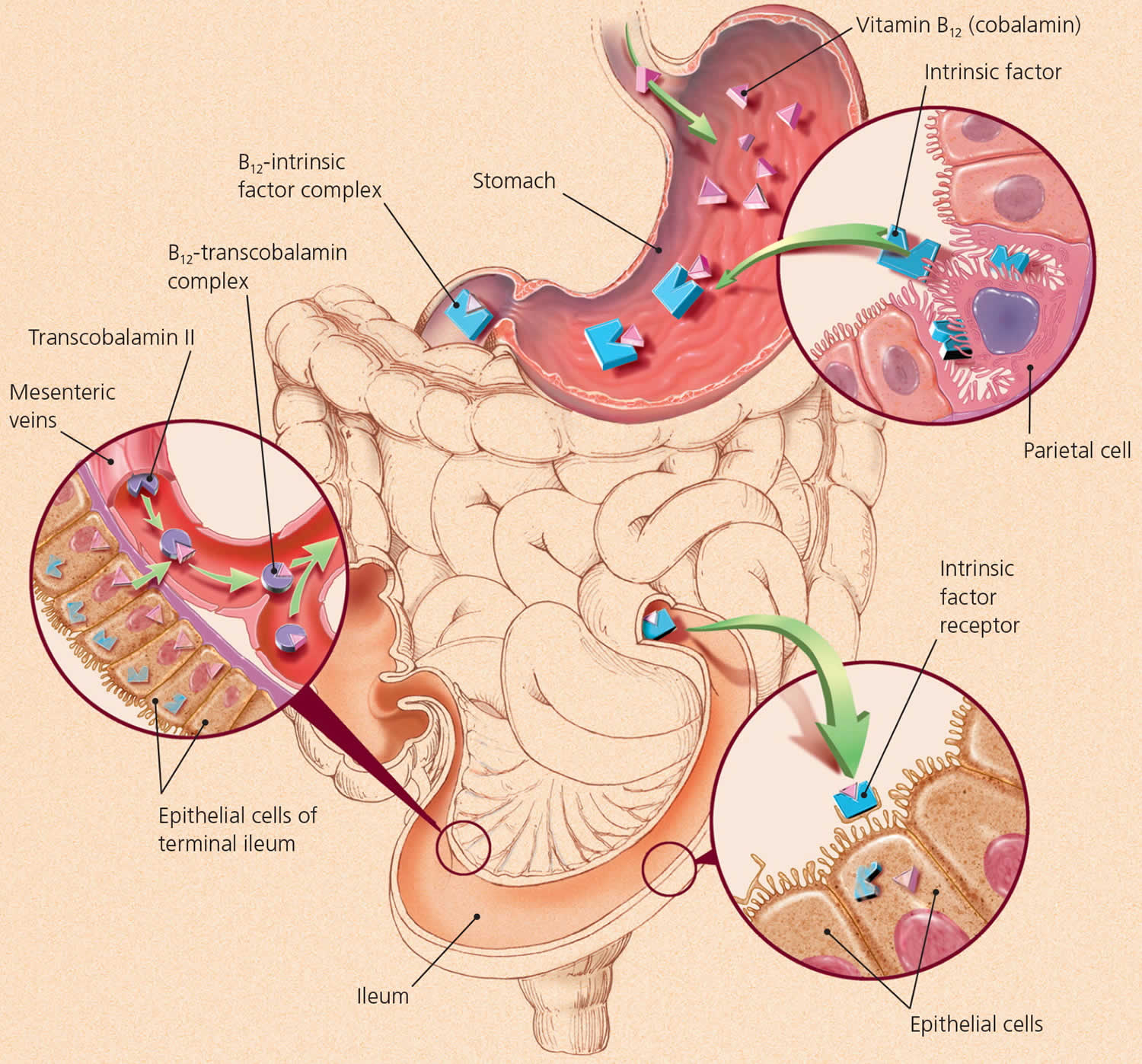
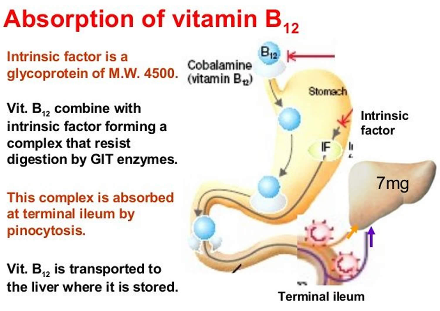
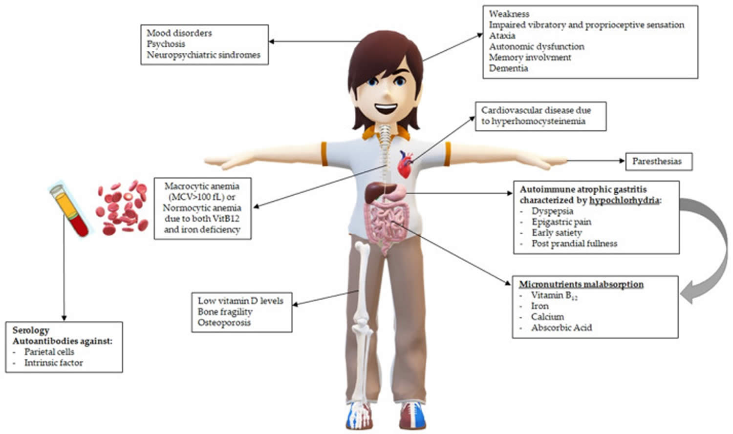
Figure 2. Vitamin B12 deficiency pathophysiology

What does vitamin B12 do?
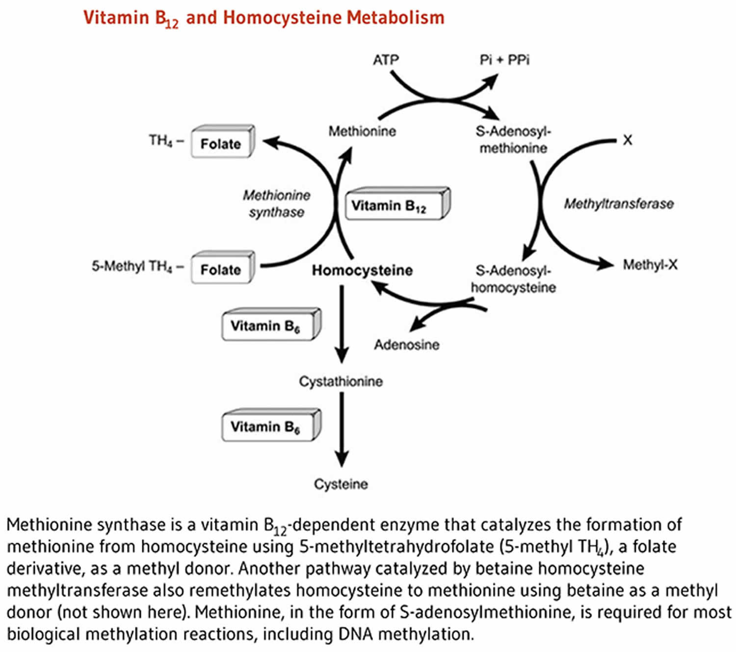
Vitamin B12 is required for the development, myelination, and function of the central nervous system; healthy red blood cell formation; and helps make DNA, the genetic material in all cells 53, 54. Vitamin B12 functions as a cofactor for two enzymes, methionine synthase and L-methylmalonyl-CoA mutase (see more below) 53, 46, 55. Methionine synthase catalyzes the conversion of homocysteine to the essential amino acid methionine 56, 46. Methionine is required for the formation of S-adenosylmethionine, a universal methyl donor for almost 100 different substrates, including DNA, RNA, proteins, and lipids 53, 55. L-methylmalonyl-CoA mutase converts L-methylmalonyl-CoA to succinyl-CoA in the metabolism of propionate, a short-chain fatty acid 46.
Vitamin B12 functions as a cofactor for methionine synthase
Methylcobalamin is required for the function of the folate-dependent enzyme, methionine synthase. The methionine synthase enzyme is required for the synthesis of the amino acid, methionine, from homocysteine. Methionine in turn is required for the synthesis of S-adenosylmethionine (SAMe), a methyl group donor used in many biological methylation reactions, including the methylation of a number of sites within DNA, RNA, and proteins 57. Aberrant methylation of DNA and proteins, which causes alterations in chromatin structure and gene expression, are a common feature of cancer cells. Inadequate function of methionine synthase can lead to an accumulation of homocysteine, which has been associated with increased risk of cardiovascular disease (Figure 3).
Figure 3. Vitamin B12 functions as a cofactor for methionine synthase
Vitamin B12 functions as a cofactor for L-methylmalonyl-coenzyme A mutase
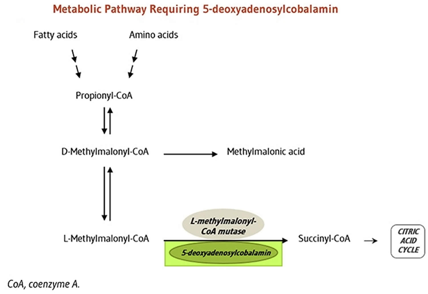
5-Deoxyadenosylcobalamin is required by the enzyme that catalyzes the conversion of L-methylmalonyl-coenzyme A to succinyl-coenzyme A (succinyl-CoA), which then enters the citric acid cycle (Figure 5). Succinyl-CoA plays an important role in the production of energy from lipids and proteins and is also required for the synthesis of hemoglobin, the oxygen-carrying pigment in red blood cells 57.
Figure 4. Vitamin B12 functions as a cofactor for L-methylmalonyl-coenzyme A mutase
How much Vitamin B12 do you need?
The amount of Vitamin B12 you need each day depends on your age. Average daily recommended amounts for different ages are listed below in micrograms (mcg). Table 1 lists the current Recommended Dietary Allowance (RDA) for Vitamin B-12 in micrograms (mcg). For infants aged 0 to 12 months, the Food and Nutrition Board established an adequate intake (AI) for vitamin B-12 that is equivalent to the mean intake of Vitamin B-12 in healthy, breastfed infants.
- Recommended Dietary Allowance (RDA): average daily level of intake sufficient to meet the nutrient requirements of nearly all (97%–98%) healthy individuals.
- Adequate Intake (AI): established when evidence is insufficient to develop an RDA and is set at a level assumed to ensure nutritional adequacy.
Table 1. Vitamin B-12 Recommended Intake
| Life Stage | Recommended Amount |
|---|---|
| Birth to 6 months | 0.4 mcg |
| Infants 7–12 months | 0.5 mcg |
| Children 1–3 years | 0.9 mcg |
| Children 4–8 years | 1.2 mcg |
| Children 9–13 years | 1.8 mcg |
| Teens 14–18 years | 2.4 mcg |
| Adults | 2.4 mcg |
| Pregnant teens and women | 2.6 mcg |
| Breastfeeding teens and women | 2.8 mcg |
What are food sources of vitamin B12?
Vitamin B12 is found naturally in a wide variety of foods of animal origin (such as fish, meat, poultry, eggs, and dairy products) and manufacturers add it to some fortified foods (e.g., fortified breakfast cereals and fortified nutritional yeasts) 53. Plant foods have no vitamin B12 unless they are fortified 59. You can get recommended amounts of vitamin B12 by eating a variety of foods including the following:
- Fish, meat, poultry, eggs, milk, and other dairy products contain vitamin B12.
- Clams and beef liver are some of the best source of vitamin B12.
- Some breakfast cereals, nutritional yeasts, and other food products are fortified with vitamin B12.
The U.S. Department of Agriculture’s FoodData Central (https://fdc.nal.usda.gov) lists the nutrient content of many foods and provides a comprehensive list of foods containing vitamin B12 arranged by nutrient content (https://ods.od.nih.gov/pubs/usdandb/VitaminB12-Content.pdf) and by food name (https://ods.od.nih.gov/pubs/usdandb/VitaminB12-Food.pdf).
The average vitamin B12 level in the breast milk of women with vitamin B12 intakes above the RDA is 0.44 mcg/L 60. The U.S. Food and Drug Administration (FDA) specifies that infant formulas sold in the United States must provide at least 0.15 mcg vitamin B12 per 100 kcal 61.
The estimated bioavailability of vitamin B12 from food varies by vitamin B12 dose because absorption decreases drastically when the capacity of intrinsic factor is exceeded (at 1–2 mcg of vitamin B12) 62. Bioavailability also varies by type of food source. For example, the bioavailability of vitamin B12 appears to be about three times higher in dairy products than in meat, fish, and poultry, and the bioavailability of vitamin B12 from dietary supplements is about 50% higher than that from food sources 63.
A variety of foods and their vitamin B12 levels per serving are listed in Table 4.
Table 2. Vitamin B12 Food Sources
| Food | Micrograms per serving | Percent DV* |
| Beef liver, cooked, pan-fried, 3 ounces | 70.7 | 2944 |
| Clams (without shells), cooked, 3 ounces | 17 | 708 |
| Tuna, bluefin, cooked, dry heat, 3 ounces | 9.3 | 385 |
| Nutritional yeast, fortified, from several brands (check label), about ¼ cup | 8.3 to 24 | 346 to 1,000 |
| Salmon, Atlantic, cooked, 3 ounces | 2.6 | 108 |
| Beef, ground, 85% lean meat/15% fat, pan-browned, 3 ounces | 2.4 | 100 |
| Milk, 2% milkfat, 1 cup | 1.3 | 54 |
| Yogurt, plain, fat free, 6-ounce container | 1 | 43 |
| Breakfast cereals, fortified with 25% of the DV for vitamin B12, 1 serving | 0.6 | 25 |
| Cheese, cheddar, 1½ ounces | 0.5 | 19 |
| Egg, whole, cooked, 1 large | 0.5 | 19 |
| Turkey, breast meat, roasted, 3 ounces | 0.3 | 14 |
| Tempeh, 1/2 cup | 0.1 | 3 |
| Banana, 1 medium | 0 | 0 |
| Bread, whole-wheat, 1 slice | 0 | 0 |
| Strawberries, raw, halved, 1/2 cup | 0 | 0 |
| Beans, kidney, boiled, 1/2 cup | 0 | 0 |
| Spinach, boiled, drained, 1/2 cup | 0 | 0 |
Footnote: *DV = Daily Value. DVs were developed by the U.S. Food and Drug Administration (FDA) to help consumers determine the level of various nutrients in a standard serving of food in relation to their approximate requirement for it. The DV for Vitamin B-12 is 6.0 mcg. However, the FDA does not require food labels to list Vitamin B-12 content unless a food has been fortified with this nutrient. Foods providing 20% or more of the DV are considered to be high sources of a nutrient, but foods providing lower percentages of the DV also contribute to a healthful diet.
[Source 64 ]Pernicious anemia causes
Pernicious anemia is an autoimmune disorder. Typically, pernicious anemia is associated with the presence of autoantibodies against intrinsic factor (anti-intrinsic factor antibody or IFA) and anti-parietal cell antibodies (PCA), thus supporting the autoimmune origin of this condition 65. Anti-parietal cell antibodies (PCA) are seen in up to 85% of patients with pernicious anemia 10. However, the anti-parietal cell antibody (PCA) is not specific for pernicious anemia and can be seen in 3 to 10% of normal healthy populations without any evidence of megaloblastic anemia 10. Antibodies against parietal cells (PCA) are class M, G, and A immunoglobulins directed towards the alpha and beta subunits of the gastric proton pump (hydrogen-potassium ATP-ase) 66. Anti-intrinsic factor antibodies (IFA) are seen in 40 to 60% of the patients with pernicious anemia and are highly specific for the disease 10. Antibodies against intrinsic factor (IFA) are class G immunoglobulins that target the binding site for cobalamin (type I) or the binding site for ileal epithelial mucosa (type II) 67. These autoantibodies are released from plasma cells activated by autoreactive CD4+ T cell lymphocytes in perigastric lymph nodes 68. These triggered CD4+ T cells target the proton pump ATPases, which leads to their immune destruction 10. Atrophic gastritis with loss of parietal cells and subsequent intrinsic factor (IF) deficiency develops. This leads to vitamin B12 deficiency and the onset of symptoms associated with pernicious anemia. Gastric dendritic cells are responsible for the activation of lymphocytes that lead to the production of these antibodies. The cause and mechanism of dendritic cell activation are not yet clarified 10. Some research studies suggest Helicobacter pylori (H. pylori) infection is a trigger in genetically susceptible individuals 69. The studies propose molecular mimicry and immune cross-reactivity between the proton pump ATPase and the H. pylori antigens as a triggering event 69. A 2017 review evaluating H. pylori antigenic mimicry stated that these antigens play an important role in the induction of humoral and cellular immune responses, which may predispose patients to pathological inflammatory responses 70.
The pathogenesis of pernicious anemia has not been clarified, but it is likely linked to the autoimmune destruction of gastric glands due to autoreactive T lymphocytes in genetically predisposed individuals 9. The role of previous Helicobacter pylori (H. pylori) infection as a supposed but not yet definitely proven trigger of gastric autoimmunity cannot be excluded 9. Hershko et al. 7 have reported that H. pylori might serve as a trigger of autoimmune metaplastic atrophic gastritis and pernicious anemia, based on their observation that the prevalence of H. pylori infection was 87.5% in patients under 20 years of age. In addition, one theory regarding the initiating event of autoimmune metaplastic atrophic gastritis is molecular mimicry between H. pylori antigens and gastric H+/K+−ATPase 71.
In pernicious anemia, the underlying pathogenetic mechanism is autoimmune gastritis, an organ-specific immune-mediated disorder featuring the damage of the gastric parietal cells involved in the secretion of intrinsic factor (IF) and hydrochloric acid by the gastric proton pump 72. The presence of anti-parietal cell antibodies (PCAs) directed towards the gastric proton pump (gastric hydrogen potassium adenosine triphosphatase (H+/K+ ATPase)) as well as antibodies against intrinsic factor (IF) (although in a lower percentage) are commonly associated with gastric corpus atrophy and intrinsic factor deficiency 9, 73. Atrophic corpus gastritis is a chronic disease defined as a decrease in or loss of the original gastric glands, replaced by pseudo-pyloric or intestinal metaplasia or fibrosis 74. Gastric corpus atrophy is a necessary but insufficient condition for the onset of pernicious anemia, as gastric corpus atrophy may also take its course without pernicious anemia.
The destruction of parietal cells leads to decreased acid production and intrinsic factor secretion, and autoantibodies against intrinsic factor inhibit the absorption of vitamin B12. As a result, gastrin secretion from antral G cells increases, and hypergastrinemia induces proliferation of oxyntic mucosal cells including enterochromaffin‐like cells and parietal cells 26. The clinical manifestations are similar to other vitamin B12 deficiencies, but pernicious anemia is sometimes associated with other autoimmune diseases such as type 1 diabetes, autoimmune thyroiditis, and Addison’s disease. Sensitivity and specificity of the anti‐intrinsic factor antibody test were 50%‐70%, and greater than 95%, respectively 22. Sensitivity and specificity of the antigastric parietal cell antibody test were more than 90% and 50%, respectively 75. The treatment for pernicious anemia is lifelong administration of vitamin B12. Patients with pernicious anemia are at high risk of developing gastric adenocarcinoma and carcinoid tumors 25. Significant risk factors for the development of gastric carcinoma in autoimmune metaplastic atrophic gastritis include the presence of pernicious anemia, severity of mucosal atrophy, intestinal metaplasia, disease duration, and over 50 years of age 26. Periodic stomach examinations are recommended for patients with pernicious anemia.
Pernicious anemia can be associated with other autoimmune diseases and in patients with polyglandular autoimmune disorders. Autoimmune diseases associated with pernicious anemia include type 1 diabetes (3 to 4%), vitiligo (2 to 8%), and autoimmune thyroid disease (3 to 32%) 65. Type III polyglandular autoimmune syndrome is characterized by the presence of autoimmune thyroiditis, vitiligo, alopecia, type 1A diabetes mellitus, pernicious anemia, and chronic atrophic gastritis 76. HLA alleles are thought to play a role in the pathogenesis of these autoimmune disorders, but the mechanism is not entirely understood. HLA-DRB1/03 and HLA-DRB1/04 alleles may predispose to autoimmune gastritis and subsequent pernicious anemia 65.
There is an overlap in patients infected with Helicobacter pylori (H. pylori) and the development of chronic atrophic gastritis associated with pernicious anemia 77. Researchers propose H. pylori peptide-induced gastric T-cell proliferation as the cause of pernicious anemia in some patients 69. They were able to demonstrate the presence of activated T cells in the gastric mucosa of patients with autoimmune chronic atrophic gastritis and H. pylori infection. These T cells reacted to both hydrogen-potassium-ATPase and H. pylori 69. Recent experimental and clinical data suggest long-standing H. pylori infection plays a pivotal role in developing atrophic gastritis and subsequent pernicious anemia; however, convincing data to support H. pylori infection as a definite cause of pernicious anemia is still lacking 65. Molecular analyses have revealed hydrogen-potassium-ATPase epitopes that are similar to, or cross-reactive with, epitopes of H. pylori antigens. Thus suggesting that in genetically susceptible individuals, H. pylori infection can trigger gastric autoimmunity via molecular mimicry 69.
In rare cases, pernicious anemia is passed down through families (inherited disorder). This is called congenital pernicious anemia or “childhood pernicious anemia” 9. Babies with congenital pernicious anemia do not make enough intrinsic factor (IF) or abnormal intrinsic factor (IF) formation 9. Or they cannot properly absorb vitamin B12 in the small intestine. Congenital pernicious anemia is quite rare and distinguishable from the usual form of pernicious anemia due to the early age of onset and the absence of gastric corpus atrophy.
Lack of Intrinsic Factor
Pernicious anemia is an autoimmune disease that affects the gastric mucosa and results in gastric atrophy. This leads to the destruction of parietal cells, achlorhydria, and failure to produce intrinsic factor, resulting in vitamin B12 malabsorption 78, 79, 80, 81, 82. An autoimmune response occurs if the body’s immune system makes antibodies (proteins) that mistakenly attack and damage the body’s tissues or cells. In pernicious anemia, the body makes antibodies that attack and destroy the parietal cells. These cells line the stomach and make intrinsic factor. Why this autoimmune response occurs isn’t known.
As a result of this attack, the stomach stops making intrinsic factor. Without intrinsic factor, your body can’t move vitamin B12 through the small intestine, where it’s absorbed. This leads to vitamin B12 deficiency.
A lack of intrinsic factor also can occur if you’ve had part or all of your stomach surgically removed. This type of surgery reduces the number of parietal cells available to make intrinsic factor.
Rarely, children are born with an inherited disorder that prevents their bodies from making intrinsic factor. This disorder is called congenital pernicious anemia.
If pernicious anemia is left untreated, it causes vitamin B12 deficiency, leading to megaloblastic anemia and neurological disorders, even in the presence of adequate dietary intake of vitamin B12.
You are more likely to develop this disease if you:
- Are Scandinavian or Northern European
- Have a family history of the condition
Certain diseases can also raise your risk. They include:
- Addison disease
- Chronic thyroiditis
- Graves disease
- Hypoparathyroidism
- Hypopituitarism
- Myasthenia gravis
- Secondary amenorrhea
- Type 1 diabetes
- Testicular dysfunction
- Vitiligo
Other Causes
Pernicious anemia also has other causes, besides a lack of intrinsic factor. Malabsorption in the small intestine and a diet lacking vitamin B12 both can lead to pernicious anemia.
Malabsorption in the Small Intestine
Sometimes pernicious anemia occurs because the body’s small intestine can’t properly absorb vitamin B12. This may be the result of:
- Too many of the wrong kind of bacteria in the small intestine. This is a common cause of pernicious anemia in older adults. The bacteria use up the available vitamin B12 before the small intestine can absorb it.
- Diseases that interfere with vitamin B12 absorption. One example is celiac disease. This is a genetic disorder in which your body can’t tolerate a protein called gluten. Another example is Crohn’s disease, an inflammatory bowel disease. HIV also may interfere with vitamin B12 absorption.
- Certain medicines that alter bacterial growth or prevent the small intestine from properly absorbing vitamin B12. Examples include antibiotics and certain diabetes and seizure medicines.
- Surgical removal of part or all of the small intestine.
- A tapeworm infection. The tapeworm feeds off of the vitamin B12. Eating undercooked, infected fish may cause this type of infection.
Diet Lacking Vitamin B12
Some people get pernicious anemia because they don’t have enough vitamin B12 in their diets. This cause of pernicious anemia is less common than other causes.
Good food sources of vitamin B12 include:
- Breakfast cereals with added vitamin B12
- Meats such as beef, liver, poultry, and fish
- Eggs and dairy products (such as milk, yogurt, and cheese)
- Foods fortified with vitamin B12, such as soy-based beverages and vegetarian burgers
Strict vegetarians who don’t eat any animal or dairy products and don’t take a vitamin B12 supplement are at risk for pernicious anemia.
Breastfed infants of strict vegetarian mothers also are at risk for pernicious anemia. These infants can develop anemia within months of being born. This is because they haven’t had enough time to store vitamin B12 in their bodies. Doctors treat these infants with vitamin B12 supplements.
Other groups, such as the elderly and people who suffer from alcoholism, also may be at risk for pernicious anemia. These people may not get the proper nutrients in their diets.
Table 2. Vitamin B12 Food Sources
| Food | Micrograms per serving | Percent DV* |
| Beef liver, cooked, pan-fried, 3 ounces | 70.7 | 2944 |
| Clams (without shells), cooked, 3 ounces | 17 | 708 |
| Tuna, bluefin, cooked, dry heat, 3 ounces | 9.3 | 385 |
| Nutritional yeast, fortified, from several brands (check label), about ¼ cup | 8.3 to 24 | 346 to 1,000 |
| Salmon, Atlantic, cooked, 3 ounces | 2.6 | 108 |
| Beef, ground, 85% lean meat/15% fat, pan-browned, 3 ounces | 2.4 | 100 |
| Milk, 2% milkfat, 1 cup | 1.3 | 54 |
| Yogurt, plain, fat free, 6-ounce container | 1 | 43 |
| Breakfast cereals, fortified with 25% of the DV for vitamin B12, 1 serving | 0.6 | 25 |
| Cheese, cheddar, 1½ ounces | 0.5 | 19 |
| Egg, whole, cooked, 1 large | 0.5 | 19 |
| Turkey, breast meat, roasted, 3 ounces | 0.3 | 14 |
| Tempeh, 1/2 cup | 0.1 | 3 |
| Banana, 1 medium | 0 | 0 |
| Bread, whole-wheat, 1 slice | 0 | 0 |
| Strawberries, raw, halved, 1/2 cup | 0 | 0 |
| Beans, kidney, boiled, 1/2 cup | 0 | 0 |
| Spinach, boiled, drained, 1/2 cup | 0 | 0 |
Footnote: *DV = Daily Value. DVs were developed by the U.S. Food and Drug Administration (FDA) to help consumers determine the level of various nutrients in a standard serving of food in relation to their approximate requirement for it. The DV for Vitamin B-12 is 6.0 mcg. However, the FDA does not require food labels to list Vitamin B-12 content unless a food has been fortified with this nutrient. Foods providing 20% or more of the DV are considered to be high sources of a nutrient, but foods providing lower percentages of the DV also contribute to a healthful diet.
[Source 64 ]Risk Factors for pernicious anemia
Pernicious anemia is more common in people of Northern European and African descent than in other ethnic groups.
Older people also are at higher risk for the condition. This is mainly due to a lack of stomach acid and intrinsic factor, which prevents the small intestine from absorbing vitamin B12. As people grow older, they tend to make less stomach acid.
Pernicious anemia also can occur in younger people and other populations. You’re at higher risk for pernicious anemia if you:
- Have a family history of the condition.
- Have had part or all of your stomach surgically removed. The stomach makes intrinsic factor. This protein helps your body absorb vitamin B12.
- Have an autoimmune disorder that involves the endocrine glands, such as Addison’s disease, type 1 diabetes, Graves’ disease, or vitiligo. Research suggests a link may exist between these autoimmune disorders and pernicious anemia that’s caused by an autoimmune response.
- Have had part or all of your small intestine surgically removed. The small intestine is where vitamin B12 is absorbed.
- Have certain intestinal diseases or other disorders that may prevent your body from properly absorbing vitamin B12. Examples include Crohn’s disease, intestinal infections, and HIV.
- Take medicines that prevent your body from properly absorbing vitamin B12. Examples of such medicines include antibiotics and certain seizure medicines.
- Are a strict vegetarian who doesn’t eat any animal or dairy products and doesn’t take a vitamin B12 supplement, or if you eat poorly overall.
Pernicious anemia prevention
You can’t prevent pernicious anemia caused by a lack of intrinsic factor. Without intrinsic factor, you won’t be able to absorb vitamin B12 and will develop pernicious anemia.
Because an increased familial incidence of pernicious anemia exists, family members should be aware that they are at greater risk of developing this disease and should seek medical attention promptly if they develop anemia or mental and neurologic symptoms 83. Monitor siblings and children of patients with a hereditary abnormality of vitamin B12 deficiency for evidence of the specific defect in cobalamin transport or metabolism.
Determine whether vitamin B12 deficiency is the cause in patients who recently developed evidence of mental deterioration.
Although uncommon, some people develop pernicious anemia because they don’t get enough vitamin B12 in their diets. You can take steps to prevent pernicious anemia caused by dietary factors.
Eating foods high in vitamin B12 can help prevent low vitamin B12 levels. Good food sources of vitamin B12 include:
- Breakfast cereals with added vitamin B12
- Meats such as beef, liver, poultry, and fish
- Eggs and dairy products (such as milk, yogurt, and cheese)
- Foods fortified with vitamin B12, such as soy-based beverages and vegetarian burgers
If you’re a strict vegetarian, talk with your doctor about having your vitamin B12 level checked regularly, particularly during pregnancy and while nursing a newborn infant 83.
Vitamin B12 also is found in multivitamins and B-complex vitamin supplements. Doctors may recommend supplements for people at risk for vitamin B12 deficiency, such as strict vegetarians or people who have had stomach surgery.
Elderly people are at risk for developing pernicious anemia due to achlorhydria. Therefore, serum vitamin B-12 levels should be checked. If low or if cobalamin deficiency is suspected, they should be treated with vitamin B-12 supplementation.
Prophylactically treat patients with vitamin B12 when they have undergone total gastrectomy, bypass procedures for weight reduction, ileectomy, pancreatectomy, or when they have atrophic gastritis or chronic inflammatory disease of the ileum 83.
Pernicious anemia signs and symptoms
The onset of pernicious anemia usually is insidious and vague. Some people do not have symptoms. Symptoms may be mild. The main signs and symptoms of pernicious anemia are hematological and neurological consequences of vitamin B12 deficiency, and both require several years for their development. The classic presentation consists of a triad of jaundice, glossitis, and myeloneuropathy 84. However, with advances in clinical detection and often routine laboratory testing, this classic triad of jaundice, glossitis, and myeloneuropathy is now a rarity.
Without enough vitamin B12, your body can’t make enough healthy red blood cells, which causes anemia. Many pernicious anemia patients are incidentally noted to have macrocytic anemia and are ultimately diagnosed with pernicious anemia. Others may either present with symptoms attributable to anemia, such as lethargy and inability to concentrate, or with symptoms attributable to neuronal damage, such as paresthesias, imbalance, and spasticity 10.
Figure 5. Pernicious anemia signs and symptoms (related to hypochlorhydria and vitamin B12 deficiency)
General symptoms
Weight loss of 10-15 lb occurs in about 50% of patients and probably is due to anorexia, which is observed in most patients. Low-grade fever occurs in one third of newly diagnosed patients and promptly disappears with treatment.
Signs and symptoms of Anemia
The most common symptom of all types of anemia is fatigue (tiredness). Fatigue occurs because your body doesn’t have enough red blood cells to carry oxygen to its various parts.
A low red blood cell count also can cause shortness of breath, dizziness, headache, coldness in your hands and feet, pale or yellowish skin, and chest pain.
A lack of red blood cells also means that your heart has to work harder to move oxygen-rich blood through your body. This can lead to irregular heartbeats called arrhythmias, heart murmur, an enlarged heart, or even heart failure.
Signs and symptoms of anemia
- Fatigue
- Weakness
- Pale or yellowish skin
- Irregular heartbeats
- Shortness of breath
- Dizziness or lightheadedness
- Chest pain
- Cold hands and feet
- Headache
At first anemia can be so mild that it goes unnoticed. But symptoms worsen as anemia worsens.
Other signs and symptoms of pernicious anemia include:
- Desire to eat ice or other non-food things (pica)
- Diarrhea or constipation
- Fatigue, lack of energy, or lightheadedness when standing up or with exertion
- Loss of appetite
- Pale skin
- Problems concentrating
- Shortness of breath, mostly during exercise
- Swollen, red tongue or bleeding gums
Figure 6. Pernicious anemia tongue

Figure 7. Glossitis secondary to vitamin B12 deficiency anemia
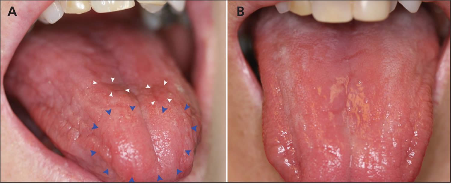
Footnotes: (A) Generalized dryness of the tongue of a 61-year-old woman with vitamin B12 deficiency, with atrophy (blue arrowheads) and erythematous plaques (white arrowheads). (B) Normal appearance of the tongue 3 days after the patient received a single injection of vitamin B12.
[Source 85 ]Signs and Symptoms of Vitamin B12 Deficiency
Vitamin B12 deficiency may lead to nerve damage. This can cause tingling and numbness in your hands and feet, muscle weakness, and loss of reflexes. You also may feel unsteady, lose your balance, and have trouble walking. Vitamin B12 deficiency can cause weakened bones and may lead to hip fractures.
Severe vitamin B12 deficiency can cause neurological problems, such as confusion, dementia, depression, and memory loss.
Other symptoms of vitamin B12 deficiency involve the digestive tract. These symptoms include nausea (feeling sick to your stomach) and vomiting, heartburn, abdominal bloating and gas, constipation or diarrhea, loss of appetite, and weight loss. An enlarged liver is another symptom.
A smooth, thick, red tongue also is a sign of vitamin B12 deficiency and pernicious anemia.
Infants who have vitamin B12 deficiency may have poor reflexes or unusual movements, such as face tremors. They may have trouble feeding due to tongue and throat problems. They also may be irritable. If vitamin B12 deficiency isn’t treated, these infants may have permanent growth problems.
If you have a low vitamin B12 level for a long time, you can have nervous system damage. Symptoms can include:
- Confusion
- Depression
- Loss of balance
- Numbness and tingling that start first in the hands and feet (from nerve damage)
- Muscle weakness
- Slow reflexes
- Loss of balance
- Unsteady walking
- Confusion, memory loss, depression, and/or dementia in severe cases.
Heart symptoms
Individuals with pernicious anemia often tolerate the anemia well, and many are ambulatory with hematocrit levels in the mid-teens. However, the cardiac output is usually increased when hematocrit levels fall below 20%, with associated accerations in heart rate. Congestive heart failure and coronary insufficiency can occur, most particularly in patients with preexisting heart disease.
Gastrointestinal symptoms
Approximately 50% of patients with pernicious anemia develop atrophic glossitis, presenting with a smooth tongue that may be painful and beefy red, with loss of papillae that is usually most marked along the edges of the tongue 86. These patients report burning or soreness, most particularly on the anterior third of the tongue, associated with changes in taste and loss of appetite 85.
Patients may report either constipation or having several semisolid bowel movements daily. These symptoms have been attributed to megaloblastic changes of the cells of the intestinal mucosa.
Nonspecific gastrointestinal symptoms are not unusual and include anorexia, nausea, vomiting, heartburn, pyrosis, flatulence, and a sense of fullness 87. Rarely, patients present with severe abdominal pain associated with abdominal rigidity; this has been attributed to spinal cord pathology. Venkatesh and colleagues 88 report the case of a patient who presented with epigastric pain, diarrhea, and vomiting and was found to have thrombosis of the portal, superior mesenteric, and splenic veins due to hyperhomocysteinemia secondary to pernicious anemia.
Neurologic symptoms
The most common neurologic symptoms in vitamin B12 deficiency include paresthesias, weakness, clumsiness, and an unsteady gait. The last two symptoms are exacerbated in dark environments due to the loss of visual cues that patients often rely on, in concert with the loss of proprioception. These neurologic symptoms are due to myelin degeneration and loss of nerve fibers in the dorsal and lateral columns of the spinal cord and cerebral cortex (subacute combined degeneration).
Neurologic symptoms and findings may be present in the absence of anemia. This is more common in patients taking folic acid or on a high-folate diet.
Older patients may present with symptoms suggesting senile dementia or Alzheimer disease; memory loss, irritability, and personality changes are commonplace 10. Common psychiatric manifestations include depression, mania, chronic fatigue syndrome, and psychosis 9. Cognitive symptoms include memory impairment, attention deficit, and dementia 9. So-called megaloblastic madness—delusions, hallucinations, outbursts, and paranoid schizophrenic ideation—is less common. Identifying the cause is important because significant reversal of these symptoms and findings can occur with vitamin B12 administration.
While neurologic symptoms usually occur in the elderly, they can rarely occur in the young 89. Kocaoglu et al. 90 reported a case of vitamin B12 deficiency and cerebral atrophy in a 12-month-old infant whose development had slowed since 6 months of age; the infant was exclusively breastfed and his mother was a long-time vegetarian. Neurologic recovery began within days after the infant received an intramuscular cobalamin injection.
Genitourinary symptoms
Urinary retention and impaired micturition may occur because of spinal cord damage. This can predispose patients to urinary tract infections.
Symptoms of thrombotic complications
A study of four patients revealed that pernicious anemia can lead to hyperhomocysteinemia that is significant enough to lead to venous thrombosis, even in the absence of any other risk factors for thromboembolism 91.
Pernicious anemia complications
One of the most dreaded complications of pernicious anemia is the development of stomach cancer 16. A 2013 systematic review of over 22,000 patients with pernicious anemia found a pooled gastric cancer incidence of 0.27% per person-year, with a nearly sevenfold increased risk of gastric cancer in these patients 92. The same study showed a pooled gastric cancer recurrence rate of 6.8 92. For this reason, an upper gastrointestinal endoscopy is recommended when pernicious anemia is diagnosed 65. Repeat endoscopies without any evidence of gastrointestinal symptoms are generally not recommended. Surveillance endoscopy every three years is recommended by some experts in patients with documented evidence of advanced chronic autoimmune atrophic gastritis 93.
According to a large United States population study based on a Surveillance, Epidemiology, and End Results (SEER) Medicare database, patients with pernicious anemia are at higher risk of 10 cancer types 16:
- Gastric adenocarcinoma
- Gastric carcinoid tumors
- Tonsillar cancer
- Hypopharyngeal cancer
- Esophageal squamous cell carcinoma
- Myeloma
- Acute myeloid leukemia
- Myelodysplastic syndrome
The authors state, “The most striking of our findings was an 11-fold increase in the risk of gastric carcinoid tumors for both men and women with pernicious anemia, a risk that was higher among cases occurring 6 or more years after the pernicious anemia report/diagnosis” 16. They also point out these are uncommon cancers with low absolute risk: “Of the 17,076 cancers in our study among people with pernicious anemia, just 83 (0.5%) were gastric carcinoid tumors” 16.
Brain and nervous system problems may continue or be permanent if treatment is delayed.
A woman with a low B12 level may have a false positive Pap smear. This is because vitamin B12 deficiency affects the way certain cells (epithelial cells) in the cervix look.
Pernicious anemia diagnosis
Your doctor will diagnose pernicious anemia based on your medical and family histories, a physical exam, and test results.
Your doctor will want to find out whether the condition is due to a lack of intrinsic factor or another cause. He or she also will want to find out the severity of the condition, so it can be properly treated.
When pernicious anemia is suspected, the first step is usually a full blood panel to test for anemia and/or macrocytosis, together with testing for vitamin B12 deficiency and increased levels of homocysteine and/or methylmalonic acid (MMA). Next, the positivity of gastric autoantibodies towards parietal cells and/or intrinsic factor is commonly assessed. In any case, the hematological and/or serological suspicion of pernicious anemia always needs to be confirmed by histological assessment of gastric antral and corpus biopsies obtained during gastroscopy 9.
Specialists Involved
Primary care doctors—such as family doctors, internists, and pediatricians (doctors who treat children)—often diagnose and treat pernicious anemia. Other kinds of doctors also may be involved, including:
- A neurologist (nervous system specialist)
- A cardiologist (heart specialist)
- A hematologist (blood disease specialist)
- A gastroenterologist (digestive tract specialist)
Medical and Family Histories
Your doctor may ask about your signs and symptoms. He or she also may ask:
- Whether you’ve had any stomach or intestinal surgeries
- Whether you have any digestive disorders, such as Celiac disease or Crohn’s disease
- About your diet and any medicines you take
- Whether you have a family history of anemia or pernicious anemia
- Whether you have a family history of autoimmune disorders (such as Addison’s disease, type 1 diabetes, Graves’ disease, or vitiligo). Research suggests a link may exist between these autoimmune disorders and pernicious anemia that’s caused by an autoimmune response.
Physical Exam
During the physical exam, your doctor may check for pale or yellowish skin and an enlarged liver. He or she may listen to your heart for rapid or irregular heartbeats or a heart murmur.
Your doctor also may check for signs of nerve damage. He or she may want to see how well your muscles, eyes, senses, and reflexes work. Your doctor may ask questions or do tests to check your mental status, coordination, and ability to walk.
The health care provider will perform a physical exam.
Pernicious anemia test
The workup for pernicious anemia may include the following 94:
- Complete blood cell count (CBC)
- Peripheral blood smear
- Indirect bilirubin and lactate dehydrogenase assays
- Evaluation of gastric secretions. Total gastric secretions are decreased to about 10% of the reference range. Most patients with pernicious anemia are achlorhydric, even with histamine stimulation. Intrinsic factor (IF) is either absent or markedly decreased.
- Serum vitamin B12 (cobalamin), folic acid, methylmalonic acid (MMA) and homocysteine assays.
- Serum cobalamin reference ranges may vary slightly among different laboratories, but are generally from 200–900 pg/mL. Values of 180-250 pg/mL are considered bordeline, while less than 150 pg/mL is considered diagnostic of vitamin B12 deficiency. In these cases, elevated levels of methylmalonic acid (MMA) and total homocysteine can confirm the diagnosis 95.
- The serum cobalamin level is usually low in patients with pernicious anemia. However, up to a third of patients can present with normal vitamin B12 levels and normocytic anemia, which often delays diagnosis 96. Certain patients with other forms of cobalamin deficiency, such as some inborn forms of cobalamin deficiency, transcobalamin 2 deficiency, and cobalamin deficiency due to nitrous oxide, can also present with normal serum cobalamin levels.
- Serum cobalamin levels may also be low in patients with no clinical or identifiable metabolic abnormality 97. Causes of falsely low serum cobalamin levels inclue the following:
- Pregnancy
- Oral contraceptives and hormone replacement therapy
- Multiple myeloma
- Transcobalamin 1 (TC1) deficiency
- Severe folic acid deficiency
- Ascorbic acid in high doses
- A serum folic acid assay is useful for ruling out folic acid deficiency. The reference range is 2.5-20 ng/mL. Blood should be drawn before patients have a single hospital meal since food can restore serum folic acid levels to normal. Red blood cell folic acid level is not influenced by food.
- Levels of antibodies against intrinsic factor (IF) or the cells which make intrinsic factor (anti-parietal cell antibodies [PCAs]).
- Schilling test (no longer available in most medical centers)
- A clinical trial of vitamin B12
- Patients with diagnosis of pernicious anemia should undergo gastroscopy with biopsies to ascertain the presence of autoimmune gastritis 9. Biopsies should be collected following the updated Sydney system 98: two biopsies of the antrum and two biopsies from the corpus should be obtained and sent in separate vials. Another biopsy should be performed from incisura angularis and sent in the same vial of antrum biopsies.
- Bone marrow aspiration and biopsy (only needed if diagnosis is unclear)
Complete blood cell count (CBC) and peripheral blood smear may show the mean corpuscular volume (MCV) and mean cell hemoglobin (MCH) are increased, with a mean corpuscular hemoglobin concentration (MCHC) within the reference range 94. However, up to 30% of patients with pernicious anemia may lack macrocytosis 9. A normal MCV (mean corpuscular volume) does not rule out megaloblastic anemia, and pathognomonic megaloblasts are rarely seen. The hematocrit must fall by 20% before megaloblasts appear in the blood 84. Anisocytosis and an increase in the red cell distribution width is the earliest measurable change in red cell indices to hint toward the diagnosis 84.
The peripheral blood usually shows a macrocytic anemia with a mild leukopenia and thrombocytopenia. The leukopenia and thrombocytopenia usually parallel the severity of the anemia. The peripheral smear shows oval macrocytes, hypersegmented granulocytes, and anisopoikilocytosis. In severe anemia, red blood cell inclusions may include Howell-Jolly bodies, Cabot rings, and punctate basophilia. The macrocytosis can be obscured by the coexistence of iron deficiency, thalassemia minor, or inflammatory
The indirect bilirubin level may be elevated because pernicious anemia is a hemolytic disorder associated with increased turnover of bilirubin 94. The serum lactate dehydrogenase (LDH) concentration usually is markedly increased 94. Increased values for other red blood cells, enzymes, and serum iron saturation also are observed. The serum potassium, cholesterol, and skeletal alkaline phosphatase often are decreased.
A significantly decreased serum cobalamin level along with a typical clinical presentation, a characteristic peripheral smear, and an increased indirect bilirubin and lactate dehydrogenase (LDH) level is sufficient evidence for the diagnosis of a megaloblastic anemia.
Serum methylmalonic acid (MMA) and homocysteine tests are important confirmatory tests but are not first-line tests. Elevated serum methylmalonic acid (MMA) and homocysteine levels are found in patients with pernicious anemia. They probably are the most reliable test for cobalamin deficiency in patients who do not have a congenital metabolism disorder. In the absence of an inborn error of methylmalonic acid metabolism, methylmalonic aciduria is a sign of cobalamin deficiency.
Table 3. Serum methylmalonic acid (MMA) and homocysteine values used in differentiating between vitamin B12 deficiency and folic acid deficiency
| Patient Condition | Methylmalonic Acid | Homocysteine |
|---|---|---|
| Healthy | Normal | Normal |
| Vitamin B12 deficiency | Elevated | Elevated |
| Folate deficiency | Normal | Elevated |
Testing for B12 Deficiency
A B12 level below 200 pg/mL (ng/L) is consistent with vitamin B12 deficiency 77. Levels between 200 to 400 pg/mL are considered borderline 10. Serum B12 measurement alone has poor sensitivity and specificity for detecting B12 deficiency 84. In patients with pernicious anemia, this level will be falsely elevated in 22 to 35% of the patients due to the interaction of IF antibody (IFA) with the “IF reagent” used to detect B12 levels in current assays 10. Falsely low serum vitamin B12 levels can occur in patients with underlying multiple myeloma and pregnancy 97, 10.
Methylmalonic acid (MMA) and homocysteine levels can be obtained in patients when vitamin B12 levels are borderline or nondiagnostic to confirm the diagnosis of B12 deficiency 100. These assays are considered more sensitive and specific for detecting B12 deficiency when compared to serum B12 levels 84. Methylmalonic acid (MMA) can also help differentiate between vitamin B12 and folate deficiency, as it is elevated in vitamin B12 deficiency but not in folate deficiency 84. Homocysteine levels are elevated in folate deficiency, vitamin B6 deficiency, and patients with hypothyroidism 97. MMA level can be falsely elevated in patients with bacterial overgrowth, especially when there are blind loops of the bowel (following gastric surgery) 84. Both levels can be falsely elevated in patients with renal failure 84, 97.
Serum holotranscobalamin (holoTC) level measures the metabolically active fraction of serum vitamin B12 and is considered a more accurate test for detecting B12 deficiency 97. Transcobalamin is a transport protein that binds only 10 to 30% of the total plasma B12; however, this constitutes all of the “active fraction” used for metabolic activity 84. Limitations of this test include a large window with indeterminate values 100. In addition, according to one study, approximately 63% of patients with low holoTC levels had normal methylmalonic acid levels, raising concerns regarding the utility of this test as a true measure of B12 deficiency 100. A 2013 study measuring the utility of biomarkers for B12 deficiency compared serum B12 levels to holotranscobalamin and recommended holotranscobalamin as the initial screening test for the detection of B12 deficiency, followed by MMA levels 101. They also suggested that an indeterminate holotranscobalamin level between 23 and 75 pmol should be followed by methylmalonic acid testing. Of note, this study was conducted in patients with normal renal function.
Definitive Testing for Pernicious Anemia
Traditionally, vitamin B12 absorption was measured using the Schilling test. This test is now considered obsolete, and there is no available assay for detecting B12 absorption at this time 84. In the absence of reliable B12 absorption assays, definitive testing for pernicious anemia relies on the detection of circulating antibodies to intrinsic factors (IFA) and gastric parietal cells (PCA).
Demonstration of circulating intrinsic factor autoantibodies is almost diagnostic of type A (autoimmune) gastritis and pernicious anemia. Intrinsic factor (IF) antibodies are specific for this disorder and can be used to confirm the diagnosis 84. There are two types of IF antibodies (IFA). Type 1 IF antibodies block binding of vitamin B12 to intrinsic factor and are found in 70% to 90% of patients with pernicious anemia. Type 2 IF antibodies prevent attachment of the vitamin B12–IF complex to ileal receptors and are present in approximately 35% to 50% of patients with pernicious anemia; they rarely occur in the absence of type 1 IF antibodies. Both type 1 and type 2 antibodies are detected more often in gastric juice than in the serum 102.
In one case report, the presence of antibodies to intrinsic factors (IFA) was used to diagnose vitamin B12 deficiency in a patient with severe leukoencephalopathy 103. Interestingly, serum vitamin B12, homocysteine, and methylmalonic acid levels were normal. The patient responded to intensive cobalamin therapy 103.
Parietal cell antibodies occurs in 90% of patients with pernicious anemia. However, antibodies to parietal cells (PCA) are not specific for pernicious anemia 10. Some experts advise against routine testing for antibodies to parietal cells (PCA); others recommend routine testing with anti-IF antibodies (IFA) because the combined sensitivity for pernicious anemia approaches 73% 11, 97. Dual testing for intrinsic factors antibodies (IFA) and parietal cell antibodies (PCA) with proof of atrophic gastritis is 100% specific for pernicious anemia 10.
In ambiguous cases, a bone marrow biopsy showing megaloblastic erythropoiesis and arrested maturation of myeloid precursor cells will establish the diagnosis 11. An alternative approach in difficult cases is to establish the presence of atrophic gastritis with endoscopic evaluation and biopsy and/or showing the presence of hypergastrinemia 11. In rare situations, an empiric trial of vitamin B12 replacement can be used to make the diagnosis 11. In this scenario, a rise in the reticulocyte count (which occurs within 5 to 14 days) confirms the diagnosis.
Alternative and new approaches to the diagnosis of pernicious anemia are under evaluation. One of these is a newer cobalamin absorption test, which has its basis in measuring the change in serum holotranscobalamin following oral ingestion of non-radiolabeled cobalamin. Another approach has been described using accelerator mass spectrometry to quantify 14C in the blood following an orally administered dose of [14C]-cyanocobalamin 84. Recently an ELISA test measuring serum concentration of human IF has been developed and may prove to be an alternate measure of impaired IF production/absorption 104.
Once the diagnosis of pernicious anemia is established, confirmatory testing with gastroscopy and histologic assessment of the gastric mucosa to assess for the presence of atrophic gastritis is indicated 9. Pernicious anemia is recognized as a late-stage complication of autoimmune gastritis, with an increased risk of gastric cancers in this population 9. Therefore, a new diagnosis of pernicious anemia warrants endoscopy with biopsies to detect the presence of atrophic gastritis and to rule out gastric cancers 93. The presence of intestinal metaplasia on gastric biopsy confers a diagnosis of atrophic gastritis 93. Pale gastric mucosa and increased visibility of vasculature are typical endoscopic features of atrophic gastritis. With metaplasia, light-blue crests and white opaque fields are present 93.
Recent advances in endoscopic techniques have led to the development of an endoscopic grading of gastric intestinal metaplasia (EGGIM) using noninvasive techniques to assess the presence of metaplasia during endoscopy without any need for biopsies. This system has shown acceptable sensitivity and specificity compared to biopsies and can be used when evaluating patients with pernicious anemia 9. In a cross-sectional study of 210 patients with atrophic gastritis, endoscopic grading of gastric intestinal metaplasia (EGGIM) was found to reliably identify more than 90% of patients with gastric corpus intestinal metaplasia. This method was shown to overestimate intestinal metaplasia when pseudopyloric metaplasia was present 9.
Bone Marrow Tests
Bone marrow tests can show whether your bone marrow is healthy and making enough red blood cells. The two bone marrow tests are aspiration and biopsy.
For aspiration, your doctor removes a small amount of fluid bone marrow through a needle. For a biopsy, your doctor removes a small amount of bone marrow tissue through a larger needle. The samples are then examined under a microscope.
In pernicious anemia, the bone marrow cells that turn into blood cells are larger than normal.
Additional testing
Testing for iron deficiency is indicated for all patients with pernicious anemia 9. Up to 20% of the patients with pernicious anemia have concomitant iron deficiency anemia and, in severe cases, may have microcytic red blood cells 11. Serum levels of iron, transferrin, and ferritin should be measured in patients with pernicious anemia, especially when macrocytosis is not present. It is important to remember that patients with pernicious anemia and associated megaloblastic anemia may also develop severe thrombocytopenia 84. Platelet production can be reduced by around 10%, and patients may also have abnormal platelet function 84. Leukopenia may be present but rarely causes any clinical issues 84. Indirect bilirubin and lactate dehydrogenase levels are usually elevated due to the rapid breakdown of red blood cells and intramedullary hemolysis. It is imperative to remember that vitamin B12 and folate levels should be tested simultaneously in patients with macrocytic anemia to ensure both deficiencies are diagnosed if present 97.
Pernicious anemia treatment
Doctors treat pernicious anemia by replacing the missing vitamin B12 in the body. People who have pernicious anemia may need lifelong treatment.
The goals of treating pernicious anemia are to increase your vitamin B12 level:
- Treatment involves a shot of vitamin B12 once a month. People with severely low levels of B12 may need more shots in the beginning.
- Some people may also need to take vitamin B12 supplements by mouth.
- A certain type of vitamin B12 may be given through the nose.
- Preventing or treating the anemia and its signs and symptoms
- Preventing or managing complications, such as heart and nerve damage
- Treating the cause of the pernicious anemia (if a cause can be found)
Pernicious anemia usually is easy to treat with vitamin B12 shots or pills. If you have severe pernicious anemia, your doctor may recommend shots first. Shots usually are given in a muscle every day or every week until the level of vitamin B12 in your blood increases. After your vitamin B12 blood level returns to normal, you may get a shot only once a month.
For less severe pernicious anemia, your doctor may recommend large doses of vitamin B12 pills. A vitamin B12 nose gel and spray also are available. These products may be useful for people who have trouble swallowing pills, such as older people who have had strokes.
Your signs and symptoms may begin to improve within a few days after you start treatment. Your doctor may advise you to limit your physical activity until your condition improves.
If your pernicious anemia is caused by something other than a lack of intrinsic factor, you may get treatment for the cause (if a cause can be found). For example, your doctor may prescribe medicines to treat a condition that prevents your body from absorbing vitamin B12.
If medicines are the cause of your pernicious anemia, your doctor may change the type or dose of medicine you take. Infants of strict vegetarian mothers may be given vitamin B12 supplements from birth.
Your provider will also recommend eating a variety of foods.
Specific pernicious anaemia treatment
Doctors treat pernicious anemia by replacing the missing vitamin B12 in the body. People who have pernicious anemia will need lifelong vitamin B12 treatment 97. Lifelong treatment for patients with confirmed pernicious anemia starts with an intramuscular (IM) injection of 1000 micrograms of B12 (hydroxocobalamin in Europe or cyanocobalamin in the United States) administered daily or every other day for 1 to 2 weeks, followed by weekly injections for 1 to 2 months, then a monthly injection (cyanocobalamin) or every 2 to 3 months (hydroxocobalamin) 95, 51, 24, 105, 1, 106.
An alternate approach is an initial dose of 1000 mcg IM injection every other day for 1 to 2 weeks, followed by weekly injections for one month and then monthly injections thereafter 84. Response should be monitored by reticulocyte counts, lactic dehydrogenase (LDH), and an appropriate rise in hemoglobin levels. LDH levels decrease and hemoglobin levels increase by about 1 g/dL/week. A rise in LDH might indicate a relapse.
Following the initial intensive treatment phase, patients can continue IM injections, or they can be offered high-dose oral vitamin B12 supplementation for the lifelong maintenance phase. In a 2016 review of oral vitamin B12 replacement for treating pernicious anemia, the authors concluded that “oral vitamin B12 is an effective alternative to vitamin B12 IM injections” 107. High-dose cyanocobalamin (1000–2000 micrograms) is most commonly taken daily as an oral tablet. Alternate formulations include sublingual and intranasal, but they are not routinely recommended 95, 51, 24, 105.
Recent data have suggested that high-dose oral replacement is as effective as IM replacement because passive absorption of vitamin B12 can occur in the absence of intrinsic factor (IF) 108. Even with a total absence of intrinsic factor (IF), about 1% of an oral dose is absorbed, and the daily requirement for vitamin B12 is 1 µg/day. A study by Zhang and colleagues 109 found evidence that using orally ingested soy protein isolate (SPI) nanoparticles as a carrier can improve the intestinal transport and absorption of vitamin B12. A 2018 Cochrane review reported oral route was as effective as IM vitamin B12 replacement; however, this was supported by very low-quality evidence 40. Therefore, current guidelines advise against the use of oral replacement in the initial phase of treatment 97. High-dose oral replacement (with 1000 to 2000 mcg of vitamin B12 daily) can be considered in patients with pernicious anemia for maintenance therapy 97.
The oral route may be necessary in the rare patients who have allergic reactions to parenteral administration, or in patients receiving anticoagulant or antiplatelet agent therapy, in whom intramuscular injections are contraindicated 110. If the oral route is used, obtain serum cobalamin measurements at periodic intervals to ensure that adequate quantities of cobalamin have been absorbed. Oral cobalamin therapy should not be used in patients with neurologic symptoms 97.
A randomized, placebo-controlled trial of oral cobalamin therapy in 50 patients with borderline serum vitamin B12 levels (125-200 pg/mL) and nonspecific symptoms compatible with subtle vitamin B12 deficiency found that after 1 month, serum methylmalonic acid (MMA) levels were corrected more often in patients receiving oral cobalamin than in those receiving placebo. However, the benefit to the MMA level disappeared after 3 additional months without cobalamin therapy 111.
A study found that oral cobalamine was more effective than parenteral therapy in some circumstances 111.
The earliest sign of treatment response is an increase in reticulocyte count that occurs within 5 to 14 days of treatment 11. A decrease in methylmalonic acid (MMA) and plasma homocysteine levels has also been observed in the first five days of treatment. Sustained normalization of serum cobalamin subsequently follows. Neuropsychiatric symptoms take a longer time to recover. As noted before, some neurologic symptoms may be irreversible.
Alternate formulations of vitamin B12 have been approved and include sublingual and intranasal formulations. However, data regarding their clinical efficacy is still limited, and they are not routinely recommended.
Patients whose vitamin B12 deficiency is due to underlying diseases involving the intestine or pancreas may require additional therapy 112. Examples of additional therapy are surgical correction of anatomic abnormalities of the gut that produce small bowel bacterial overgrowth, or the treatment of fish tapeworm anemia or pancreatitis. Elderly patients who also have hypokalemia should receive oral potassium supplements, to prevent severe hypokalemia and possible arrhythmias.
Blood transfusions are rarely required in patients with a megaloblastic anemia that is due to vitamin B12 deficiency 113. The likelihood of obtaining a dramatic response to cobalamin therapy within a few days of initiating treatment makes it unnecessary to subject the patient to the hazards of blood transfusion. Usually, mild-to-moderate congestive heart failure secondary to anemia abates with bed rest and low-dosage diuretic therapy. However, if the congestive heart failure is severe or the patient has coronary insufficiency, transfusion of packed red blood cells may be necessary 113. Transfuse the blood slowly because patients who are transfused for severe anemia often develop circulatory overload. For this reason, low-dose diuretic therapy is often employed with the blood transfusion 113.
Pernicious Anemia Management Guidelines
British Society of Gastroenterology (2019): “We suggest that a baseline endoscopy with biopsies should be considered in individuals aged ≥50 years, with laboratory evidence of pernicious anemia, defined by vitamin B12 deficiency and either positive gastric parietal cell or intrinsic factor antibodies. As gastric adenocarcinoma affects the corpus in pernicious anemia, biopsies should be taken from the greater and lesser curves (evidence level: low quality; grade of recommendation: weak; level of agreement: 93%)” 114.
European Society of Gastrointestinal Endoscopy (ESGE), European Helicobacter and Microbiota Study Group (EHMSG), European Society of Pathology (ESP), and Sociedade Portuguesa de Endoscopia Digestiva (SPED) (2019): “Patients with autoimmune gastritis (includes those with pernicious anemia) may benefit from endoscopic follow-up every 3 to 5 years. Low-quality evidence, weak recommendation (82% agree [76% strongly or moderately agree])” 115.
American Gastroenterological Association (2021): “Providers should recognize pernicious anemia as a late-stage manifestation of autoimmune gastritis that is characterized by vitamin B-12 deficiency and macrocytic anemia. Patients with a new diagnosis of pernicious anemia who have not had a recent endoscopy should undergo endoscopy with topographical biopsies to confirm corpus-predominant atrophic gastritis for risk stratification and to rule out prevalent gastric neoplasia, including neuroendocrine tumors” 93.
Endoscopic Follow-Up
Due to the risk of developing gastric cancer and type 1 gastric neuroendocrine tumors, endoscopic surveillance is the mainstay of the follow-up in pernicious anemia, as the early detection of neoplastic lesions at follow-up could permit a curative resection. European guidelines 116 suggest a 3-year follow-up in patients with metaplastic atrophic gastritis only in the antrum or only in the corpus, if risk factors, namely autoimmune gastritis, first-degree family history of gastric cancer, incomplete intestinal metaplasia, or persistent H. pylori infection, are present. In case of atrophic gastritis or intestinal metaplasia involving both antrum and corpus, a 3-year follow-up is recommended unless a first-degree family history is present. In this case, endoscopy should be performed every 1–2 years. The 3-year follow-up was shown to be cost-effective in intermediate-risk gastric cancer countries 117 and was confirmed in another Italian cost-effective study 118. However, only a few studies were performed to investigate the best interval to follow-up patients harboring this condition. In two studies conducted proposing a 2-year interval, the development of neoplastic lesions was present earlier than the 3-year interval 119, 120. However, other studies showed that even in patients with non-extensive atrophic gastritis, namely OLGA I-II/OLGIM I-II, neoplastic lesions could develop 121, 122, 123, 124. Furthermore, a recent longitudinal cohort study, conducted on 122 patients with AAG and 38 patients with multifocal atrophic gastritis in a follow-up program with a 3-year interval, showed the presence of 16 gastric neoplastic lesions at follow-up: 18.7% gastric cancers, 25.0% low-grade dysplasia, 12.5% low-grade dysplasia adenomas and 43.7% type-1 neuroendocrine tumors 125. All the diagnosed lesions were endoscopically (87.5%) or surgically (12.5%) treated with favorable outcomes 125.
The management of type 1 neuroendocrine tumors and resection strategy have still not been clarified 126. The European Neuroendocrine Tumor Society guidelines suggest removing tumors larger than 10 mm as they represent neuroendocrine tumors with potentially more aggressive behavior 127. In a recent prospective study conducted on 80 patients presenting 127 gastric type 1 neuroendocrine tumors, the authors concluded that resection strategy could be planned based on the tumor size. Neuroendocrine tumors smaller than 5 mm could be surveilled, and could be removed by cold snare polypectomy when the size was between 5 and 10 mm and by endoscopic mucosal resection (EMR) or endoscopic submucosal dissection (ESD) when bigger than 10 mm, even if with low evidence 128.
European guidelines suggest that surveillance in patients with gastric precancerous conditions should be performed with high-definition electronic chromoendoscopy to better recognize areas at risk for gastric cancer and to perform gastric biopsies. Three meta-analyses showed the high accuracy of conventional and virtual chromoendoscopy for the diagnosis of early gastric cancer and dysplasia in patients harboring precancerous conditions 129, 130, 131. Moreover, in pilot studies, virtual chromoendoscopy seemed to be useful for the characterization of type-1 neuroendocrine tumors 132.
Living with pernicious anemia
With proper treatment, people who have pernicious anemia can recover, feel well, and live normal lives. If you have complications of pernicious anemia, such as nerve damage, early treatment may help reverse the damage.
Ongoing Care
If you have pernicious anemia, you may need lifelong treatment. See your doctor regularly for checkups and ongoing care. Take vitamin B12 supplements as your doctor advises. This may help prevent symptoms and complications.
During your followup visits, your doctor may check for signs of vitamin B12 deficiency. He or she also may adjust your treatment as needed.
- If you have pernicious anemia, you’re at higher risk for stomach cancer. See your doctor regularly so he or she can check for this complication.
Also, tell your family members, especially your children and brothers and sisters, that you have pernicious anemia. Pernicious anemia can run in families, so they may have a higher risk for the condition.
Pernicious anemia prognosis
Most people with pernicious anemia do well with treatment, except for patients diagnosed with subacute combined degeneration (SCD) 133. It is important to start treatment early. Nerve damage can be permanent if treatment does not start within 6 months of symptoms. Although B12 supplementation stops progression and improves neurologic deficits in most patients with subacute combined degeneration, evidence shows complete resolution only occurs in a small percentage of them 134. A 2006 observational study evaluating 57 patients with subacute combined degeneration reported only 14% clinical resolution after B12 treatment 134. Still, the study reported that of these patients, 86% had at least some clinical improvement. Subgroup analysis revealed that the absence of sensory dermatomal deficit, negative Romberg and Babinski signs, age less than 50 years, and less than or equal to 7-segment involvement on magnetic resonance imaging correlated with complete resolution of neurologic symptoms 134. This study highlights the importance of early diagnosis and treatment of vitamin B12 deficiency, as patients with severe or prolonged neurological symptoms tend to have persistent symptoms despite treatment.
After treatment initiation for pernicious anemia, reticulocytosis begins approximately 5 days later, followed by red blood cell count normalization within 4 to 6 weeks 95. Typically, neurological symptom improvement is slower than hematological improvement, and the degree of neurological recovery is inversely proportional to the severity and duration of symptoms before treatment. Psychiatric symptoms such as emotional lability and psychosis may rapidly improve 135.
With ongoing care and proper treatment, most people who have pernicious anemia can recover, feel well, and live normal lives.
Without treatment, pernicious anemia can lead to serious problems with the heart, nerves, and other parts of the body. Some of these problems may be permanent.
Although vitamin B12 therapy resolves the anemia, it will not cure atrophic gastritis, and patients with pernicious anemia remain at higher risk of gastric cancers throughout their life 16. There is an increased risk of gastric adenocarcinoma in these patients 93. A longitudinal study from China reported that patients with pernicious anemia and anti-IF or anti-parietal cell antibodies had an unsatisfactory neurologic response to treatment and, over time, developed newly diagnosed hypothyroidism as well as alimentary canal malignancies 136. Twenty percent of the cancers noted in this study were gastric cancers. This study reported a mean survival of 64 months for patients who developed a malignancy compared to 129 months for those without malignancy. The mortality rate was reported as 31%, with cancer-related deaths representing 37%. Interestingly, the authors reported a higher risk of cancer in patients who were positive for anti-IF antibodies compared to those without these antibodies 136.
- Rojas Hernandez CM, Oo TH. Advances in mechanisms, diagnosis, and treatment of pernicious anemia. Discov Med. 2015 Mar;19(104):159-68. https://www.discoverymedicine.com/Cristhiam-M-Rojas-Hernandez/2015/03/advances-in-mechanisms-diagnosis-and-treatment-of-pernicious-anemia[↩][↩][↩][↩]
- Annibale B., Lahner E., Fave G.D. Diagnosis and management of pernicious anemia. Curr. Gastroenterol. Rep. 2011;13:518–524. doi: 10.1007/s11894-011-0225-5[↩]
- Stabler SP. Clinical practice. Vitamin B12 deficiency. N Engl J Med 2013;368:149–60. http://www.nejm.org/doi/full/10.1056/NEJMcp1113996[↩][↩]
- Green R, Allen LH, Bjørke-Monsen AL, Brito A, Guéant JL, Miller JW, Molloy AM, Nexo E, Stabler S, Toh BH, Ueland PM, Yajnik C. Vitamin B12 deficiency. Nat Rev Dis Primers. 2017 Jun 29;3:17040. doi: 10.1038/nrdp.2017.40. Erratum in: Nat Rev Dis Primers. 2017 Jul 20;3:17054[↩][↩]
- Allen LH, Miller JW, de Groot L, Rosenberg IH, Smith AD, Refsum H, Raiten DJ. Biomarkers of Nutrition for Development (BOND): Vitamin B-12 Review. J Nutr. 2018 Dec 1;148(suppl_4):1995S-2027S. doi: 10.1093/jn/nxy201[↩][↩][↩][↩]
- Nagao T, Hirokawa M. Diagnosis and treatment of macrocytic anemias in adults. Journal of General and Family Medicine. 2017;18(5):200-204. doi:10.1002/jgf2.31. https://www.ncbi.nlm.nih.gov/pmc/articles/PMC5689413/[↩]
- Hershko C, Ronson A, Souroujon M, et al. Variable hematologic presentation of autoimmune gastritis: age‐related progression from iron deficiency to cobalamin depletion. Blood 2006;107:1673. http://www.bloodjournal.org/content/107/4/1673.long[↩][↩]
- Amedai A, Bergman MP, Appelmelk BJ, et al. Molecular mimicry between Helicobacter pylori antigens and H+, K+‐adenosine triphosphatase in human gastric autoimmunity. J Exp Med 2003;198:1147–56. https://www.ncbi.nlm.nih.gov/pmc/articles/PMC2194239/[↩]
- Esposito G, Dottori L, Pivetta G, Ligato I, Dilaghi E, Lahner E. Pernicious Anemia: The Hematological Presentation of a Multifaceted Disorder Caused by Cobalamin Deficiency. Nutrients. 2022 Apr 17;14(8):1672. doi: 10.3390/nu14081672[↩][↩][↩][↩][↩][↩][↩][↩][↩][↩][↩][↩][↩][↩][↩][↩][↩][↩][↩][↩][↩][↩][↩][↩]
- Htut, TW, Thein, KZ, Oo, TH. Pernicious anemia: pathophysiology and diagnostic difficulties. J Evid Based Med. 2021; 14: 161- 169. https://doi.org/10.1111/jebm.12435[↩][↩][↩][↩][↩][↩][↩][↩][↩][↩][↩][↩][↩]
- Oo TH. Diagnostic difficulties in pernicious anemia. Discov Med. 2019 Nov-Dec;28(155):247-253. https://www.discoverymedicine.com/Thein-H-Oo/2019/12/diagnostic-difficulties-in-pernicious-anemia[↩][↩][↩][↩][↩][↩][↩][↩][↩]
- Carabotti M., Annibale B., Lahner E. Common Pitfalls in the Management of Patients with Micronutrient Deficiency: Keep in Mind the Stomach. Nutrients. 2021;13:208. doi: 10.3390/nu13010208[↩]
- Loukili NH, Noel E, Blaison G, et al. Update of pernicious anemia. A retrospective study of 49 cases. Rev Med Interne 2004;25:556–61. https://www.ncbi.nlm.nih.gov/pubmed/15276287[↩]
- Stabler SP, Allen RH. Vitamin B12 deficiency as a worldwide problem. Annu Rev Nutr 2004;24:299–326. https://www.ncbi.nlm.nih.gov/pubmed/15189123[↩]
- Toh B.H., van Driel I.R., Gleeson P.A. Pernicious anemia. N. Engl. J. Med. 1997;337:1441–1448. doi: 10.1056/NEJM199711133372007[↩]
- Murphy G, Dawsey SM, Engels EA, Ricker W, Parsons R, Etemadi A, Lin SW, Abnet CC, Freedman ND. Cancer Risk After Pernicious Anemia in the US Elderly Population. Clin Gastroenterol Hepatol. 2015 Dec;13(13):2282-9.e1-4. doi: 10.1016/j.cgh.2015.05.040[↩][↩][↩][↩][↩][↩][↩]
- Pernicious Anemia. https://emedicine.medscape.com/article/204930-overview#a6[↩][↩]
- Chan JC, Liu HS, Kho BC, Lau TK, Li VL, Chan FH, Leong IS, Pang HK, Lee CK, Liang YS. Longitudinal study of Chinese patients with pernicious anaemia. Postgrad Med J. 2008 Dec;84(998):644-50. doi: 10.1136/pgmj.2007.06742[↩][↩][↩][↩]
- Carmel R. Megaloblastic anemias. Curr Opin Hematol. 1994 Mar;1(2):107-12.[↩]
- Banka S, Ryan K, Thomson W, Newman WG. Pernicious anemia – genetic insights. Autoimmun Rev. 2011 Jun;10(8):455-9. doi: 10.1016/j.autrev.2011.01.009[↩]
- Checchi S, Montanaro A, Ciuoli C, Brusco L, Pasqui L, Fioravanti C, Sestini F, Pacini F. Prevalence of parietal cell antibodies in a large cohort of patients with autoimmune thyroiditis. Thyroid. 2010 Dec;20(12):1385-9. doi: 10.1089/thy.2010.0041[↩]
- Carmel R. How I treat cobalamin (vitamin B12) deficiency. Blood 2008;112:2214–21. https://www.ncbi.nlm.nih.gov/pmc/articles/PMC2532799[↩][↩]
- Andrès E, Loukili NH, Noel E, et al. Vitamin B12 (cobalamin) deficiency in elderly patients. CMAJ 2004;171:251–9. https://www.ncbi.nlm.nih.gov/pmc/articles/PMC490077[↩]
- Carmel R. How I treat cobalamin (vitamin B12) deficiency. Blood. 2008 Sep 15;112(6):2214-21. doi: 10.1182/blood-2008-03-040253[↩][↩][↩][↩][↩]
- Hirokawa M. Recent progress of diagnosis and treatment for immune‐mediated hematological diseases. Topics: III. Diagnosis and treatment: 3. Pernicious anemia. Nihon Naika Gakkai Zasshi. 2014;103:1609–12. https://www.ncbi.nlm.nih.gov/pubmed/25154255[↩][↩]
- Park JY, Lam‐Himlin D, Vemulapalli R. Review of autoimmune metaplastic atrophic gastritis. Gastrointest Endosc 2013;77:284–92. https://www.ncbi.nlm.nih.gov/pubmed/23199649[↩][↩][↩]
- Vitamin B12. https://lpi.oregonstate.edu/mic/vitamins/vitamin-B12[↩][↩][↩][↩][↩]
- Herbert V. Vitamin B-12 in Present Knowledge in Nutrition. 17th ed. Washington, DC: International Life Sciences Institute Press, 1996.[↩][↩]
- Herbert V, Das K. Vitamin B-12 in Modern Nutrition in Health and Disease. 8th ed. Baltimore, MD: Williams & Wilkins, 1994.[↩][↩]
- Combs G. Vitamin B-12 in The Vitamins. New York: Academic Press, Inc., 1992.[↩][↩][↩][↩]
- Zittoun J, Zittoun R. Modern clinical testing strategies in cobalamin and folate deficiency. Semin Hematol. 1999 Jan;36(1):35-46.[↩][↩]
- Thakkar K, Billa G. Treatment of vitamin B12 deficiency-methylcobalamine? Cyancobalamine? Hydroxocobalamin?-clearing the confusion. Eur J Clin Nutr. 2015 Jan;69(1):1-2. doi: 10.1038/ejcn.2014.165[↩]
- Brody T. Nutritional Biochemistry. 2nd ed. San Diego: Academic Press; 1999.[↩]
- Institute of Medicine, Food and Nutrition Board. Dietary Reference Intakes for Thiamin, Riboflavin, Niacin, Vitamin B(6), Folate, Vitamin B(12), Pantothenic Acid, Biotin, and Choline. Washington, DC: National Academies Press; 1998. Available from: https://www.ncbi.nlm.nih.gov/books/NBK114310/ doi: 10.17226/6015[↩][↩][↩][↩][↩][↩][↩][↩]
- Carmel R. Cobalamin (Vitamin B-12). In: Shils ME, Shike M, Ross AC, Caballero B, Cousins RJ, eds. Modern Nutrition in Health and Disease. Philadelphia: Lippincott Williams & Wilkins; 2006:482-497.[↩]
- Paul C, Brady DM. Comparative Bioavailability and Utilization of Particular Forms of B12 Supplements With Potential to Mitigate B12-related Genetic Polymorphisms. Integr Med (Encinitas). 2017 Feb;16(1):42-49. https://www.ncbi.nlm.nih.gov/pmc/articles/PMC5312744[↩]
- Clarke R. B-vitamins and prevention of dementia. Proc Nutr Soc. 2008 Feb;67(1):75-81. doi: 10.1017/S0029665108006046[↩][↩]
- Vitamin B12. https://ods.od.nih.gov/factsheets/VitaminB12-HealthProfessional[↩][↩]
- Al Amin ASM, Gupta V. Vitamin B12 (Cobalamin) [Updated 2023 Jan 9]. In: StatPearls [Internet]. Treasure Island (FL): StatPearls Publishing; 2023 Jan-. Available from: https://www.ncbi.nlm.nih.gov/books/NBK559132[↩]
- Wang H, Li L, Qin LL, Song Y, Vidal-Alaball J, Liu TH. Oral vitamin B12 versus intramuscular vitamin B12 for vitamin B12 deficiency. Cochrane Database Syst Rev. 2018 Mar 15;3(3):CD004655. doi: 10.1002/14651858.CD004655.pub3[↩][↩][↩]
- Klee GG. Cobalamin and folate evaluation: measurement of methylmalonic acid and homocysteine vs vitamin B(12) and folate. Clin Chem. 2000 Aug;46(8 Pt 2):1277-83.[↩]
- Guéant JL, Safi A, Aimone-Gastin I, Rabesona H, Bronowicki JP, Plénat F, Bigard MA, Haertlé T. Autoantibodies in pernicious anemia type I patients recognize sequence 251-256 in human intrinsic factor. Proc Assoc Am Physicians. 1997 Sep;109(5):462-9.[↩]
- Kapadia CR. Vitamin B12 in health and disease: part I–inherited disorders of function, absorption, and transport. Gastroenterologist. 1995 Dec;3(4):329-44.[↩]
- Johnson MA. If high folic acid aggravates vitamin B12 deficiency what should be done about it? Nutr Rev. 2007 Oct;65(10):451-8. doi: 10.1111/j.1753-4887.2007.tb00270.x[↩]
- Hall CA. Transcobalamins I and II as natural transport proteins of vitamin B12. J Clin Invest. 1975 Nov;56(5):1125-31. https://www.ncbi.nlm.nih.gov/pmc/articles/PMC301974/pdf/jcinvest00173-0061.pdf[↩]
- Carmel R. Cobalamin (vitamin B12). In: Ross AC, Caballero B, Cousins RJ, Tucker KL, Ziegler TR, eds. Modern Nutrition in Health and Disease. 11th ed. Baltimore, MD: Lippincott Williams & Wilkins; 2014:369-89.[↩][↩][↩][↩]
- Langan RC, Goodbred AJ. Vitamin B12 Deficiency: Recognition and Management. Am Fam Physician. 2017 Sep 15;96(6):384-389. https://www.aafp.org/pubs/afp/issues/2017/0915/p384.html[↩][↩]
- Maruvada P, Stover PJ, Mason JB, Bailey RL, Davis CD, Field MS, Finnell RH, Garza C, Green R, Gueant JL, Jacques PF, Klurfeld DM, Lamers Y, MacFarlane AJ, Miller JW, Molloy AM, O’Connor DL, Pfeiffer CM, Potischman NA, Rodricks JV, Rosenberg IH, Ross SA, Shane B, Selhub J, Stabler SP, Trasler J, Yamini S, Zappalà G. Knowledge gaps in understanding the metabolic and clinical effects of excess folates/folic acid: a summary, and perspectives, from an NIH workshop. Am J Clin Nutr. 2020 Nov 11;112(5):1390-1403. doi: 10.1093/ajcn/nqaa259[↩]
- Hannibal L, Lysne V, Bjørke-Monsen AL, Behringer S, Grünert SC, Spiekerkoetter U, Jacobsen DW, Blom HJ. Biomarkers and Algorithms for the Diagnosis of Vitamin B12 Deficiency. Front Mol Biosci. 2016 Jun 27;3:27. doi: 10.3389/fmolb.2016.00027. Erratum in: Front Mol Biosci. 2017 Aug 08;4:53.[↩][↩]
- Mineva EM, Sternberg MR, Zhang M, Aoki Y, Storandt R, Bailey RL, Pfeiffer CM. Age-specific reference ranges are needed to interpret serum methylmalonic acid concentrations in the US population. Am J Clin Nutr. 2019 Jul 1;110(1):158-168. doi: 10.1093/ajcn/nqz045[↩]
- Green R, Allen LH, Bjørke-Monsen AL, Brito A, Guéant JL, Miller JW, Molloy AM, Nexo E, Stabler S, Toh BH, Ueland PM, Yajnik C. Vitamin B12 deficiency. Nat Rev Dis Primers. 2017 Jun 29;3:17040. doi: 10.1038/nrdp.2017.40. Erratum in: Nat Rev Dis Primers. 2017 Jul 20;3:17054.[↩][↩][↩]
- https://calgaryguide.ucalgary.ca/vitamin-b12-deficiency/[↩]
- Stabler SP. Vitamin B12. In: Marriott BP, Birt DF, Stallings VA, Yates AA, eds. Present Knowledge in Nutrition. 11th ed. Washington, DC: Elsevier; 2020:257-71.[↩][↩][↩][↩]
- Allen LH. Vitamin B-12. Adv Nutr. 2012 Jan;3(1):54-5. doi: 10.3945/an.111.001370[↩]
- Allen LH. Vitamin B12. In: Coates PM, Betz JM, Blackman MR, et al., eds. Encyclopedia of Dietary Supplements. 2nd ed. London and New York: Informa Healthcare; 2010:812-20.[↩][↩]
- Institute of Medicine (US) Standing Committee on the Scientific Evaluation of Dietary Reference Intakes and its Panel on Folate, Other B Vitamins, and Choline. Dietary Reference Intakes for Thiamin, Riboflavin, Niacin, Vitamin B6, Folate, Vitamin B12, Pantothenic Acid, Biotin, and Choline. Washington (DC): National Academies Press (US); 1998. Available from: https://www.ncbi.nlm.nih.gov/books/NBK114310[↩]
- Shane B. Folic acid, vitamin B-12, and vitamin B-6. In: Stipanuk M, ed. Biochemical and Physiological Aspects of Human Nutrition. Philadelphia: W.B. Saunders Co.; 2000:483-518.[↩][↩]
- National Institute of Health. Vitamin B-12. https://ods.od.nih.gov/factsheets/VitaminB12-HealthProfessional/[↩]
- Damayanti D, Jaceldo-Siegl K, Beeson WL, Fraser G, Oda K, Haddad EH. Foods and Supplements Associated with Vitamin B12 Biomarkers among Vegetarian and Non-Vegetarian Participants of the Adventist Health Study-2 (AHS-2) Calibration Study. Nutrients. 2018 Jun 4;10(6):722. doi: 10.3390/nu10060722[↩]
- Henjum S, Manger M, Hampel D, Brantsæter AL, Shahab-Ferdows S, Bastani NE, Strand TA, Refsum H, Allen LH. Vitamin B12 concentrations in milk from Norwegian women during the six first months of lactation. Eur J Clin Nutr. 2020 May;74(5):749-756. doi: 10.1038/s41430-020-0567-x[↩]
- U.S. Food and Drug Adminstration. Code of Federal Regulations Title 21 § 107 – Infant Formula. https://www.accessdata.fda.gov/scripts/cdrh/cfdocs/cfcfr/cfrsearch.cfm?fr=107.100[↩]
- Doets EL, In ‘t Veld PH, Szczecińska A, Dhonukshe-Rutten RA, Cavelaars AE, van ‘t Veer P, Brzozowska A, de Groot LC. Systematic review on daily vitamin B12 losses and bioavailability for deriving recommendations on vitamin B12 intake with the factorial approach. Ann Nutr Metab. 2013;62(4):311-22. doi: 10.1159/000346968[↩]
- Allen LH. Bioavailability of vitamin B12. Int J Vitam Nutr Res. 2010 Oct;80(4-5):330-5. doi: 10.1024/0300-9831/a000041[↩]
- U.S. Department of Agriculture, Agricultural Research Service. USDA National Nutrient Database for Standard Reference, Release 27. Nutrient Data Laboratory home page, 2014. https://ndb.nal.usda.gov/ndb/[↩][↩]
- Lahner E, Annibale B. Pernicious anemia: new insights from a gastroenterological point of view. World J Gastroenterol. 2009 Nov 7;15(41):5121-8. doi: 10.3748/wjg.15.5121[↩][↩][↩][↩][↩]
- Bunn H.F. Vitamin B12 and pernicious anemia–the dawn of molecular medicine. N. Engl. J. Med. 2014;370:773–776. doi: 10.1056/NEJMcibr1315544[↩]
- Conn DA. Detection of type I and type II antibodies to intrinsic factor. Med Lab Sci. 1986 Apr;43(2):148-51.[↩]
- Toh B.H., Chan J., Kyaw T., Alderuccio F. Cutting edge issues in autoimmune gastritis. Clin. Rev. Allergy Immunol. 2012;42:269–278. doi: 10.1007/s12016-010-8218-y[↩]
- Amedei A, Bergman MP, Appelmelk BJ, Azzurri A, Benagiano M, Tamburini C, van der Zee R, Telford JL, Vandenbroucke-Grauls CM, D’Elios MM, Del Prete G. Molecular mimicry between Helicobacter pylori antigens and H+, K+ –adenosine triphosphatase in human gastric autoimmunity. J Exp Med. 2003 Oct 20;198(8):1147-56. doi: 10.1084/jem.20030530[↩][↩][↩][↩][↩]
- Chmiela M, Gonciarz W. Molecular mimicry in Helicobacter pylori infections. World J Gastroenterol. 2017 Jun 14;23(22):3964-3977. doi: 10.3748/wjg.v23.i22.3964[↩]
- Amedai A, Bergman MP, Appelmelk BJ, et al. Molecular mimicry between Helicobacter pylori antigens and H+, K+‐adenosine triphosphatase in human gastric autoimmunity. J Exp Med 2003;198:1147–56. https://www.ncbi.nlm.nih.gov/pmc/articles/PMC2194239[↩]
- Lenti M.V., Rugge M., Lahner E., Miceli E., Toh B.H., Genta R.M., De Block C., Hershko C., Di Sabatino A. Autoimmune gastritis. Nat. Rev. Dis. Primers. 2020;6:56. doi: 10.1038/s41572-020-0187-8[↩]
- Nagao T, Hirokawa M. Diagnosis and treatment of macrocytic anemias in adults. Journal of General and Family Medicine. 2017;18(5):200-204. doi:10.1002/jgf2.31. https://www.ncbi.nlm.nih.gov/pmc/articles/PMC5689413[↩]
- Lenti M.V., Miceli E., Cococcia S., Klersy C., Staiani M., Guglielmi F., Giuffrida P., Vanoli A., Luinetti O., De Grazia F., et al. Determinants of diagnostic delay in autoimmune atrophic gastritis. Aliment. Pharmacol. Ther. 2019;50:167–175. doi: 10.1111/apt.15317[↩]
- Andrès E, Loukili NH, Noel E, et al. Vitamin B12 (cobalamin) deficiency in elderly patients. CMAJ 2004;171:251–9. https://www.ncbi.nlm.nih.gov/pmc/articles/PMC490077/[↩]
- Castoro C, Le Moli R, Arpi ML, Tavarelli M, Sapuppo G, Frittitta L, Squatrito S, Pellegriti G. Association of autoimmune thyroid diseases, chronic atrophic gastritis and gastric carcinoid: experience from a single institution. J Endocrinol Invest. 2016 Jul;39(7):779-84. doi: 10.1007/s40618-016-0445-5[↩]
- Vaqar S, Shackelford K. Pernicious Anemia. [Updated 2022 Nov 21]. In: StatPearls [Internet]. Treasure Island (FL): StatPearls Publishing; 2022 Jan-. Available from: https://www.ncbi.nlm.nih.gov/books/NBK540989[↩][↩]
- Combs G. Vitamin B12 in The Vitamins. New York: Academic Press, Inc., 1992.[↩]
- Institute of Medicine. Food and Nutrition Board. Dietary Reference Intakes: Thiamin, Riboflavin, Niacin, Vitamin B6, Folate, Vitamin B12, Pantothenic Acid, Biotin, and Choline. Washington, DC: National Academy Press, 1998.[↩]
- Gueant JL, Safi A, Aimone-Gastin I, Rabesona H, Bronowicki J P, Plenat F, et al. Autoantibodies in pernicious anemia type I patients recognize sequence 251-256 in human intrinsic factor. Proc Assoc Am Physicians 1997;109:462-9. https://www.ncbi.nlm.nih.gov/pubmed/9285945?dopt=Abstract[↩]
- Kapadia CR. Vitamin B12 in health and disease: part I—inherited disorders of function, absorption, and transport. Gastroenterologist 1995;3:329-44. https://www.ncbi.nlm.nih.gov/pubmed/8775094?dopt=Abstract[↩]
- Johnson MA. If high folic acid aggravates vitamin B12 deficiency what should be done about it? Nutr Rev 2007;65:451-8. https://www.ncbi.nlm.nih.gov/pubmed/17972439?dopt=Abstract[↩]
- Pernicious Anemia Treatment & Management. https://emedicine.medscape.com/article/204930-treatment#d12[↩][↩][↩]
- Green R. Vitamin B12 deficiency from the perspective of a practicing hematologist. Blood. 2017 May 11;129(19):2603-2611. doi: 10.1182/blood-2016-10-569186[↩][↩][↩][↩][↩][↩][↩][↩][↩][↩][↩][↩][↩][↩][↩][↩]
- Stoopler ET, Kuperstein AS. Glossitis secondary to vitamin B12 deficiency anemia. CMAJ. 2013 Sep 3;185(12):E582. doi: 10.1503/cmaj.120970[↩][↩]
- Carabotti M, Lahner E, Esposito G, Sacchi MC, Severi C, Annibale B. Upper gastrointestinal symptoms in autoimmune gastritis: A cross-sectional study. Medicine (Baltimore). 2017 Jan;96(1):e5784. doi: 10.1097/MD.0000000000005784[↩]
- Pernicious Anemia Clinical Presentation. https://emedicine.medscape.com/article/204930-clinical[↩]
- Venkatesh P, Shaikh N, Malmstrom MF, Kumar VR, Nour B. Portal, superior mesenteric and splenic vein thrombosis secondary to hyperhomocysteinemia with pernicious anemia: a case report. J Med Case Rep. 2014 Aug 25;8:286. doi: 10.1186/1752-1947-8-286[↩]
- Ekabe CJ, Kehbila J, Abanda MH, Kadia BM, Sama CB, Monekosso GL. Vitamin B12 deficiency neuropathy; a rare diagnosis in young adults: a case report. BMC Res Notes. 2017 Jan 28;10(1):72. doi: 10.1186/s13104-017-2393-3[↩]
- Kocaoglu C, Akin F, Caksen H, Böke SB, Arslan S, Aygün S. Cerebral atrophy in a vitamin B12-deficient infant of a vegetarian mother. J Health Popul Nutr. 2014 Jun;32(2):367-71. https://www.ncbi.nlm.nih.gov/pmc/articles/PMC4216972[↩]
- Ammouri W, Tazi ZM, Harmouche H, Maamar M, Adnaoui M. Venous thromboembolism and hyperhomocysteinemia as first manifestation of pernicious anemia: a case series. J Med Case Rep. 2017 Sep 2;11(1):250. doi: 10.1186/s13256-017-1415-z[↩]
- Vannella, L., Lahner, E., Osborn, J. and Annibale, B. (2013), Systematic review: gastric cancer incidence in pernicious anaemia. Aliment Pharmacol Ther, 37: 375-382. https://doi.org/10.1111/apt.12177[↩][↩]
- Shah SC, Piazuelo MB, Kuipers EJ, Li D. AGA Clinical Practice Update on the Diagnosis and Management of Atrophic Gastritis: Expert Review. Gastroenterology. 2021 Oct;161(4):1325-1332.e7. doi: 10.1053/j.gastro.2021.06.078[↩][↩][↩][↩][↩][↩]
- Pernicious Anemia Workup. https://emedicine.medscape.com/article/204930-workup#c8[↩][↩][↩][↩]
- Stabler SP. Clinical practice. Vitamin B12 deficiency. N Engl J Med. 2013 Jan 10;368(2):149-60. doi: 10.1056/NEJMcp1113996[↩][↩][↩][↩]
- Mohamed M, Thio J, Thomas RS, Phillips J. Pernicious anaemia. BMJ. 2020 Apr 24;369:m1319. doi: 10.1136/bmj.m1319[↩]
- Devalia, V., Hamilton, M.S., Molloy, A.M. and (2014), Guidelines for the diagnosis and treatment of cobalamin and folate disorders. Br J Haematol, 166: 496-513. https://doi.org/10.1111/bjh.12959[↩][↩][↩][↩][↩][↩][↩][↩][↩][↩][↩]
- Dixon M.F., Genta R.M., Yardley J.H., Correa P. Classification and grading of gastritis. The updated Sydney System. International Workshop on the Histopathology of Gastritis, Houston 1994. Am. J. Surg. Pathol. 1996;20:1161–1181. doi: 10.1097/00000478-199610000-00001[↩]
- Pernicious Anemia Workup. https://emedicine.medscape.com/article/204930-workup#c12[↩]
- Wolffenbuttel BHR, Wouters HJCM, Heiner-Fokkema MR, van der Klauw MM. The Many Faces of Cobalamin (Vitamin B12) Deficiency. Mayo Clin Proc Innov Qual Outcomes. 2019 May 27;3(2):200-214. doi: 10.1016/j.mayocpiqo.2019.03.002[↩][↩][↩]
- Utility and limitations of biochemical markers of vitamin B12 deficiency. Eur J Clin Invest 2013; 43 ( 3): 231– 237. https://doi.org/10.1111/eci.12034[↩]
- Toh BH, van Driel IR, Gleeson PA. Pernicious anemia. N Engl J Med. 1997 Nov 13;337(20):1441-8. doi: 10.1056/NEJM199711133372007[↩]
- Graber JJ, Sherman FT, Kaufmann H, Kolodny EH, Sathe S. Vitamin B12-responsive severe leukoencephalopathy and autonomic dysfunction in a patient with “normal” serum B12 levels. J Neurol Neurosurg Psychiatry. 2010 Dec;81(12):1369-71. doi: 10.1136/jnnp.2009.178657[↩][↩]
- Greibe E, Nexo E. Development of a Sensitive ELISA for Gastric Intrinsic Factor and Detection of Intrinsic Factor Immunoreactivity in Human Serum. Nutrients. 2022 Sep 28;14(19):4043. doi: 10.3390/nu14194043[↩]
- Devalia V, Hamilton MS, Molloy AM; British Committee for Standards in Haematology. Guidelines for the diagnosis and treatment of cobalamin and folate disorders. Br J Haematol. 2014 Aug;166(4):496-513. doi: 10.1111/bjh.12959[↩][↩]
- Erkurt MA, Aydogdu I, Dikilitaş M, Kuku I, Kaya E, Bayraktar N, Ozhan O, Ozkan I, Sonmez A. Effects of cyanocobalamin on immunity in patients with pernicious anemia. Med Princ Pract. 2008;17(2):131-5. doi: 10.1159/000112967[↩]
- Chan CQ, Low LL, Lee KH. Oral Vitamin B12 Replacement for the Treatment of Pernicious Anemia. Front Med (Lausanne). 2016 Aug 23;3:38. doi: 10.3389/fmed.2016.00038[↩]
- Christopher C Butler, Josep Vidal-Alaball, Rebecca Cannings-John, Andrew McCaddon, Kerenza Hood, Alexandra Papaioannou, Ian Mcdowell, Andrew Goringe, Oral vitamin B12 versus intramuscular vitamin B12 for vitamin B12 deficiency: a systematic review of randomized controlled trials, Family Practice, Volume 23, Issue 3, June 2006, Pages 279–285, https://doi.org/10.1093/fampra/cml008[↩]
- Zhang J, Field CJ, Vine D, Chen L. Intestinal uptake and transport of vitamin B12-loaded soy protein nanoparticles. Pharm Res. 2015 Apr;32(4):1288-303. doi: 10.1007/s11095-014-1533-x[↩]
- Andres E, Serraj K. Optimal management of pernicious anemia. J Blood Med. 2012;3:97-103. doi: 10.2147/JBM.S25620[↩]
- Favrat B, Vaucher P, Herzig L, Burnand B, Ali G, Boulat O, Bischoff T, Verdon F. Oral vitamin B12 for patients suspected of subtle cobalamin deficiency: a multicentre pragmatic randomised controlled trial. BMC Fam Pract. 2011 Jan 13;12:2. doi: 10.1186/1471-2296-12-2[↩][↩]
- Pernicious Anemia Treatment & Management. https://emedicine.medscape.com/article/204930-treatment[↩]
- Pernicious Anemia Treatment & Management. https://emedicine.medscape.com/article/204930-treatment#d10[↩][↩][↩]
- Banks M, Graham D, Jansen M, Gotoda T, Coda S, di Pietro M, Uedo N, Bhandari P, Pritchard DM, Kuipers EJ, Rodriguez-Justo M, Novelli MR, Ragunath K, Shepherd N, Dinis-Ribeiro M. British Society of Gastroenterology guidelines on the diagnosis and management of patients at risk of gastric adenocarcinoma. Gut. 2019 Sep;68(9):1545-1575. doi: 10.1136/gutjnl-2018-318126[↩]
- Pimentel-Nunes P, Libânio D, Marcos-Pinto R, Areia M, Leja M, Esposito G, Garrido M, Kikuste I, Megraud F, Matysiak-Budnik T, Annibale B, Dumonceau JM, Barros R, Fléjou JF, Carneiro F, van Hooft JE, Kuipers EJ, Dinis-Ribeiro M. Management of epithelial precancerous conditions and lesions in the stomach (MAPS II): European Society of Gastrointestinal Endoscopy (ESGE), European Helicobacter and Microbiota Study Group (EHMSG), European Society of Pathology (ESP), and Sociedade Portuguesa de Endoscopia Digestiva (SPED) guideline update 2019. Endoscopy. 2019 Apr;51(4):365-388. doi: 10.1055/a-0859-1883[↩]
- Pimentel-Nunes P, Libânio D, Marcos-Pinto R, et al. Management of epithelial precancerous conditions and lesions in the stomach (MAPS II): European Society of Gastrointestinal Endoscopy (ESGE), European Helicobacter and Microbiota Study Group (EHMSG), European Society of Pathology (ESP), and Sociedade Portuguesa de Endoscopia Digestiva (SPED) guideline update 2019. Endoscopy. 2019 Apr;51(4):365-388. doi: 10.1055/a-0859-1883[↩]
- Areia M., Dinis-Ribeiro M., Goncalves F.R. Cost-utility analysis of endoscopic surveillance of patients with gastric premalignant conditions. Helicobacter. 2014;19:425–436. doi: 10.1111/hel.12150[↩]
- Lahner E., Hassan C., Esposito G., Carabotti M., Zullo A., Dinis-Ribeiro M., Annibale B. Cost of detecting gastric neoplasia by surveillance endoscopy in atrophic gastritis in Italy: A low risk country. Dig. Liver. Dis. 2017;49:291–296. doi: 10.1016/j.dld.2016.12.004[↩]
- Rugge M., Meggio A., Pravadelli C., Barbareschi M., Fassan M., Gentilini M., Zorzi M., Pretis G., Graham D.Y., Genta R.M. Gastritis staging in the endoscopic follow-up for the secondary prevention of gastric cancer: A 5-year prospective study of 1755 patients. Gut. 2019;68:11–17. doi: 10.1136/gutjnl-2017-314600[↩]
- Zullo A., Hassan C., Repici A., Annibale B. Intestinal metaplasia surveillance: Searching for the road-map. World J. Gastroenterol. 2013;19:1523–1526. doi: 10.3748/wjg.v19.i10.1523[↩]
- Lahner E., Esposito G., Pilozzi E., Purchiaroni F., Corleto V.D., Di Giulio E., Annibale B. Occurrence of gastric cancer and carcinoids in atrophic gastritis during prospective long-term follow up. Scand. J. Gastroenterol. 2015;50:856–865. doi: 10.3109/00365521.2015.1010570[↩]
- Rugge M., Genta R.M., Fassan M., Valentini E., Coati I., Guzzinati S., Savarino E., Zorzi M., Farinati F., Malfertheiner P. OLGA Gastritis Staging for the Prediction of Gastric Cancer Risk: A Long-term Follow-up Study of 7436 Patients. Am. J. Gastroenterol. 2018;113:1621–1628. doi: 10.1038/s41395-018-0353-8[↩]
- de Vries A.C., van Grieken N.C., Looman C.W., Casparie M.K., de Vries E., Meijer G.A., Kuipers E.J. Gastric cancer risk in patients with premalignant gastric lesions: A nationwide cohort study in the Netherlands. Gastroenterology. 2008;134:945–952. doi: 10.1053/j.gastro.2008.01.071[↩]
- Cho S.J., Choi I.J., Kook M.C., Nam B.H., Kim C.G., Lee J.Y., Ryu K.W., Kim Y.W. Staging of intestinal- and diffuse-type gastric cancers with the OLGA and OLGIM staging systems. Aliment. Pharmacol. Ther. 2013;38:1292–1302. doi: 10.1111/apt.12515[↩]
- Esposito G., Dilaghi E., Cazzato M., Pilozzi E., Conti L., Carabotti M., Di Giulio E., Annibale B., Lahner E. Endoscopic surveillance at 3 years after diagnosis, according to European guidelines, seems safe in patients with atrophic gastritis in a low-risk region. Dig. Liver Dis. 2021;53:467–473. doi: 10.1016/j.dld.2020.10.038[↩][↩]
- Panzuto F., Magi L., Esposito G., Rinzivillo M., Annibale B. Comparison of Endoscopic Techniques in the Management of Type I Gastric Neuroendocrine Neoplasia: A Systematic Review. Gastroenterol. Res. Pract. 2021;2021:6679397. doi: 10.1155/2021/6679397[↩]
- Fave G.D., O’Toole D., Sundin A., Taal B., Ferolla P., Ramage J.K., Ferone D., Ito T., Weber W., Zheng-Pei Z., et al. ENETS Consensus Guidelines Update for Gastroduodenal Neuroendocrine Neoplasms. Neuroendocrinology. 2016;103:119–124. doi: 10.1159/000443168[↩]
- Esposito G., Cazzato M., Rinzivillo M., Pilozzi E., Lahner E., Annibale B., Panzuto F. Management of type-I gastric neuroendocrine neoplasms: A 10-years prospective single centre study. Dig. Liver Dis. 2021 doi: 10.1016/j.dld.2021.11.012[↩]
- Kikuste I., Marques-Pereira R., Monteiro-Soares M., Pimentel-Nunes P., Areia M., Leja M., Dinis-Ribeiro M. Systematic review of the diagnosis of gastric premalignant conditions and neoplasia with high-resolution endoscopic technologies. Scand. J. Gastroenterol. 2013;48:1108–1117. doi: 10.3109/00365521.2013.825315[↩]
- Zhao Z., Yin Z., Wang S., Wang J., Bai B., Qiu Z., Zhao Q. Meta-analysis: The diagnostic efficacy of chromoendoscopy for early gastric cancer and premalignant gastric lesions. J. Gastroenterol. Hepatol. 2016;31:1539–1545. doi: 10.1111/jgh.13313[↩]
- Rodriguez-Carrasco M., Esposito G., Libânio D., Pimentel-Nunes P., Dinis-Ribeiro M. Image-enhanced endoscopy for gastric preneoplastic conditions and neoplastic lesions: A systematic review and meta-analysis. Endoscopy. 2020;52:1048–1065. doi: 10.1055/a-1205-0570[↩]
- Esposito G., Angeletti S., Cazzato M., Galli G., Conti L., Di Giulio E., Annibale B., Lahner E. Narrow band imaging characteristics of gastric polypoid lesions: A single-center prospective pilot study. Eur. J. Gastroenterol. Hepatol. 2020;32:701–705. doi: 10.1097/MEG.0000000000001697[↩]
- Vaqar S, Shackelford K. Pernicious Anemia. [Updated 2023 May 8]. In: StatPearls [Internet]. Treasure Island (FL): StatPearls Publishing; 2023 Jan-. Available from: https://www.ncbi.nlm.nih.gov/books/NBK540989[↩]
- Vasconcelos OM, Poehm EH, McCarter RJ, Campbell WW, Quezado ZM. Potential outcome factors in subacute combined degeneration: review of observational studies. J Gen Intern Med. 2006 Oct;21(10):1063-8. doi: 10.1111/j.1525-1497.2006.00525.x[↩][↩][↩]
- Kumar N. Neurologic aspects of cobalamin (B12) deficiency. Handb Clin Neurol. 2014;120:915-26. doi: 10.1016/B978-0-7020-4087-0.00060-7[↩]
- Chan JC, Liu HS, Kho BC, Lau TK, Li VL, Chan FH, Leong IS, Pang HK, Lee CK, Liang YS. Longitudinal study of Chinese patients with pernicious anaemia. Postgrad Med J. 2008 Dec;84(998):644-50. doi: 10.1136/pgmj.2007.067421[↩][↩]


