Contents
What is phlebitis
Phlebitis is inflammation of a vein without a blood clot. Superficial thrombophlebitis, is a common inflammatory disorder of a superficial vein with a blood clot (thrombosis), found just under the skin 1. Phlebitis most commonly occurs in the veins in the leg but can happen in other veins around the body such as the arms, penis and breasts (Mondor disease). Superficial thrombophlebitis can also develop anywhere that medical interventions occur, such as in the arm or neck (external jugular vein) when intravenous (IV) catheters are used.
Superficial thrombophlebitis is not usually a serious condition and often settles down and goes away on its own within 2-6 weeks. However, the condition can be recurrent and tenaciously persistent and cause significant pain and immobility. In addition, complications may occur if the affected veins become infected or the blood clot moves further up the vein to where the superficial and deep veins join; leading to a more serious condition called deep vein thrombosis (DVT). Most superficial veins that develop thrombosis also have phlebitis, in contrast to deep venous thrombosis (DVT), a sometimes asymptomatic condition in which phlebitis may be absent.
When thrombophlebitis affects the great saphenous vein (also referred to as the greater or long saphenous vein), thrombophlebitis will sometimes progress into the deep venous system. Damage to deep venous valves leads to chronic deep venous insufficiency (often referred to as postphlebitic syndrome), as well as to recurrent pulmonary embolism (PE) and an increased risk of death 2.
Pulmonary embolism (PE). If part of a deep vein clot becomes dislodged, it can travel to your lungs, where it can block an artery (embolism) and become potentially life-threatening.
Post-phlebetic syndrome. This condition, also known as post-thrombotic syndrome, can develop months or even years after you’ve had DVT. Post-phlebetic syndrome can cause lasting and possibly disabling pain, swelling and a feeling of heaviness in the affected leg.
Complications of Phlebitis and Superficial thrombophlebitis
Complications in superficial thrombophlebitis include the following:
- Extension into the deep venous system
- Hyperpigmentation over the affected vein
- Persistent, firm nodule in subcutaneous tissues at the site of the affected vein
- Conversion to suppurative thrombophlebitis
Complications of suppurative phlebitis include the following:
- Metastatic abscess formation
- Septicemia
- Septic emboli
Death from superficial thrombophlebitis without complication is unusual; however, if superficial thrombophlebitis extends into the deep venous system, it can be a source of pulmonary emboli (PE) 3.
Complications can also result if the recanalization of thrombosed veins results in a valveless channel from destruction of the fragile valves by the inflammatory process. The lack of valves in the vein can lead to a prolonged venous circulation time and often to chronically elevated ambulatory venous pressure within the legs. This often results in a clinical postphlebitic syndrome of chronic pain, edema, hyperpigmentation, ulceration, and an increased risk of recurrent thrombophlebitis.
When to see a doctor
See your doctor right away if you have a red, swollen or tender vein — especially if you have one or more risk factors for thrombophlebitis.
If you have leg swelling and pain and develop shortness of breath or chest pain that worsens when you breathe, go to an emergency room. These might indicate that you have a dislodged blood clot traveling through your veins to your lungs (pulmonary embolism).
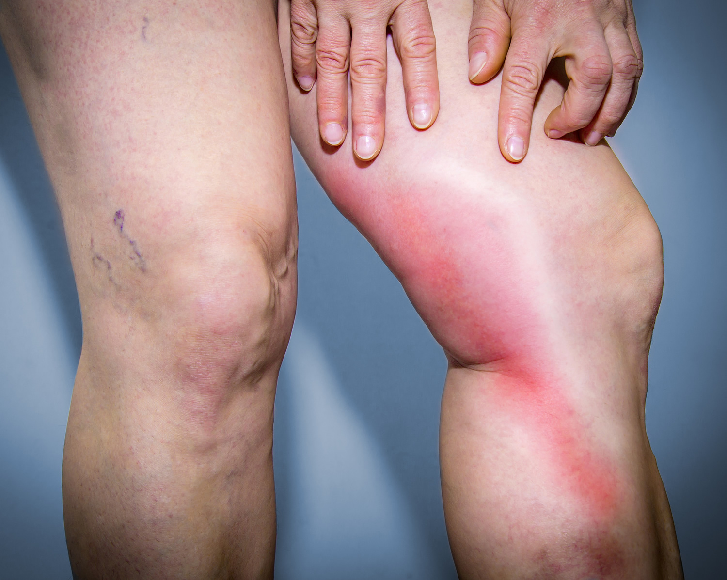
Figure 1. Phlebitis and thrombophlebitis
Phlebitis causes
Superficial thrombophlebitis can occur spontaneously, especially in the lower extremities in the great saphenous vein, or as a complication of medical or surgical interventions. Although the cause of superficial thrombophlebitis is not completely clear but it is believed to be associated with a change in the dynamic balance of hemostasis (stopping of bleeding). In 1846, the German pathologist Virchow showed that damage to a blood vessel wall, abnormal blood flow, or a change in blood constituents causing abnormal blood clotting, could lead to inflammation or formation of blood clots in the veins. Therefore, superficial venous thrombosis is most often associated with one of the components of the Virchow triad; that is, intimal (blood vessel wall) damage (which can result from trauma, infection, or inflammation), stasis or turbulent flow, or changes in blood constituents (presumably causing increased coagulability).
In each type of superficial thrombophlebitis, the condition presents as redness and tenderness along the course of the vein, usually accompanied by swelling. Bleeding also can occur at the site of a varicose vein.
Although unusual, superficial thrombophlebitis may occur in the lesser saphenous vein, which empties into the popliteal vein.
Superficial thrombophlebitis can also occur in the external jugular vein, if it has been used for an infusion site. Superficial thrombophlebitis of the upper extremities usually occurs at infusion sites or sites of trauma.
Who gets phlebitis ?
Superficial thrombophlebitis can occur spontaneously and without apparent reason. However, there are risk factors that make it more likely for the condition to occur.
The most important clinically identifiable risk factors for thrombophlebitis are a prior history of superficial phlebitis, deep vein thrombosis (DVT) and pulmonary embolism (PE). Some common risk markers include recent surgery or pregnancy, prolonged immobilization, and underlying malignancy.
Phlebitis also occurs in diseases associated with vasculitis, such as polyarteritis nodosa (periarteritis nodosa) and Buerger disease (thromboangiitis obliterans). Buerger noted phlebitis in 8 of 19 patients, and Shionoya reported it in 43% of the 255 patients he followed 4. After 2010, a medically emergent cutaneous thombophlebitis began to be noted more frequently, after an increase in use of levamisole (an antihelminth) for bulking cocaine in the US. In a review of the literature by Pearson et al 5 cutaneous thrombosis was noted in 84% of patients presenting with levamisole-induced vasculopathy.
Pregnancy
The increased likelihood of developing thrombophlebitis occurs through most of pregnancy and for approximately 6 weeks after delivery. This is partly due to increased platelet stickiness and partly due to reduced fibrinolytic activity.
The association between pregnancy and thrombophlebitis is of particular concern to women who carry the factor V Leiden or prothrombin C-20210-a gene, because they already have a predisposition to clotting, which would also be exacerbated by pregnancy 6.
Estrogen therapy
High-dose estrogen therapy is another risk factor. Case-controlled and cohort studies based on clinical signs and symptoms of thrombosis suggest that by taking high-estrogen oral contraceptives, a woman may increase her risk of thrombosis by a factor of 3-12 times, though the absolute risk remains low. Newer low-dose oral contraceptives are associated with a much lower risk of thrombophlebitis, though the absolute risk has not been well quantified 7.
Other common risk factors for phlebitis:
- History of superficial phlebitis, deep vein thrombosis, and pulmonary embolism – much greater chance of getting superficial thrombophlebitis.
- Varicose veins – people with varicose veins are more prone to minor injuries of the blood vessels which can lead to inflammation.
- Intravenous injection or infusion sites – superficial thrombophlebitis in the arm or neck region may occur at infusion sites or sites of trauma.
- Blood clotting abnormalities – there are various conditions or drugs that can make blood clot more easily and cause superficial thrombophlebitis:
- Pregnancy – through most of pregnancy and for about 6 weeks after delivery. Especially of concern in pregnant women who carry prothrombin 20210 gene mutation, as they have a predisposition to clotting.
- Oral contraceptive pill and hormone replacement therapy – high dose eostrogen treatments may increase the risk of thrombosis by 3-12 times.
- Diseases associated with vasculitis such as Buerger disease and polyarteritis nodosa
- Smoking
- Underlying cancer
- Reduced blood flow (stasis) – this may occur in veins in people during long air flights, those that are immobile, or had recent major surgery.
Additional risk factors
Other recognized markers of risk for venous thromboembolic disease include the following:
- Obesity
- Age older than 60 years (however, there are fewer complications in this age group)
- IV drug abuse
- Hypercoagulable states (eg, factor V Leiden mutation, prothrombin gene mutation, and protein S deficiency)
- Systemic lupus erythematosus
- Acquired immunodeficiency syndrome (AIDS)
- Lupus anticoagulant
- Drug-induced lupus anticoagulant
- Antithrombin III deficiency
- Behçet disease
- Blood type A
- Burns
- Catheters (indwelling venous infusion catheters)
- Chemotherapy
- Congestive heart failure
- Estrogen replacements (high dose only)
- Fibrinogen abnormality
- Fractures
- Hemolytic anemias
- Heparin-associated thrombocytopenia
- Homocysteinemia
- Homocystinuria
- Hyperlipidemias
- Immobilization
- Malignancy
- Myocardial infarction (heart attack)
- Phenothiazines
- Plasminogen abnormality
- Plasminogen activator abnormality
- Polycythemia
- Postoperative
- Protein C deficiency
- Protein S deficiency
- Resistance to activated protein C
- Thrombocytosis
- Trauma
- Ulcerative colitis
- Venography
- Venous pacemakers
- Venous stasis
- Warfarin – First few days of therapy
- Caustic materials, such as lighter fluid, injected intravenously 8.
Prevention of phlebitis
Sitting during a long flight or car ride can cause your ankles and calves to swell and increases your risk of thrombophlebitis. To help prevent a blood clot:
- Take a walk. If you’re flying or riding a train or bus, walk up and down the aisle once an hour or so. If you’re driving, stop every hour or so and move around.
- Move your legs regularly. Flex your ankles, or carefully press your feet against the floor or footrest in front of you at least 10 times each hour.
- Wear loose clothing.
- Drink plenty of nonalcoholic fluids to avoid dehydration.
- Eliminate or reduce risk factors
What are the signs and symptoms of phlebitis ?
Characteristic signs and symptoms include:
- Slight swelling, redness and tenderness along a part of the affected vein.
- Veins on the foot, ankle and area just behind the knee are swollen and pop-out.
- Other veins in the affected area may appear blue colour.
- If a blood clot develops the vein may feel hard or knobbly.
- If the condition has been present for a while and the swelling has resolved, the skin may appear stained or darkened.
Phlebitis Diagnosis
Superficial thrombophlebitis is a clinical diagnosis in which the clinician identifies tender and inflamed superficial veins. However, ruling out deep vein thrombosis (DVT) in the clinical setting is difficult; further testing is often required to evaluate for this condition.
Patients with superficial thrombophlebitis often give a history of a gradual onset of localized tenderness, followed by the appearance of an area of erythema along the path of a superficial vein.
Patients may also have a history of the following:
- Local trauma
- Similar prior episodes
- Varicose veins
- Prolonged travel
- Hormone use
- Tobacco use
- Family history of blood coagulopathies
- Enforced stasis
- IV drug use
Although patients should be asked about these risk factors for hypercoagulability, the absence of identifiable risk factors has no prognostic value.
Traumatic thrombophlebitis
- History of trauma, needlesticks, indwelling intravenous (IV) catheters, drug (eg, phenytoin) or hypertonic solution (10% calcium chloride) infusion, and sclerotherapy.
Thrombosed varicose veins
- History of varicose veins, previous history of thrombosed varices, and any injury to the leg that has the varices. One should ascertain whether there was a thrombosed vein and should determine the timing of erythema and pain.
Thrombosed hemorrhoids
- History should focus on previous occurrences of thromboses and surgical intervention, as well as on the timing of symptoms.
Migratory thrombophlebitis
- Also known as Trousseau’s sign of malignancy, migratory thrombophlebitis is described as thrombophlebitis that travels, often from one leg to the other. It has a strong association with adenocarcinoma of the pancreas and lung; therefore, the history should be directed toward finding malignancy.
To determine whether you have superficial thrombophlebitis or deep vein thrombosis, your doctor might choose one of these tests:
- Ultrasound. Duplex ultrasonographic evaluation is the diagnostic study of choice for venous thrombosis. A wandlike device (transducer) moved over the affected area of your leg sends sound waves into your leg. As the sound waves travel through your leg tissue and reflect back, a computer transforms the waves into a moving image on a video screen. Thrombosed veins may appear thickened or inflamed on ultrasonography, but the most accurate diagnostic finding is a lack of compressibility of the vein using the scan head. An experienced ultrasonographer should be able to diagnose superficial thrombophlebitis with a high sensitivity and specificity 9.
- All patients with superficial thrombophlebitis above the knee should undergo duplex ultrasonography as the initial diagnostic modality of choice to rule out DVT. When the patient has superficial thrombophlebitis below the knee, duplex ultrasonography is only indicated for signs and symptoms consistent with DVT (eg, asymmetrical swelling, erythema, pain). Superficial thrombophlebitis in lower-extremity varicose veins has an extremely low incidence of DVT 10.After an initial diagnosis of superficial thrombophlebitis, especially in the thigh region, a follow-up duplex ultrasonographic examination in 48 to 72 hours should be performed to look for progression of disease after treatment is initiated. A finding of no clot extension indicates successful therapy. Thrombus extension or encroachment toward the deep venous system should prompt more aggressive treatment.Results from a study by Quéré et al indicated that in patients with symptomatic superficial venous thrombosis, compression ultrasonography of an affected limb’s entire venous system provides valuable treatment information 11. The investigation involved 844 patients from the Prospective Observational Superficial Thrombophlebitis (POST) study, all of whom had symptomatic superficial venous thrombosis. A complete lower limb scan of both legs using compression ultrasonography was performed on all of the patients, with imaging of the superficial and deep venous systems.
Among the report’s findings were that concomitant DVT was present in 198 patients (23.5%) and that 41.8% of those individuals had proximal DVT 11. Superficial venous thrombosis was not contiguous with DVT in 83 (41.9%) of the 198 patients.
This test can confirm the diagnosis and distinguish between superficial and deep vein thrombosis.
- Blood test. Almost everyone with a blood clot has an elevated blood level of a naturally occurring, clot-dissolving substance called D dimer. D-dimer is a unique degradation product produced by plasmin-mediated proteolysis of cross-linked fibrin that is often measured in the evaluation for DVT and pulmonary embolism (PE). But D dimer levels can be elevated in other conditions, thus it is of little clinical use for detecting thrombophlebitis 12. So a test for D dimer isn’t conclusive, but can indicate the need for further testing.
- Blood tests rarely are helpful in the diagnosis of thrombophlebitis, except in those patients at risk for an underlying hypercoagulable state. Several common hypercoagulable states, including the following, can be identified through laboratory studies:
- Resistance to activated protein C (most often due to factor V Leiden)
- Protein C deficiency
- Protein S deficiency
- Antithrombin III deficiency
- Antiphospholipid antibodies
- Prothrombin gene 2010-a mutation (factor II mutation)
- Schönauer et al reported a high factor VIII concentration to be an independent risk factor for superficial thrombophlebitis 13. In another study, de Godoy and Braile reported that 5.5% of patients with repetitive superficial thrombophlebitis were positive for protein S deficiency 14. Other authors have reported that factor V Leiden and the prothrombin gene mutation significantly increase the risk of superficial thrombophlebitis.
It’s also useful for ruling out DVT and for identifying people at risk of developing thrombophlebitis repeatedly.
- Migratory thrombophlebitis, especially without good cause, may be an indication for a more detailed evaluation of the patient to determine whether a malignant lesion exists. This evaluation should include selective application of serum carcinoembryonic antigen (CEA) testing, prostate-specific antigen (PSA) testing, colonoscopy, computed tomography (CT), and mammography.
Phlebitis treatment
Treatment for superficial thrombophlebitis is aimed at patient comfort and at preventing superficial phlebitis from involving the deep veins.
- Superficial phlebitis with infection, such as phlebitis originating at an IV catheter site, is referred to as septic thrombophlebitis, a clinical entity requiring diagnostic and therapeutic approaches that are different from those applied to sterile phlebitis.
For superficial thrombophlebitis, your doctor might recommend applying heat to the painful area, elevating the affected leg, using an over-the-counter nonsteroidal anti-inflammatory drug (NSAID) and possibly wearing compression stockings. The condition usually improves on its own.
Your doctor might also recommend these treatments for both types of thrombophlebitis:
- Blood-thinning medications. If you have deep vein thrombosis, injection of a blood-thinning (anticoagulant) medication, such as low molecular weight heparin or fondaparinux (Arixtra), will prevent clots from enlarging. After the initial treatment, taking the oral anticoagulant warfarin (Coumadin, Jantoven, others) or the newer rivaroxaban (Xarelto) for several months continues to prevent clots from enlarging.
- A Cochrane review published in 2013 added, among others, a large randomized control study that included more than 3000 patients with superficial thrombophlebitis and compared fondaparinux (Arixtra) with placebo. The investigators found fondaparinux to be a good option for treatment of superficial thrombophlebitis and prevention of some of its associated complications 15.
- Fondaparinux. Fondaparinux is a newer anticoagulant that was derived from the binding region of heparin and antithrombin. It is an inhibitor of factor Xa, and its main uses are the same as those of heparin—more specifically, prevention and treatment of venous thrombosis and pulmonary embolism (PE). Fondaparinux is not shown to interact with platelets and platelet factor 4 and thus theoretically should not cause heparin-induced thrombocytopenia (HIT). Its main advantage over heparin or low molecular weight heparin is that its bioavailability and half-life (15-17 hours) allow once-daily dosing.Fondaparinux has been shown to achieve significant reductions in the extension of superficial thrombophlebitis into the deeper venous systems and the rate of recurrence in general, as well as to reduce the symptoms of venous thromboembolism when compared to placebo 15; however, there was no difference with respect to the rates of major bleeding. To date, no studies have been done to compare the efficacy of fondaparinux with that of heparin or low molecular weight heparin in superficial thrombophlebitis.Use of the lowest dosage of fondaparinux (2.5 mg/day subcutaneously) for 45 days has been shown by Decousus et al to suffice for prevention of extension, recurrence, and embolization of superficial venous thrombosis 16. Another advantage to this anticoagulant medication is that the dosage studied for superficial venous thrombosis does not have to be monitored. At this dosage, fondaparinux has not been shown to affect activated partial thromboplastin time (aPTT), prothrombin time (PT), or bleeding time 17.
Fondaparinux should be avoided in patients with kidney function compromise, active bleeding, bacterial endocarditis, and body weight below 50 kg. One downside to the use of fondaparinux is that there is currently no antidote, especially for the low dosage used for superficial thrombophlebitis treatment. Also, because this agent is cleared by the renal system, prolonged administration warrants renal function monitoring and should be discontinued if creatinine clearance is less than 30 mL/min.
If your doctor prescribes a blood thinner, follow directions carefully. Their most serious side effect can be excessive bleeding.
- Clot-dissolving medications. Treatment with medications such as alteplase (Activase) dissolves blood clots. Also known as thrombolysis, this treatment is used for extensive DVT, including some cases that involve a blood clot in the lungs (pulmonary embolus).
- Compression stockings. Prescription-strength compression stockings help prevent swelling and reduce the chances of complications of DVT.Gradient compression hose are highly elastic stockings that provide a gradient of compression that is highest at the toes (at least 30-40 mm Hg) and gradually decreases to the level of the thigh. This amount of compression reduces capacitive venous volume by approximately 70% and increases the measured velocity of blood flow in the deep veins by a factor of 5 or more. Gradient compression hose also have been shown to increase local and regional intrinsic fibrinolytic activity.In the early phases of superficial thrombophlebitis in the leg, dangling the extremity without external support from stockings or elastic bandages leads to leg swelling and increased pain.
- Filter. In some instances, especially if you can’t take blood thinners, a filter can be inserted into the main vein in your abdomen (vena cava) to prevent clots that break loose in leg veins from lodging in your lungs. Usually, the filter is removed when it’s no longer needed. If you have a filter placed, ask your doctor if and when it should be removed.
- Varicose vein stripping. Your doctor can surgically remove varicose veins that cause pain or recurrent thrombophlebitis in this procedure. It involves removing a long vein through small incisions. Removing the vein won’t affect circulation in your leg because veins deeper in the leg take care of the increased volumes of blood.
Treatment of Septic and Suppurative Thrombophlebitis
If thrombophlebitis is associated with a cannula or a catheter, the device should be immediately removed and cultured. If the patient is in a septic state, appropriate antibiotics should be given. If suppurative thrombophlebitis is suspected, immediate and complete excision of all of the involved veins is indicated. The wound may be left packed open for secondary closure or skin grafting at a later date. The use of appropriate systemic antibiotics is always indicated.
If the suppurative process involves one of the deep veins, aggressive antimicrobial and anticoagulant therapy are necessary.
If a venous segment involved in superficial thrombophlebitis is suspected to be a source of bacteremia but does not require excision, it can be aspirated in order to culture the contents of the venous lumen. This may be helpful in immunocompromised patients with phlebothrombosis and positive blood cultures.
Phlebitis treatment at home
In addition to medical treatments, here are self-care measures to help improve thrombophlebitis.
If you have superficial thrombophlebitis:
- Use a warm washcloth to apply heat to the involved area several times daily
- Elevate your leg
- Use a nonsteroidal anti-inflammatory drug (NSAID), such as ibuprofen (Advil, Motrin IB, others) or naproxen sodium (Aleve, others), if recommended by your doctor
If you have deep vein thrombosis:
- Take prescription anticoagulant medications as directed to prevent complications
- Elevate your leg if it’s swollen
- Wear your prescription compression stockings as directed
Let your doctor know if you’re taking another blood thinner, such as aspirin.
Prognosis of phlebitis
The prognosis in superficial thrombophlebitis is usually good. Superficial phlebitis is rarely associated with pulmonary embolism (PE), although it can occur, particularly if the process extends into a deep vein. However, individuals with superficial venous thrombosis do not seem to have a great tendency to develop deep vein thrombosis (DVT). In contrast, patients with DVT are frequently found to have superficial venous thrombosis.
The patient should be told to expect the disease process to persist for 3-4 weeks or longer. If it occurs in the lower extremity in association with varicose veins, it has a high likelihood of recurrence unless excision is performed.
- https://www.dermnetnz.org/topics/superficial-thrombophlebitis/[↩]
- Verlato F, Zucchetta P, Prandoni P, Camporese G, Marzola MC, Salmistraro G, et al. An unexpectedly high rate of pulmonary embolism in patients with superficial thrombophlebitis of the thigh. J Vasc Surg. 1999 Dec. 30(6):1113-5.[↩]
- Wichers IM, Di Nisio M, Buller HR, et al. Treatment of superficial vein thrombosis to prevent deep vein thrombosis and pulmonary embolism: a systematic review. Haematologica. 2005 May. 90(5):672-7.[↩]
- Shionoya S. Buerger’s Disease: Pathology, Diagnosis and Treatment. Nagoya, Japan: University of Nagoya Press; 1990.[↩]
- Pearson T, Bremmer M, Cohen J, Driscoll M. Vasculopathy related to cocaine adulterated with levamisole: A review of the literature. Dermatol Online J. 2012 Jul. 18(7):1.[↩]
- McColl MD, Ramsay JE, Tait RC, et al. Superficial vein thrombosis: incidence in association with pregnancy and prevalence of thrombophilic defects. Thromb Haemost. 1998 Apr. 79(4):741-2.[↩]
- Rosendaal FR, Helmerhorst FM, Vandenbroucke JP. Oral contraceptives, hormone replacement therapy and thrombosis. Thromb Haemost. 2001 Jul. 86(1):112-23.[↩]
- Rush MD, Schoenfeld CN, Watson WA, et al. Skin necrosis and venous thrombosis from subcutaneous injection of charcoal lighter fluid (naptha). Am J Emerg Med. 1998 Sep. 16(5):508-11.[↩]
- Lutter KS, Kerr TM, Roedersheimer LR, et al. Superficial thrombophlebitis diagnosed by duplex scanning. Surgery. 1991 Jul. 110(1):42-6.[↩]
- Bergqvist D, Jaroszewski H. Deep vein thrombosis in patients with superficial thrombophlebitis of the leg. Br Med J (Clin Res Ed). 1986 Mar 8. 292(6521):658-9.[↩]
- Quéré I, Leizorovicz A, Galanaud JP, Presles E, Barrellier MT, Becker F, et al. Superficial venous thrombosis and compression ultrasound imaging. J Vasc Surg. 2012 Oct. 56 (4):1032-8.e1.[↩][↩]
- Gillet JL, Ffrench P, Hanss M, Allaert FA, Chleir F. [Predictive value of D-dimer assay in superficial thrombophlebitis of the lower limbs]. J Mal Vasc. 2007 Apr. 32(2):90-5.[↩]
- Schonauer V, Kyrle PA, Weltermann A, et al. Superficial thrombophlebitis and risk for recurrent venous thromboembolism. J Vasc Surg. 2003 Apr. 37(4):834-8.[↩]
- de Godoy JM, Braile DM. Protein S deficiency in repetitive superficial thrombophlebitis. Clin Appl Thromb Hemost. 2003 Jan. 9 (1):61-2.[↩]
- Di Nisio M, Wichers IM, Middeldorp S. Treatment for superficial thrombophlebitis of the leg. Cochrane Database Syst Rev. 2013 Apr 30. 4:CD004982.[↩][↩]
- Decousus H, Prandoni P, Mismetti P, et al. Fondaparinux for the treatment of superficial-vein thrombosis in the legs. N Engl J Med. 2010 Sep 23. 363(13):1222-32.[↩]
- Bijsterveld NR, Moons AH, Boekholdt SM, et al. Ability of recombinant factor VIIa to reverse the anticoagulant effect of the pentasaccharide fondaparinux in healthy volunteers. Circulation. 2002 Nov 12. 106(20):2550-4.[↩]


