Contents
- What is polymorphic light eruption
- Polymorphic light eruption causes
- Polymorphic light eruption prevention
- Polymorphic light eruption signs and symptoms
- What does polymorphic light eruption rash look like?
- Polymorphic light eruption diagnosis
- Polymorphous light eruption treatment
- Polymorphic light eruption outlook (prognosis)
- What is photosensitivity
- Photosensitivity lupus
What is polymorphic light eruption
Polymorphic light eruption, also known as polymorphous light eruption, PMLE and prurigo aestivalis, is a common form of primary photosensitivity that mainly occurs in young adult women in temperate climates during spring and summer. Polymorphic light eruption is a fairly common skin rash triggered by exposure to sunlight or artificial ultraviolet (UV) light.
‘Polymorphic’ refers to the fact that the rash can take many forms, although in one individual it usually looks the same every time it appears.
An itchy or burning rash appears within hours, or up to two to three days after exposure to sunlight. It lasts for up to two weeks, healing without scarring.
The rash usually appears on the parts of the skin exposed to sunlight – typically the head and neck, chest and arms (the face isn’t always affected).
The polymorphic light eruption skin rash may be a rare occurrence, or may happen every time your skin is exposed to sunlight. It ranges from mild to severe.
Sometimes, as little as 30 minutes of sun exposure is enough to cause the problem, and it can develop even through window glass or thin clothing.
However, for most people with polymorphic light eruption, the rash develops after several hours outside on a sunny day. If further sun is avoided, the rash may settle and go without a trace within a couple of weeks. It may or may not return when skin is re-exposed to sunlight.
If the skin is exposed to more sunlight before the rash has cleared up, it will probably get much worse and spread.
For many affected people, the skin rash appears every spring and remains a problem throughout summer, settling by autumn.
- Polymorphic light eruption rash not infectious – there’s no risk of catching polymorphic light eruption from another person.
Polymorphic light eruption generally affects adult females aged 20–40, although it sometimes affects children and males in 25% of cases 1. Polymorphic light eruption is particularly common in places where sun exposure is uncommon, such as Northern Europe, where it is said to affect 10–20% of women holidaying in the Mediterranean area 1. It is less common in Australasia. It has also been reported to be relatively common at higher altitudes compared to sea level.
Polymorphic light eruption can affect all skin phototypes, though it is more often diagnosed in white skin than in skin of color. There is a genetic tendency to polymorphic light eruption and it is sometimes associated with or confused with photosensitivity due to lupus erythematosus, which generally is more persistent than polymorphic light eruption.
Some patients experience polymorphic light eruption during phototherapy, which is used to treat skin conditions such as psoriasis and dermatitis.
Severe polymorphic light eruption can lead to emotional distress, anxiety and depression.
Polymorphic light eruption causes
Genetic factors may be important with many affected individuals reporting a family history of polymorphic light eruption. Polymorphic light eruption can run in families and about a fifth of affected people will have an affected relative.
Polymorphic light eruption is caused by a delayed hypersensitivity reaction to a compound in the skin that is altered by exposure to ultraviolet radiation (UVR). Ultraviolet radiation leads to impaired T–cell function and altered production of cytokines in affected individuals. There is a reduction in the normal UV–induced immune suppression in the skin. This has been suggested to be related to estrogen, insufficiency of vitamin D or to the microbiome and antimicrobial factors.
The rash is usually provoked by UVA (ultraviolet A radiation) in 75–90% of cases. This means the rash can occur when the sunlight is coming through window glass, and that standard sunscreens may not prevent it. Occasionally, UVB (ultraviolet B radiation) and/or visible light provoke polymorphic light eruption.
Risk factors for polymorphic light eruption
Anyone can develop polymorphous light eruption, but several factors are associated with an increased risk of the condition:
- Being female
- Experiencing the first episode during the teenage years or 20s
- Having light skin and living in northern regions
- Having a family history of the condition
Polymorphic light eruption prevention
It is not known how to prevent polymorphic light eruption altogether. However, many people can avoid developing a rash by using effective sun protection during the middle hours of the day during summer.
- Cover affected areas with densely woven sun protective clothing. Choose UPF 40+ clothing where available.
- Apply SPF 50+ semi-opaque sunscreen frequently to all uncovered skin.
- Stay in the shade.
Many patients find antioxidant nutritional supplements as a helpful preventative measure. These include:
- Nicotinamide
- Polypodium leucotomas extract. Polypodium leucotomos is a fern extract supplement for which there is some evidence of benefit in polymorphic light eruption. Additionally, there is relatively good evidence from multiple studies of benefit for sunburn. The risk of sunburn is decreased and the MED (minimal erythema dose) is increased. The usual dose is 240 mg oral twice daily.
- Carotenes: beta carotene, astaxathanin and canthaxanthin.
Some people with polymorphic light eruption successfully manage to gradually harden their skin to sun exposure.
- Slowly increasing time spent outdoors with uncovered skin, starting with a few minutes’ exposure during spring.
- Phototherapy: UVB narrowband UVB, or PUVA 2–3 times per week for 4–6 weeks in early spring.
A short course of oral steroids may be prescribed to reduce the likelihood of breaking out during a summer holiday, but are unsuitable long-term because of their adverse effects and risks.
Hydroxychloroquine is the most common on-going treatment prescribed for severely affected individuals. Immune suppressing drugs such as azathioprine are sometimes used.
Polymorphic light eruption signs and symptoms
Polymorphic light eruption may be a rare occurrence or may occur every time the skin is exposed to sunlight. The term “eruption” refers to the rash, which usually appears 30 minutes to several hours after exposure to sunlight. The polymorphic light eruption rash typically appears on areas of the body that tend to be covered during winter but exposed in summer: the upper chest, front of the neck and arms. In many affected individuals, polymorphic light eruption occurs each spring, provoked by several hours outside on a sunny day. If further sun exposure is avoided, the rash settles in a few days and is gone without a trace within a couple of weeks. However, polymorphic light eruption may recur the next time the sun shines on the skin. If the affected area is exposed to more sun before it has cleared up, the condition tends to get more severe and extensive.
The arms, the back of the hands, the V of the neck, the chest and lower legs/feet may be affected, but the face is usually spared. A few people complain of ocular and/or lip lesions. Juvenile spring eruption is a variant of polymorphic light eruption that is confined to the ears of children (usually boys).
The most common variety of polymorphic light eruption presents as crops of 2–5 mm pink or red papules. Other presentations include:
- Erythema (red macules)
- Erythematous plaques
- Dry, red patches or plaques (dermatitis)
- Vesicles (blisters)
- Pinpoint papules (especially in skin of colour)
- Lichenoid plaques
- Targetoid lesions (bull’s-eye appearance)
- Prurigo confined to exposed areas
- Itch without visible signs (polymorphic light eruption sine eruptione).
Rarely people may have other signs or symptoms, such as fever, chills, headache or nausea. These conditions may be the result of an associated sunburn rather than polymorphous light eruption.
Polymorphic light eruption persists for several days, and often longer if the affected skin is exposed to more sunlight. It resolves without scarring.
Polymorphic light eruption usually causes a burning sensation or itch. A few individuals also report fever and malaise following sun exposure.
Polymorphic light eruption can be the first sign of lupus erythematosus, but this is not usually the case.
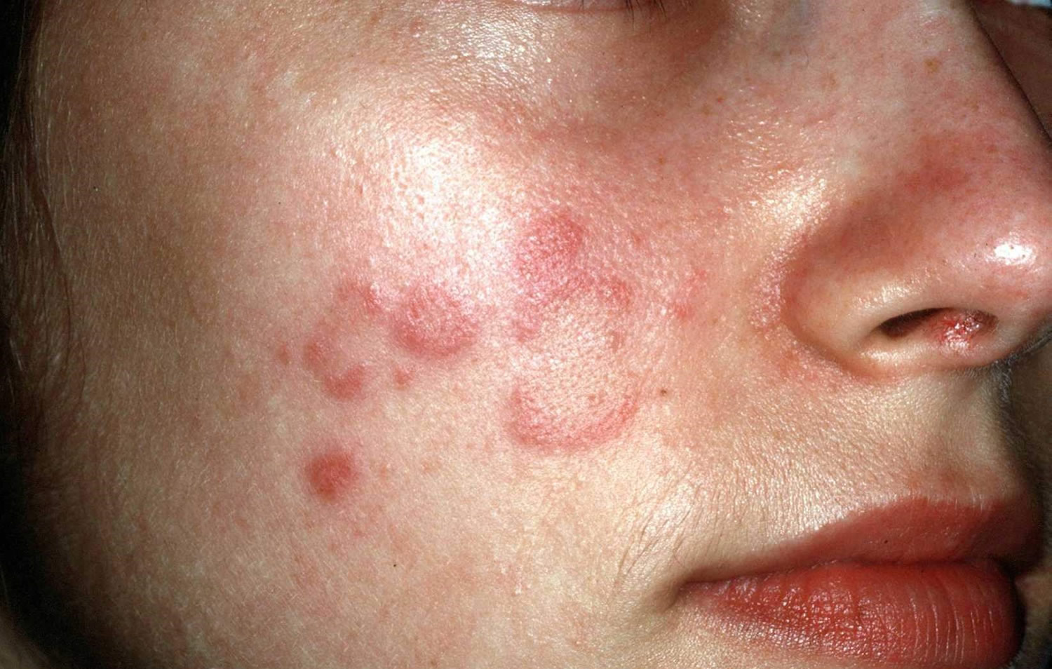
What does polymorphic light eruption rash look like?
The rash can take many different forms (known as being “polymorphic”):
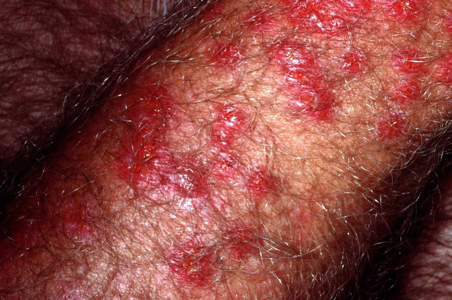
- most people get crops of 2-5mm pink or red raised spots (see Figure 4)
- some people get blisters that turn into larger, dry red patches (looks a bit like eczema)
- less commonly, the skin patches look like targets or bulls-eyes, a bit like the skin rash erythema multiforme
It can be easy to mistake polymorphic light eruption for prickly heat. Prickly heat is caused by warm weather or overheating, rather than sunlight or UV light. The skin in prickly heat doesn’t “harden” or desensitize, as it can do in polymorphic light eruption.
Figure 1. Polymorphic light eruption rash (forearm)
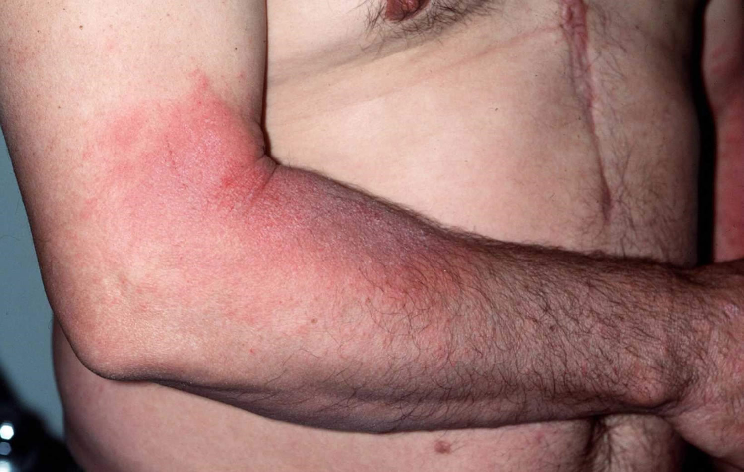
Figure 2. Polymorphic light eruption rash (back of thigh)
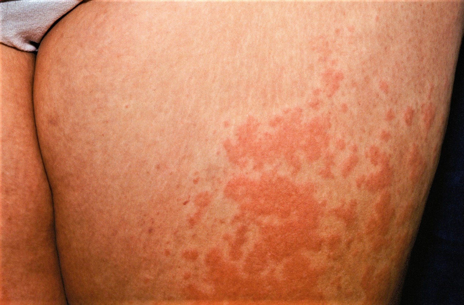
Figure 3. Polymorphic light eruption rash (face)
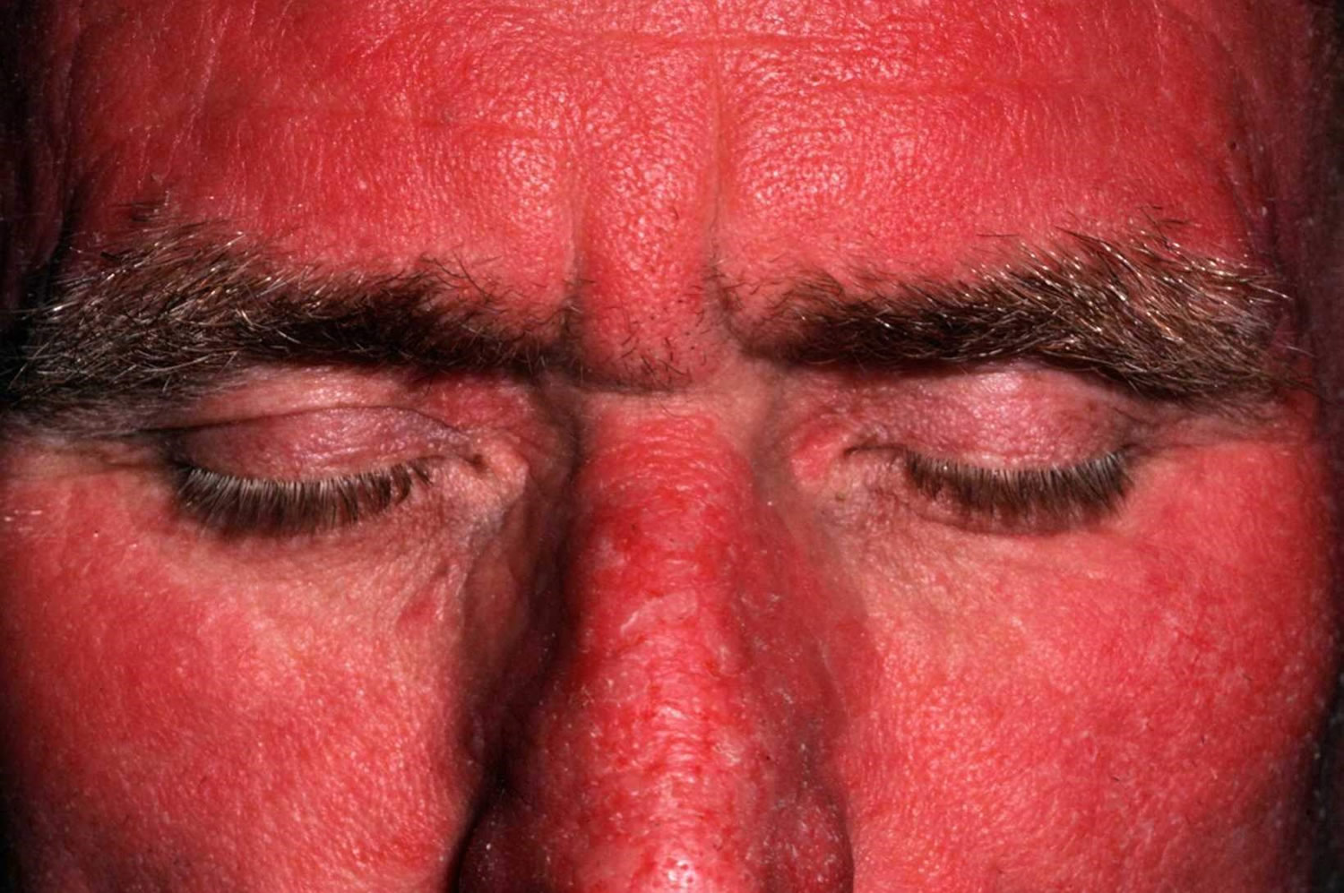
Figure 4. Polymorphic light eruption rash
Polymorphic light eruption diagnosis
Polymorphic light eruption is diagnosed clinically by its typical onset within hours of exposure to sunlight, and clearance after a few days. The rash is confined to exposed sites, and is often composed of erythematous papules and plaques.
Your doctor may need to rule out other disorders characterized by light-induced skin reactions. These conditions include:
- Chemical photosensitivity. A number of chemicals — drugs, medicated lotions, fragrances, plant products — can induce photosensitivity. When this occurs, your skin reacts each time it’s exposed to sunlight after ingesting or coming into contact with a particular chemical.
- Solar urticaria. Solar urticaria is a sun-induced allergic reaction that produces hives — raised, red, itchy welts that appear and disappear on your skin. The welts can appear within a few minutes of sun exposure and last for a few minutes to hours. Solar urticaria is a chronic condition that can last for years.
- Lupus rash. Lupus is an inflammatory disorder that affects a number of body systems. One symptom is the appearance of a discolored, bumpy rash on areas of skin exposed to sunlight, such as the face, neck or upper chest.
Sometimes a skin biopsy is necessary to make a diagnosis. Polymorphic light eruption has characteristic histopathological features, with upper dermal edema and a dense perivascular lymphocytic infiltrate. Eczematous changes may be present. Direct immune fluorescence is negative, unless the patient has cutaneous lupus erythematosus.
It is usual to have a blood count and a check for circulating antinuclear antibodies (ANA) and extractable nuclear antigens (ENA) in case of photosensitive cutaneous lupus erythematosus.
Phototesting is not usually carried out in patients with polymorphic light eruption, but provocation tests of exposure to UVA daily for 3 days to a small area of skin can confirm the diagnosis.
Polymorphous light eruption treatment
There’s no cure for polymorphic light eruption, but careful avoidance of the sun and using sunscreens will help you manage the rash.
Treatment of polymorphous light eruption usually isn’t necessary. The polymorphic light eruption rash typically goes away on its own within 10 days.
Generally, avoid the sun between 11am and 3pm, and wear protective clothing when outdoors (unless you’re hardening your skin; see below). Introduce your skin to sunlight gradually in the spring.
The following treatments may reduce the severity and duration of outbreaks of polymorphic light eruption:
- Topical corticosteroid creams
- Oral antihistamines
- Oral corticosteroids, eg, prednisone or prednisolone.
Sunscreen
You may be prescribed sunscreens, which may prevent the rash developing.
Use a broadspectrum sunscreen that is SPF 30+ or above with a good UVA rating. Apply it thickly and evenly, reapplying often.
Steroid creams and ointments
Your doctor can prescribe corticosteroid (steroid) cream or ointment, to be applied only when the rash appears. You should apply this sparingly, as often as your doctor advises, and never when there’s no rash.
Desensitization or UV treatment
Sometimes you can increase the resistance of your skin to the sun. This involves visiting a hospital department three times a week for four to six weeks in the spring.
Your skin is gradually exposed to a little more UV light every visit, to try to build up your skin’s resistance. The effects of desensitization are lost in the winter, so you’ll have to build up your resistance again in the spring.
Phototherapy
Your doctor may suggest phototherapy to prevent seasonal episodes of polymorphous light eruption in people who have experienced disabling signs and symptoms. Phototherapy exposes your skin to small doses of UVA or UVB light, which helps your skin be less sensitive to light. Basically, it’s a controlled version of the increased exposure you would experience over the course of the summer.
One type of light therapy called psoralen plus ultraviolet A (PUVA) combines UVA with a medicine called psoralen, which makes the skin more sensitive to this light. Short-term side effects of this therapy may include nausea, headache and itching.
Hardening or toughening
You may be able to increase the resistance of your skin at home, which is known as “hardening”. This involves going outside for short periods in the spring to build up your resistance.
You might find the time is as short as a few minutes at first, but you may be able to gradually build up to longer times. You’ll have to be careful not to overdo it, but as you understand more about how much light triggers your rash, you’ll be able to judge how long to stay out.
The effects of hardening are lost in the winter, so you’ll have to build up your resistance again in the spring.
Vitamin D
People with polymorphic light eruption are at greater risk of vitamin D deficiency, as a certain amount of sun exposure is needed to make your own vitamin D. Your doctor will advise whether you need treatment with vitamin D supplements.
Polymorphous light eruption treatment natural
Many patients find antioxidant nutritional supplements as a helpful preventative measure. These include:
- Nicotinamide
- Polypodium leucotomas extract. Polypodium leucotomos is a fern extract supplement for which there is some evidence of benefit in polymorphic light eruption. Additionally, there is relatively good evidence from multiple studies of benefit for sunburn. The risk of sunburn is decreased and the MED (minimal erythema dose) is increased. The usual dose is 240 mg oral twice daily.
- Carotenes: beta carotene, astaxathanin and canthaxanthin.
Lifestyle and home remedies
Self-care measures that may help ease your signs and symptoms include:
- Applying anti-itch cream. Try an over-the-counter (nonprescription) anti-itch cream, which may include products containing at least 1 percent hydrocortisone.
- Taking antihistamines. If itching is a problem, oral antihistamines may help.
- Using cold compresses. Apply a towel dampened with cool tap water to the affected skin, or take a cool bath.
- Leaving blisters alone. To speed healing and avoid infection, leave blisters intact. If needed, you can lightly cover blisters with gauze.
- Taking a pain reliever. An over-the-counter pain medication may help reduce redness or pain. These include ibuprofen (Advil, Motrin IB, others), acetaminophen (Tylenol, others) and naproxen sodium (Aleve, others).
To lessen the likelihood of recurring episodes of polymorphous light eruption, take the following precautions:
- Avoid the sun between 10 a.m. and 2 p.m. Because the sun’s rays are most intense during this time, try to schedule outdoor activities for other times of the day.
- Use sunscreen. Fifteen to 30 minutes before going outdoors, apply a broad-spectrum sunscreen, one that provides protection from both UVA and UVB light. Use a sunscreen with a sun protection factor (SPF) of at least 30. Apply sunscreen generously, and reapply every two hours — or more often if you’re swimming or perspiring. If you’re using a spray sunscreen, be sure to cover the entire area completely. Cover up. For protection from the sun, wear tightly woven clothing that covers your arms and legs and a broad-brimmed hat, which provides more protection than does a baseball cap or golf visor.Consider wearing clothing designed to provide sun protection. Look for clothes labeled with an ultraviolet protection factor (UPF) of 40 to 50. Follow care instructions on the label of UV-blocking clothes to maintain their protective feature.
Polymorphic light eruption outlook (prognosis)
Many people with polymorphic light eruption find the skin complaint improves over years – the skin may harden (become more resistant to sunlight) during the summer, which means more and more sun can be tolerated without the skin reacting. It may even eventually clear up on its own, although this is unusual and some very sensitive individuals even develop polymorphic light eruption in the winter.
However, hardening of the skin doesn’t always happen, and some people with very sensitive skin may even get the rash in the winter. For these people, it may be a long-term condition to manage with lifestyle changes and creams.
It has been noted that polymorphic light eruption appears to be less frequent and severe in women after menopause.
What is photosensitivity
Photosensitivity refers to various symptoms, diseases and conditions caused or aggravated by exposure to sunlight.
- A rash due to photosensitivity is a photodermatosis (plural photodermatoses).
- If the rash is eczematous, it is a photodermatitis.
- A chemical or drug that causes photosensitivity is a photosensitizer.
- A phototoxic reaction to a photosensitiser results in an exaggerated sunburn reaction and no immune reaction is involved.
- A photoallergic reaction to a photosensitiser results in photodermatitis and is due to delayed hypersensitivity reaction.
- A photoexacerbated condition describes a flare of an underlying skin disease on exposure to sunlight.
Photosensitivity occurs in males and females of all races and at all ages. Different types of photosensitivity may be prevalent at different times of life. Genetic and environmental factors are involved.
People with very white skin who don’t tan on exposure to the sun (Fitzpatrick skin type 1), especially if they also have red hair and blue eyes, are often considered photosensitive, relative to people with darker skin phototypes that tan more easily. These fair skinned individuals do not have a photodermatosis.
Figure 5. Photosensitivity rash
Complications of photosensitivity
Severe photosensitivity can lead to a person being unable to go outdoors during the day unless completely covered (including face). This leads to social isolation and depression.
Some photodermatoses cause permanent scarring.
Photosensitivity classification
Photosensitivity is classified into the following groups:
Primary photodermatoses
The causes of primary or idiopathic photodermatoses have not yet been discovered. Exposure to the sun produces a clearly defined disease entity. These include:
- Polymorphic light eruption
- Juvenile spring eruption
- Actinic folliculitis
- Actinic prurigo
- Solar urticaria
- Chronic actinic/photosensitivity dermatitis
- Hydroa vacciniforme (associated with Epstein Barr virus)
With the exception of polymorphic light eruption and juvenile spring eruption, these disorders are rare.
Exogenous photodermatoses
Exogenous photodermatoses are those in which phototoxic or photoallergic reaction is caused by an external photosensitiser. These include:
- Drug-induced photosensitivity: common photosensitising drugs are thiazides, tetracyclines (e.g., doxycycline photosensitivity), non-steroidal anti-inflammatory drugs (NSAIDs), phenothiazines, voriconazole, quinine, vemurafenib.
- Photocontact dermatitis: due to phototoxic chemicals such as psoralens in plants, vegetables, fruit; fragrances in cosmetics; sunscreen chemicals; dyes and disinfectants
- Pseudoporphyria: induced by drugs and/or renal insufficiency
Photoexacerbated dermatoses
Photoexacerbated dermatoses include:
- Lupus erythematosus
- Dermatomyositis
- Darier disease
- Rosacea
- Pemphigus vulgaris
- Pemphigus foliaceus
- Atopic dermatitis
- Psoriasis
Metabolic photodermatoses
Photosensitivity can be caused by a metabolic defect. The most common disorders of this type are porphyrias, in which phototoxic porphyrins accumulate in the skin. There are genetic defects in various enzymes, and the diseases may be activated by exposure to certain medications or toxins. Clinical presentation depends on which enzyme is defective.
- Porphyria cutanea tarda
- Erythropoeitic protoporphyria
- Variegate porphyria
- Erythropoeitic porphyria (Gunther disease)
Genetic photodermatoses
Photosensitivity can be associated with a pre-existing genetic disorder. These are rare.
- Xeroderma pigmentosum
- Bloom syndrome
- Rothmund Thomson syndrome
- Cockayne syndrome
Photosensitivity causes
Photosensitivity is caused by an abnormal reaction to some component of the electromagnetic spectrum of sunlight and a chromophore (reactive compound) within the skin.
The electromagnetic spectrum ranges from cosmic rays, invisible rays called ultraviolet radiation (UVR), through visible light, to infrared, microwaves and radiowaves. Ultraviolet radiation has 3 portions.
- UVA (ultraviolet A radiation): longer wavelength rays 320–400 nm that cause tanning and also suppress immune reactions in the skin
- UVB (ultraviolet B radiation): short wavelength rays 290–320 nm that cause sunburn and tan
- UVC (ultraviolet C radiation): ultrashort wavelength rays 200–290 nm that do not reach the earth’s surface
Patients can be sensitive to one kind of sunlight (i.e. only to UVB, UVA or visible light) or to a wider range of radiation. The most common photosensitivity is to UVA (ultraviolet A radiation). Properties of UVA (ultraviolet A radiation) include:
- Present throughout the year, but there is more UVA during summer
- Present throughout the day, but there is more UVA around the solar noon that earlier or later
- UVA is of lower energy than UVB, thus photon for photon, UVA is less damaging than UVB to DNA in skin cells
- UVA is however 100 times more prevalent at the earth’s surface than UVB
- UVA can penetrate through the epidermis into the dermis thus UVA damages more deeply than UVB
- UVA can penetrate untreated and untinted glass, which blocks UVB
- UVA is blocked by polycarbonate and densely woven fabric
Porphyria is mainly triggered by exposure to visible light.
Photosensitivity reaction
The photosensitivity reaction clinical features depend on the specific photodermatosis.
- Photodermatoses affect areas exposed to sunlight (face, neck, hands) and do not affect areas not exposed to the light (covered at least by underwear), or are less severe in covered areas.
- Sometimes they spare areas that are habitually exposed to the light, eg the face in polymorphic light eruption.
- Sometimes they only affect certain parts of the body, eg juvenile spring eruption is confined to the tops of the ears.
- Photodermatoses may also occur following indoor exposure to artificial sources of ultraviolet radiation (e.g., fluorescent lamps) or visible radiation.
Rashes on exposed sites may be due to another cause. For example:
- Facial acne due to follicular occlusion by sebum and keratin
- Contact dermatitis due to makeup applied to the face
- Airborne contact dermatitis due to plant pollens, such as sesquiterpene lactone
Clues to photosensitivity include:
- Summer exacerbation; note that many photodermatoses are present year round
- Sharp cut-off between affected area and skin covered by clothing or jewellery (eg watch strap, ring)
- Sparing of folds of upper eyelids
- Sparing of deep furrows on face and neck
- Sparing of skin covered by hair
- Sparing of skin shadowed by the ears, under the nose and under the chin
- Sparing of the web spaces between the fingers
Photosensitivity reactions prevention
Photosensitivity reactions are mainly prevented by careful protection from sun exposure and avoidance of exposure to artificial sources of ultraviolet radiation. However:
- The degree of protection needed from ultraviolet radiation depends on the severity of the disorder and the geographic location of the patient.
- Protection from ultraviolet radiation using sunscreens alone is not effective for porphyria. Affected areas must be covered when outdoors.
- Graduated exposure to small amounts of ultraviolet radiation may reduce sun-induced reactions in polymorphous light eruption.
Protection from ultraviolet radiation
When considering sun protective measures:
- Take note of the time of year and time of day. ultraviolet radiation is greater when the sun is overhead.
- Find out local ultraviolet levels.
- Note that the temperature and to some extent weather conditions make little difference to environmental UV.
- Be aware of greater ultraviolet radiation at altitude, and when reflected by bright surfaces, like snow, concrete and sand.
- Do not rely on shade from tree, umbrella or sail. ultraviolet radiation is scattered by dust particles, meaning may they may provide only partial protection.
- Watch out for a small amount of ultraviolet radiation released from some unguarded fluorescent daylight lamps.
Sunscreens are essential
- Sunscreens often fail to completely prevent photodermatoses.
- Sunscreens are best at filtering out UVB. To be most effective, they need to be applied thickly and frequently to all exposed skin.
- Select a sunscreen with a very high Sun Protection Factor (SPF 50+), which is a water resistant and broad spectrum product that complies with current United States FDA Standard for Sunscreens or the equivalent in other countries.
- Sunscreens containing reflectants such as zinc may be more effective than pure chemical sunscreens, as they filter out more UVA. They can be messy to use and cosmetically unappealing.
- Contact allergic dermatitis to sunscreens or contact photodermatitis to sunscreen chemicals can occur uncommonly, particularly to benzophenone or butyl methoxy dibenzoylmethane, and in the past, to para-aminobenzoic acid (PABA).
People with photodermatoses also may need to:
- Take vitamin D supplements
- Confine summer excursions out of doors to early in the morning or late in the evening.
- Cover up, wearing shirts with high collar and long sleeves, trousers or a long skirt, socks and shoes, a wide-brimmed hat, and if possible gloves.
- Wear opaque sun protective clothing. Dark coloured and densely woven fabric is the most effective. Some clothes are now labelled with UPF, the sun protection factor for fabrics. Choose those with a UPF of 40+.
- Protect their skin indoors and when in a vehicle.
- Apply ultraviolet radiation-absorbing film to windows at home or in the car.
- Wear a clear plastic mask to protect the face
Oral antioxidants such as polyphenols have been reported to provide limited extra protection, particularly Polypodium leucotomas and carotenoids. Nicotinamide may also provide benefit.
Photosensitivity diagnosis
Photosensitivity is diagnosed by the history of a skin problem arising on exposure to sunlight. The specific type is determined by examination of the skin and specific tests.
Photosensitivity is sometimes confirmed by phototests — artificial light from various different sources and at different doses is shone on small areas of the skin to see whether the rash can be reproduced, or if sunburn occurs more easily than expected. These tests can be difficult to perform and to interpret, and are only available in specialized centres.
Contact photosensitivity can be tested by photopatch tests, in association with standard patch tests. Adhesive patches containing known photosensitising materials are applied to the upper back, removed after two days, and light is shone on the area. The reaction is observed two days later.
Investigations may include:
- Full blood count
- Connective tissue antibodies including antinuclear antibodies (ANA), extractable nuclear antigens (ENA)
- Porphyrins in blood, urine and faeces
Patients suspected of porphyria cutanea tarda may also have liver function and iron tests.
Photosensitivity treatment
Management of photosensitivity involves sun protection and treatment of the underlying disorder.
Photosensitivity outlook (prognosis)
The prognosis depends on the specific disorder, its treatment, where the patient lives, and how carefully they protect their skin from exposure to sunlight.
For the most severely light sensitive patients, normal activities may be severely curtailed. Some find night work and sleep during the day, others put up with the rash.
Photosensitivity lupus
Systemic lupus erythematosus (SLE), also called lupus, is a chronic inflammatory disease that can affect almost any part of the body, especially the skin, joints, kidneys, heart, lungs, bones, blood, or brain. The clinical features of systemic lupus erythematosus (SLE) are highly variable and may overlap with other diseases and conditions. Skin involvement or cutaneous lupus (CLE) affects 80% of patients with SLE. Systemic lupus erythematosus is sometimes called acute lupus erythematosus, and the cutaneous features may be described as acute cutaneous lupus. Systemic lupus erythematosus (SLE) is considered an autoimmune disorder, meaning that a person’s own immune system attacks his or her own healthy cells and tissues, causing inflammation and damage.
Because systemic lupus erythematosus can affect any organ system, no two people have identical forms of the disease. However, most people with systemic lupus erythematosus report periods of time in which their symptoms seem to be mild or absent (remission) and other periods of time when the inflammation is more severe (flare or relapse).
Systemic lupus erythematosus can occur in people of all ages, all races, and both sexes. However, it is far more common in women, especially those between 15–45 years old. In America, it is also more commonly seen in people with darker skin than in light-skinned people. Every year about 2–7 new cases are diagnosed in a population of 100,000 people.
SLE is more prevalent and more severe in smokers. Smoking also reduces the effectiveness of antimalarials and other therapies.
Autoantibodies can be present in people that have no manifestation of the disease.
Although it is not directly inherited, lupus and other autoimmune diseases may run in families. Inheriting certain genes may make some people more susceptible to developing lupus.
In addition, certain environmental factors may trigger lupus in those who have a family (genetic) tendency toward the disease, including:
- Ultraviolet light, especially sunlight
- Certain medications, especially hydralazine and procainamide
- Infections
- Antibiotics, especially penicillins or sulfa-containing medicines
- Stress
- Hormonal changes, especially related to pregnancy and menstrual cycles.
Systemic lupus erythematosus causes
Factors leading to SLE include:
- Genetic predisposition, incuding haplotype HLA-B8, -DR3
- Exposure to sunlight
- Viral infection, particularly Epstein-Barr virus
- Hormones
- Toxins such as cigarette smoke
- Drugs in drug-induced LE
- Emotional upset
The manifestations of SLE are due to loss of regulation of the patient’s immune system.
- Nuclear proteins are not processed properly.
- Nuclear debris accumulates within the cell.
- This leads to production of autoantibodies against nuclear proteins.
- Immune complexes are not removed.
- The complement system is activated.
- Inflammation leads to cell and/or tissue injury.
Photosensitivity lupus prevention
If you know you have systemic lupus erythematosus, several measures can help prevent flares:
- Avoid intense sun exposure.
- Apply sunscreen with SPF of 30 or higher every day.
- Maintain healthy habits such as resting well, eating a balanced diet, and exercising regularly.
- Reduce stress.
- Avoid smoking and limit alcohol use.
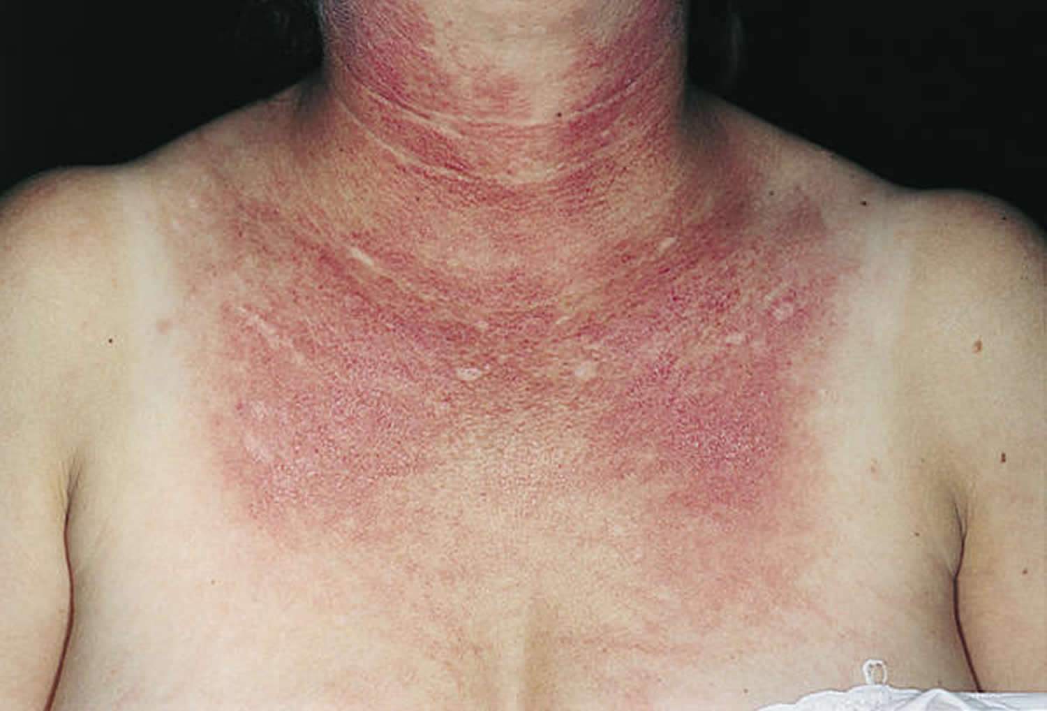
Figure 6. Photosensitivity lupus (Systemic Lupus Erythematosus)
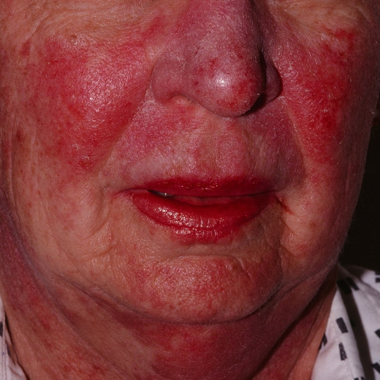
Figure 7. Systemic lupus erythematosus rash (hand)
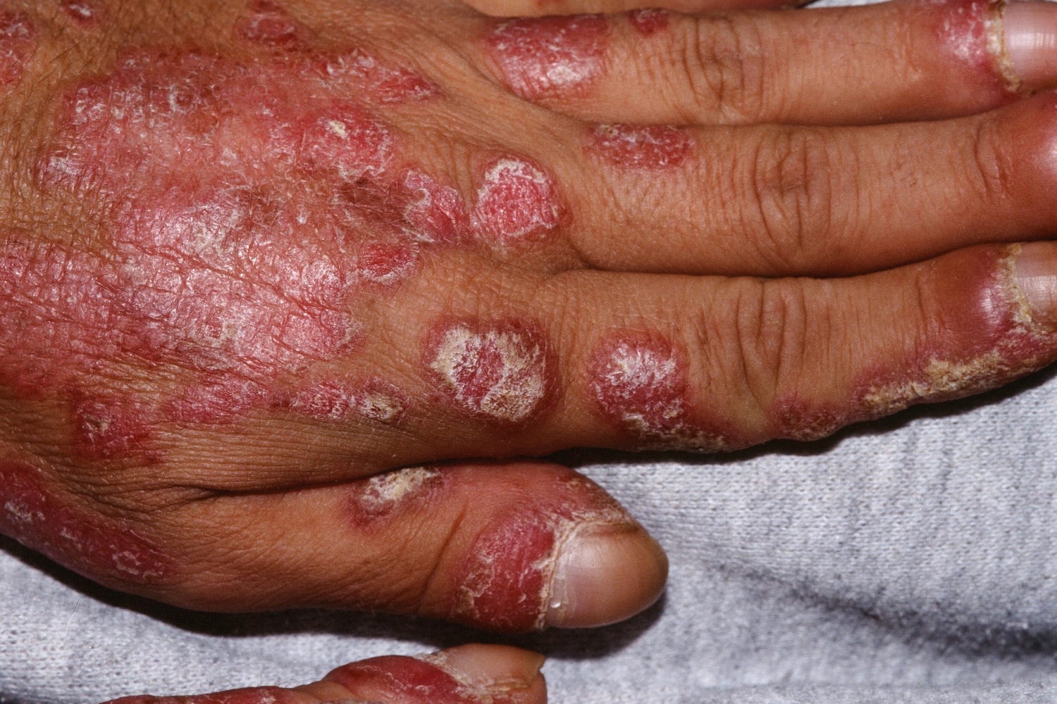
Figure 8. Cutaneous lupus erythematosus (face)
Photosensitivity lupus signs and symptoms
More than 90% of people with systemic lupus erythematosus have skin symptoms or skin involvement (cutaneous lupus erythematosus) and it is the first sign of SLE in about one quarter of them. It can present as lupus erythematosus-specific or lupus erythematosus-nonspecific manifestations. Lupus erythematosus-specific lesions tend to be induced or aggravated by exposure to ultraviolet radiation and are localised in sun-exposed sites such as face, neck, V of neck and upper back.
Specific cutaneous SLE
Cutaneous lupus erythematosus (CLE) has specific acute, subacute and chronic manifestations.
- Typically, SLE presents with acute cutaneous lupus erythematosus face.
- About half of patients with subacute cutaneous lupus erythematosus face develop mild SLE
- Only 5% of patients with chronic cutaneous lupus erythematosus face to have SLE, ie it presents as a skin problem without involvement of other organs.
Acute cutaneous lupus erythematosus
- Central face malar or “butterfly” violaceous erythema with sharp cutoff at lateral margins, resolves without scarring (may result in persistent telangiectasia)
- Bullous lupus: a blistering rash, if severe may resemble toxic epidermal necrolysis
- Maculopapular rash resembling morbilliform drug eruption
- Mucosal erosions and ulcerations (lips, nose, mouth, genitals)
- Photosensitivity: lupus rashes are mainly on sun-exposed sites. Photosensitivity can be mild to very severe with rash appearing after minimal light exposure.
- Diffuse hair loss (nonscarring alopecia) with brittle hair shafts
Subacute cutaneous lupus erythematosus
- Flat scaly patches resembling psoriasis, often in network pattern
- Annular (ring-shaped) polycyclic (overlapping circular) lesions
- Lesions resolve with minimal scarring
- Affects trunk and arms
- Flares on exposure to sun, but usually spares face and hands
Chronic cutaneous lupus erythematosus
- Chronic cutaneous lupus erythematosus affects 25% of patients with SLE
- Classic discoid lupus is most common: indurated hyperpigmented plaques
- Localised (above neck in 80%) or generalised (above and below neck in 20%)
- Hypertrophic (warty) lupus
- Tumid lupus
- Lupus panniculitis/profundus
- Mucosal lupus (lips, nose, mouth, genitals)
- Chilblain lupus
- Discoid lupus/lichen planus overlap
- Discoid lesions and panniculitis resolve with scarring
Nonspecific cutaneous SLE
Nonspecific cutaneous SLE refers to features relating to underlying illness rather than an autoimmune attack. These features may occur in other autoimmune or connective tissue diseases.
- Nail fold capillary telangiectasia
- Raynaud phenomenon (white fingers and toes on exposure to the cold)
- Vasculopathy of tips of digits (occlusion of small blood vessels by thrombus)
- Diffuse hair thinning without brittle hair
- Urticaria, which may be neutrophilic on biopsy
- Cutaneous vasculitis: palpable purpura or urticarial vasculitis
- Livedo reticularis (a network pattern of blood vessels) in 20–30% patients with SLE
- Papular mucinosis (deposits of mucin in skin)
- Calcinosis cutis (deposits of calcium in skin)
The most common locations for the skin lesions of systemic lupus erythematosus include:
- Face, especially cheeks and nose (Figure 6)
- Sun-exposed skin on arms, backs of hands, upper chest, and upper back due to increased sensitivity to sunlight (photosensitivity) (Figure 7)
- Fingers and fingernails
- Mouth or nose
- Scalp
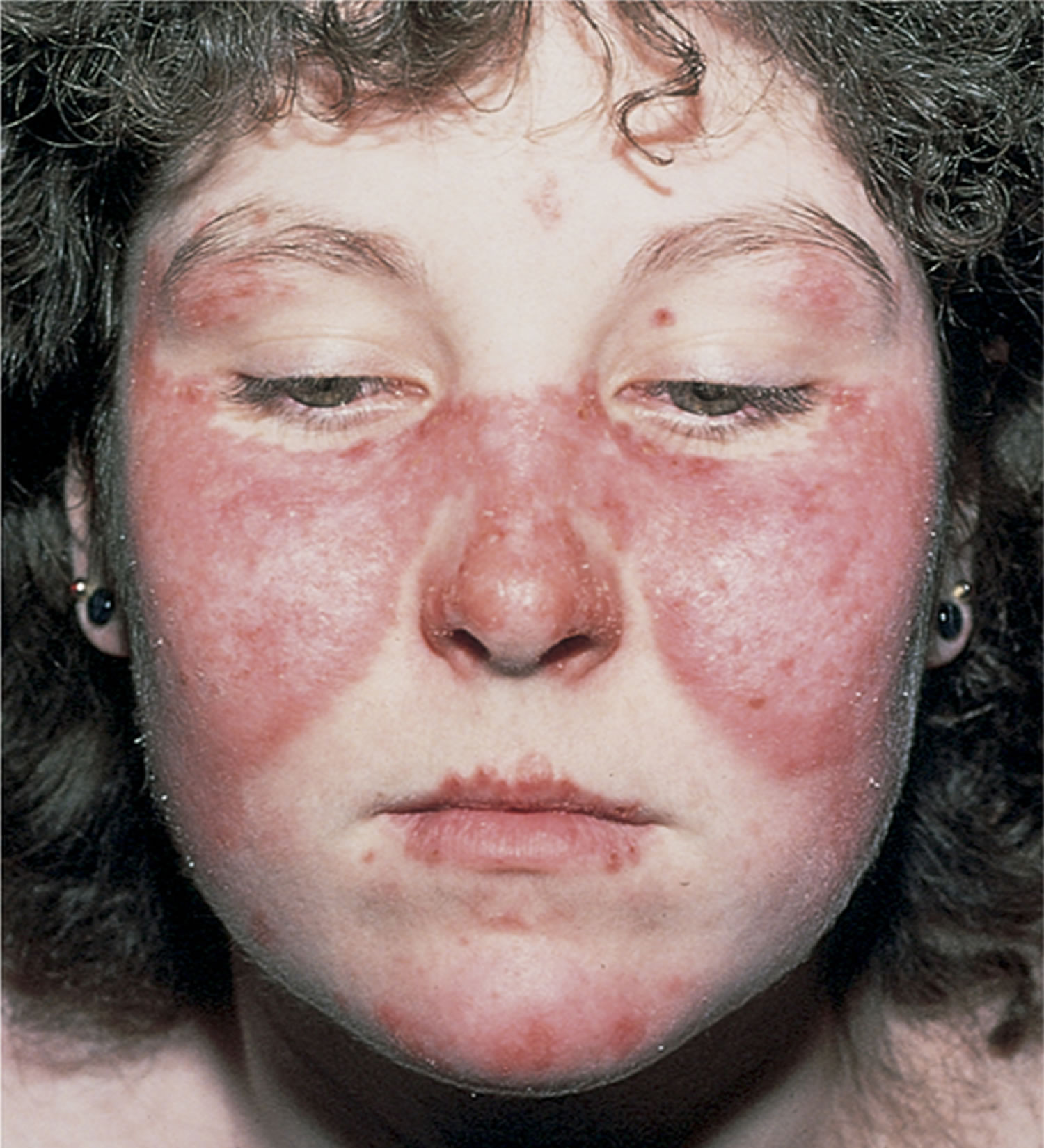
The classic skin finding in systemic lupus erythematosus is the butterfly rash (malar blush). Redness across the cheeks and bridge of the nose can occur after sun exposure and may appear as much as several weeks before other symptoms develop.
Figure 9. Systemic lupus erythematosus butterfly rash (malar blush)
A rash can develop in sun-exposed skin (photo-distribution), especially on the backs of the hands and fingers. This rash, which appears as red, scaly patches, can also affect the arms and trunk.
The skin around fingernails (nail folds) can be red and inflamed, and tiny, dilated blood vessels (telangiectasia) may be seen. In addition, people may develop Raynaud phenomenon, in which the fingers (and sometimes toes) turn pale and numb after exposure to cold temperatures.
Small, painless ulcers can develop in the nose or, more commonly, in the mouth, especially on the roof of the mouth.
When lupus affects the scalp skin, you may notice hair loss. It may be patchy, or there may be thinning across the scalp, especially at the temples.
In addition to the skin lesions of lupus, people may have:
- Joint pain or swelling, especially in hands, wrists, and knees
- Blood problems, including anemia and clotting disorders
- Kidney disorders
- Lung problems, such as painful breathing
- Seizures or other brain disorders
- Swollen lymph glands
- Fever
- Fatigue
Numerous systemic features may occur in SLE and can result in critical illness.
- General: tiredness, malaise, chronic pain, fever with flares
- Joints: arthritis or synovitis causing swelling, pain and morning stiffness
- Lungs: pleurisy or pleural effusions
- Heart: pericarditis or pericardial effusions
- Kidneys: protein, casts in urine, glomerulonephritis
- Brain: seizures, psychosis, confusion
- Nervous system: mononeuritis multiplex, myelitis, peripheral neuropathy
- Blood: reduced numbers of red cells, white cells and platelets
Systemic lupus erythematosus diagnosis
Lupus can be difficult to diagnose for 3 reasons: systemic lupus erythematosus can affect so many different organ systems, its symptoms can come and go, and no 2 people have exactly the same form of the disease. In addition to a careful review of your medical history, your doctor may perform blood tests, urinalysis, chest X-ray, or an electrocardiogram (ECG) before confirming the diagnosis of lupus.
Several attempts have been made to help clinicians reach the diagnosis, including the American College of Rheumatology criteria for the classification of SLE (revised in 1997). In 2012, the criteria were revised by the Systemic Lupus International Collaborating Clinics 2.
Using the Systemic Lupus International Collaborating Clinics Classification Criteria 2, SLE is diagnosed if the patient has either of the following over time:
- 4 criteria including ≥ 1 clinical criterion and ≥ 1 immunological criterion
- Biopsy-proven lupus nephritis and antinuclear antibodies or anti-double-stranded DNA antibodies
These criteria depend on history, clinical examination, exclusion of other causes of the symptoms, and the results of investigations—including blood tests and biopsy of affected tissue. Four of the 17 Systemic Lupus International Collaborating Clinics Classification Criteria criteria relate to the skin.
Clinical criteria
- Acute or subacute cutaneous lupus
- Chronic cutaneous lupus
- Oral ulcers
- Nonscarring alopecia
- Synovitis involving 2 or more joints
- Serositis involving lungs or heart
- Renal involvement
- Neurological involvement
- Haemolytic anaemia
- Leukopenia or lymphopaenia
- Thrombocytopenia
Immunological criteria
- Raised ANA level
- Raised anti-dsDNA antibody level
- Presence of anti-Sm
- Positive antiphospholipid antibody (lupus anticoagulant, false positive rapid plasma reagin, high-titre anticardiolipin antibody, positive anti–2-glycoprotein I)
- Low complement levels
- Positive direct Coombs’ test
Note: ANA antinuclear antibody; anti-dsDNA anti–double-stranded DNA.
Biopsy findings
Patients with SLE often undergo skin biopsy.
Acute CLE: nonspecific dermatitis.
Subacute CLE: features of lupus noted in epidermis and superficial dermis
Chronic discoid CLE: typical features of lupus with atrophy and scarring
Direct immunofluorescence is positive in sun-protected normal skin in SLE
Blood tests
Multiple autoantibodies are typically present in SLE, often in high titre (see immunological criteria above). Relating to skin disease in SLE:
- About 70% of patients with subacute CLE have positive extractable nuclear antibodies anti-Ro (also called anti-SSA) and/or anti-La (also called anti-SSB).
- Anti Ro/La is also associated with Sjogren syndrome and neonatal lupus.
- Low serum complement in SLE has been associated with urticarial vasculitis and renal disease.
- Antiphospholipid antibodies are associated with livedo reticularis, thrombosis and pregnancy complications (antiphospholipid syndrome).
- Anti-annexin 1 antibodies may be a diagnostic marker for discoid CLE
Patients with SLE should also have renal, liver and thyroid function and markers of inflammation performed, such as C-reactive protein (CRP), immunoglobulins and rheumatoid factor.
Photoprovocation tests
Photoprovocation tests are sometimes carried out to confirm that a skin eruption is precipitated by exposure to particular wavelengths of ultraviolet or visible radiation.
Other tests
Other tests depend on which organ is affected. They may, for example, include:
- Urine tests for hyaline casts, creatinine, protein and blood
- Blood pressure
- Chest X-ray, ultrasound, CT and MRI scans
- Electrocardiograph (ECG) and echocardiograph
- Nerve and muscle testing
- Ophthalmological examination
- Endoscopy of the gastrointestinal tract
- Kidney biopsy.
If you have a rash that is suspicious for lupus, you nay need a skin biopsy. The procedure involves:
- Numbing the skin with an injectable anesthetic.
- Sampling a small piece of skin by using a flexible razor blade, a scalpel, or a tiny cookie cutter (called a “punch biopsy”). If a punch biopsy is taken, a suture or two may be placed and will need to be removed 6–14 days later.
- Having the skin sample examined under the microscope by a specially trained physician (dermatopathologist).
Even with a confirmed diagnosis of lupus, treatments vary as much as the disease itself. Treatments depend greatly on which organs are affected and how severe your symptoms are. In general, however, the following oral medications are frequently used for lupus:
- Anti-malarial drugs such as hydroxychloroquine, chloroquine, or quinacrine
- Corticosteroids
- Anti-inflammatory medications such as aspirin, ibuprofen, naproxen, or indomethacin
- Immune-suppressing medications including azathioprine, cyclophosphamide, methotrexate, cyclosporine, chlorambucil, or mycophenolate mofetil
As yet, there is no cure for lupus.
Systemic lupus erythematosus treatment
Preventative measures
The following measures are important to reduce the chance of flares and organ damage.
- Careful protection from sun exposure using clothing, accessories and SPF 50+ broad-spectrum sunscreens. Sunscreens alone are not adequate.
- Smoking cessation
- Rest when needed.
Topical therapy
Intermittent courses of potent topical corticosteroids are important in the treatment of cutaneous lupus erythematosus. They should be applied accurately to the skin lesions.
The calcineurin inhibitors tacrolimus ointment and pimecrolimus cream can also be used.
Systemic therapy
Treatment of SLE depends on which are the predominant organs involved in the disease. Typically, any of the following drugs may be used alone or in combination.
- Systemic corticosteroids such as prednisone or prednisolone. These are the mainstay of treatment in a seriously ill patient with acute LE.
- Hydroxychloroquine and other antimalarials—response rates are about 80% in cutaneous lupus erythematosus.
- Methotrexate—best response in subacute cutaneous lupus erythematosus and discoid cutaneous lupus erythematosus
- Immunosuppressives such as azathioprine, mycophenolate and cyclophosphamide
- Intravenous immunoglobulin
- Aspirin is recommended for antiphospholipid syndrome.
- Targeted biologic therapies under evaluation for SLE include belimumab (intravenous and subcutaneous formulations were registered by FDA for use in SLE in 2017) and off-label rituximab, abatacept, tociluzimab and eculizumab 3.
Cutaneous lupus erythematosus is also sometimes treated with:
- Retinoids (isotretinoin and acitretin)
- Dapsone.
- Polymorphic light eruption. https://www.dermnetnz.org/topics/polymorphic-light-eruption/[↩][↩]
- Petri M, Orbai A-M, Alarcón GS, et al. Derivation and Validation of Systemic Lupus International Collaborating Clinics Classification Criteria for Systemic Lupus Erythematosus. Arthritis and rheumatism. 2012;64(8):2677-2686. doi:10.1002/art.34473. https://www.ncbi.nlm.nih.gov/pmc/articles/PMC3409311/[↩][↩]
- Belmont HM. Treatment of systemic lupus erythematosus – 2013 update. Bull Hosp Jt Dis 2013; 71: 208–13. Review.[↩]


