Contents
What is Prune Belly syndrome
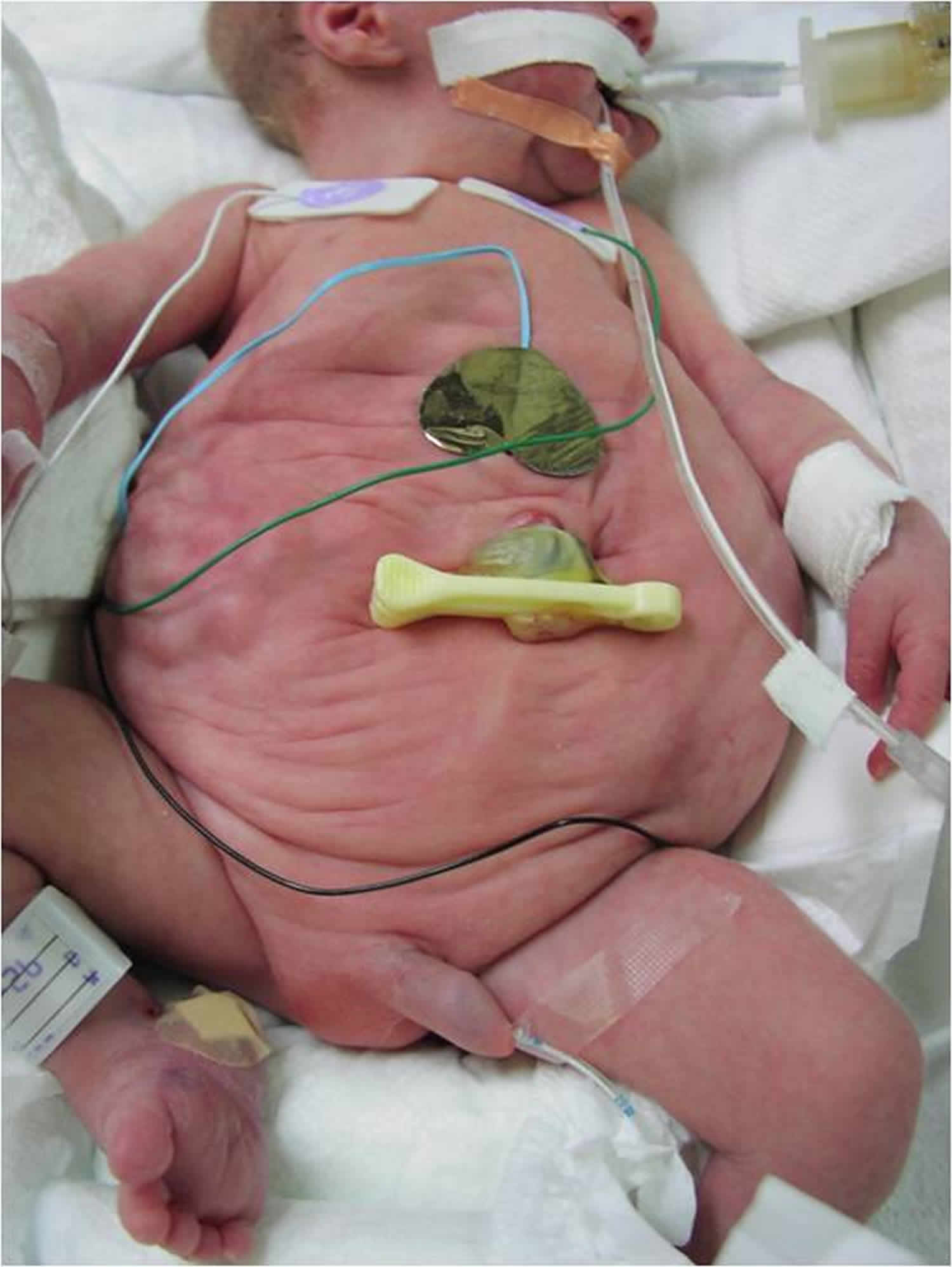
Prune belly syndrome is also known as Eagle-Barrett syndrome or Triad syndrome, is a rare birth defects characterized by partial or complete absence of the stomach (abdominal) muscles causing the skin on the abdominal area to wrinkle and appear “prune-like”, failure of both testes to descend into the scrotum (bilateral cryptorchidism), and/or urinary tract malformations 1. The urinary malformations may include abnormal widening (dilation) of the tubes that bring urine to the bladder (ureters), accumulation of urine in the ureters (hydroureter) and the kidneys (hydronephrosis), and/or backflow of urine from the bladder into the ureters (vesicoureteral reflux). Complications associated with Prune-Belly syndrome may include underdevelopment of the lungs (pulmonary hypoplasia) and/or chronic renal failure. The exact cause of Prune-Belly syndrome is not known. Prune belly syndrome is more common in males but a few female cases have been described in the medical literature.
Prune belly syndrome affects 1 per 30,000-40,000 live births 2. Approximately 3-4% of all prune belly syndrome cases occur in females. Twinning is associated with prune belly syndrome; 4% of all cases are products of twin pregnancies.
Prune belly syndrome involves these three main problems:
- Poor development of the abdominal muscles, causing the skin of the belly area to wrinkle like a prune
- Undescended testicles
- Urinary tract problems
Children with prune belly syndrome can present with myriad renal, ureteral, and urethral abnormalities. Obstruction and/or upper urinary tract dilatation is not unusual in these children. The site of obstruction can vary from as high as the pelviureteral junction to as low as the prostatic membranous urethra.
A lack of abdominal muscles leads to a poor cough mechanism, which, in turn, leads to increased pulmonary secretions. Weak abdominal muscles lead to constipation because of an inability to perform the Valsalva maneuver, which helps push the stool out of the rectum during defecation.
In a review of 46 patients (44 boys and 2 girls) with prune belly syndrome seen at a pediatric urology clinic, the following were the most common clinical manifestations 3:
- Hydroureteronephrosis – 97.8%
- Vesicoureteral reflux – 78.3%
- Significant pulmonary insufficiency – 10.9%
- Congenital malformations – 39.1%
- Chronic kidney disease – 39.1%; 17.4% underwent renal transplantation
The mortality rate associated with prune belly syndrome is 20%.
Treatment options depend on the individual’s age, health, medical history, extent of disease, tolerance for certain treatments or procedures, the expected course of the disease, and the parent’s and/or guardian’s opinions and preferences. Timing of therapy may vary from patient to patient.
Early surgery is recommended to fix weak abdominal muscles, urinary tract problems, and undescended testicles.
The baby may be given antibiotics to treat or help prevent urinary tract infections.
Prune belly syndrome causes
The exact causes of prune belly syndrome are unknown. There are several theories. Prune belly syndrome affects mostly boys. Prune belly syndrome is associated with trisomy 18 (Edwards syndrome) and trisomy 21 (Down syndrome) 2. Patients with prune belly syndrome also have an increased incidence of tetralogy of Fallot and ventriculoseptal defects 2. Additionally, a mutation in the CHRM3 gene has been reported in one family with a history of prune belly syndrome. Otherwise, an underlying genetic cause has not been identified 4.
Prune belly syndrome may be caused by an abnormality in the bladder during fetal development. Accumulation of urine can distend the bladder, the ureters, and the kidney. As the bladder enlarges, it causes wasting (atrophy) of the abdominal muscles. Retention of the testes in the abdomen (cryptorchidism) may be attributed to obstruction by an unusually large bladder or to obliteration of the groin (inguinal) canals. By the time of birth, the obstruction at the bladder outlet or the urethral obstruction may have been resolved, so that no mechanical obstacle can be identified after birth.
Other researchers consider the urinary abnormalities as secondary to the incomplete development of abdominal muscles. Incomplete emptying of the bladder leading to urinary retention and infection can occur as a result. Constipation and symptoms of indigestion are additional possible complications. Since the abdominal muscles are important for respiration, deformity of the chest could be explained by their absence.
A third possibility is that the muscle deficiency and the urinary abnormalities have a common cause that has not yet been discovered. A nervous system defect that could be responsible for early malfunction of abdominal muscles may be the cause. Association with a congenital open spinal canal (spina bifida) has been identified in some children, and the presence of clubfeet is also fairly commonly associated with Prune Belly syndrome.
While in the womb, the developing baby’s abdomen swells with fluid. Often, the cause is a problem in the urinary tract. The fluid disappears after birth, leading to a wrinkled abdomen that looks like a prune. This appearance is more noticeable due to the lack of abdominal muscles.
Prune belly syndrome symptoms
The severity of symptoms in individuals with prune belly syndrome can vary greatly.
Weak abdominal muscles can cause:
- Constipation
- Delay in sitting and walking
- Difficulties coughing
Urinary tract problems can cause difficulty urinating.
Common symptoms include 5:
- Poorly developed and/or absent abdominal muscles
- Undescended testicles in males (cryptorchidism)
- Urinary tract problems such enlarged or blocked ureters (tube that carries urine from the bladder outside the body)
- Enlarged bladder
- Enlarged kidney (hydronephrosis)
Other symptoms might include 5:
- Cardiac defects
- Spine malformations
- Club foot
- Gastrointestinal anomalies
Prune Belly syndrome is characterized by partial absence of some or most abdominal muscles giving rise to a wrinkled or prune-like appearance. Often, the attachments of the muscles to the bones are present, but the muscles diminish in size and thickness over the bladder. The abdomen appears large and lax, the abdominal wall is thin and the intestinal loops can be seen through the thin abdominal wall. Skin folds may radiate from the navel or occur as transverse folds across the abdomen. A midline crease from the navel to pubic area may be present in some cases. The navel may appear as a vertical slit, or as a linear central scar, but it can also appear normal. Sometimes the navel is connected with the bladder through a canal (urachus) or a cyst. The chest is often deformed. Flaring of the rib margins or a horizontal depression under the chest (Harrison groove) can appear in many children born with Prune Belly Syndrome. Narrowing of the chest in the transverse direction (pigeon breast) may also occur.
Enlargement of the bladder is present in almost all cases. Obstruction of the neck of the bladder is the primary problem, resulting in bladder distention and urine retention. The connection between the kidney and bladder (ureter) may be abnormal; the opening between ureter and bladder may be narrowed or closed. Obstruction may also occur at the junction of the ureter and kidney. Usually, the ureters are greatly widened. Occasionally this enlargement occurs only on one side or decreases as the ureter nears the bladder. Distention of the kidney with urine (hydronephrosis), on one or both sides, may also occur. In some cases, hydronephrosis occurs on one side while the kidney is underdeveloped on the other side. Kidney cysts may also be present. The canal that carries urine from the bladder to the outside of the body (urethra) usually is unobstructed. In males, absence of an opening (atresia) in the urethra, folds acting as valves below the entrance of the semen and prostate ducts (verumontanum), compression by a pouch and overdevelopment of the prostatic urethra also have been noted in some cases.
Musculoskeletal abnormalities, especially club foot, are present in about 20% of cases, while cardiovascular abnormalities are seen in about 10% of cases.
Blood and pus in the urine (hematuria and pyuria) often signal infection. Undescended testes (cryptorchidism) and testes that may be attached to a ureter, often occur in males with Prune Belly syndrome. Abnormal fixation of the gastrointestinal tract and failure to rotate during fetal development (malrotation) have also been described in the medical literature.
Prune belly syndrome possible complications
Complications depend on the related problems. The most common are:
- Constipation
- Bone deformities (clubfoot, dislocated hip, missing limb, finger, or toe, funnel chest)
- Disease of the urinary tract (may need dialysis and a kidney transplant)
Undescended testicles can lead to infertility or cancer.
Prune belly syndrome diagnosis
The diagnosis is usually obvious from birth, but care and time are required to determine the location and number of abnormalities. A full understanding of the complications will involve imaging tests such as ultrasound, X-ray, and, in order to determine the extent of involvement of the genitourinary tract, intravenous pyelogram (IVP). An IVP makes use of a dye to map the degree of involvement of the kidneys and their ducts.
The following tests may be performed on the baby after birth to diagnose the condition:
- Blood tests
- Intravenous pyelogram (IVP)
- Ultrasound
- Voiding cystourethrogram (VCUG)
- X-ray
- CT scan
A woman who is pregnant with a baby who has prune belly syndrome may not have enough amniotic fluid (the fluid that surrounds the fetus). This can cause the infant to have lung problems from being compressed in the womb.
An ultrasound done during pregnancy may show that the baby has a swollen bladder or enlarged kidney.
In some cases, a pregnancy ultrasound may also help determine if the baby has:
- Heart problems
- Abnormal bones or muscles
- Stomach and intestinal problems
- Underdeveloped lungs
Prune belly syndrome prognosis
Prune belly syndrome is a serious and often life-threatening problem. The prognosis associated with prune belly syndrome varies depending several factors including the severity of the underlying tract anomaly, how well the kidneys are developed, and the likelihood of renal failure. The condition can become life threatening in severely affected children; however, mild cases might be limited to undescended testicles and a small amount of abdominal wall laxity. Studies have found that 30% of individuals with prune belly syndrome require kidney transplantation in their lifetime 6.
Despite these concerns, many individuals with prune belly syndrome report having good physical and mental health as well as a good overall quality of life 6.
Prune belly syndrome life expectancy
Many infants with prune belly syndrome are either stillborn or die within the first few weeks of life. The cause of death is from severe lung or kidney problems, or from a combination of birth problems.
Some newborns survive and can develop normally. Others continue to have many medical and developmental problems. The prognosis associated with prune belly syndrome varies depending several factors including the severity of the underlying tract anomaly, how well the kidneys are developed, and the likelihood of renal failure. The condition can become life threatening in severely affected children; however, mild cases might be limited to undescended testicles and a small amount of abdominal wall laxity. Studies have found that 30% of individuals with prune belly syndrome require kidney transplantation in their lifetime 6.
Prune belly syndrome treatment
Treatment will depend upon the severity of the symptoms. Some children will require rather modest surgical procedures such as the creation of a small opening in the bladder through the abdomen (vesicostomy) that will facilitate voiding of urine, or a procedure to help the testicles descend into the scrotum (orchiopexy). More extensive surgical procedures such a. bladder reconstruction (cystoplasty), surgical widening of the urethra, and augmentation of the muscles that contract the bladder (detrusor augmentation) using a paired graft of a hip muscle (rectus femoris) have been successfully undertaken on children with prune belly syndrome. In rare cases, kidney transplantation may be necessary.
Surgical Therapy
Surgical treatment for prune belly syndrome includes repair of the abdominal wall and urinary tract abnormalities and correction of cryptorchidism. These procedures can be performed in a single comprehensive approach or in multiple steps. A review by Lopes et al concluded that comprehensive surgical treatment is feasible and has good long-term results. However, many patients require reoperations because of complications or progression of disease 7.
Undescended testis
Management of undescended testis in a patient with prune belly syndrome should be left to the experienced pediatric urologist. In some instances, patients with undescended testis do not require any urologic work and the testis can be brought down using laparoscopic techniques. The laparoscopic technique reduces the morbidity associated with intra-abdominal surgery. The author has performed reconstructive surgery in several patients using this technique, with superior results. In other cases, the testis can be brought down during open surgery for the reconstruction of the urinary tract.
Abdominal wall reconstruction
Abdominal wall reconstruction is performed in most patients with prune belly syndrome to improve respiratory function and to improve cosmesis. Several innovations in abdominal wall reconstruction have been developed. The Monfort 8:639-40.)) and Ehrlich 9 variations of the Randolph operation have improved results, with decreased morbidity and the ability to preserve the umbilicus. Furness et al have described a new technique that allows for improved results over other techniques, without opening the abdominal cavity 10. Most recently, Franco modified this technique further to obtain even better results by using laparoscopic guidance to ensure that wall tension and cosmetic results persist postoperatively 11. These innovations have significantly reduced the morbidity of abdominal wall reconstruction 12.
Placement of a percutaneous nephrostomy
Obstruction at the ureteropelvic junction (UPJ) has been observed in patients with prune belly syndrome. In some cases, diagnosing this can be difficult; however, the diagnosis can be confirmed with the placement of a percutaneous nephrostomy. This procedure can be performed under ultrasound guidance with relative ease in a dilated system and provides the opportunity to perform a renal biopsy, which could help with the later management of the urinary tract.
Placement of a percutaneous nephrostomy provides the surgeon with several options. The surgeon can confirm the diagnosis of ureteropelvic junction obstruction. The urinary tract can be decompressed in an ill child or in a child who is too small to safely undergo reconstructive surgery at the time. Finally, the technique also allows the surgeon to evaluate the renal function of the obstructed unit.
Standard pyeloplasty
If conditions are right and the child is stable, a standard pyeloplasty can be performed as another means of treating the obstructed ureteropelvic junction. The surgeon should be meticulous in the dissection of the upper ureter. The proximal ureteral blood supply should be preserved as much as possible in case ureteral tapering or reimplantation becomes necessary later.
Recent studies indicate that the more distal lower ureter is abnormal in several ways, while the more proximal portion of the ureter is more anatomically normal. Histologically, the ureter has a smooth muscle deficiency with fibrous degeneration and a poor blood supply. In addition, a decrease in nerve plexuses is reported, with irregularity in degeneration of nonmyelinated Schwann fibers.
In patients who may have supravesical obstruction and functional vesical obstruction, cutaneous pyelostomy has been recommended as the preferred means of diverting the ureter and the pelvis for several reasons. The proximal ureter is believed to less compromised with this procedure than with a high-loop ureterostomy, as had been performed previously. In addition, by performing a pyelostomy, surgeons avoid the undesirable attachment of the ureters to the abdominal wall, thereby preventing the common problem of a prolapsed ureterostomy. When ureterostomy is performed, the distal ureter is typically used because this ureter would most likely be discarded when the reconstruction is performed.
Cutaneous pyelostomy still carries the risk of resultant scarring from excessive dissection, which may be required to take down the pyelostomy at a later stage. At the time of reconstruction, ureteral tapering or reimplantation that is necessary creates a difficult situation. Recently, these problems have been overcome with the increasing use of percutaneous nephrostomy drainage and with the use of vesicostomy as a means of draining the upper tracts. Some believe that vesicostomy can decompress the upper tracts just as well as high diversion. Subsequently, since the introduction of these 2 procedures, the use of high diversion has declined significantly over the last few years, and reconstruction in these patients has become easier.
Infravesical obstruction or obstruction at the prostatic urethra
Patients with documented obstruction can be treated with several means.
Blocksom vesicostomy performed in the early newborn period is the simplest and best treatment in patients with documented obstruction. Bringing the dome of the bladder out to the skin is essential when creating the vesicostomy; this approach prevents the resultant herniation of the bladder in an improperly created vesicostomy.
Herniation of the bladder is quite prevalent in patients with prune belly syndrome because the bladder is quite large and redundant. A patent urachus can be found frequently in patients with urethral obstruction. The patent urachus is the means by which patients are able to survive; early deaths usually are observed in patients with urethral obstruction without a patent urachus. In these patients, once a vesicostomy is performed, the surgeon should try to identify the urachus and ligate it at that time.
Some patients with prune belly syndrome have posterior urethral valves, and the valves are best managed with transurethral resection. Some advocate the use of sphincterotomy, transurethral resection of the bladder neck, or internal urethrotomy to manage the functionally obstructed system. Snyder and associates believe that judicious use of urethrotomy can lower urethral resistance and improve voiding dynamics without causing incontinence 13. Opinions are mixed regarding the use of these modalities because few actual results have been seen. Passerini-Glazel et al used soft catheter dilatation to dilate the functionally obstructed urethra in patients with prune belly syndrome 14. They left soft indwelling catheters for extended periods while they gradually increased the size of the catheter until they achieved the desired caliber. Radiologic and functional results are needed.
Ureteral reimplantation in patients with megaureters, reflux, and a ureteral transplant requires an aggressive approach; as much of the abnormal distal ureter as possible should be removed and the blood supply to the proximal ureter should be preserved. Some authors advocate shortening, tapering, and reimplanting the better of the 2 ureters with a long tunnel and psoas hitch and draining the contralateral side with a transureteroureterostomy . These authors reason that the bladder may be thick and fibrous in patients with prune belly syndrome, thereby complicating placement 2 good long reimplants. Ureters are preferentially tapered over an 8F or 10F red rubber catheter and excess tissue is excised. Most recently, Starr has reimplanted the ureter using the folding cross-reimplantation. He has not reported obstruction or reflux. This technique may prevent interruption of the critical blood supply to the distal ureter; this is not possible with traditional excisions.
In patients with megacystitis, reduction cystoplasty has met with mixed reviews. Reduction cystoplasty is generally recognized to be unnecessary as a primary procedure but may be useful and is frequently performed at the time of ureteral reimplantation and tapering.
- Prune Belly Syndrome. https://rarediseases.org/rare-diseases/prune-belly-syndrome[↩]
- Prune belly syndrome. https://emedicine.medscape.com/article/447619-overview[↩][↩][↩]
- Seidel NE, Arlen AM, Smith EA, Kirsch AJ. Clinical manifestations and management of prune-belly syndrome in a large contemporary pediatric population. Urology. 2015 Jan. 85 (1):211-5.[↩]
- Prune belly syndrome. https://rarediseases.info.nih.gov/diseases/7479/prune-belly-syndrome/cases/23732[↩]
- Natan E. Seidel, Angela M. Arlen, Edwin A. Smith, Andrew J. Kirsch. Clinical Manifestations and Management of Prune-belly Syndrome in a Large Contemporary Pediatric Population. Pediatric Urology. Jan 2015; 85(1):211-215. http://www.ncbi.nlm.nih.gov/pubmed/25444629[↩][↩]
- Prune Belly Syndrome. https://emedicine.medscape.com/article/447619-overview[↩][↩][↩]
- Lopes RI, Tavares A, Srougi M, Dénes FT. 27 years of experience with the comprehensive surgical treatment of prune belly syndrome. J Pediatr Urol. 2015 Oct. 11 (5):276.e1-7.[↩]
- Monfort G, Guys JM, Bocciardi A, Coquet M, Chevallier D. A novel technique for reconstruction of the abdominal wall in the prune belly syndrome. J Urol. 1991 Aug. 146(2 ( Pt 2[↩]
- Ehrlich RM, Lesavoy MA. Umbilicus preservation with total abdominal wall reconstruction in prune-belly syndrome. Urology. 1993 Mar. 41(3):231-2.[↩]
- Furness PD 3rd, Cheng EY, Franco I, Firlit CF. The prune-belly syndrome: a new and simplified technique of abdominal wall reconstruction. J Urol. 1998 Sep. 160(3 Pt 2):1195-7; discussion 1216.[↩]
- Franco I. Laparoscopic assisted modification of the firlit abdominal wall plication. J Urol. 2005 Jul. 174(1):280-3.[↩]
- Lesavoy MA, Chang EI, Suliman A, Taylor J, Kim SE, Ehrlich RM. Long-term follow-up of total abdominal wall reconstruction for prune belly syndrome. Plast Reconstr Surg. 2012 Jan. 129(1):104e-9e.[↩]
- Snyder HM, Harrison NW, Whitfield HN, Williams INNES. Urodynamics in the prune belly syndrome. Br J Urol. 1976. 48(7):663-70.[↩]
- Passerini-Glazel G, Araguna F, Chiozza L, Artibani W, Rabinowitz R, Firlit CF. The P.A.D.U.A. (progressive augmentation by dilating the urethra anterior) procedure for the treatment of severe urethral hypoplasia. J Urol. 1988 Nov. 140(5 Pt 2):1247-9.[↩]


