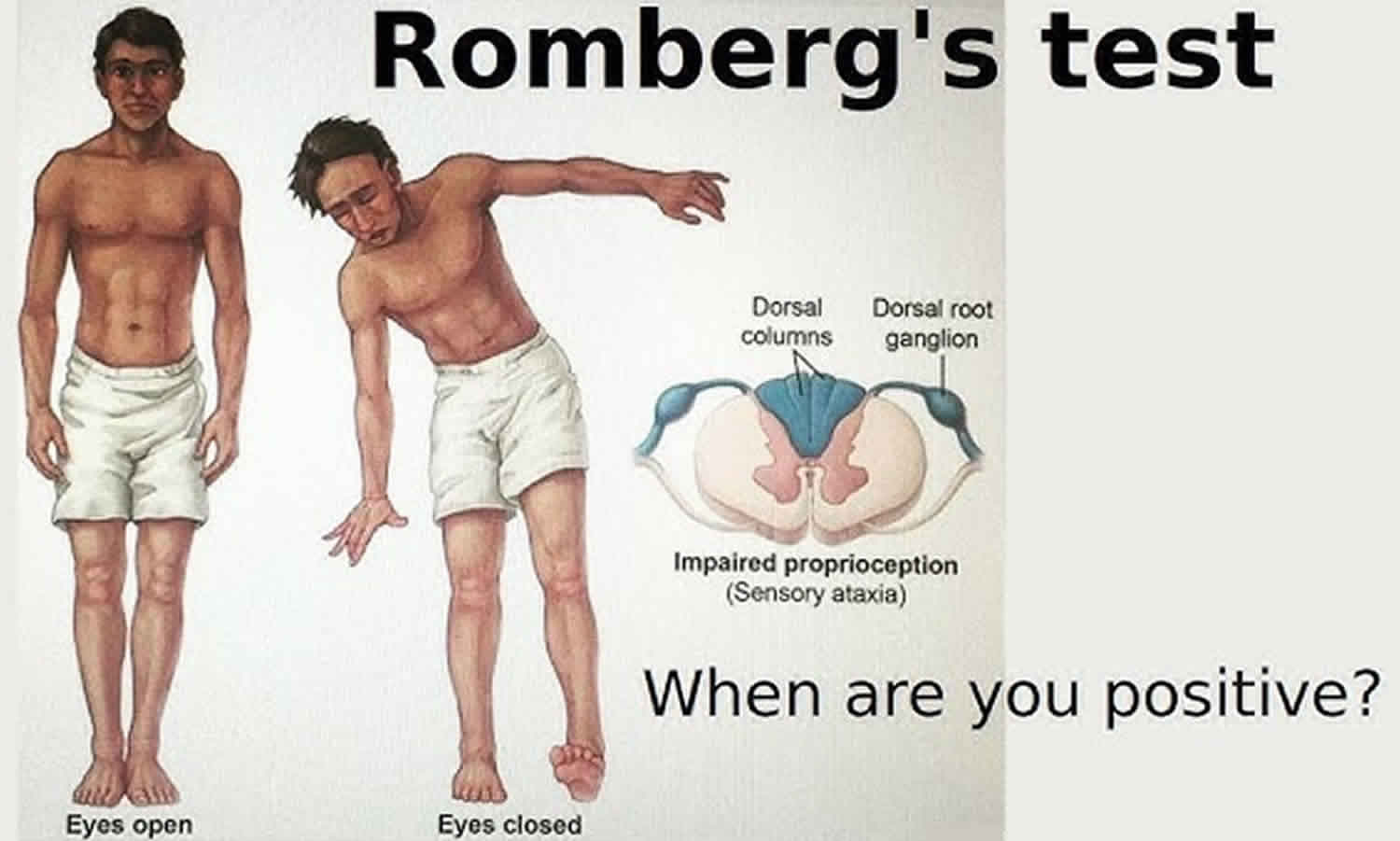
Romberg test
Romberg test also called Romberg’s sign, is a neurological exam that measures a persons sense of balance where your doctor will ask you to close your eyes while standing with your feet together and your arms to your side, this removes the visual and vestibular components that contribute to maintaining balance 1, 2, 3. Normal stance in a person is governed by the integrity of three sensory inputs: vision, proprioception, and vestibular apparatus 4, 3. The Romberg test removes the visual and vestibular components that contribute to maintaining balance and can therefore identify a proprioception-related neurologic disease. The Romberg test should be performed on all patients presenting with imbalance, dizziness, and unprovoked falls. Romberg’s test detects the integrity of the posterior dorsal columns and proprioception, the body’s awareness of its own movement and position in space.
Romberg’s test is a simple bedside test to determine the integrity of the dorsal column pathway of your brain and spinal cord (the neural pathways that carry proprioception sense by which sensory information from the peripheral nerves is transmitted to the cerebral cortex), which controls proprioception 2. Proprioception is the sense that lets you perceive the location, movement, and action of parts of your body 5. Proprioception encompasses a complex of sensations, including perception of joint position and movement, muscle force, and effort 5. These sensations arise from signals of sensory receptors in your muscle, skin, and joints, and from central signals related to motor output. Proprioception enables you to judge limb movements and positions, force, heaviness, stiffness, and viscosity. It combines with other senses to locate external objects relative to the body and contributes to body image.
Romberg sign is positive in a patient who can stand with his feet placed together and eyes open but paradoxically sways or falls while closing his eyes, thereby eliminating his visual cues 6, 1.
The sensitivity of Romberg test can be increased by:
- “Sharpened Romberg test”- narrowing the patient’s base of support with feet in a heel-to-toe tandem position or
- Conducting the Romberg test in foam rubber to nullify the proprioceptive inputs from the foot 7. Standing with their eyes closed on a compliant instead of a firm surface is a test of the vestibular system rather than that of proprioception 4.
Romberg sign is positive in 7, 2:
- Loss of proprioception among patients with myelopathies and sensory neuropathies
- Uncompensated unilateral or bilateral vestibular dysfunction, and
- Pathology involving the anterior vermis and the paravermis of the anterior cerebellar lobe.
However, a positive Romberg test may also result from inherited, metabolic, toxic, immunologic, or other disorders 3.
The Romberg test is an appropriate tool to diagnose sensory ataxia (a gait disturbance caused by abnormal proprioception involving information about the location of the joints). Examples of proprioception-related neurological disease include: subacute combined degeneration of the spinal cord (vitamin B12 deficiency); posterior cord syndrome (posterior spinal artery infarction); hemisection of spinal cord (Brown Sequard syndrome) and tertiary syphilis patients who exhibited neurologic signs of late-stage disease referred to as neurosyphilis or tabes dorsalis 7, 8, 9. It is important to note that both the dorsal column and cerebellar damage may result in ataxia 9.
Romberg’s sign or Romberg’s test was first described in the 19th century as a useful indicator of proprioceptive loss due to neurosyphilis (tabes dorsalis) 7, 8. In cases of neurosyphilis, or tabes dorsalis, there is demyelination of the axonal fibers of the posterior column or dorsal pathway of the spinal cord. This demyelination leads to severe sensory deficits of the pathway, including position sense or proprioception 9
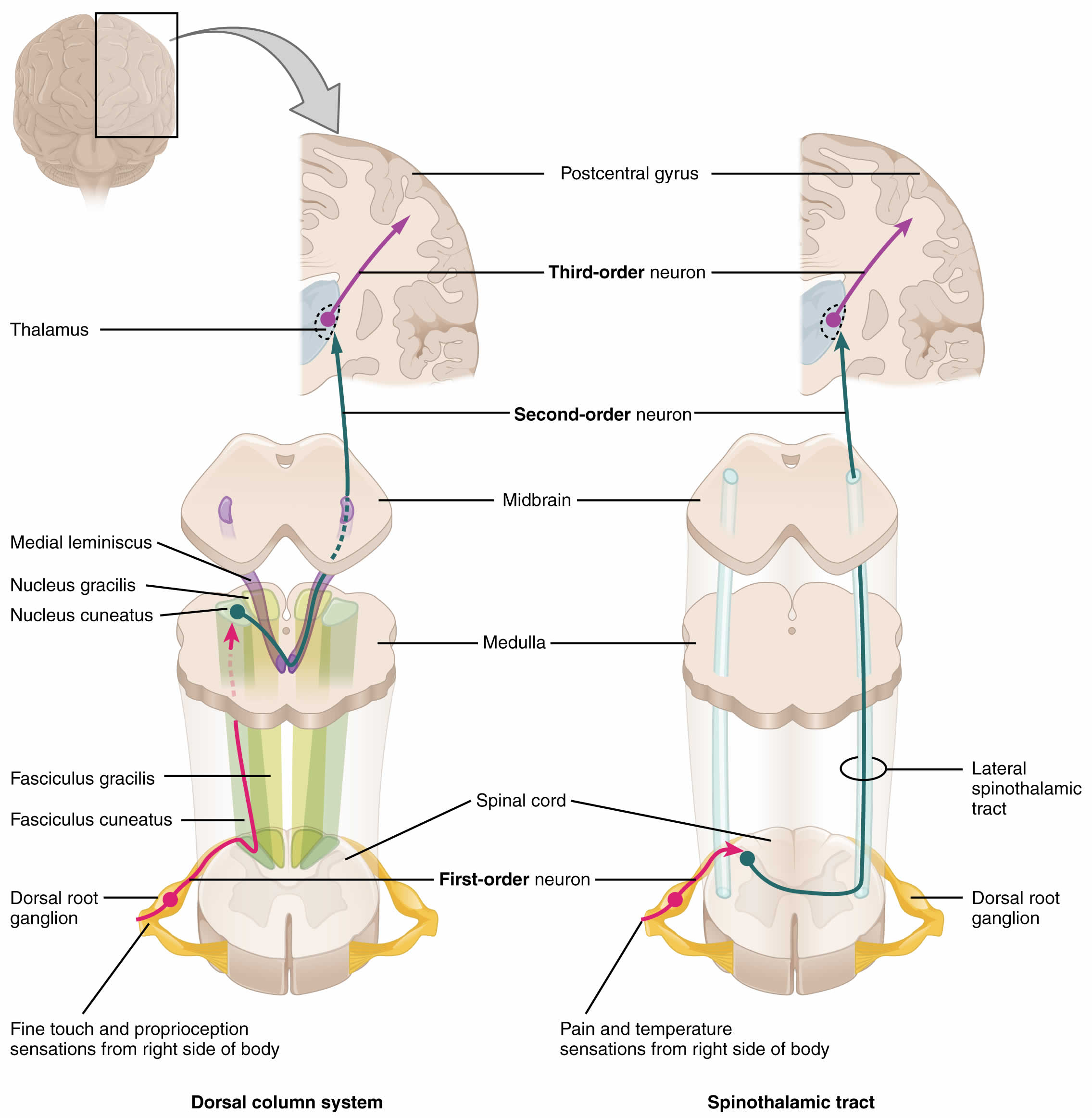
The dorsal column also called dorsal column medial lemniscus (DCML) or posterior column-medial lemniscus pathway is a three-order neuronal pathway that functions as a method of signal transmission throughout the spinal cord to the brainstem (Figure 1). The dorsal column medial lemniscus (DCML) pathway specifically controls conscious appreciation of vibration, fine touch, 2-point discrimination, and proprioception 9, 10, 11, 12, 13, 14.
Anatomically, the cerebellum is also involved in truncal and extremity balance and coordination. The cerebellum contains two lateral hemispheres lesions and one medial vermis. The cerebellum is associated with the movement of the body and is part of the human brain, which allows the body to make voluntary and coordinated movements and balance 15. It is important to understand that a negative Romberg test does not confirm cerebellar dysfunction, but the clinician may rule in the cause of ataxia due to cerebellar damage. In cerebellar disease, the patient is often unsteady with the eyes open as well. The patient significantly tends to fall to the side of the affected labyrinth during the test.
Figure 1. Dorsal column medial lemniscus tract
Modified Romberg test
Modified versions of the Romberg’s test provide a wide range of clinical applications for balance assessment. These variations of the Romberg test mentioned below are utilized depending on the discretion of the physician and patient presentation. Not one test is deemed superior to the others, but slight variations in each test may provide a more specific method of detecting a postural imbalance that caters to a particular patient profile.
Some modified versions of the Romberg’s test include:
- Sharpened Romberg Test used in assessing ataxia in patients recovering the severity of decompression sickness, such as in scuba divers 16. The Sharpened Romberg Test procedure differs from the traditional Romberg Test in the positioning of the feet. Instead of standing with feet shoulder-width apart, the sharpened Romberg test dictates that the feet of the patient align in a strict tandem heel-to-toe position.
- Single-legged Stance Test is mainly used to assess postural stability and control in the elderly and those with Parkinson’s disease 17. Static balance test serves as a balanced assessment of Parkinson’s disease. The process entails assessing how long the patient can maintain a single-leg stance with eyes open. The Single-legged Stance Test ends after 60 seconds, and each leg is tested three times 17.
- Sitting-rising Test is an easily administered test focused on the assessment of the elderly population as well as stroke victims as a tool to predict mortality risk. The examiner instructs the patient to try to sit, rise, and then stand to a position using the minimum amount of support needed. The Sitting-rising Test is on a point scale, with measures for strength, balance, and integration aspects, with 10 points being the maximum 18.
- Get-Up and Go Test is an assessment of frailty and predictor of a geriatric patient’s ability to go outside alone safely 19.
- Berge JE, Goplen FK, Aarstad HJ, Storhaug TA, Nordahl SHG. The Romberg sign, unilateral vestibulopathy, cerebrovascular risk factors, and long-term mortality in dizzy patients. Front Neurol. 2022 Aug 5;13:945764. doi: 10.3389/fneur.2022.945764[↩][↩]
- Forbes J, Munakomi S, Cronovich H. Romberg Test. [Updated 2023 Aug 13]. In: StatPearls [Internet]. Treasure Island (FL): StatPearls Publishing; 2024 Jan-. Available from: https://www.ncbi.nlm.nih.gov/books/NBK563187[↩][↩][↩]
- Khasnis A, Gokula RM. Romberg’s test. J Postgrad Med. 2003 Apr-Jun;49(2):169-72.[↩][↩][↩]
- Halmágyi GM, Curthoys IS. Vestibular contributions to the Romberg test: Testing semicircular canal and otolith function. Eur J Neurol. 2021 Sep;28(9):3211-3219. doi: 10.1111/ene.14942[↩][↩]
- Proprioception. Encyclopedia of Neuroscience 2009, Pages 1143-1149. https://doi.org/10.1016/B978-008045046-9.01907-0[↩][↩]
- Lanska DJ, Goetz CG. Romberg’s sign: development, adoption, and adaptation in the 19th century. Neurology. 2000 Oct 24;55(8):1201-6. https://doi.org/10.1212/WNL.55.8.1201[↩]
- Lanska DJ. The Romberg sign and early instruments for measuring postural sway. Semin Neurol. 2002 Dec;22(4):409-18. doi: 10.1055/s-2002-36763[↩][↩][↩][↩]
- Wilkins RH, Brody IA. Romberg’s sign. Arch Neurol. 1968 Jul;19(1):123-6. doi: 10.1001/archneur.1968.00480010141013[↩][↩]
- Al-Chalabi M, Reddy V, Alsalman I. Neuroanatomy, Posterior Column (Dorsal Column) [Updated 2023 Apr 8]. In: StatPearls [Internet]. Treasure Island (FL): StatPearls Publishing; 2024 Jan-. Available from: https://www.ncbi.nlm.nih.gov/books/NBK507888[↩][↩][↩][↩]
- MacDonald DB, Dong C, Quatrale R, Sala F, Skinner S, Soto F, Szelényi A. Recommendations of the International Society of Intraoperative Neurophysiology for intraoperative somatosensory evoked potentials. Clin Neurophysiol. 2019 Jan;130(1):161-179. doi: 10.1016/j.clinph.2018.10.008[↩]
- Gonschorek O, Hauck S, Weiß T, Bühren V. Frakturen der Brust- und Lendenwirbelsäule [Fractures of the thoracic and lumbar spine]. Chirurg. 2015 Sep;86(9):901-14; quiz 915-6. German. doi: 10.1007/s00104-015-0045-5[↩]
- Freund HJ. Somatosensory and motor disturbances in patients with parietal lobe lesions. Adv Neurol. 2003;93:179-93.[↩]
- Willis WD Jr, Westlund KN. The role of the dorsal column pathway in visceral nociception. Curr Pain Headache Rep. 2001 Feb;5(1):20-6. doi: 10.1007/s11916-001-0006-1[↩]
- Westlund KN. Visceral nociception. Curr Rev Pain. 2000;4(6):478-87. doi: 10.1007/s11916-000-0072-9[↩]
- Cohen HS. A review on screening tests for vestibular disorders. J Neurophysiol. 2019 Jul 1;122(1):81-92. doi: 10.1152/jn.00819.2018[↩]
- Johnson BG, Wright AD, Beazley MF, Harvey TC, Hillenbrand P, Imray CH; Birmingham Medical Research Expeditionary Society. The sharpened Romberg test for assessing ataxia in mild acute mountain sickness. Wilderness Environ Med. 2005 Summer;16(2):62-6. doi: 10.1580/pr02-04.1[↩]
- Chomiak T, Pereira FV, Hu B. The single-leg-stance test in Parkinson’s disease. J Clin Med Res. 2015 Mar;7(3):182-5. doi: 10.14740/jocmr1878w[↩][↩]
- Ng SS, Fong SS, Chan WL, Hung BK, Chung RK, Chim TH, Kwong PW, Liu TW, Tse MM, Chung RC. The sitting and rising test for assessing people with chronic stroke. J Phys Ther Sci. 2016 Jun;28(6):1701-8. doi: 10.1589/jpts.28.1701[↩]
- Podsiadlo D, Richardson S. The timed “Up & Go”: a test of basic functional mobility for frail elderly persons. J Am Geriatr Soc. 1991 Feb;39(2):142-8. doi: 10.1111/j.1532-5415.1991.tb01616.x[↩]


