
Contents
What is tularemia
Tularemia is a rare bacterial disease of animals and humans that is caused by the bacterium Francisella tularensis, usually acquired from handling infected animals, bites of infected ticks or deer flies or from contaminated food or water.
Various tick species are responsible for 9% to 57% of transmission. Other animals that can transmit Francisella tularensis include rabbits, squirrels, opossums, cats, muskrats, and mosquitoes.
People can get tularemia many different ways:
- Being bitten by an infected tick, deerfly or other insect
- Handling infected animal carcasses
- Eating or drinking contaminated food or water
- Breathing in the bacteria, Francisella tularensis
- Laboratory exposure
- In addition, humans could be exposed as a result of bioterrorism. Francisella tularensis is very infectious. A small number (10-50 or so organisms) can cause disease. If F. tularensis were used as a weapon, the bacteria would likely be made airborne for exposure by inhalation. People who inhale an infectious aerosol would generally experience severe respiratory illness, including life-threatening pneumonia and systemic infection, if they are not treated. The bacteria that cause tularemia occur widely in nature and could be isolated and grown in quantity in a laboratory, although manufacturing an effective aerosol weapon would require considerable sophistication.
Francisella tularensis is mainly found in the Northern hemisphere and has been reported in North America, Russia, Europe, the Middle East, China, and Japan. Tularemia has recently received attention due to its potential as a biological weapon.
Tularemia is not known to be spread from person to person. People who have tularemia do not need to be isolated. People who have been exposed to the tularemia bacteria should be treated as soon as possible. The disease can be fatal if it is not treated with the right antibiotics.
Rabbits, hares, and rodents are especially susceptible and often die in large numbers during outbreaks.
Two predominant strains of Francisella tularensis have been isolated; type A and type B.
- Type A causes more severe disease, causing death in up to 5% to 7% of untreated patients.
- Type B generally causes a milder illness, which can occasionally be symptom-free.
Symptoms vary depending on the route of infection. Although tularemia can be life-threatening, most infections can be treated successfully with antibiotics.
Steps to prevent tularemia include:
- Use of insect repellent
- Wearing gloves when handling sick or dead animals
- Avoiding mowing over dead animals
In the United States, naturally occurring infections have been reported from all states except Hawaii.
Tularemia was much more common in the early part of the 20th century than it is now. In 1950 there were 927 cases and in 2016 there were 230 cases 1.
- Tularemia is more common in males, possibly because of a greater likelihood of exposure through hunting and landscaping. Tularemia occurs in persons of all ages, but is most common in children.
- Tularemia is more common in the months of May through September. Bites from infected ticks or deer flies usually occur in the summer months, but illness due to animal handling and hunting can occur at any time of the year.
Tularemia transmission
The bacterium that causes tularemia is highly infectious and can enter the human body through the skin, eyes, mouth, or lungs. Symptoms of infection vary depending on the route of entry. Usual sources of infection are described below. Transmission of tularemia from person to person has not been reported.
Tick or deer fly bites
In the United States, ticks that transmit tularemia to humans include the dog tick (Dermacentor variabilis), the wood tick (Dermacentor andersoni), and the lone star tick (Amblyomma americanum). Deer flies (Chrysops spp.) have been shown to transmit tularemia in the western United States. Infections due to tick and deer fly bites usually take the form of ulceroglandular or glandular tularemia.
Figure 1. American dog tick (Dermacentor variabilis)

Figure 2. Lone star tick (Amblyomma americanum)
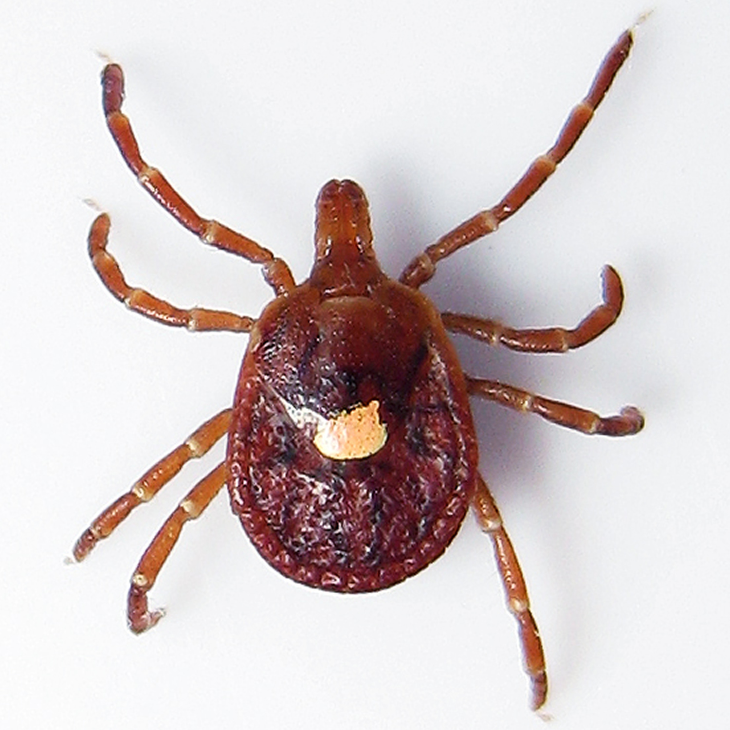 Figure 3. Deer fly (Chrysops spp.)
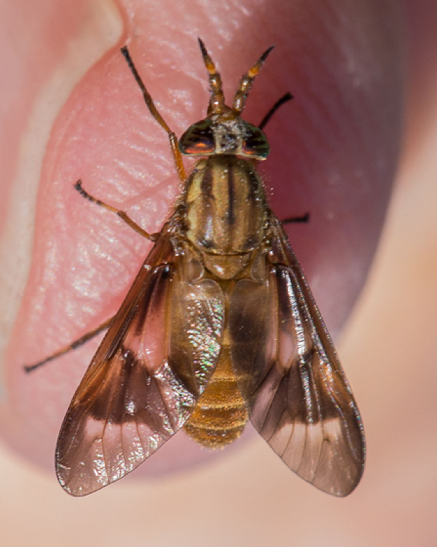
| Tick-borne transmission | Transmission due to other animals | |
|---|---|---|
| Season | Summer | Winter |
| Primary lesion | Usually a single primary lesion at the site of the bite. Lesion occurs on a part of the body commonly affected by tick bites. | Multiple primary lesions, often on the arms and hands. |
Handling infected animals
Francisella tularensis bacteria can be transmitted to humans via the skin when handling infected animal tissue. In particular, this can occur when hunting or skinning infected rabbits, muskrats, prairie dogs and other rodents. Many other animals have also been known to become ill with tularemia. Domestic cats are very susceptible to tularemia and have been known to transmit the bacteria to humans. Care should be taken when handling any sick or dead animal. Outbreaks of tularemia have occurred among hamsters purchased from pet stores. At least one child in the U.S. has developed tularemia after being bitten by a pet hamster. Infection due to handling animals can result in glandular, ulceroglandular and oculoglandular tularemia. Oropharyngeal tularemia can result from eating under-cooked meat of infected animals.
Other exposures
Humans can acquire tularemia by inhaling dust or aerosols contaminated with F. tularensis bacteria. This can occur during farming or landscaping activities, especially when machinery (e.g. tractors or mowers) runs over infected animals or carcasses. Although rare, this type of exposure can result in pneumonic tularemia, one of the most severe forms of the disease. Water can also become contaminated with the bacteria through contact with infected animals. Humans who drink contaminated water that has not been treated may contract oropharyngeal tularemia. This mode of transmission appears to be much more common in Europe than the U.S.
Heat kills F. tularensis, so cook meat to the right temperature — a minimum of 165° F (73.8° C) for ground meat and game meat — to make it safe to eat.
Risk factors for tularemia
Although anyone of any age can develop tularemia, engaging in certain occupations or activities or living in certain areas pose a greater risk.
Living in or visiting certain areas
In the United States, people living in or visiting areas of Arkansas, Missouri and Oklahoma may be at greater risk because of the concentration of ticks in those areas.
Having certain hobbies or occupations
The following can increase your risk of developing tularemia:
- Hunting and trapping. Because hunters are exposed to wild animal blood and may eat their flesh, they’re at risk of tularemia.
- Gardening or landscaping. Gardeners and landscapers may also be at risk of tularemia. It’s possible that gardeners inhale bacteria that are stirred up while working the soil or when using mowers and weed trimmers.
- Working in wildlife management or veterinary medicine. People who work with wildlife are at increased risk of tularemia.
Tularemia prevention
- Avoid insect bites by using permethrin-impregnated clothing and DEET insect repellants.
- Avoid sick or dead animals and thoroughly cook wild meat.
- If animal carcasses must be handled, use care and wear gloves when handling sick or dead animals. Wash hands thoroughly afterwards.
- Ensure drinking water is from a safe source.
- A vaccine for tularemia is under review by the Food and Drug Administration and is not currently available in the United States.
Tularemia signs and symptoms
The signs and symptoms of tularemia vary depending on how the bacteria enter the body.
Symptoms usually appear 3 to 5 days after exposure to the bacteria, but can take as long as 14 days.
Illness ranges from mild to life-threatening. All forms are accompanied by fever, which can be as high as 104 °F (40 °C).
- In adults – fever, headache, skin changes, malaise, and enlarged lymph nodes of the head and neck.
- In children – fever, sore throat, enlarged liver and spleen, fatigue and malaise.
Other symptoms of tularemia may include:
- Chills
- Headaches
- Diarrhea
- Muscle aches
- Joint pain
- Dry cough
- Progressive weakness
People can also develop pneumonia with chest pain, cough, and difficulty breathing.
Other symptoms of tularemia depend on how a person was exposed to the tularemia bacteria. These symptoms can include ulcers on the skin or mouth, swollen and painful lymph glands, swollen and painful eyes, and a sore throat.
Various clinical subtypes of tularemia have been described depending on the mode of transmission and organ systems involved.
Clinical subtypes of tularemia:
- Ulceroglandular. This is the most common form of tularemia and usually occurs following a tick or deer fly bite or after handing of an infected animal. A painful skin ulcer appears at the site where the bacteria entered the body. The ulcer is accompanied by swelling of regional lymph glands or nodular lymphangitis (swellings beneath the skin that track along the course of lymph channels, swellings may be painful and ulcerate). Lymph nodes may become fluctuant (soft, fluid-like) and rupture, usually in the armpit or groin.
- Glandular. Causes enlarged lymph nodes without an ulcer. Also generally acquired through the bite of an infected tick or deer fly or from handling sick or dead animals.
- Oculoglandular. This form occurs when the bacteria enter through the eye. This can occur when a person is butchering an infected animal and touches his or her eyes. Symptoms include irritation and inflammation of the eye, eye swelling and discharge, an ulcer on the inside of the eyelid, sensitivity to light and swelling of lymph glands in front of the ear.
- Oropharyngeal. This form results from eating or drinking contaminated food or water. Patients with orophyangeal tularemia may have sore throat, mouth ulcers, tonsillitis, diarrhea and swelling of lymph glands in the neck.
- Pneumonic. This is the most serious form of tularemia. Symptoms include cough, chest pain, and difficulty breathing. This form results from breathing dusts or aerosols containing the organism. It can also occur when other forms of tularemia (e.g. ulceroglandular) are left untreated and the bacteria spread through the bloodstream to the lungs.
- Typhoidal. This form is most lethal and is characterized by any combination of the general symptoms (without the localizing symptoms of other syndromes). This rare and serious form of the disease usually causes: high fever, extreme exhaustion, vomiting and diarrhea, enlarged spleen (splenomegaly), enlarged liver (hepatomegaly) and pneumonia
What are the skin manifestations of tularemia?
Tularemia can cause primary skin lesions (seen in ulceroglandular subtype) and secondary skin lesions (seen in all forms of tularaemia).
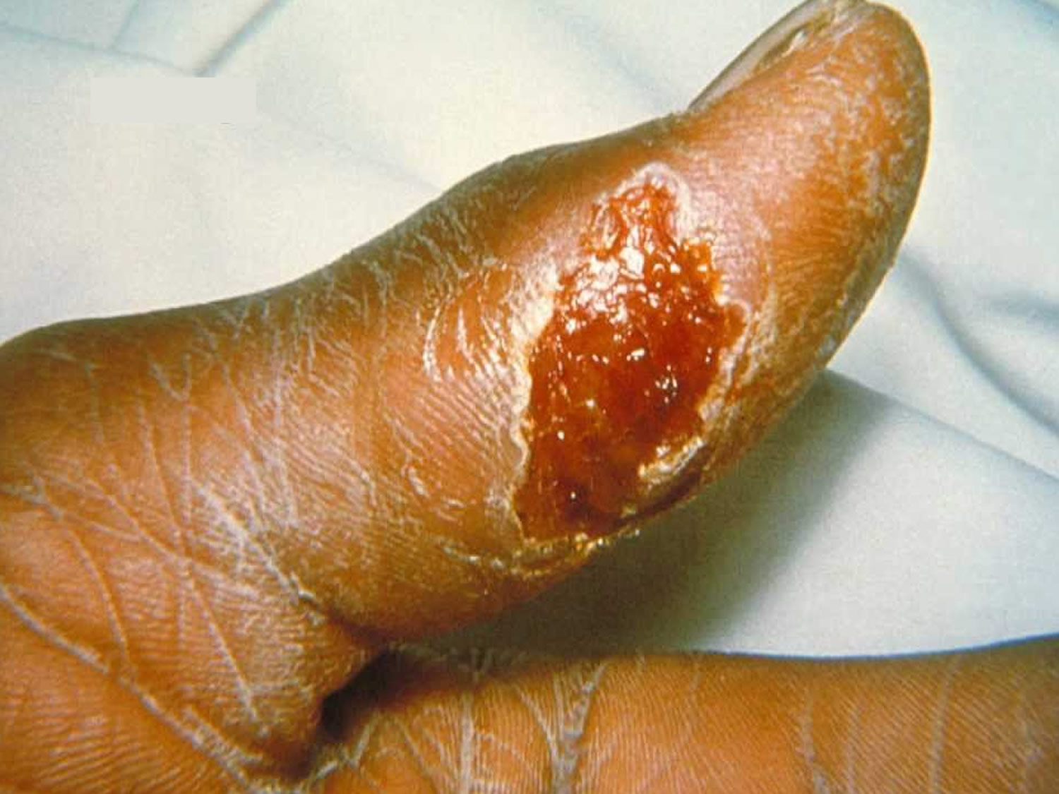
Figure 4. An ulcer caused by Francisella tularensis
Primary lesion
This develops at the point of entry after a 2 to 5 day incubation period. The lesion is a painful red papule (lump) that slowly enlarges and ulcerates within a few days. The ulcer has raised, hardened, ragged edges, and a sensitive base. A discharge may be present, and the ulcer may be covered by an eschar (scab) and/or be itchy. Over weeks to months the ulcer heals and is replaced by scar tissue.
The primary lesion caused by type B tularemia infection may be less severe, e.g. crusting, but no ulcer.
Secondary lesions
Secondary lesions (called tularemids) develop in 8% to 20% of cases of tularemia and take various forms:
- The most common secondary lesions are papular (small elevated lumps) or papulovesicular (small elevated lumps and blisters). Occasionally the lesions are macular (flat discolouration), maculopapular (small discolored lumps), vesicular, pustular (pus-filled blister), pimple-like, nodular (larger, solid papule), or plaque-like (broad, flat lesion). These lesions appear around 11 days after the onset of symptoms and may be widespread, symmetrically distributed on both sides of the body, and itchy.
- Erythema nodosum occurs in 1% to 13% of cases and appears at the end of the second week of illness.
- Erythema multiforme occurs in 0.5% to 2.0% of cases and predominantly affects the trunk and extremities. Erythema nodosum may also be present.
- Less common secondary skin lesions include herpes simplex labialis (cold sores), urticaria, and lymphangitis (red streaks visible along the path of lymph channels) and swollen, tender lymph nodes.
Tularemia complications
Left untreated, tularemia can be fatal. Other possible complications include:
- Inflammation of the lungs (pneumonia). Pneumonia can lead to respiratory failure — a condition in which the lungs don’t take in enough oxygen, don’t release enough carbon dioxide or both.
- Infection around the brain and spinal cord (meningitis). Meningitis is a serious and sometimes life-threatening infection of the fluid and membranes (meninges) surrounding the brain and spinal cord.
- Irritation around the heart (pericarditis). This is swelling and irritation of the pericardium, the thin membrane that surrounds the heart. Mild pericarditis may improve without treatment, but more-serious cases may require antibiotic therapy.
- Bone infection (osteomyelitis). Tularemia bacteria sometimes spread to the bones.
Tularemia diagnosis
Tularemia can be difficult to diagnose. It is a rare disease, and the symptoms can be mistaken for other, more common, illnesses. For this reason, it is important to share with your health care provider any likely exposures, such as tick and deer fly bites, or contact with sick or dead animals.
The diagnosis of tularemia is usually made by serologic testing (detection of antibodies against Francisella tularensis in the blood). A rising level of antibodies between the acute and convalescent stages of disease confirms the diagnosis. Because this process can take 2 to 4 weeks, treatment may be started based on the patient’s history and clinical features.
Francisella tularensis can be isolated from blood, biopsy samples, or other bodily fluids and tissues. A specific culture medium is required to grow this organism in the laboratory. Francisella tularensis is highly infectious to laboratory staff so infection control precautions are required.
Blood tests show disturbed liver function in around half of all patients.
Tularemia treatment
Tularemia is treated with antibiotics such as streptomycin, gentamicin, tetracyclines, chloramphenicol, and tobramycin. Streptomycin is generally the drug of choice, as it appears to offer the highest rate of cure with the lowest rate of relapse.
Treatment usually lasts 10 to 21 days depending on the stage of illness and the medication used. Although symptoms may last for several weeks, most patients completely recover.
Secondary skin manifestations may require topical corticosteroids.
- Tularemia Statistics. https://www.cdc.gov/tularemia/statistics/index.html[↩]


