Contents
What is venous ulcer
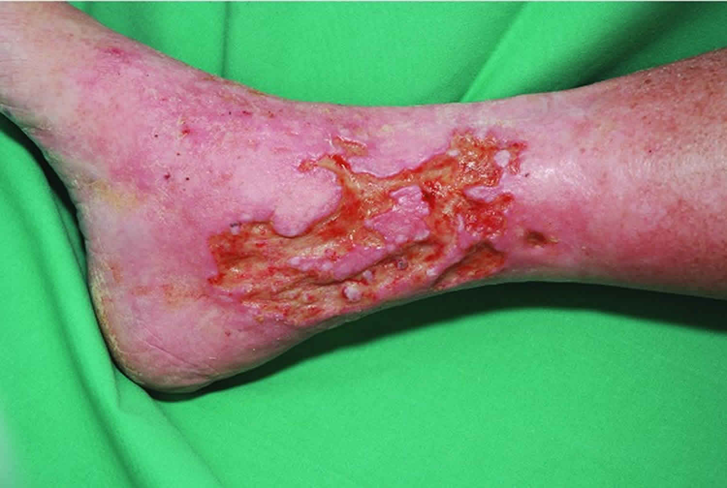
A venous leg ulcer is a long-lasting (chronic) sore that takes more than 4 to 6 weeks to heal. Venous leg ulcers usually develop on the inside of the leg, just above the ankle. A venous leg ulcer is the most common type of leg ulcer, accounting for more than 90% of all cases. Venous leg ulcers can develop after a minor injury, where persistently high pressure in the veins (venous hypertension) of the legs has damaged the skin. The symptoms of a venous leg ulcer include pain, itching and swelling in the affected leg. There may also be discolored or hardened skin around the ulcer, and the sore may produce a foul-smelling discharge.
It’s estimated around 1 in 50 people over the age of 80 has one venous ulcer.
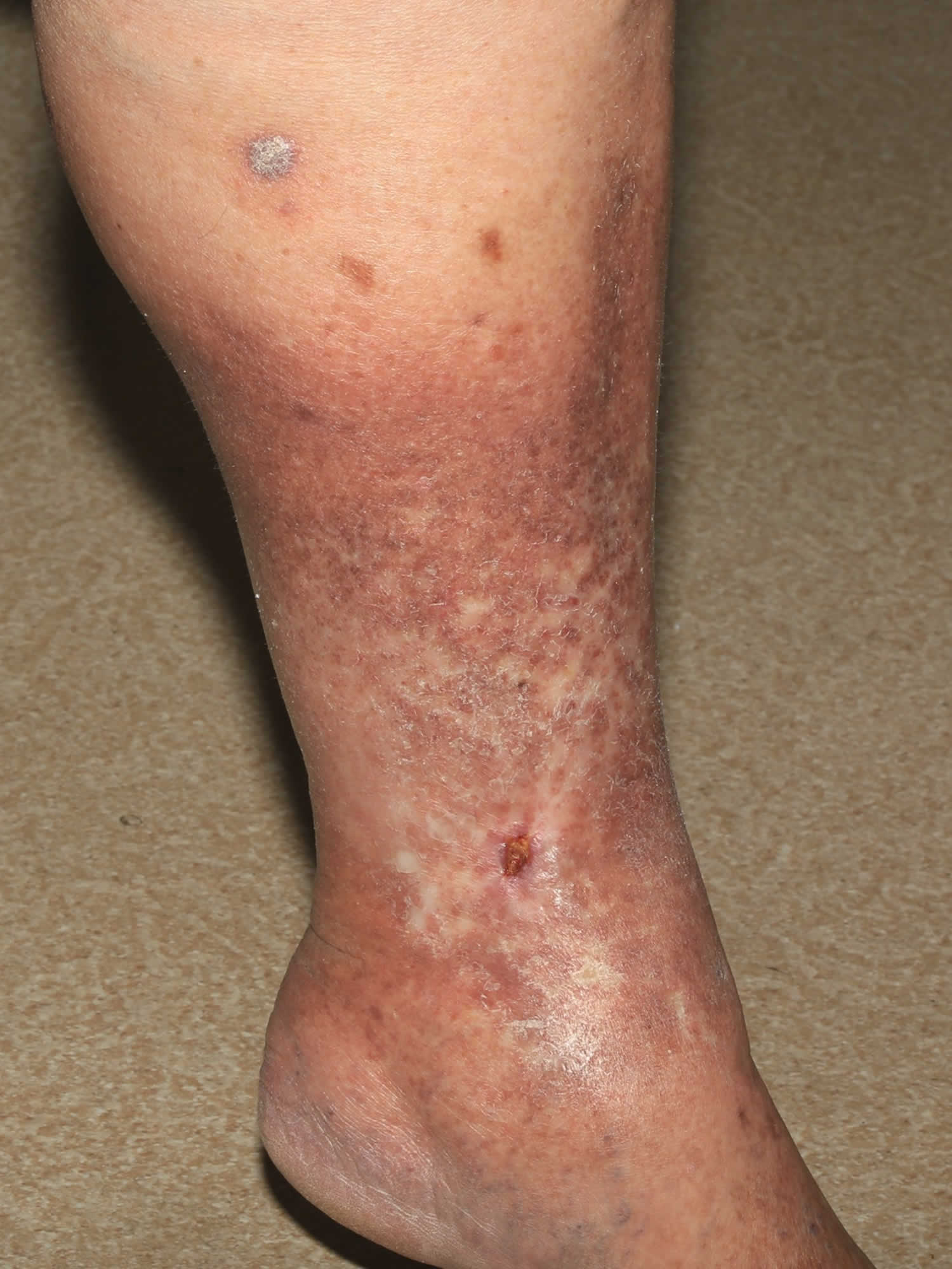
Venous ulcers are usually accompanied by other clinical signs of venous disease such as swelling, pigmentation, varicose dermatitis, atrophie blanche and lipodermatosclerosis. High blood pressure in the leg veins causes leakage of fluid from the abnormal veins into the surrounding tissues. This leads to swelling, especially around the ankles. Leakage of red blood cells leads to iron staining and skin pigmentation in the lower legs. The subsequent inflammation will result in varicose eczema and may lead to scarring (known as atrophie blanche) and hardening of the skin and fat (known as lipodermatosclerosis) in affected areas. This will eventually lead to skin breakdown and ulceration.
You’re more at risk of developing venous ulcer if you previously had deep vein thrombosis (DVT) or find it difficult to walk because of a problem such as:
- osteoarthritis
- a leg injury
- obesity
- paralysis
You’re also more at risk if you recently had an operation on your leg, such as a hip replacement or knee replacement.
There are several ways to help prevent developing a venous leg ulcer in people at risk.
These include:
- wearing compression stockings. Compression is needed to control the underlying venous hypertension. Leg elevation, compression stockings and occasional, intermittent pneumatic compression pumps should be used to prevent recurrence.
- losing weight if you’re overweight
- exercising regularly
- elevating your leg when possible
- stopping smoking if you smoke
These measures are particularly important if you previously had a leg ulcer. This is because you’re at increased risk of having another one in the same leg within months or years.
People with varicose veins (swollen and enlarged veins) also have a higher risk of developing venous leg ulcers.
See your doctor if you think you have a venous leg ulcer, as it’ll need specialist treatment to help it heal.
Your doctor will examine your leg and may carry out additional tests to rule out other conditions.
Most venous leg ulcers heal within 3 to 4 months if they’re treated by a healthcare professional trained in compression therapy for leg ulcers.
But some ulcers may take longer to heal, and a very small number never heal.
Treatment usually involves:
- Cleaning and dressing the wound.
- Use elastic or compression stockings or bandages, to improve the flow of blood in the legs and to reduce swelling.
- Avoid standing or sitting for long periods of time
- Keep your leg raised when you sit
- If the underlying veins need to be treated, the ideal treatment in elderly people involves non-invasive techniques such as endovenous laser ablation and foam sclerotherapy to achieve long-term healing. Surgery may be necessary in selected cases.
Oral antibiotics may also be used if the ulcer becomes infected, but they do not help ulcers to heal.
But unless the underlying cause of the ulcer is addressed, there’s a high risk of a venous leg ulcer coming back after treatment.
Underlying causes could include immobility, obesity, previous deep vein thrombosis (DVT) or varicose veins.
Treatments your provider may suggest include:
- Wet dressings (use only when instructed)
- Topical steroid creams or ointments
- Oral antibiotics
Some skin care treatments can make the problem worse. Talk with your doctor before using any lotions, creams, or antibiotic ointments.
Things to avoid:
- Topical antibiotics, such as neomycin
- Drying lotions, such as calamine
- Lanolin
- Benzocaine and other products meant to numb the skin
See your doctor if you develop leg swelling or symptoms of stasis dermatitis.
Watch for signs of infection such as:
- Drainage that looks like pus
- Open skin sores (ulcers)
- Redness, increased warmth, or swelling around the wound
- More drainage than before or drainage that is yellowish or cloudy
- Bleeding
- Odor
- Fever or chills
- Increased pain
Figure 1. Lipodermatosclerosis (often a precursor of venous ulcer)
Venous ulcer vs Arterial ulcer
Arterial ulcers are painful and most often arise over bony prominences such as between the toes or on the heels, following minor trauma. A well-demarcated purple patch progresses to blackened slough or dry gangrene. The slough sheds to reveal a punched out ulcer with a sharp border. It may be very deep exposing tendons.
Arterial ulcers are less common than venous ulcers. They are more common in men, smokers and people with diabetes. In some people, leg ulcers may be related to both arterial and venous diseases being present at the same time.
Arterial ulcers are caused by lack of normal blood flow to the tissues. Leg arteries that carry blood to the tissues are partially or completely blocked. Arterial ulcers are most often due to arterial insufficiency caused by atherosclerosis. Atherosclerosis is a disease in which plaque builds up inside your arteries. Plaque is a sticky substance made up of fat, cholesterol, calcium, and other substances found in the blood. Over time, plaque hardens and narrows your arteries. That limits the flow of oxygen-rich blood to your body.
Atherosclerosis can lead to serious problems, including:
- Coronary artery disease. These arteries supply blood to your heart. When they are blocked, you can suffer angina or a heart attack.
- Carotid artery disease. These arteries supply blood to your brain. When they are blocked you can suffer a stroke.
- Peripheral arterial disease. These arteries are in your arms, legs and pelvis. When they are blocked, you can suffer from numbness, pain and sometimes infections.
Atherosclerosis usually doesn’t cause symptoms until it severely narrows or totally blocks an artery. Many people don’t know they have it until they have a medical emergency.
Ischemia and infarction may also be due to vasculitis, chilblains, Raynaud’s phenomenon, disseminated intravascular coagulation (DIC), cryoglobulinaemia, hyperviscosity syndrome, septic embolisation, aneurysms, drug-induced necrosis (warfarin, heparin, ergot, intra-arterial injection), connective tissue disease, external compression or entrapment.
Atherosclerosis results in leg ulceration because of acute or chronic ischaemia and/or atheroembolisation of fibrin, platelet and cholesterol debris from a distal source into small vessels of the skin.
Risk factors for arterial ulcers include:
- Smoking
- Hyperlipidemia
- Hypertension
- Diabetes mellitus (large and small vessel disease)
- Insulin resistance
- Obesity
- Personal or family history of ischemic heart disease, cerebrovascular disease or peripheral vascular disease.
Lower extremity arterial insufficiency results in pain on exercise (intermittent claudication) and eventually pain at rest, most marked at night. Sudden onset of localized pain and tenderness may indicate embolus, especially if arising postoperatively.
Signs of arterial insufficiency include:
- Cyanosed later pale skin (especially on elevation), often with livedo-pattern vasculature
- Loss of hair
- Shiny thin skin
- Diminished or absent pulses (may be palpable in diabetics with microangiopathy, or when arterial disease is localised due to embolisation)
- Pain relieved by placing the leg in a dependent position.
Painful ulcers on the anterior shin may be caused by uncontrolled hypertension in the absence of occlusion of larger arteries.
Poor tissue perfusion predisposes to infection particularly if there are breaks or fissures such as with athlete’s foot (interdigital maceration) or ulceration.
Investigation should include Doppler studies and arteriography. The Ankle Brachial Pressure Index (ABPI) is derived by dividing the Doppler measurement of ankle systolic pressure by the brachial systolic pressure. In the absence of arterial disease the ABPI is below 1. An index of less than 0.8 indicates significant arterial disease.
Management of arterial ulcers should include:
- Stopping smoking
- Exercise to increase arterial collateral circulation
- Correction of hyperlipidemia: low fat diet and lipid-lowering medication
- Treatment of hypertension
- Analgesics for ischemic pain
- Referral to vascular surgeon for angioplasty, endarterectomy, arterial bypass procedures and debridement of necrotic tissue or amputation as needed.
Venous ulcer causes
Normally, vein valves in your deeper leg veins keep blood moving forward toward your heart. The cause of venous ulcers is high pressure in the veins of the lower leg. The veins have one-way valves that keep blood flowing up toward your heart. When these vein valves become weak or the veins become scarred and blocked, blood can flow backward and pool in your legs. This is called venous insufficiency. This leads to the high pressure in the lower leg veins. Venous insufficiency results from venous hypertension which in turn is usually caused by reflux in the superficial venous compartment. This is called venous reflux. The increase in pressure and buildup of fluid prevents nutrients and oxygen from getting to tissues. The lack of nutrients causes cells to die, damaging the tissue, and a wound can form.
Some people with venous insufficiency develop stasis dermatitis. Blood pools in the veins of the lower leg. Fluid and blood cells leak out of the veins into the skin and other tissues. This may lead to itching and inflammation, which cause more skin changes. The skin may then break down to form open sores.
Vein valves may fail to close due to:
- Vein wall weakness that causes the vein to enlarge so that the valves can not close
- A history of blood clots or deep vein thrombosis (DVT) in the vein that damage the valve
- An absence of vein valves since birth
Less common causes include:
- deep venous compression
- post-thrombotic stenosis or occlusion
- deep venous reflux
- venous hypertension caused by vascular malformations, arteriovenous fistulae, and neuromuscular disorders (rare)
Varicose veins are hereditary most of the time and generally occur in several members of the same family. Much less commonly, varicose veins develop after
a trauma or injury.
Regardless of the cause, defective valves cause a build up of pressure of the blood in the leg, leading to venous hypertension or high blood pressure in the vein. This may result in enlargement of the varicose veins and an increased likelihood of other symptoms such as swelling, skin changes and ulcers at the ankles or lower leg. Valve failure and venous reflux can also occur in the veins that are unseen, such as the saphenous veins (which run from the foot to the thigh) or in the deep veins. Reflux in these veins is often the underlying cause of painful varicose veins. Venous reflux is a condition that is progressive. If left untreated, it can worsen and cause more advanced symptoms of chronic venous insufficiency. On occasion, the cause of the problem it not even in the legs, but is in the pelvis. Here, blockage of the veins may severely aggravate the symptoms of varicose veins, thus requiring separate treatment.
A venous leg ulcer can develop after a minor injury if there’s a problem with the circulation of blood in your leg veins. If this happens, pressure inside the veins increases. This constant high pressure can gradually damage the tiny blood vessels in your skin and make it fragile. As a result, your skin can easily break and form an ulcer after a knock or scratch.
Unless you have treatment to improve the circulation in your legs, the ulcer may not heal.
A number of factors can increase your risk of developing a venous leg ulcer, including:
- Obesity or being overweight – this increases the pressure in the leg veins
- If you have difficulty walking – this can weaken the calf muscles, which can affect circulation in the leg veins
- Previous deep vein thrombosis (DVT) – blood clots that develop in the leg can damage valves in the veins
- Varicose veins – swollen and enlarged veins caused by malfunctioning valves
- Previous injury to the leg, such as a broken or fractured bone, which may cause DVT (deep vein thrombosis) or impair walking
- Previous surgery to the leg, such as a hip replacement or knee replacement, which can prevent you moving about
- Increasing age – people find it harder to move around as they get older, particularly if they suffer from arthritis
- Older age, being female, or being tall
- Family history of venous insufficiency
- Obesity
- Pregnancy
- Smoking
- Sitting or standing for a long periods (usually for work)
- Fracture of a long bone in the leg or other serious injuries, such as burns or muscle damage
- Blockage of the lymph vessels, which causes fluid to build up in the legs
Risk factors for venous insufficiency include:
- Family history of this condition
- Aging, which results in decreased elasticity of blood vessels
- Pregnancy and other conditions that put pressure on veins in the legs
- History of deep vein thrombosis in the legs
- Female gender (related to levels of the hormone progesterone)
- A history of smoking
- Standing or sitting for long periods of time
- Being overweight or obesity, which increases pressure on the legs
- Menopause
- Weakened blood vessel walls
- Inflammation of the veins (known as phlebitis)
- Chronic constipation and in rare cases, tumors
- Tall height
Venous ulcer prevention
You can help reduce your risk of developing a venous leg ulcer in several ways, such as wearing a compression stocking, losing weight and taking care of your skin. Also, check your feet and legs every day: the tops and bottoms, ankles, and heels. Look for cracks and changes in skin color.
People most at risk of developing a venous leg ulcer are those who have previously had a leg ulcer.
Lifestyle changes can help prevent venous ulcers. The following measures may help improve blood flow and aid healing:
- Quit smoking. Smoking is bad for your blood vessels.
- If you have diabetes, keep your blood sugar level under control. This will help you heal faster.
- Exercise as much as you can. Staying active helps with blood flow.
- Eat healthy foods and get plenty of sleep at night.
- Lose weight if you are overweight.
- Manage your blood pressure and cholesterol levels.
Compression stockings
If you had a venous leg ulcer before or you’re at risk of developing one, treatment with compression stockings may be recommended by your doctor. These stockings are specially designed to squeeze your legs, improving your circulation. They’re usually tightest at the ankle and less tight further up your leg. This encourages blood to flow upwards towards your heart.
To be most effective, these stockings should be put on as you get up and only taken off at night. Compression stockings are available in a variety of different sizes, colors, styles and pressures.
A nurse can help you find a stocking that fits correctly and you can manage yourself. There are various accessories you can buy to help get the stockings on and off.
Losing weight
If you’re obese or overweight, losing weight can help treat and prevent venous leg ulcers. Excess weight leads to high pressure in the veins in your legs, which can damage your skin. Venous ulcers are much more common among people who are overweight.
To help you lose weight, regular exercise and a healthy, balanced diet are recommended.
You should also avoid sitting or standing for long periods. Elevating your legs whenever possible can also help.
Treating underlying problems
Treating severe varicose veins may help prevent leg swelling or ulcers. This may involve a procedure where a thin, flexible tube called a catheter is inserted into the affected veins, with high-frequency radio waves or lasers used to seal them. Alternatively, you may need surgery to repair the damage to your leg veins or remove the affected veins altogether.
Choices of procedures include:
- Sclerotherapy — Sclerotherapy works best for spider veins. These are small varicose veins. Salt water (saline) or a chemical solution is injected into the vein. The vein hardens and then disappears.
- Phlebectomy — Small surgical cuts (incisions) are made in the leg near the damaged vein. The vein is removed through one of the incisions. This may be done along with other procedures, such as ablation.
- Ablation uses intense heat to treat the vein. There are two methods. One uses radiofrequency energy and the other uses laser energy. During these procedures:
- Your doctor will puncture the varicose vein.
- Your doctor will thread a flexible tube (catheter) through the vein.
- The catheter will send intense heat to the vein. The heat will close off and destroy the vein and the vein will disappear over time.
- Laser therapy. Laser treatment can be used on the surface of the skin. Small bursts of light make small varicose veins disappear.
- Varicose vein stripping — Used to remove or tie off a large vein in the leg called the superficial saphenous vein.
Venous stasis ulcer signs and symptoms
Venous stasis ulcer signs and symptoms include:
- Shallow sore with a red base, sometimes covered by yellow tissue
- Unevenly shaped borders
- Surrounding skin may be shiny, tight, warm or hot, and discolored
- Leg pain
- If the sore becomes infected, it may have a bad odor and pus may drain from the wound
You may have symptoms of venous insufficiency including:
- Dull aching or heaviness in the leg
- Pain that gets worse when you stand
- Swelling in the leg
At first, the skin of the ankles and lower legs may look thin or tissue-like. You may slowly get brown stains on the skin.
The skin may become irritated or crack if you scratch it. It may also become red or swollen, crusted, or weepy.
Over time, some skin changes become permanent:
- Thickening and hardening of the skin on the legs and ankles (lipodermatosclerosis)
- A bumpy or cobblestone appearance of the skin
- Skin turns dark brown
Skin sores (ulcers) may develop (called a venous ulcer or stasis ulcer). These most often form on the inside of the ankle.
Venous stasis ulcer possible complications
Complications of venous stasis ulcers include:
- Bacterial skin infections
- Infection of bone
- Permanent scar
Venous stasis ulcer diagnosis
Venous stasis ulcer diagnosis is largely based on your symptoms and examination of your affected leg, although additional tests may be required.
Venous stasis dermatitis can also be related to heart problems or other conditions that cause leg swelling. Your provider may need to check your general health and order more tests.
Medical history and examination
Your doctor or practice nurse will ask whether you have any other symptoms associated with venous leg ulcers, such as:
- swelling in your ankles
- discolored or hard skin
Your doctor will try to determine the cause of the ulcer by asking about underlying conditions or previous injuries, such as:
- diabetes
- deep vein thrombosis (DVT)
- injury or surgery in the affected leg
- a previous leg ulcer
Your doctor will also examine your leg, both when you’re standing up and lying down.
Varicose veins will be more obvious when you’re standing up, and it’ll be easier to look at the ulcer when you’re lying down.
Your doctor will also feel your pulse at your ankles to make sure the arteries in your leg are working properly.
Doppler study
To rule out peripheral arterial disease (a condition affecting the arteries) as a possible cause of your symptoms, your doctor or nurse will carry out a test known as a Doppler study.
This involves measuring the blood pressure in the arteries at your ankles and comparing it to the pressure in your arms.
If you have peripheral arterial disease, the blood pressure in your ankles will be lower than your arms.
It’s important to carry out this check, as the main treatment for venous ulcers is compression bandages or stockings to improve the vein circulation in your legs.
It’s not safe to apply compression if the ankle artery pressures are low.
Referral to a specialist
In some cases, your doctor may decide to refer you to a specialist in conditions affecting the blood vessels (vascular specialist).
For example, you may be referred to a vascular specialist if your doctor is unsure about your diagnosis, or if they suspect your ulcer may be caused by artery diseases, diabetes or rheumatoid arthritis.
After taking your medical history and examining you, the vascular specialist may need to arrange further investigations to plan your treatment.
Venous ulcer treatment
With appropriate treatment, most venous leg ulcers heal within 3 to 4 months. Treatment should always be carried out by a healthcare professional trained in compression therapy for leg ulcers. This will usually be a practice or district nurse.
Venous stasis ulcer cleaning and dressing
The first step is to remove any debris or dead tissue from the ulcer, elimination of serious infection and applying an appropriate dressing for proper moisture balance 1. This provides the best conditions for the ulcer to heal.
A simple non-sticky dressing will be used to dress your ulcer. This usually needs to be changed once a week. Many people find they can manage cleaning and dressing their own ulcer under the supervision of a nurse.
One of the most controversial aspects is the selection of topical ulcer dressings 2. The key factor is controlling ulcer drainage and using products that can wick away the drainage to prevent maceration of the ulcer. Many practitioners are very fond of the use of specialized and expensive dressings including silver containing products. There is very little evidence in randomized trials for most of these products 3. We prefer to use simple, inexpensive, absorbing and non-adherent products in these ulcers. A very important factor in ulcer healing is proper moisture balance in the ulcer. Excessive ulcer moisture or dryness impedes healing 1. The practice of keeping ulcers open to the air that in the past was thought to promote healing actually slows down this process.
The basic wound care instructions are:
- Always keep the wound clean and bandaged to prevent infection.
- Your doctor will tell you how often you need to change the dressing.
- Keep the dressing and the skin around it dry. Try not to get healthy tissue around the wound too wet. This can soften the health tissue, causing the wound to get bigger.
- Before applying a dressing, cleanse the wound thoroughly according to your provider’s instructions.
- Protect the skin around the wound by keeping it clean and moisturized.
- You will wear a compression stocking or bandages over the dressing. Your provider will show you how to apply the bandages.
To help treat a venous ulcer, the high pressure in the leg veins needs to be relieved.
- Wear compression stockings or bandages every day as instructed. They help prevent blood from pooling, reduce swelling, help with healing, and reduce pain.
- Put your feet above your heart as often as possible. For example, you can lie down with your feet propped up on pillows.
- Take a walk or exercise every day. Being active helps improve blood flow.
- Take medicines as directed to help with healing.
If ulcers DO NOT heal well, your doctor may recommend certain procedures or surgery to improve blood flow through your veins.
Compression
The single most important aspect in the healing of venous ulcers is appropriate compression therapy 4. To improve vein circulation in your legs and treat swelling, your nurse will apply a firm compression bandage over the affected leg. These bandages are designed to squeeze your legs and encourage blood to flow upwards towards your heart.
There are many different types of bandage or elastic stockings used to treat venous leg ulcers, which may be made in 2, 3 or 4 different layers. A four-layer bandage system is quite popular and although these layers are mostly elastic, the friction between the layers increases the static stiffness index. They are much less technically demanding to apply. Another popular and effective method is the use of paste bandages (Unna’s boot) which are “zero-stretch” products that when properly applied can yield very high static stiffness index (SSI) values. Static stiffness index (SSI) is the difference between standing and resting pressure under the bandages or stockings. They can be used as the primary dressing in cases where diffuse eczema is present and in conjunction with short stretch bandages. As the edema resolves the extremity decreases in size and the bandages become loose requiring reapplication after several days.
Recently a Velcro compression system has become available that can be tailored to the individual patient in the ulcer clinic and used as an alternative to compression bandages. This system involves a large Velcro wrap that is trimmed according to the calf and ankle measurements and employs a unique measuring system that is easy to use and provides predictable pressures on the leg due to an additional pressure monitoring card. These devices are short-stretch products that have a low resting pressure and high working pressure with an average static stiffness index (SSI) of >20 in the typical patient. The advantages include self-management, enabling the patient to change the dressings frequently and maintain a consistent pressure profile with a compression system that does not require special skills to apply. Velcro band devices are used in selected patients without heavily draining ulcers. They can also be introduced after initial therapy with the standard 4 layer bandages or short-stretch bandages.
Bandages may be classified as long stretch (elastic), or short stretch (inelastic). Elastic bandages are applied with sufficient tension to make sure they do not fall down when the patient walks. Blood and fluid that leaks out of the deep muscular compartment causes the bandage to expand because of its elastic properties. This provides little or no control of edema. As the edema increases the elastic bandage expands (“gives way”) to accommodate the swelling. The bandage becomes tighter and tighter until the bandage can no longer expand. Discomfort and pain under the bandage increases until the patient can no longer tolerate the compression. A rubber band effect occurs and if the compression bandage is not removed skin breakdown can occur. A surgical stocking used in patients with edema often can cause these skin lesions when used in patients with edema. The use of elastic compression to treat venous stasis ulcers often results in “stalled” or very slow healing and in many cases the ulcers will worsen using this form of compression. The basic principle of increasing flow out of the leg to reduce venous stasis and improve arterial inflow is poorly accomplished by elastic modalities.
Short stretch products include cotton bandages (e.g. Comprilan®, Rosidal®), zinc paste bandages, Velcro strap devices (e.g. CircAid®, Farrow wraps®), and multilayer wraps (e.g. Profore®, Coban 2®). These products need to be applied with initially high pressure. There is an immediate pressure drop due to decrease in the size of the leg due to instant edema reduction resulting in a low resting pressure after short time. This allows short stretch products to be worn day and night with excellent patient comfort. Ideally good compression should always be combined with walking exercises 5. This process reduces edema, decreasing the pressure at the venous end of the capillary facilitating flow and hence removal of waste products from the tissues. This decrease of ambulatory venous hypertension allows increased arterial perfusion to the capillaries delivering nutrients and oxygen to the tissues. As this process continues healing of the ulcer is facilitated.
The most frequent cause for poor ulcer healing is the lack of a proper compression technique by the bandagers. This applies also to “experienced staff” which was obviously never trained in applying adequate bandages. Most bandages are “under dosed” 6.
Using inelastic material the initial sub-bandage pressure on the lower leg in patients with intact arterial inflow should be 40–60 mm Hg or higher.
The application of a compression bandage is a skilled procedure and should only be done by trained healthcare staff.
The bandage is changed once a week, when the dressing is changed.
When compression bandages are first applied to an unhealthy ulcer, it’s usually painful.
Ideally, you should have acetaminophen (paracetamol) or an alternative painkiller prescribed by your doctor.
The pain will lessen once the ulcer starts to heal, but this can take up to 10 to 12 days.
It’s important to wear your compression bandage exactly as instructed. If you have any problems, it’s usually best to contact your nurse instead of trying to remove it yourself.
If the compression bandage feels a little too tight and is uncomfortable in bed at night, getting up for a short walk will usually help.
But you’ll need to cut the bandage off if:
- you get severe pain at the front of your ankle
- you get severe pain on the top of your foot
- your toes become blue and swollen
Once you remove the bandage, make sure you keep your leg highly elevated and contact your doctor or nurse as soon as possible.
In some clinics, specialist teams are using new alternatives to compression bandages, such as special stockings or other compressive devices.
These may not be available in every clinic, but could change the way ulcers are treated in the future.
Your specialist will be able to advise you whether a different approach may help you.
Compression therapy in accompanying arterial disease
Several studies and meta-analyses have shown that specially designed intermittent pneumatic pressure pumps can be very useful to treat arterial and mixed arterial-venous ulcers 7. The beneficial effects of intermittent pneumatic pressure pump is mainly explained by a release of vasoactive mediators from the endothelial cells in the venules due to an intermittent increase of the shear stress in the blood capillaries by the massaging effect of the pump 8.
Inelastic bandages in combination with walking also exert a comparable massaging effect on the leg, which may explain the fact that many times venous stasis ulcers can be healed in the presence of arterial insufficiency particularly in the non-diabetic using short-stretch compression 7. In any case with mixed arterial and venous insufficiency it is critical to use a compression system where the resting pressure is less than the leg arterial pressure 9. Knowing the pressure characteristics of the bandage is critical in selecting the optimal product. The tendency to apply Stockinet type compression, elastic bandages, or low-pressure elastic hose (surgical stockings) to those with arterial insufficiency has major disadvantages. As the elastic product expands to accommodate increasing edema, a “rubber band” like effect is produced that can lead to further skin damage and ischemic changes in the extremity. A much safer approach is to use several short-stretch bandages on the extremity with reduced pressure. When properly applied this results in an initial 20–40 mm Hg resting pressure and a SSI of >10. Such bandages have been termed “modified compression” 9. In patients with arterial occlusive disease and an ankle-brachial index (ABI) > 0.6, it could be shown that modified compression consisting in inelastic bandages applied with a pressure up to 40 mm Hg are able to increase arterial flow and venous pumping function 10. The measurement of the ankle brachial index (ABI) involves recording the systolic pressures in the brachial artery at each elbow and systolic pressures in the posterior tibial and the dorsalis pedis arteries at each ankle. The result is reported as a ratio of the ankle systolic pressure in the numerator,over the higher brachial pressure in the denominator. The ABI (ankle brachial index) is calculated for each leg separately, and the lower of the two values is taken as a result for the patient. There is a definite learning curve for proper application of these products but they are very effective in reducing edema and their low resting pressure provides patient comfort.
Basic therapy of mixed, arterial-venous ulcers still consist in modified compression and walking exercises 11. Abolishment of venous reflux may also be beneficial in such patients 12.
Treating associated symptoms
Swelling in the legs and ankles
Venous leg ulcers are often accompanied by swelling of your feet and ankles (edema), which is caused by fluid. This can be controlled by compression bandages.
Keeping your leg elevated whenever possible, ideally with your toes above your hips, will also help ease swelling. You should put a block, sofa cushion or foam wedge under the bottom of your mattress to help keep your legs raised while you sleep.
You should also keep as active as possible and aim to continue with your normal activities.
Regular exercise, such as a daily walk, will help reduce leg swelling.
But you should avoid sitting or standing still with your feet down. You should elevate your feet at least every hour.
Itchy skin
Some people with venous leg ulcers develop rashes with scaly and itchy skin.
This is often caused by varicose eczema, which can be treated with a moisturiser (emollient) and occasionally a mild corticosteroid cream or ointment.
In rare cases, you may need to be referred to a dermatologist (skin specialist) for treatment.
Itchy skin can also sometimes be caused by an allergic reaction to the dressings or creams applied by your nurse. If this happens, you may need to be tested for allergies.
It’s important to avoid scratching your legs if they feel itchy as this damages the skin and may lead to further ulcers.
Treating an infected ulcer
An ulcer sometimes produces a large amount of discharge and becomes more painful. There may also be redness around the ulcer. These symptoms and feeling unwell are signs of infection.
If your ulcer becomes infected, it should be cleaned and dressed as usual. You should also elevate your leg most of the time. You’ll be prescribed a 7-day course of antibiotics. The aim of antibiotic treatment is to clear the infection. But antibiotics do not heal ulcers and should only be used in short courses to treat infected ulcers.
Looking after yourself during treatment
The following advice may help your ulcer heal more quickly.
- Try to keep active by walking regularly. Sitting and standing still without elevating your legs can make venous leg ulcers and swelling worse.
- Whenever you’re sitting or lying down, keep your affected leg elevated.
- Regularly exercise your legs by moving your feet up and down, and rotating them at the ankles. This can help encourage better circulation.
- If you’re overweight, try to reduce your weight with a healthy diet and regular exercise.
- Stop smoking and moderate your alcohol consumption. This can help the ulcer heal faster.
- Be careful not to injure your affected leg, and wear comfortable, well-fitting footwear.
Follow-up
You should visit your nurse once a week to have your dressings and compression bandages changed. They’ll also monitor the ulcer to see how well it’s healing.
Once your ulcer is healing well, your nurse will see you less often.
After the ulcer has healed
Once you have had a venous leg ulcer, another ulcer could develop within months or years. The most effective method of preventing this is to wear compression stockings at all times when you’re out of bed. This is often difficult since it requires the patient to wear some form of compression on a long-term basis. It has been traditional to prescribe compression stockings for these patients but many pitfalls may occur. A sizeable percentage of patients with venous stasis ulcers have a level of edema that is difficult to control with 30–40 mm Hg stockings. These stockings become tighter on the leg since an elastic product expands to accommodate the increased edema. The static stiffness index (SSI) of these products is close to zero. Eventually the material is stretched to the maximum and the stocking acts like a tourniquet on the leg. This can result in further skin damage including ulcers. Donning and doffing becomes more difficult and with time the patient stops wearing the stockings altogether. Another major compliance problem occurs in patients with arthritis, decreased arm or hand strength, limited flexibility, age, surgery, and joint problems to mention a few. These individuals find donning and doffing stockings a difficult or impossible task. Aids for application and stocking removal are often awkward and the techniques required hard to master. Also for these patients Velcro-band devices are frequently a better alternative.
A major obstacle to long-term edema control is the obese patient. Many individuals have a large abdominal girth that increase venous pressure in the abdominal, pelvic, and leg veins producing edema and venous stasis. In some individuals this progresses to venous insufficiency-induced lymphedema. This degree of edema is virtually impossible to control in most patients using even high-pressure support stockings. These patients often are unable to bend at the waist and are unable to apply or remove stockings. Frequently these patients are seen in the ulcer clinic with recurrent ulceration and confessing that they have not been wearing their hose. Many of us caring for these individuals feel they should never have been prescribed stockings in the first place. Fortunately Velcro compression devices represent a very effective compression solution for these individuals. These devices have a well-tolerated resting pressure and a high working pressure with an average static stiffness index (SSI) of more than 20. They are easy to apply and remove and can be adjusted by the patient. The devices can be tightened as the edema decreases, or loosened with patient discomfort occurs. Products from five companies are available and come in a variety of configurations for the calf, and foot. The disadvantage is that these products are expensive, require special fitters, and are cosmetically unappealing in some cases. Nevertheless they enable those to control edema long term where stockings are not appropriate or cannot be used. Finally these devices are ideal for those seniors who cannot manage stockings.
A number of pneumatic compression devices have appeared which can also be used in those with extreme edema or recurrent ulceration where other measures have failed, in addition to conventional compression devices. Using these devices on a part term basis during the day can be very helpful (adequate compliance provided), especially in patients who cannot walk or who are immobile 13. Very recently a new pump has become available that is an inflatable stocking providing 30–40 mm Hg calf and foot pressure 14: The device consists of a sleeve applied to the leg and then an attached miniature pump inflates the sleeve to the appropriate pressure. The pressure is readjusted by the device every 30 min. The device also converts to a pneumatic compression device when plugged in. In many cases these pumps are expensive but under certain circumstances are covered by insurance.
- Zulkowski K. Diagnosing and treating moisture-associated skin damage. Adv Skin Wound Care. 2012;25:231–236.[↩][↩]
- Venous Ulcers. J Am Coll Clin Wound Spec. 2012 Sep; 4(3): 54–60. doi: 10.1016/j.jccw.2013.11.001 https://www.ncbi.nlm.nih.gov/pmc/articles/PMC4511547/[↩]
- Palfreyman S.J., Nelson E.A., Lochiel R., Michaels J.A. Dressings for healing venous leg ulcers. Cochrane Database Syst Rev. 2006 Jul 19;(3):CD001103[↩]
- O’Meara S., Cullum N., Nelson E.A., Dumville J.C. Compression for venous leg ulcers. Cochrane Database Syst Rev. 2012 Nov 14;11:CD000265[↩]
- World Union of Ulcer Healing Societies (WUWHS) MEP Ltd; London: 2008. Principles of Best Practice: Compression in Venous Leg ulcers. A Consensus Document[↩]
- Keller A., Müller M.L., Calow T., Kern I.K., Schumann H. Bandage pressure measurement and training: simple interventions to improve efficacy in compression bandaging. Int Wound J. 2009 Oct;6(5):324–330.[↩]
- Comerota A.J. Intermittent pneumatic compression: physiologic and clinical basis to improve management of venous leg ulcers. J Vasc Surg. 2011 Apr;53(4):1121–1129.[↩][↩]
- Chen A.H., Frangos S.G., Kilaru S., Sumpio B.E. Intermittent pneumatic compression devices – physiological mechanisms of action. Eur J Vasc Endovasc Surg. 2001 May;21(5):383–392.[↩]
- World Union of Ulcer Healing Societies (WUWHS) MEP Ltd; London: 2008. Principles of Best Practice: Compression in Venous Leg ulcers. A Consensus Document.[↩][↩]
- Mosti G., Iabichella M.L., Partsch H. Compression therapy in mixed ulcers increases venous output and arterial perfusion. J Vasc Surg. 2012;55:122–128.[↩]
- Marston W.A., Davies S.W., Armstrong B. Natural history of limbs with arterial insufficiency and chronic ulceration treated without revascularization. J Vasc Surg. 2006 Jul;44(1):108–114.[↩]
- Obermayer A., Göstl K., Partsch H., Benesch T. Venous reflux surgery promotes venous leg ulcer healing despite reduced ankle brachial pressure index. Int Angiol. 2008 Jun;27(3):239–246.[↩]
- Partsch H. Intermittent pneumatic compression in immobile patients. Int Wound J. 2008 Jun;5(3):389–397[↩]
- Vanscheidt W., Ukat A., Partsch H. Dose-response of compression therapy for chronic venous edema – higher pressures are associated with greater volume reduction: two randomized clinical studies. J Vasc Surg. 2009 Feb;49(2):395–402.[↩]


