What is the esophagus ?
The esophagus, a straight, collapsible muscular tube, about 25 cm (10 in.) long, that lies posterior to the trachea. The esophagus is a food passageway from the pharynx to the stomach (see Figure 1). The esophagus begins at the base of the laryngopharynx and descends posterior to the trachea, passing through the mediastinum. It penetrates the diaphragm through an opening, the esophageal hiatus and is continuous with the stomach on the abdominal side of the diaphragm.
Mucous glands are scattered throughout the submucosa of the esophagus. Their secretions moisten and lubricate the tube’s inner lining.
Microscopic Anatomy of the Esophagus
Unlike the mouth and pharynx, the esophagus wall (Figure 2) contains all four layers of the alimentary canal: mucosa, submucosa, muscularis externa, and adventitia.
The following histological features are of interest:
- The mucosal epithelium is a nonkeratinized stratified squamous epithelium. At the junction of the esophagus and stomach, this thick, abrasion-resistant layer changes abruptly to the thin simple columnar epithelium of the stomach, which is specialized for secretion (Figure 2).
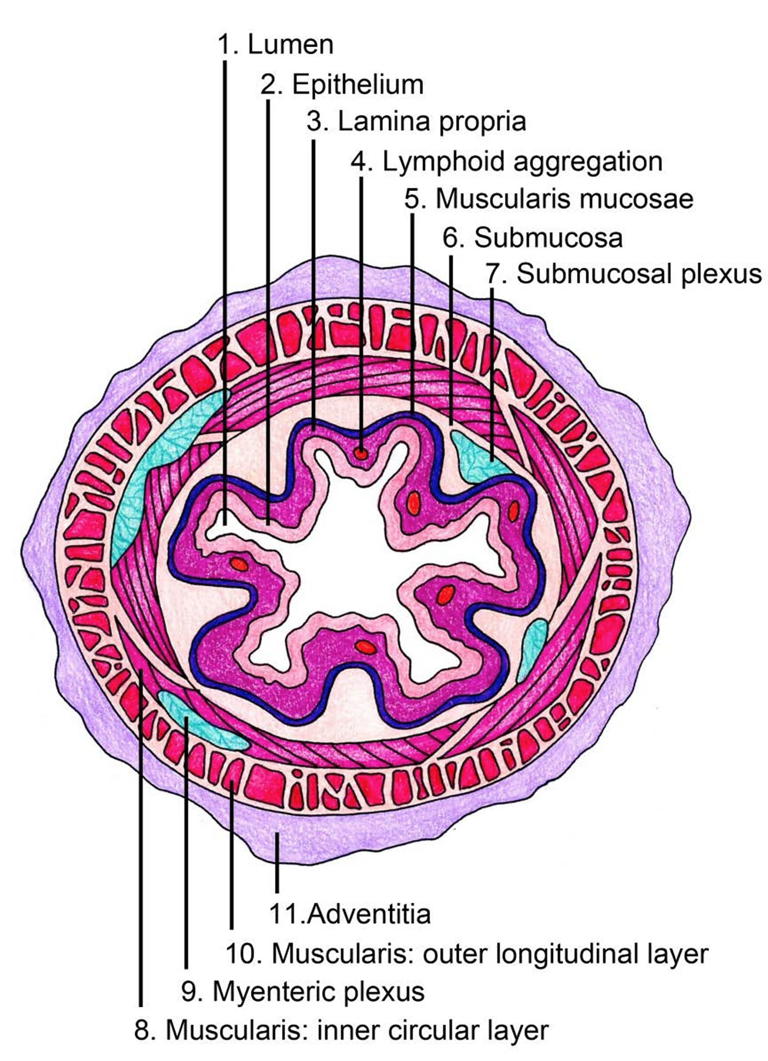
- When the esophagus is empty, its mucosa and submucosa are thrown into longitudinal folds, but during passage of a bolus, these folds flatten out (Figure 3).
- The submucosa of the wall of the esophagus contains mucous glands, primarily compound tubuloalveolar glands, that extend to the lumen. As a bolus passes, it compresses these glands, causing them to secrete a lubricating mucus. This mucus helps the bolus pass through the esophagus.
- The muscularis externa consists of skeletal muscle in the superior third of the esophagus, a mixture of skeletal and smooth muscle in the middle third, and smooth muscle in the inferior third. This arrangement is easy to remember if the esophagus is viewed as the zone where the skeletal
muscle of the mouth and pharynx gives way to the smooth muscle of the stomach and intestines. - The most external esophageal layer is an adventitia, not a serosa, because the thoracic segment of the esophagus is not suspended in the peritoneal cavity.
Figure 1. Esophagus
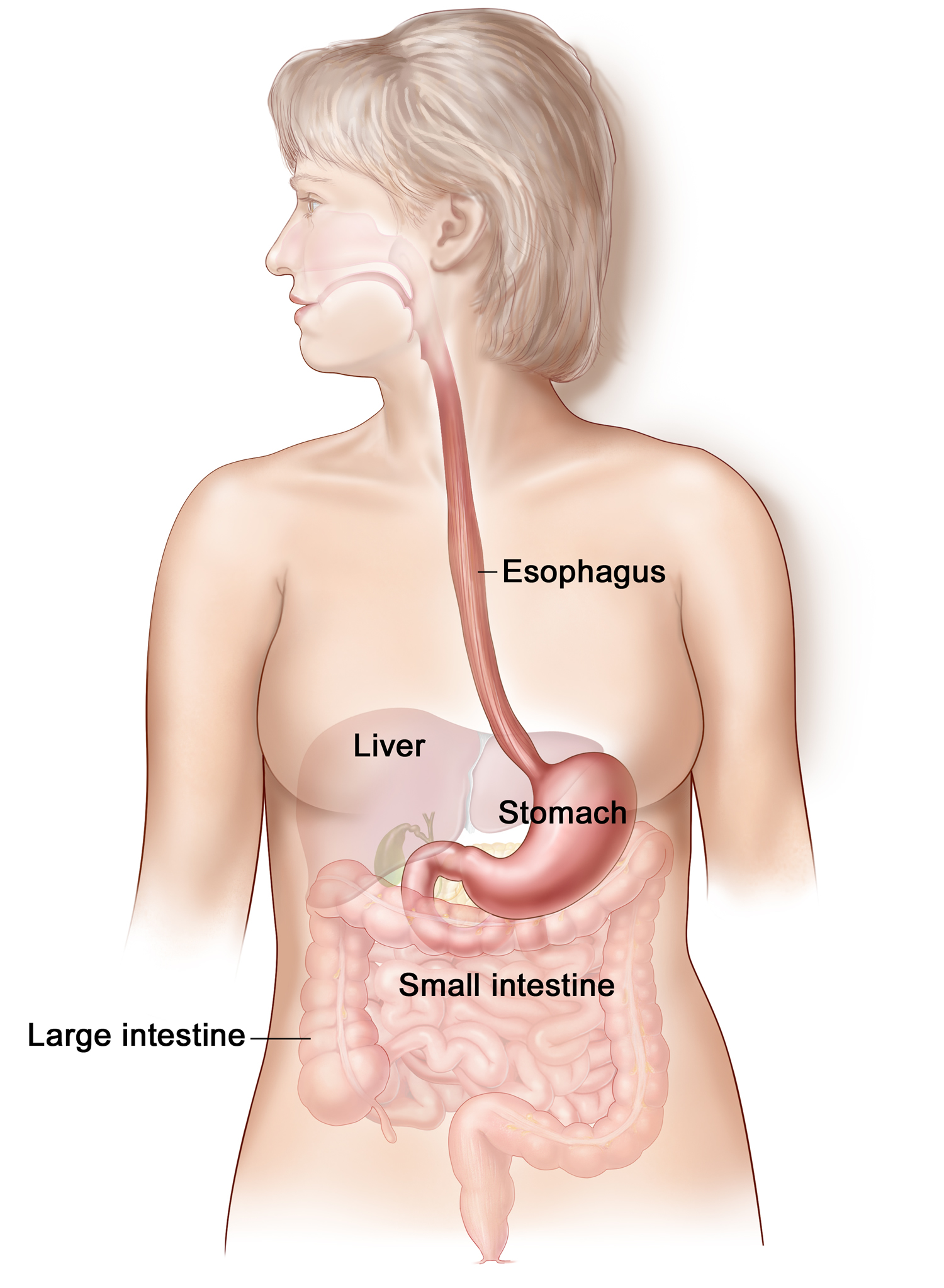
Figure 2. Esophagus anatomy
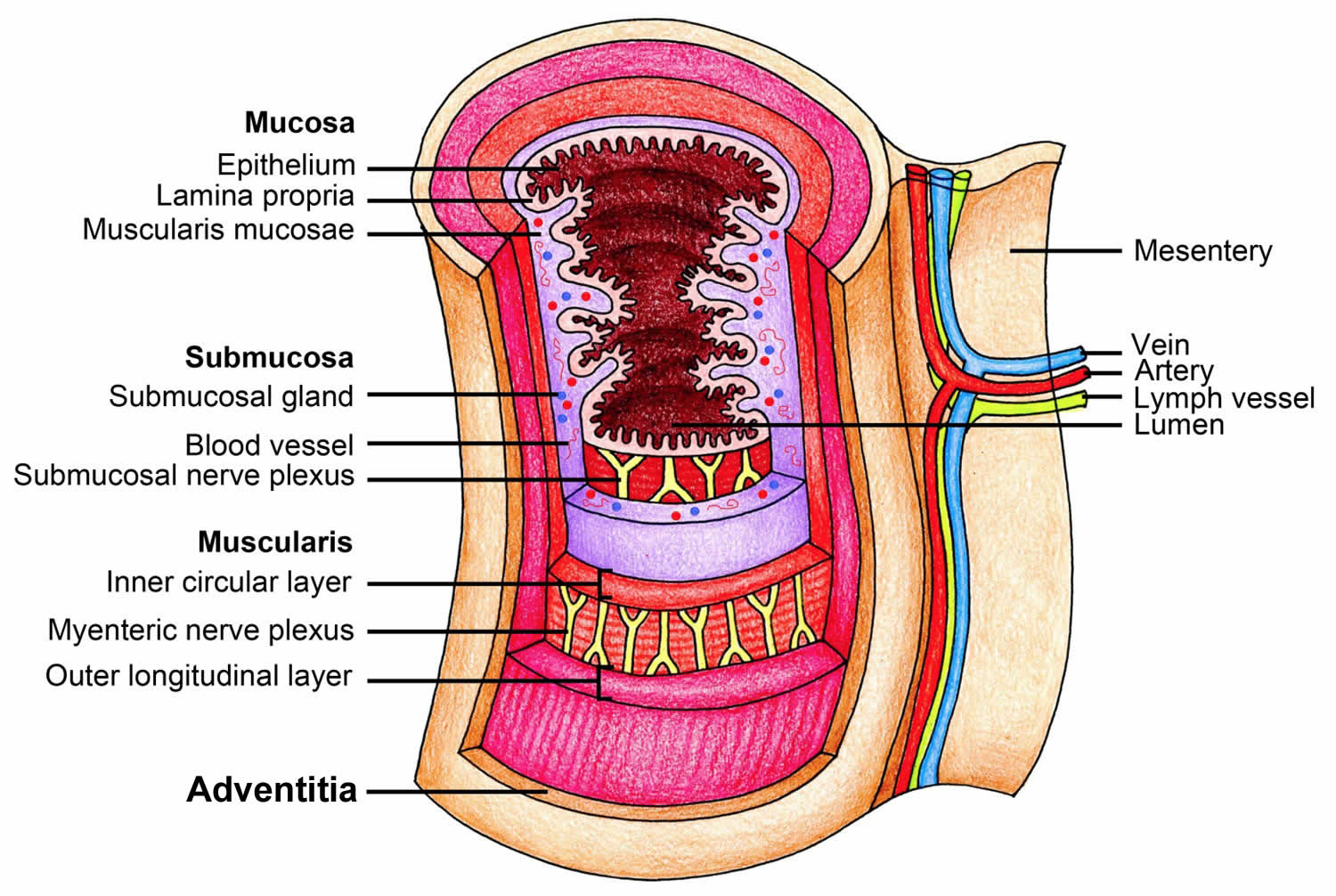
Figure 3. Cross section view of the esophagus
Some of the smooth muscle just superior to the point where the esophagus joins the stomach has increased muscle tone, forming the lower esophageal sphincter (LES), or cardiac sphincter (Figures 4 and 5). This smooth muscle usually remains contracted. The sphincter closes the entrance to the stomach, preventing the stomach contents from regurgitating into the esophagus. When peristaltic waves in the esophagus reach the stomach, the sphincter temporarily relaxes and allows the swallowed food to enter.
Figure 4. Lower esophageal sphincter
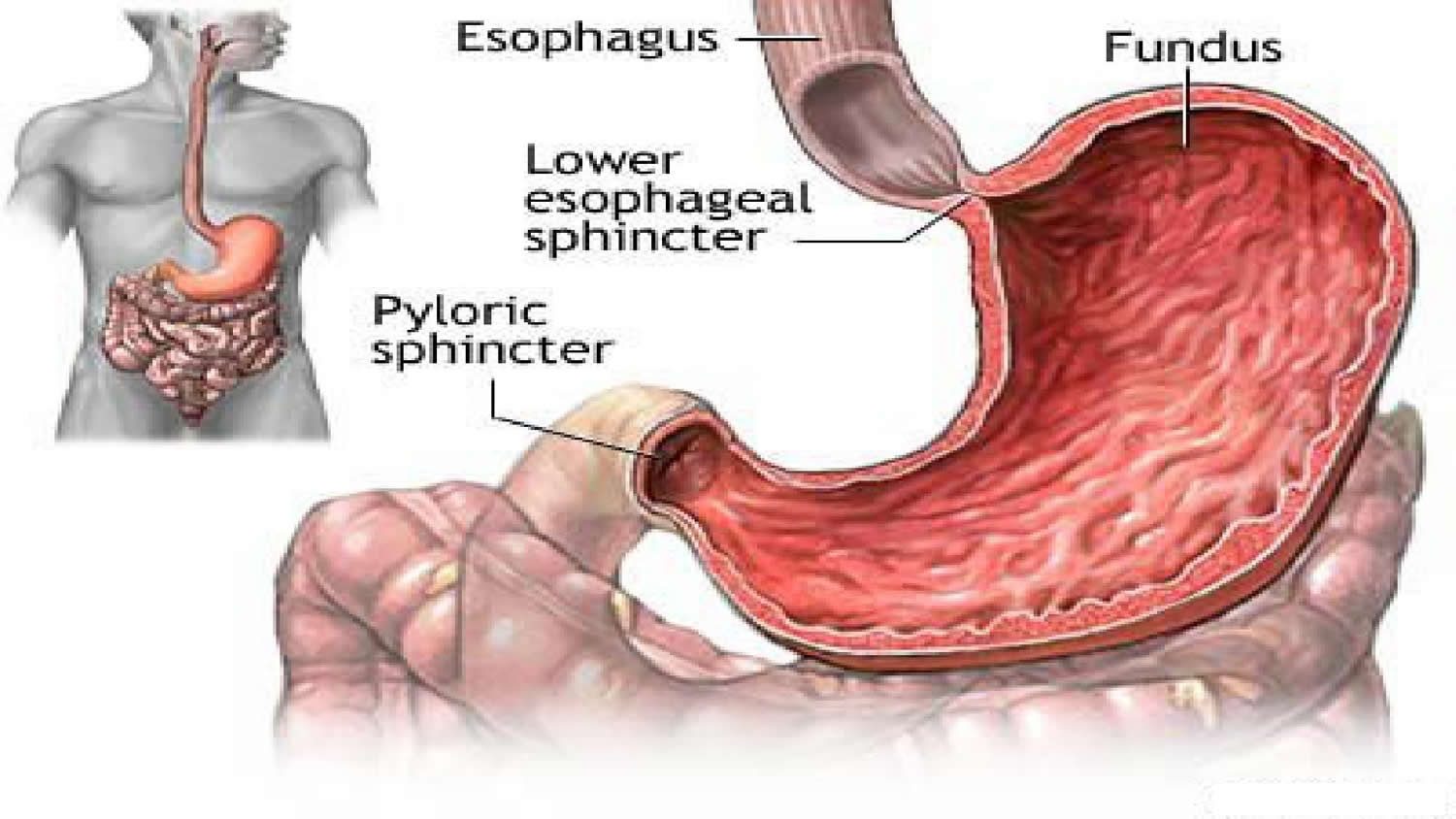
Figure 5. lower esophageal sphincter (endoscopic view)

What does the esophagus do
The esophagus is the muscular tube that carries food, and liquids from your mouth to the stomach. You may not be aware of your esophagus until you swallow something too large, too hot, or too cold. You may also notice it when something is wrong. You may feel pain or have trouble swallowing.
The most common problem with the esophagus is GERD (gastroesophageal reflux disease). With gastroesophageal reflux disease (GERD), a muscle at the end of your esophagus does not close properly. This allows stomach contents to leak back, or reflux, into the esophagus and irritate it. Over time, gastroesophageal reflux disease (GERD) can cause damage to the esophagus.
Other problems include heartburn, cancer, and esophagitis. Doctors may use various tests to make a diagnosis. These include imaging tests, an upper endoscopy, and a biopsy.
Treatment depends on the problem. Some problems get better with over-the-counter medicines or changes in diet. Others may need prescription medicines or surgery.