Contents
What is actinic keratosis
Actinic keratosis, also known as solar keratosis, are small rough or scaly areas of skin due to damage from sun exposure. An actinic keratosis is a marker of ongoing sun exposure. Actinic keratosis is considered precancerous or an early form of cutaneous squamous cell carcinoma, so it is important to perform self-examinations often and catch them early.
Actinic keratosis affect people that have often lived in the tropics or subtropics, and have predisposing factors such as:
- Other signs of photoageing skin
- Fair skin with a history of sunburn
- History of long hours spent outdoors for work or recreation
- Defective immune system
The main concern with actinic keratosis is that actinic keratosis predisposes you to squamous cell carcinoma. It is rare for a solitary actinic keratosis to evolve to squamous cell carcinoma (SCC), but the risk of squamous cell carcinoma (SCC) occurring at some stage in a patient with more than 10 actinic keratoses is thought to be about 10 to 15%. A tender, thickened, ulcerated or enlarging actinic keratosis is suspicious of squamous cell carcinoma.
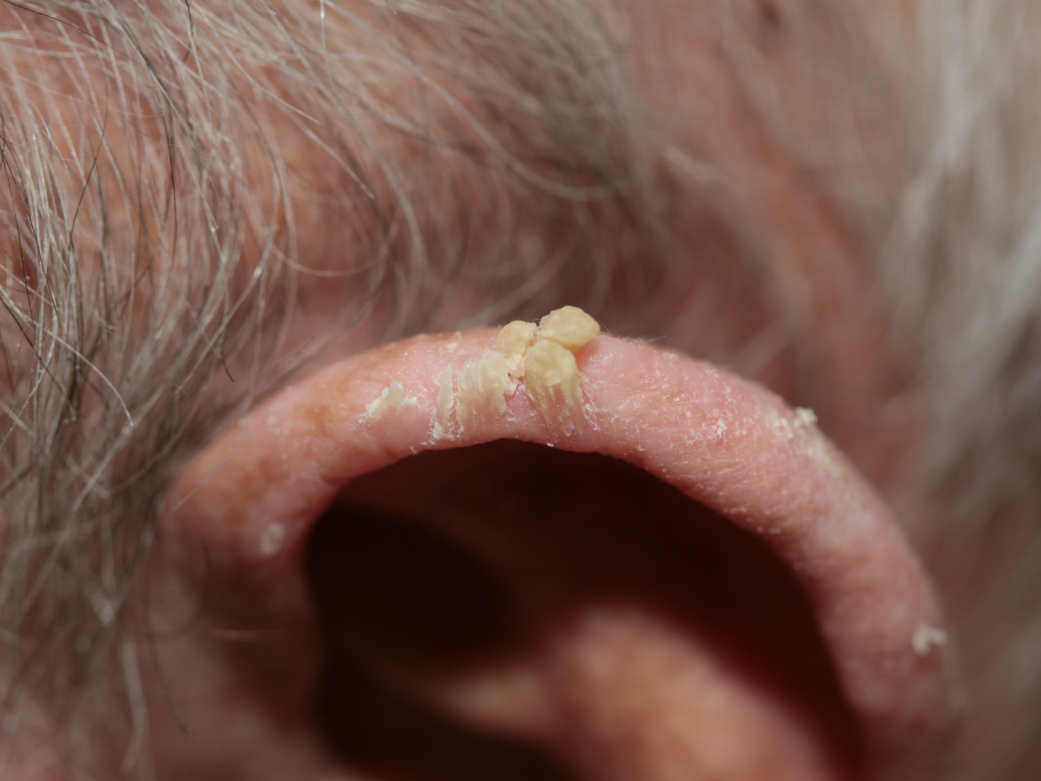
Cutaneous horn may arise from an underlying actinic keratosis or squamous cell carcinoma.
Because they are sun damaged, people with actinic keratoses are also at risk of developing actinic cheilitis, basal cell carcinoma (BCC, which is more common than squamous cell carcinoma), melanoma and rare forms of skin cancer such as Merkel cell carcinoma.
A review of all available studies looking at the rate of conversion of actinic keratosis to squamous cell carcinoma (SCC) concluded that “currently, no reliable estimates concerning the frequency of actinic keratosis developing into invasive carcinoma can be given, and further studies are needed.” 1. Detailed study results vary widely, but it is often quoted that individual actinic keratosis progress to squamous cell carcinoma at a rate of approximately 1/250 to 1/1000 per year. Part of the problems is that there is a very high turnover of actinic keratoses with large numbers developing, regressing, and recurring. For example in one detailed study of the natural history of actinic keratoses over a 1 year period (no treatment, merely observation), 74% of actinic keratoses noted at the beginning of the study spontaneously regressed at some point over the ensuing year 2. Another problems is that most studies don’t biopsy the presumed actinic keratosiss so indeed many may be squamous cell carcinomas or squamous cell carcinoma in situ to begin with. Maybe the actinic keratosis didn’t convert to squamous cell carcinoma, but instead was an squamous cell carcinoma in the first place. Furthermore, some use conversion to squamous cell carcinoma in situ as a positive event while others do not.
In one study that used accurate mapping of both squamous cell carcinomas and pre-existing actinic keratoses, it was found that 10/17 (60%) squamous cell carcinomas arose from a lesion diagnosed clinically as a solar keratosis in the previous year and the other 7 (40%) squamous cell carcinomas on what had been clinically normal skin 12 months previously. The risk of malignant transformation of an actinic keratosis to squamous cell carcinoma within 1 year in that study was less than 1/1000 3.
Figure 1. Mild actinic keratosis
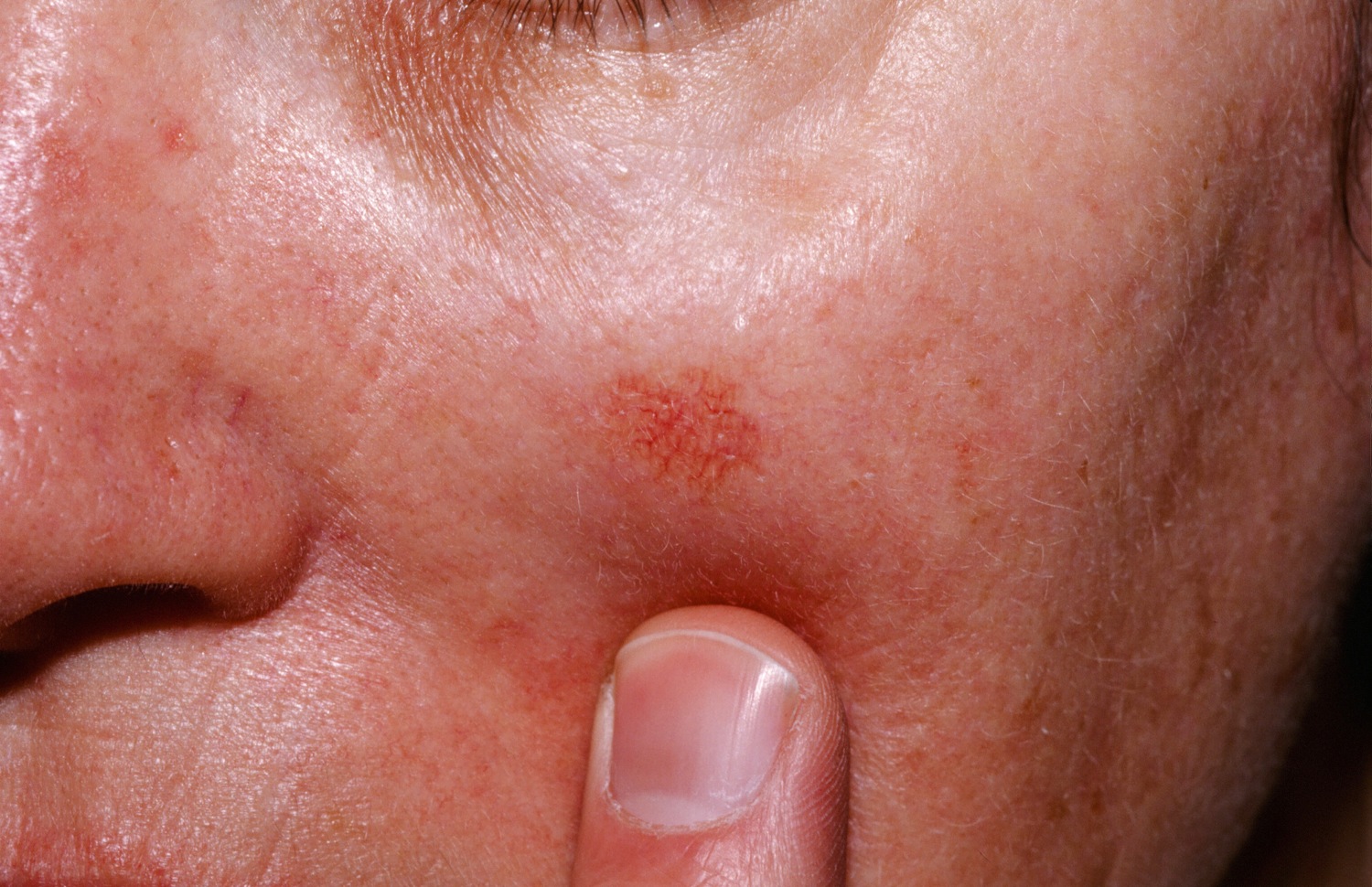
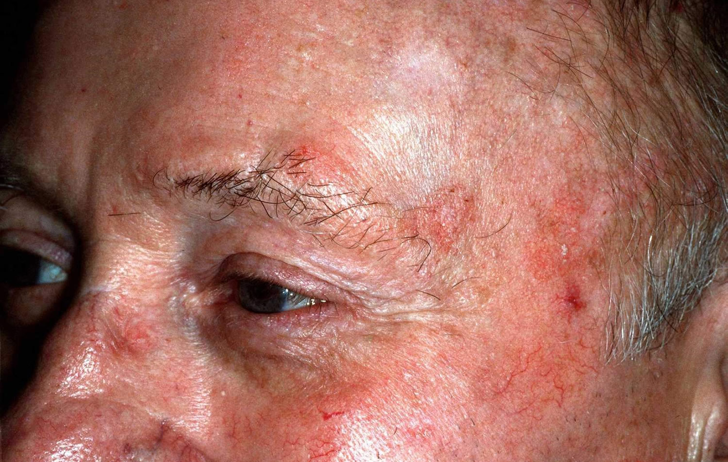
Figure 2. Hypertrophic actinic keratosis

Actinic keratosis causes
Actinic keratosis is a reflection of abnormal skin cell development due to DNA damage by short wavelength UVB.
They are more likely to appear if the immune function is poor, due to ageing, recent sun exposure, predisposing disease or certain drugs.
Actinic keratosis prevention
Actinic keratoses are prevented by strict daily sun protection. If already present, keratoses may improve with very high sun protection factor (SPF 50+) broad-spectrum sunscreen applied at least daily to affected areas, year-round.
The number and severity of actinic keratoses can also be reduced by taking nicotinamide (vitamin B3) 500 mg twice daily. Niacinamide 500 mg twice a day has been shown to reduce actinic keratoses on the order of 30% 4. It has also been shown reduce the development of new actinic keratoses 5.
Sun protection is very important and can reduce the number of new areas occurring and may help small lesions go away on their own.
- Avoid direct sun in the middle of the day (10 AM to 4 PM). Remember: snow and water reflect light to the skin, and clouds still let a lot of light through, so you may still be exposed to ultraviolet light even on cloudy days.
- Use a hat with a wide brim. A baseball hat does not give much protection.
- Cover up with tightly woven clothing. Some manufacturers make specialty clothing with a high sun protection factor (SPF) rating, or you can purchase a special ingredient to be added to your washer that can “wash” SPF into your clothing.
- Use sunscreen on all exposed skin areas, including the lips, before going outdoors. A broad spectrum (blocks UVB and UVA light), with an SPF of at least 50+, is best. Apply generously 30 minutes before going outdoors and reapply every 2 hours or after swimming or sweating a lot.
- Do not use tanning beds!
- A low-fat diet (less than 21% calories from fat) has been shown to reduce the incidence of actinic keratoses.
Dramatic improvements can be made with just daily morning sunscreen use 6. One review found that proper sunscreen use can reduce the number of actinic keratoses by up to 53% 7. Actinic keratoses are a marker of previous sun exposure, with more recent exposure, e.g., the last 2 years, having the greatest impact 8.
Once a month, you should perform a self-exam to look for signs of skin cancer. It is best to perform the exam in a well-lit area after a shower or bath. Use a full-length mirror with the added assistance of a hand mirror, when necessary. Using a hair dryer can help you examine any areas of skin covered by hair, such as your scalp.
- In front of a full-length mirror, inspect the front of your body making sure to look at the front of your neck, chest (including under breasts), legs, and genitals.
- With your arms raised, inspect both sides of your body making sure to examine your underarms.
- With your elbows bent, examine the front and back of your arms as well as your elbows, hands, fingers, area between your fingers, and fingernails.
- Inspect the tops and bottoms of your feet, the area between your toes, and toenails.
- With your back to the mirror and holding a hand mirror, inspect the back of your body, including the back of your neck, shoulders, legs, and buttocks.
- Using a hand mirror, examine your scalp and face.
As you perform your monthly self-exam, familiarize yourself with the moles, freckles, and other marks on your body, and look for any changes in them from month to month, including shape, size, color, or other changes, such as bleeding or itching.
Actinic keratosis symptoms
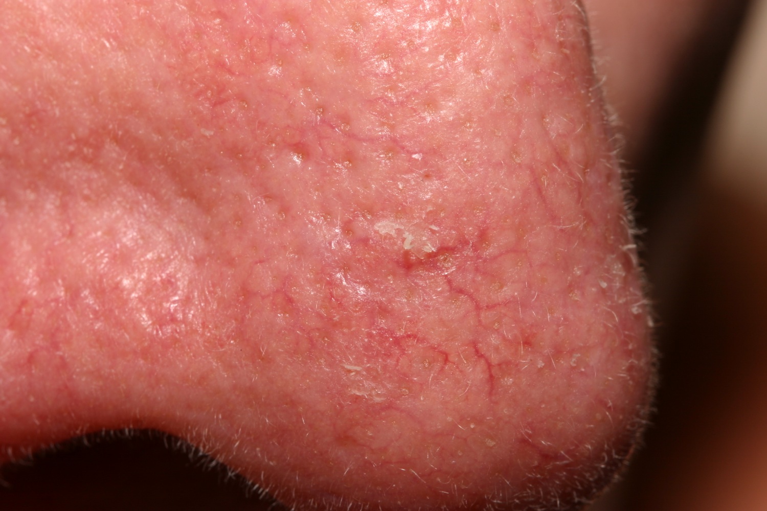
The sun-exposed areas of the face, scalp (where balding), ears, neck, forearms, and backs of the hands are most commonly affected with actinic keratoses, but any skin area frequently exposed to sun can be involved.
Patches are usually less than an inch in size with slight scale (sometimes thick like a wart) and a pink, red, or brownish color. They are slightly rough to the touch, like fine sandpaper, and may be a bit sensitive.
- Mild – one or two spots, not thick or hard
- Moderate – scattered, few spots
- Severe – numerous or thick, hard, or bleeding spots
Actinic keratosis may be solitary but there are often multiple keratoses. The appearance varies.
- A flat or thickened papule or plaque
- White or yellow; scaly, warty or horny surface
- Skin colored, red or pigmented
- Tender or asymptomatic
Actinic keratoses are very common on sites repeatedly exposed to the sun, especially the backs of the hands and the face, most often affecting the ears, nose, cheeks, upper lip, vermilion of the lower lip, temples, forehead and balding scalp. In severely chronically sun damaged individuals, they may also be found on the upper trunk, upper and lower limbs, and dorsum of feet.
Actinic keratosis diagnosis
Actinic keratosis is usually easy to diagnose clinically. Occasionally, biopsy is necessary, for example to exclude squamous cell carcinoma (SCC), or if treatment fails.
Actinic keratosis prognosis
Actinic keratoses may recur months or years after treatment. The same treatment can be repeated or another method used. Patients who have been treated for actinic keratoses are at risk of developing new keratoses. They are also at increased risk of other skin cancers, especially intraepidermal squamous cell carcinoma, invasive cutaneous squamous cell carcinoma, basal cell carcinoma and melanoma.
Actinic keratosis treatment
An actinic keratosis sometimes disappears on its own, but it typically returns after more sun exposure. Because it’s impossible to tell which patches or lesions will develop into skin cancer, actinic keratoses are usually removed as a precaution.
Actinic keratoses are also removed because they are unsightly or uncomfortable.
Treatment of an actinic keratosis requires removal of the defective skin cells. Epidermis regenerates from surrounding or follicular keratinocytes that have escaped sun damage.
Tender, thickened, ulcerated or enlarging actinic keratoses should be treated aggressively. Asymptomatic flat keratoses may not require active treatment but should be kept under observation.
Physical treatments
Physical treatments are used to destroy individual keratoses that are generally symptomatic or have a thick hard surface scale. The lesions may recur in time, in which case they may be retreated by the same or a different method.
Cryotherapy using liquid nitrogen
Liquid nitrogen spray is required to ensure adequate depth and duration of freeze. This varies according to lesion location, width and thickness. Your doctor applies the substance to the affected skin, which causes blistering or peeling. As your skin heals, the lesions slough off, allowing new skin to appear. Cryotherapy is the most common treatment. It takes only a few minutes and can be done in your doctor’s office. Healing varies from 5–10 days on face, 3–4 weeks on the hands, and 6 weeks or longer on the legs. A light freeze for a superficial actinic keratosis usually leaves no mark, but longer freeze times result in hypopigmentation or scar. Side effects may include blisters, scarring, changes to skin texture, infection and darkening of the skin at the site of treatment.
Surgery
If you have only a few actinic keratoses, your doctor may recommend removing them. The most common methods include:
- Shave, curettage and electrocautery
Scraping scraping with a sharp instrument (curettage). In this procedure, your surgeon uses a device called a curet to scrape off damaged cells. Shave, curettage and electrocautery (burning) may be necessary to remove a cutaneous horn or hypertrophic actinic keratosis. Scraping may be followed by electrosurgery, in which the doctor uses a pencil-shaped instrument to cut and destroy the affected tissue with an electric current. This procedure requires a local anesthetic. Side effects may include infection, scarring and changes in skin coloration at the site of treatment.
- Excision
Excision ensures the actinic keratosis has been completely removed, which should be confirmed by pathology. The surgical wound is sutured (stitched). The sutures are removed after a few days, the time depending on the size and location of the lesion. The procedure leaves a permanent scar.
Medications
Creams are used to treat areas of sun damage and flat actinic keratoses, sometimes after physical treatments have been carried out. If you have several actinic keratoses, you may be better served by treating the entire affected area. Prescription creams are most effective on facial skin. Pretreatment with keratolytics (such as urea cream, salicylic acid ointment or topical retinoid), and thorough skin cleansing improves response rates. Results are variable and the course of treatment may need repeating from time to time. With the exception of diclofenac gel, prescription creams all result in local inflammatory reactions such as redness, blistering and discomfort for a varying length of time.
Prescription medicines that can be applied to your skin for this purpose include:
- 5-Fluorouracil 5% cream (Carac, Fluoroplex, Efudex). 5-Fluorouracil is a cytotoxic agent. The cream formulation is applied once or twice daily for 2 to 8 weeks. 5-fluorouracil cream is sometimes combined with salicylic acid. Its effect may be enhanced by calcipotriol ointment. 2-4 weeks of 5% 5-Fluorouracil cream applied twice daily to the face and ears (along with daily SPF 30+ sunscreen) reduced by 75% the incidence of squamous cell carcinoma (SCC) over the following year in a large study 9. Additionally, that same regimen reduced actinic keratoses counts and the need for spot treatment of actinic keratoses for longer than 2 years 10. It therefore seems prudent to recommend annual 5-Fluorouracil 5% cream treatment along with daily morning sunscreen to all patients with a history of non-melanoma skin cancer.
- Imiquimod cream (Aldara, Zyclara). Imiquimod cream is an immune response modifier. It is applied 2 or 3 times weekly for 4 to 16 weeks.
- Ingenol mebutate gel (Picato). Ingenol mebutate gel is effective after only 2–3 applications.
- Diclofenac gel (Voltaren, Solaraze). Diclofenac is more often used as an anti-inflammatory drug. Applied as a gel twice daily for 3 months, it is fairly well tolerated in the treatment of actinic keratoses, but less effective than the other options listed here.
These creams may cause redness, scaling or a burning sensation for a few weeks.
Photodynamic therapy
In photodynamic therapy, your doctor applies a photosensitiser (a porphyrin chemical such as methyl aminolevulanic acid) to the affected skin that makes it sensitive to light. He or she then exposes the area to artificial light to destroy the damaged skin cells. Side effects may include redness, swelling and a burning sensation during therapy. For example, the Levulan Kerastick contains 20% aminolevulinic acid and is activated by blue light illumination (peak wavelength 417 nm) to treat actinic keratoses of the face or scalp. Hypertrophic actinic keratoses tend not to respond. The highest clearance is with two treatments e.g., 8 weeks apart.
Field-Directed Therapy
- Sunscreen SPF 30+
- 5-Fluorouracil
- 5-Fluorouracil mixed with calcipotriene
- Imiquimod
- Ingenol mebutate
- Diclofenac
Field-directed therapy is a more comprehensive treatment approach. However, it is less commonly used than freezing for a variety of reasons including more time needed, longer downtime for the patient, higher cost for the patient, etc.
5-Fluorouracil plus Calcipotriene
In a study of 131 patients, the four-day application of 5-Fluorouracil 5% cream mixed with 0.0005% calcipotriene applied twice daily removed 88% of facial actinic keratoses compared with a 26% reduction in those who used 5-fluorouracil alone 11. Redness and burning were the most common side effects but overall, the treatment was well tolerated. The majority of patients who had used the classic 5-fluorouracil treatment in the past preferred the new approach. Calcipotriol cream alone daily can reduce actinic keratoses 12.
5-Fluorouracil
If lesions are widespread, 5-Fluorouracil 5% cream (5-FU) applied twice daily for 7-14 days on the face and 10-14 days on the body is effective although the patient should be fully informed of the tenderness, erosion, crusting and pain that is involved. Older regimens are applied twice daily for 2-3 weeks but twice daily for one week has 80% of the effect with much less inflammation, etc. Concentrations range from 0.5% (e.g., Carac) to 5%. But in general, the longer the treatment duration, the higher the clearance rate.
In one study of veteran affairs patients 10, A single course of 5% topical fluorouracil cream applied twice daily for 4 weeks along with daily SPF 30+ sunscreen reduced actinic keratoses counts and the need for spot treatment for longer than 2 years. In addition, 2-4 weeks of 5-Fluorouracil 5% cream applied twice daily reduces by 75% the incidence of SCC over the following year 9.
Imiquimod
Imiquimod is a topical agent that is used to treat the entire field of sun damage including actinic keratoses. Strengths include 5%, 3.75% and 2.5%. Many treatment regimens are used. The higher the concentration used and the more applications performed, the higher the clearance 13. The manufacturer’s prescribing information for imiquimod 5% cream states that patients should be treated twice weekly for 16 weeks and that treatment should be applied to a single contiguous treatment area of approximately 25 cm2 (forehead, scalp, or one cheek). Nowadays however, few clinicians use it in this manner as the duration of therapy rather long. A typical approach (but off label) is to use daily imiquimod for 2 weeks, then 2 weeks off therapy, then 2 weeks daily imiquimod. In one study, actinic keratoses cleared in the majority (82%) with imiquimod 3 times a week for 4 weeks and repeated after a month if necessary 14. Others use the imiquimod 3x per week until a good reaction is achieved. Finally, some use imiquimod once a week indefinitely.
Ingenol Mebutate
Ingenol mebutate gel (Picato) is FDA approved to treat actinic keratoses. It is used as follows: Ingenol mebutate 0.015% daily for 3 days for the face and scalp. Ingenol mebutate 0.05% daily for 2 days on the trunk or extremities. The benefit with ingenol is the short treatment length compared with 5-Fluorouracil 5% cream and imiquimod. In one study for example 15, local skin reaction peaked at day 4 and was almost entirely gone in 2 weeks.
Diclofenac
Diclofenac (Solaraze) gel applied twice daily to the actinic keratoses for 2-3 months can clear 1/3 to 1/2 of actinic keratoses. It is a “kindler-gentler treatment,” but takes much longer.
- Br J Dermatol. 2013 Sep;169(3):502-18[↩]
- J of Invest Dermatol 2000;115;273[↩]
- Lancet 1988 Apr 9;1(8589):795[↩]
- Journal of Investigative Dermatology (2012) 132, 1497–1500[↩]
- Australian Oral Nicotinamide to Reduce Actinic Cancer (ONTRAC) study[↩]
- Br J Dermatol. 2009 Nov;161 Suppl 3:78-84[↩]
- BJD 2013;169;502[↩]
- Br J Cancer. 1996 Oct;74(8):1308-12[↩]
- JAMA Dermatol online 2018 Jan 3.[↩][↩]
- JAMA Derm 2015;151;952[↩][↩]
- Dermatology News July p. 4[↩]
- J Drugs Dermatol. 2009:451[↩]
- JAAD 2010;62;582[↩]
- JAAD 2002;47;571[↩]
- JEADV 2015;29;1822[↩]


