Contents
What is aortic stenosis
Aortic stenosis also called aortic valve stenosis, occurs when the heart’s aortic valve narrows. In aortic valve stenosis, the aortic valve between the lower left heart chamber (left ventricle) and the main artery that delivers blood from the heart to the body (aorta) is narrowed (stenosis). This narrowing prevents the valve from opening fully, which reduces or blocks blood flow from your heart into the main artery to your body (aorta) and onward to the rest of your body. The aorta is the big artery supplying your body with oxygen rich blood.
The aortic valve is like a one-way door leading out of the heart to the aorta, the artery that carries blood to the rest of the body. In aortic stenosis, the opening is narrowed, making the left ventricle work harder to pump a sufficient amount of blood through the aortic valve into the aorta and onward to the rest of your body. This means it gets harder for your heart to push blood out into the aorta. This can cause the left ventricle to thicken and enlarge. As a result of all extra work on your heart can weaken the left ventricle and your heart overall, you might be at risk of angina, irregular heart rhythms or heart failure.
Your treatment depends on the severity of your condition. If you are affected by aortic stenosis, your doctor might advise you not to overexert yourself.
If you have no symptoms from your aortic stenosis, your doctor may just want you to have regular check-ups.
You may need surgery to repair or replace the aortic valve. Left untreated, aortic valve stenosis can lead to serious heart problems.
Surgeons rarely repair an aortic valve to treat aortic valve stenosis, and generally aortic valve stenosis requires aortic valve replacement. To repair an aortic valve, surgeons may separate valve flaps (cusps) that have fused.
Some people with aortic stenosis need an operation to remove the aortic valve and replace it with an artificial one. Another operation is sometimes done where your own valve is opened up with a tiny balloon. But this doesn’t work as well as replacing the valve.
Children with aortic stenosis can sometimes get their aortic valve repaired. However, if the valve is very abnormal it is better to replace it with a new one. Using a balloon to stretch the valve can help for a while, but over time the valve can become narrow again, meaning another operation might need to be done.
Figure 1. Aortic valve
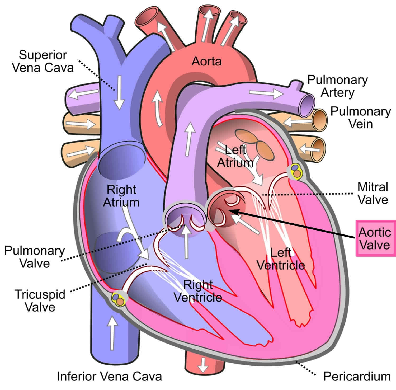
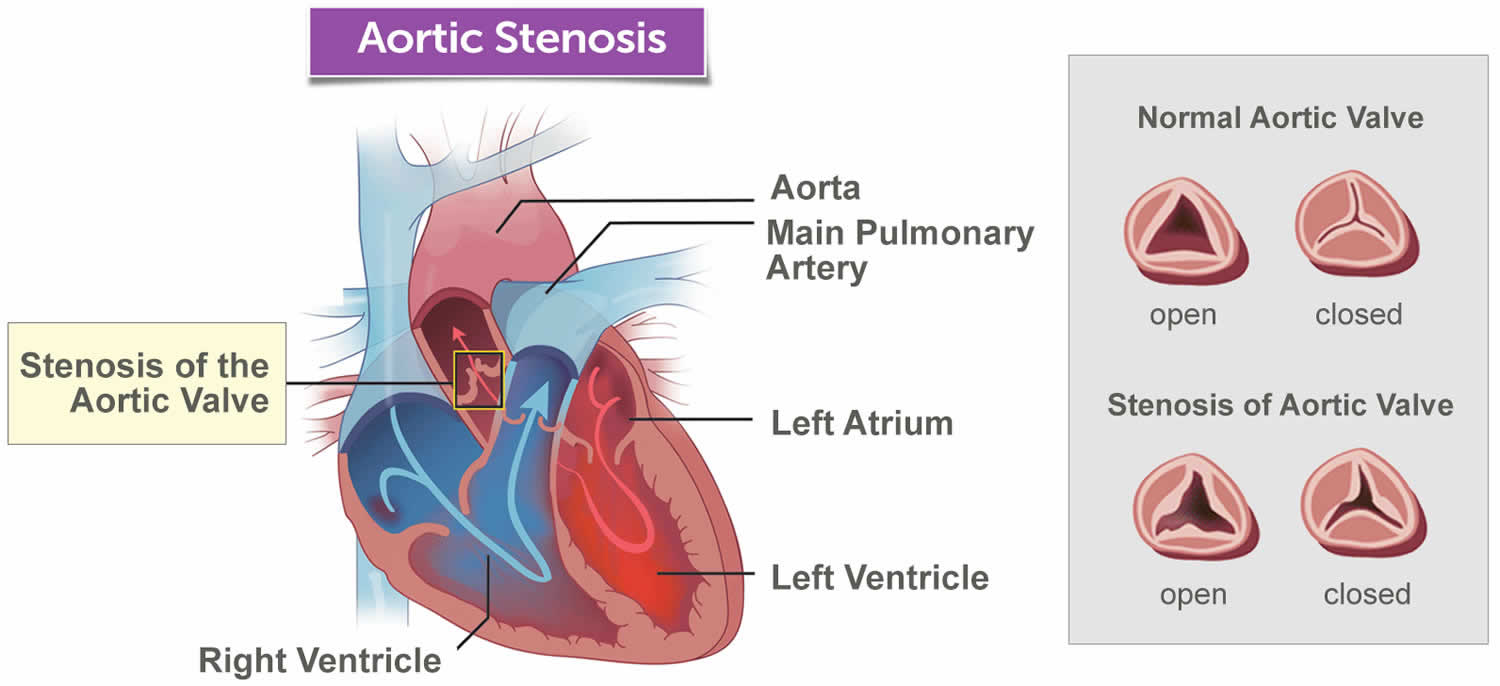
Figure 2. Aortic stenosis
Aortic stenosis causes
Around 4 in 1000 people are born with an aortic valve that is shaped differently. These people can get aortic stenosis earlier in life.
The most common cause of aortic stenosis is that the aortic valve can get hardened or scarred as people get older.
A less common cause of aortic stenosis is rheumatic heart disease.
Aortic valve stenosis can occur due to many causes, including:
Congenital heart defect
The aortic valve consists of three tightly fitting, triangular-shaped flaps of tissue called cusps. Some children are born with an aortic valve that has only two (bicuspid) cusps instead of three. People may also be born with one (unicuspid) or four (quadricuspid) cusps, but these are rare.
This defect may not cause any problems until adulthood, at which time the valve may begin to narrow or leak and may need to be repaired or replaced.
Having a congenitally abnormal aortic valve requires regular evaluation by a doctor to watch for signs of valve problems. In most cases, doctors don’t know why a heart valve fails to develop properly, so it isn’t something you could have prevented.
Calcium buildup on the valve
With age, heart valves may accumulate deposits of calcium (aortic valve calcification). Calcium is a mineral found in your blood. As blood repeatedly flows over the aortic valve, deposits of calcium can build up on the valve’s cusps. These calcium deposits aren’t linked to taking calcium tablets or drinking calcium-fortified drinks.
These deposits may never cause any problems. However, in some people — particularly those with a congenitally abnormal aortic valve, such as a bicuspid aortic valve — calcium deposits result in stiffening of the cusps of the valve. This stiffening narrows the aortic valve and can occur at a younger age.
However, aortic valve stenosis that is related to increasing age and the buildup of calcium deposits on the aortic valve is most common in older people. It usually doesn’t cause symptoms until ages 70 or 80.
Rheumatic fever
A complication of strep throat infection, rheumatic fever may result in scar tissue forming on the aortic valve. Scar tissue alone can narrow the aortic valve and lead to aortic valve stenosis. Scar tissue can also create a rough surface on which calcium deposits can collect, contributing to aortic valve stenosis later in life.
Rheumatic fever may damage more than one heart valve, and in more than one way. A damaged heart valve may not open fully or close fully — or both. While rheumatic fever is rare in the United States, some older adults had rheumatic fever as children.
Risk factors for aortic stenosis
Risk factors of aortic valve stenosis include:
- Older age
- Certain heart conditions present at birth (congenital heart disease) such as a bicuspid aortic valve
- History of infections that can affect the heart
- Having cardiovascular risk factors, such as diabetes, high cholesterol and high blood pressure
- Chronic kidney disease
- History of radiation therapy to the chest.
Aortic stenosis prevention
Some possible ways to prevent aortic valve stenosis include:
- Taking steps to prevent rheumatic fever. You can do this by making sure you see your doctor when you have a sore throat. Untreated strep throat can develop into rheumatic fever. Fortunately, strep throat can usually be easily treated with antibiotics. Rheumatic fever is more common in children and young adults.
- Addressing risk factors for coronary artery disease. These include high blood pressure, obesity and high cholesterol levels. These factors may be linked to aortic valve stenosis, so it’s a good idea to keep your weight, blood pressure and cholesterol levels under control if you have aortic valve stenosis.
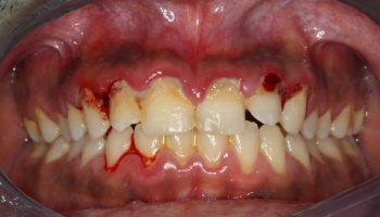
- Taking care of your teeth and gums. There may be a link between infected gums (gingivitis) and infected heart tissue (endocarditis). Inflammation of heart tissue caused by infection can narrow arteries and aggravate aortic valve stenosis.
Once you know that you have aortic valve stenosis, your doctor may recommend that you limit strenuous activity to avoid overworking your heart.
Aortic stenosis symptoms
Some people with aortic stenosis don’t have any symptoms, especially if the aortic valve is only a little narrower.
Others have:
- palpitations (heart racing or skipping a beat)
- fainting during exercise
- feeling tired or worn out
- chest pain
- feeling short of breath
Aortic valve stenosis ranges from mild to severe. Aortic valve stenosis signs and symptoms generally develop when narrowing of the valve is severe. Some people with aortic valve stenosis may not experience symptoms for many years. Signs and symptoms of aortic valve stenosis may include:
- Abnormal heart sound (heart murmur) heard through a stethoscope
- Chest pain (angina) or tightness with activity
- Feeling faint or dizzy or fainting with activity
- Shortness of breath, especially when you have been active
- Fatigue, especially during times of increased activity
- Heart palpitations — sensations of a rapid, fluttering heartbeat
- Not eating enough (mainly in children with aortic valve stenosis)
- Not gaining enough weight (mainly in children with aortic valve stenosis)
The heart-weakening effects of aortic valve stenosis may lead to heart failure. Heart failure signs and symptoms include fatigue, shortness of breath, and swollen ankles and feet.
If you’re having symptoms like this, it’s a good idea to see your doctor. If you have a heart murmur, your doctor may recommend that you visit a cardiologist.
Aortic stenosis complications
Aortic valve stenosis can cause complications, including:
- Heart failure
- Stroke
- Blood clots
- Bleeding
- Heart rhythm abnormalities (arrhythmias)
- Infections that affect the heart, such as endocarditis
- Death
Aortic stenosis diagnosis
To diagnose aortic valve stenosis, your doctor may review your signs and symptoms, discuss your medical history, and conduct a physical examination. Your doctor may listen to your heart with a stethoscope to determine if you have a heart murmur that may indicate an aortic valve condition. A doctor trained in heart disease (cardiologist) may evaluate you.
Your doctor may order several tests to diagnose your condition and determine the cause and severity of your condition. Tests may include:
- Echocardiogram. This test uses sound waves to produce video images of your heart in motion. During this test, specialists hold a wand like device (transducer) on your chest. Doctors may use this test to evaluate your heart chambers, the aortic valve and the blood flow through your heart. A doctor generally uses this test to diagnose your condition if he or she suspects you have a heart valve condition. This test can help doctors closely look at the condition of the aortic valve, and the cause and severity of your condition. It can also help doctors determine if you have additional heart valve conditions. Doctors may conduct another type of echocardiogram called a transesophageal echocardiogram (TEE) to get a closer look at the aortic valve. In this test, a small transducer attached to the end of a tube is inserted down the tube leading from your mouth to your stomach (esophagus).
- Electrocardiogram (ECG). In this test, wires (electrodes) attached to pads on your skin measure the electrical activity of your heart. An ECG can detect enlarged chambers of your heart, heart disease and abnormal heart rhythms.
- Chest X-ray. A chest X-ray can help your doctor determine whether your heart is enlarged, which can occur in aortic valve stenosis. It can also show whether you have an enlarged blood vessel (aorta) leading from your heart or any calcium buildup on your aortic valve. A chest X-ray can also help doctors determine the condition of your lungs.
- Exercise tests or stress tests. Exercise tests help doctors see whether you have signs and symptoms of aortic valve disease during physical activity, and these tests can help determine the severity of your condition. If you are unable to exercise, medications that have similar effects as exercise on your heart may be used.
- Cardiac computerized tomography (CT) scan. A cardiac CT scan uses a series of X-rays to create detailed images of your heart and heart valves. Doctors may use this test to measure the size of your aorta and look at your aortic valve more closely.
Cardiac MRI. A cardiac MRI uses magnetic fields and radio waves to create detailed images of your heart. This test may be used to determine the severity of your condition and evaluate the size of your aorta. - Cardiac catheterization. This test isn’t often used to diagnose aortic valve disease, but it may be used if other tests aren’t able to diagnose the condition or to determine its severity. In this procedure, your doctor threads a thin tube (catheter) through a blood vessel in your arm or groin and guides it to an artery in your heart. Doctors may inject a dye through the catheter, which helps your arteries become visible on an X-ray (coronary angiogram). This provides your doctor with a detailed picture of your heart arteries and how your heart functions. It can also measure the pressure inside your heart chambers.
Aortic stenosis treatment
Treatment for aortic valve stenosis depends on the severity of your condition, whether you’re experiencing signs and symptoms, and if your condition is getting worse.
If your symptoms are mild or you aren’t experiencing symptoms, your doctor may monitor your condition with regular follow-up appointments. Your doctor may recommend you make healthy lifestyle changes and take medications to treat symptoms or reduce the risk of complications.
You may eventually need surgery to repair or replace the diseased aortic valve. In some cases, your doctor may recommend surgery even if you aren’t experiencing symptoms. If you’re having another heart surgery, doctors may perform aortic valve surgery at the same time.
Surgery to repair or replace an aortic valve is usually performed through a cut (incision) in the chest. Less invasive approaches may be available, and your doctor will evaluate you to determine if you’re a candidate for these procedures.
If you have aortic valve stenosis, consider being evaluated and treated at a medical center with a multidisciplinary team of cardiologists and other doctors and medical staff trained and experienced in evaluating and treating heart valve disease. This team can work closely with you to determine the most appropriate treatment for your condition.
Aortic stenosis surgery
Many young people with aortic stenosis will need aortic valve replacement surgery at some point in their lives. Some of you will have needed this surgery in early childhood, while others could wait until now or will need surgery in the future.
The timing of your surgery depends on how narrow your aortic valve was at birth, how much further it has narrowed as you’ve grown (it almost always gets narrower with age), and how your heart is coping with the extra work. The surgery is usually delayed as long as possible, because an artificial valve will not grow as you do and will need to be replaced again.
You may need to have one or more of these procedures.
Balloon valvuloplasty
If your aortic valve was very narrow when you were a young child, you may have had balloon valvuloplasty. This stretched open your narrowed valve. Balloon valvuloplasty does not make your aortic valve normal and it does not always work, but in many cases it can widen your narrowed valve, helping to delay surgery.
Doctors may conduct a balloon valvuloplasty procedure using a long, thin tube (catheter) to repair aortic valve stenosis. In this procedure, a doctor inserts a catheter with a balloon on the tip into an artery in your arm or groin and guides it to the aortic valve. The doctor performing the procedure then inflates the balloon, which expands the opening of the valve. The balloon is then deflated, and the catheter and balloon are removed.
Balloon valvuloplasty procedure can treat aortic valve stenosis in infants and children. However, the aortic valve tends to narrow again in adults who’ve had the procedure, so it’s usually only performed in adults who are too ill for surgery or who are waiting for a valve replacement, as they typically need additional procedures to treat the narrowed valve over time.
Valvotomy
Valvotomy is an operation to open your narrowed aortic valve. Cutting your aortic valve open will almost always make your valve a bit leaky. It is not possible
to predict how big your leak will be but if very severe you will need a valve replacement.
Aortic valve replacement surgery
One option is to replace your aortic valve with an artificial metal one, because they last longer. This means that you will need to take an anticoagulant drug called warfarin to reduce the risk of a blood clot forming across your metal valve.
The second option is to use an animal or human tissue valve. You may have had the Ross Procedure. This used your pulmonary valve to replace your narrowed aortic valve, then a ‘tissue valve’ (usually from a pig, or a human valve) was used to replace your pulmonary valve.
Many children and teenagers lead normal, active lives after surgery. However, replacement tissues do not last as long as your own valve, so checkups in cardiac clinics are very important throughout your adult life.
Biological tissue valves degenerate over time and may eventually need to be replaced. People with mechanical valves will need to take blood-thinning medications for life to prevent blood clots. Your doctor will discuss with you the benefits and risks of each type of valve and discuss which valve may be appropriate for you.
Doctors may perform a less invasive procedure called transcatheter aortic valve replacement (TAVR) to replace a narrowed aortic valve. Transcatheter aortic valve replacement may be an option for people who are considered to be at intermediate or high risk of complications from surgical aortic valve replacement.
In transcatheter aortic valve replacement, doctors insert a catheter in your leg or chest and guide it to your heart. A replacement valve is then inserted through the catheter and guided to your heart. A balloon may expand the valve, or some valves can self-expand. When the valve is implanted, doctors remove the catheter from your blood vessel.
Doctors may also conduct a catheter procedure to insert a replacement valve into a failing biological tissue valve that is no longer working properly. Other catheter procedures to repair or replace aortic valves continue to be researched.
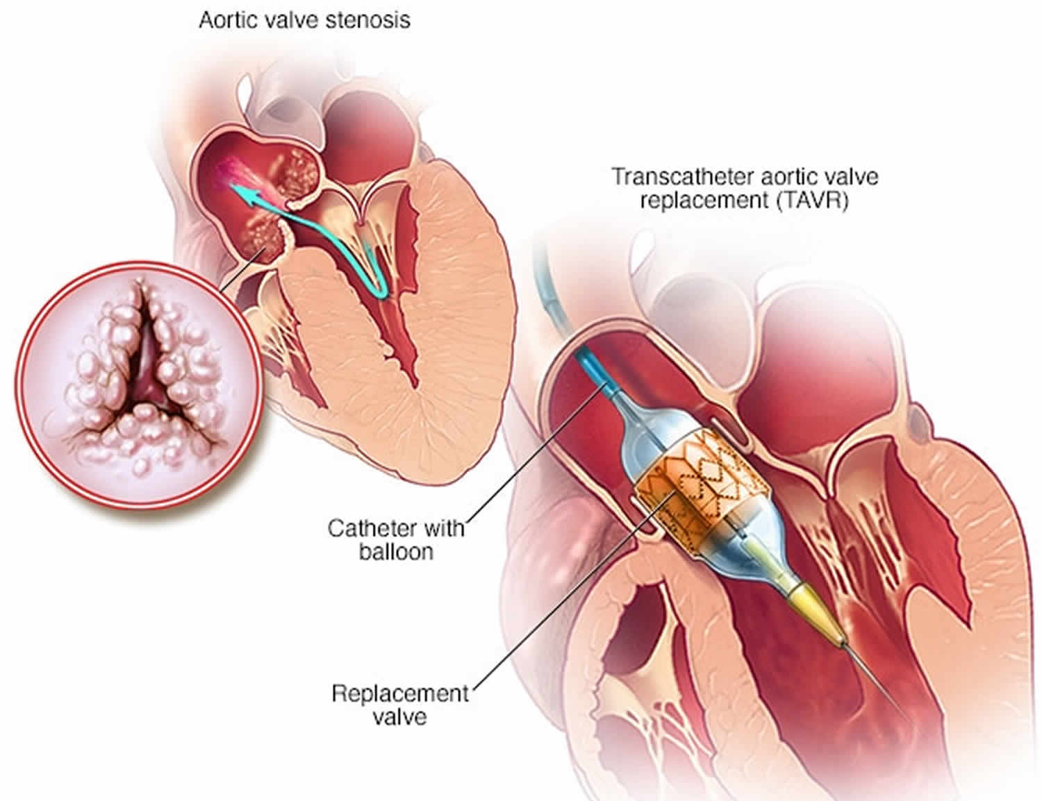
Figure 3. Transcatheter aortic valve replacement
Lifestyle and home remedies
You’ll have regular follow-up appointments with your doctor to monitor your condition. You’ll need to continue taking all your medications as prescribed.
Your doctor may suggest you incorporate several heart-healthy lifestyle changes into your life, including:
- Eating a heart-healthy diet. Eat a variety of fruits and vegetables, low-fat or fat-free dairy products, poultry, fish, and whole grains. Avoid saturated and trans fat, and excess salt and sugar.
- Maintaining a healthy weight. Aim to keep a healthy weight. If you’re overweight or obese, your doctor may recommend that you lose weight.
- Getting regular physical activity. Aim to include about 30 minutes of physical activity, such as brisk walks, into your daily fitness routine.
- Managing stress. Find ways to help manage your stress, such as through relaxation activities, meditation, physical activity, and spending time with family and friends.
- Avoiding tobacco. If you smoke, quit. Ask your doctor about resources to help you quit smoking. Joining a support group may be helpful.
Pregnancy
For women with aortic valve stenosis, it’s important to talk with your doctor before you become pregnant. Your doctor can discuss with you which medications you can safely take, and whether you may need a procedure to treat your valve condition prior to pregnancy.
If you are a girl who has a metal valve it is important that you do not have an unplanned pregnancy as taking warfarin anticoagulant during a pregnancy can harm your unborn baby.
You’ll likely require close monitoring by your doctor during pregnancy. Doctors may recommend that women with severe valve stenosis avoid pregnancy to avoid the risk of complications.
Endocarditis
To reduce your risk of getting endocarditis:
- Keep your teeth and mouth clean and have regular check-ups with a dentist
- Avoid body piercing and tattooing
- Never inject recreational drugs.


