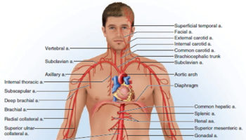
Brachiocephalic artery
The arch of the aorta is 4–5 cm (almost 2 in.) in length and is the continuation of the ascending aorta. Three major elastic arteries originate along the aortic arch (see Figure 2) and they are:
- The Brachiocephalic trunk,
- The Left Common Carotid artery, and
- The Left Subclavian artery.
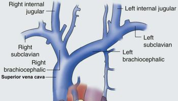
The first and largest branch from the arch of the aorta is the brachiocephalic trunk. It extends superiorly for a short distance, bending slightly to the right, and posterior to the right sternoclavicular joint, it divides to form the right subclavian artery and right common carotid artery.
There is only one brachiocephalic trunk; the left common carotid and left subclavian arteries arise separately from the aortic arch.
Figure 1. Brachiocephalic artery (brachiocephalic trunk)
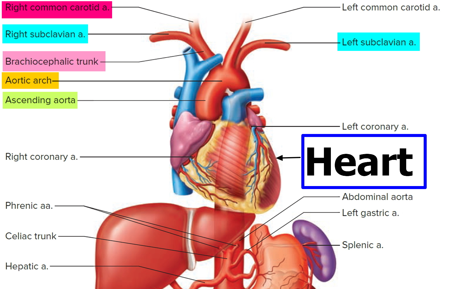
Figure 2. Brachiocephalic artery (brachiocephalic trunk) coming off the Aortic arch
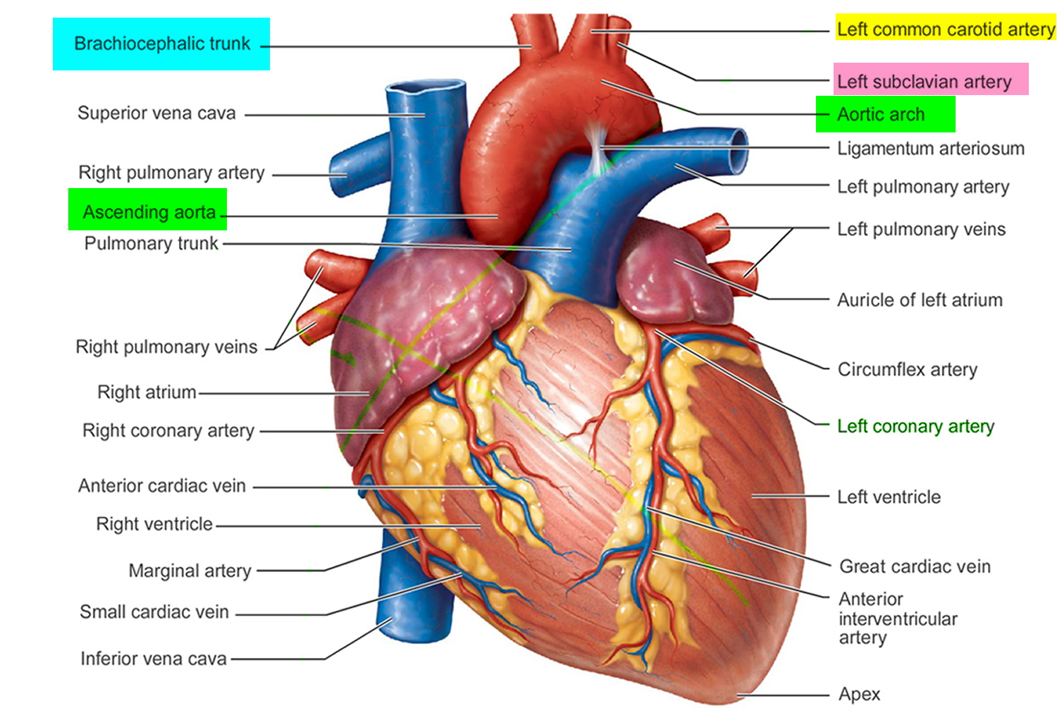
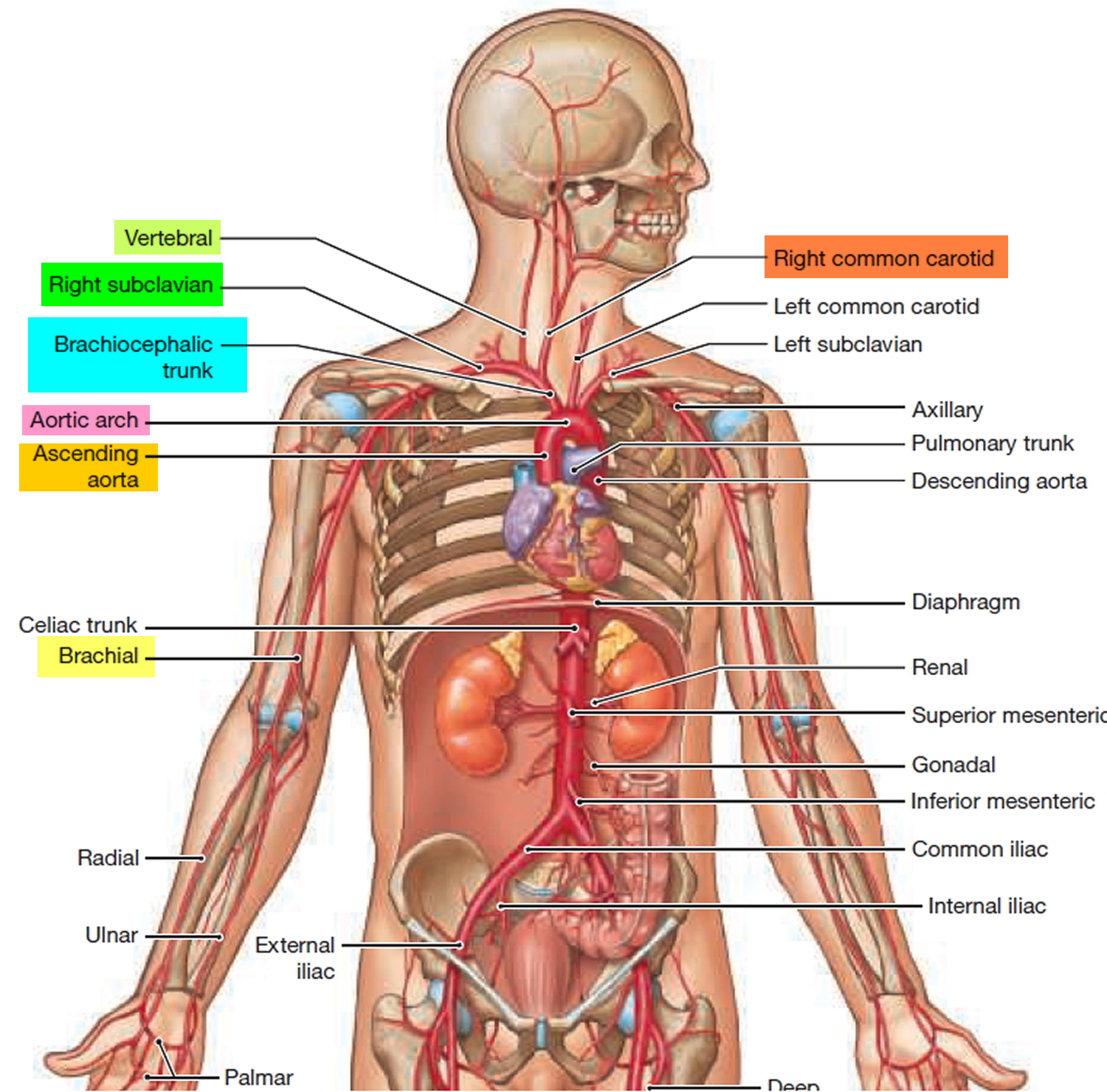
Figure 3. Brachiocephalic artery (brachiocephalic trunk) location
Brachiocephalic artery function
The brachiocephalic artery (brachiocephalic trunk) divides to form:
- Right subclavian artery and
- Right common carotid artery.
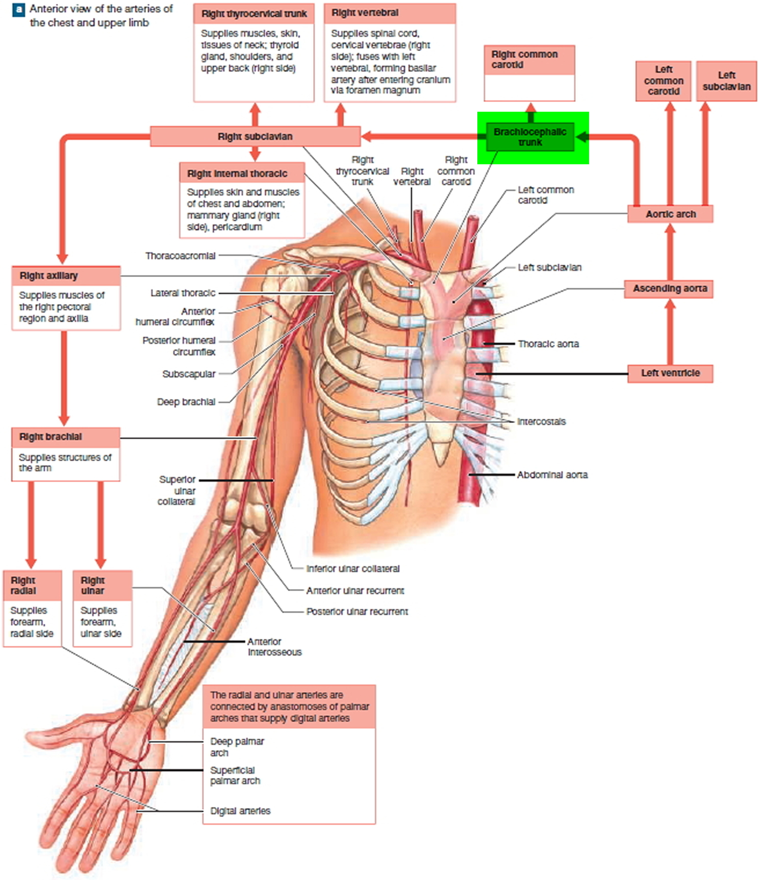
The right subclavian artery supply the upper limbs, chest wall, shoulders, back, brain, and spinal cord with blood. Three major branches arise from the right subclavian artery before they leave the thoracic cavity: (1) a right thyrocervical trunk, which supplies blood to muscles, skin, tissues of neck; thyroid gland, shoulders, and upper back (right side); (2) an right internal thoracic artery, supplying the skin and muscles of chest and abdomen; mammary gland (right
side), pericardium; and (3) a right vertebral artery, which supplies blood to the spinal cord, cervical vertebrae (right side); fuses with left vertebral, forming basilar artery after entering cranium via foramen magnum.
The right common carotid artery originates from the brachiocephalic trunk immediately posterior to the right sternoclavicular joint and is entirely in the neck throughout its course. Both right and left common carotid arteries ascend through the neck, just lateral to the trachea and esophagus, within a fascial compartment (the carotid sheath). They give off no branches as they pass through the neck. Near the superior edge of the thyroid cartilage each common carotid artery divides into its two terminal branches-the external and internal carotid arteries. At the bifurcation, the common carotid artery and the beginning of the internal carotid artery are dilated. This dilation is the carotid sinus and contains receptors that monitor changes in blood pressure and are innervated by a branch of the glossopharyngeal nerve [cranial nerve IX]. Another accumulation of receptors in the area of the bifurcation is responsible for detecting changes in blood chemistry, primarily oxygen content. This is the carotid body and is innervated by branches from both the glossopharyngeal [IX] and vagus [X] nerves.
Figure 4. Brachiocephalic artery major branches and functions
Internal carotid arteries
After its origin, the internal carotid artery ascends toward the base of the skull. It gives off no branches in the neck and enters the cranial cavity through the carotid canal in the petrous part of the temporal bone. The internal carotid arteries supply the cerebral hemispheres, the eyes and the contents of the orbits, and the forehead.
External carotid arteries
The external carotid arteries begin giving off branches immediately after the bifurcation of the common carotid arteries as follows:
- Superior thyroid artery: supplies the thyrohyoid muscle, internal structures of the larynx, sternocleidomastoid and cricothyroid muscles, thyroid gland
- Ascending pharyngeal artery: supplies pharyngeal constrictors and stylopharyngeus muscle, palate, tonsil, pharyngotympanic tube, meninges in posterior cranial fossa
- Lingual artery: Muscles of the tongue, palatine tonsil, soft palate, epiglottis, floor of mouth, sublingual gland
- Facial artery: All structures in the face from the inferior border of the mandible anterior to the masseter muscle to the medial corner of the eye, the soft palate, palatine tonsil, pharyngotympanic tube, submandibular gland
- Occipital artery: Sternocleidomastoid muscle, meninges in posterior cranial fossa, mastoid cells, deep muscles of the back, posterior scalp
- Posterior auricular artery: Parotid gland and nearby muscles, external ear and scalp posterior to ear, middle and inner ear structures
- Superficial temporal artery: Parotid gland and duct, masseter muscle, lateral face, anterior part of external ear, temporal is muscle, parietal and temporal fossae
- Maxillary artery: External acoustic meatus, lateral and medial surface of tympanic membrane, temporomandibular joint, dura mater on lateral wall of skull and inner table of cranial bones, trigeminal ganglion and dura in vicinity, mylohyoid muscle, mandibular teeth, skin on chin , temporalis muscle, outer table of bones of skull in temporal fossa, structures in infratemporal fossa, maxillary sinus, upper teeth and gingivae, infra-orbital skin, palate, roof of pharynx, nasal cavity.
Figure 5. Carotid arteries
