Supplements

Red yeast rice reviews

red yeast rice

What is red yeast rice

Red yeast rice also known as Hongqu, red Koji, red fermented rice, red kojic rice, red koji rice, anka, or ang-kak, is a bright reddish purple fermented rice, which acquires its color from being cultivated with the mold Monascus purpureus (red yeast of Aspergillaceae family). Red yeast rice is a remedy belonging to Traditional Chinese Medicine, largely used nowadays as dietary supplement in Western countries to improve blood circulation by decreasing cholesterol and triglyceride levels 1. China is the world’s largest producer of red yeast rice. Red yeast rice supplements contain monacolin K (also known as Mevinolin), which is chemically identical to lovastatin, a licensed drug 2. Lovastatin is associated with various adverse effects such as myopathy and abnormal liver function test results, which can lead to serious problems if patients are not monitored and treated 3. Red yeast rice is often used by statin‐intolerant patients, although there are very few studies testing red yeast rice safety profile as compared to statins. Red yeast rice products may not be safe; some may have the same side effects as certain cholesterol-lowering drugs, and some may contain a potentially harmful contaminant. Clinical trials have reported no significant serious adverse events, but minor events include heartburn 4, flatulence 5, dizziness 4, myalgia 6 and loose stools 6.

Red yeast rice is produced by first soaking in water until the rice grains are fully saturated. The raw soaked rice can then either be directly inoculated or it can be steamed for the purpose of sterilizing and cooking the grains prior to inoculation. Inoculation is done by mixing a food fungus of the Monascus genus, mainly Monascus purpureus spores or powdered red yeast rice together with the rice that is being treated. The mix is then incubated in an environment around room temperature for 3–6 days. During this period of time, the rice should be fully cultured with Monascus purpureus, with each rice grain turning bright red in its core and reddish purple on the outside. During fermentation the naturally produced pigments give the characteristic red color to the rice and monacolins are produced. As part of the Chinese diet, Red yeast rice is used as food additive to increase the color of meat, fish and soybean products; some preparations of red yeast rice are used in food products in Chinese cuisine, including Peking duck. Moreover, it is recognized as a folk medicine for improving food digestion and blood circulation 7. The fully cultured rice is then either sold as the dried grain, or cooked and pasteurized to be sold as a wet paste, or dried and pulverized to be sold as a fine powder.

Key facts

  • Some red yeast rice products contain substantial amounts of monacolin K, which is chemically identical to the active ingredient in the cholesterol-lowering drug lovastatin. Lovastatin is one of the drugs in the category known as statins. These drugs lower blood cholesterol levels by reducing the production of cholesterol by the liver. These products may lower blood cholesterol levels and can cause the same types of side effects and drug interactions as lovastatin.
  • Other red yeast rice products contain little or no monacolin K. It is not known whether these products have any effect on blood cholesterol levels.
  • Consumers have no way of knowing how much monacolin K is present in most red yeast rice products. The labels on these products usually state only the amount of red yeast rice that they contain, not the amount of monacolin K.
  • The composition of red yeast rice products varies depending on the yeast strains and culture conditions used to manufacture them. The strains and conditions used to produce culinary red yeast rice differ from those used to produce products that are intended to lower cholesterol. Tests performed by the U.S. Food and Drug Administration (FDA) indicate that the red yeast rice sold as a food product contains only traces of monacolin K or none at all.
  • The U.S. Food and Drug Administration (FDA) has determined that red yeast rice products that contain more than trace amounts of monacolin K are unapproved new drugs and cannot be sold legally as dietary supplements.
  • Some red yeast rice products contain a contaminant called citrinin, which can cause kidney failure.
  • Tell all your health care providers about any complementary health approaches you use. Give them a full picture of what you do to manage your health. This will help ensure coordinated and safe care.

In 1998, the FDA determined that a red yeast rice product that contained a substantial amount of monacolin K was an unapproved new drug, not a dietary supplement. On several occasions since then, the FDA has taken action against companies selling red yeast rice products that contain more than trace amounts of monacolin K, warning them that it is against the law to market these products as dietary supplements.

Despite the FDA actions, some red yeast rice products currently on the market in the United States may contain monacolin K. (Some products tested as recently as 2011 have been found to contain it in substantial amounts.) Other products may contain little or none of this component. Consumers have no way of knowing how much monacolin K is present in most red yeast rice products, and therefore have no way of knowing whether a particular product is safe, effective, or legal. The labels on these products usually state only the amount of red yeast rice that they contain, not the amounts of monacolin K or other monacolins.

Several studies have shown the lipid‐lowering effects of red yeast rice in humans, especially in comparison with placebo in patients intolerant to statins 8, 9, although only a minority of trials compared head‐to‐head red yeast rice with statins or ezetimibe 10, 11, and some quality issues (e.g., allocation concealment and blinding) have been raised 12.

If you are considering red yeast rice

  • Do not use red yeast rice to replace conventional care or to postpone seeing your health care provider about a health problem.
  • Do not use red yeast rice dietary supplements if you are pregnant, trying to become pregnant, or nursing a child. If you are considering giving a child a red yeast rice dietary supplement, it is especially important to consult the child’s health care provider.
  • Do not take red yeast rice in addition to prescription statin drugs.
  • Many Web sites, including sales sites, have information about red yeast rice. Be cautious when you evaluate information from the Web; not all of it is trustworthy.
  • Federal regulations for dietary supplements are very different from—and less strict than—those for drugs.
  • Tell all your health care providers about any complementary health approaches you use. Give them a full picture of what you do to manage your health. This will help ensure coordinated and safe care.

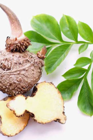
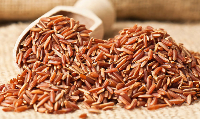
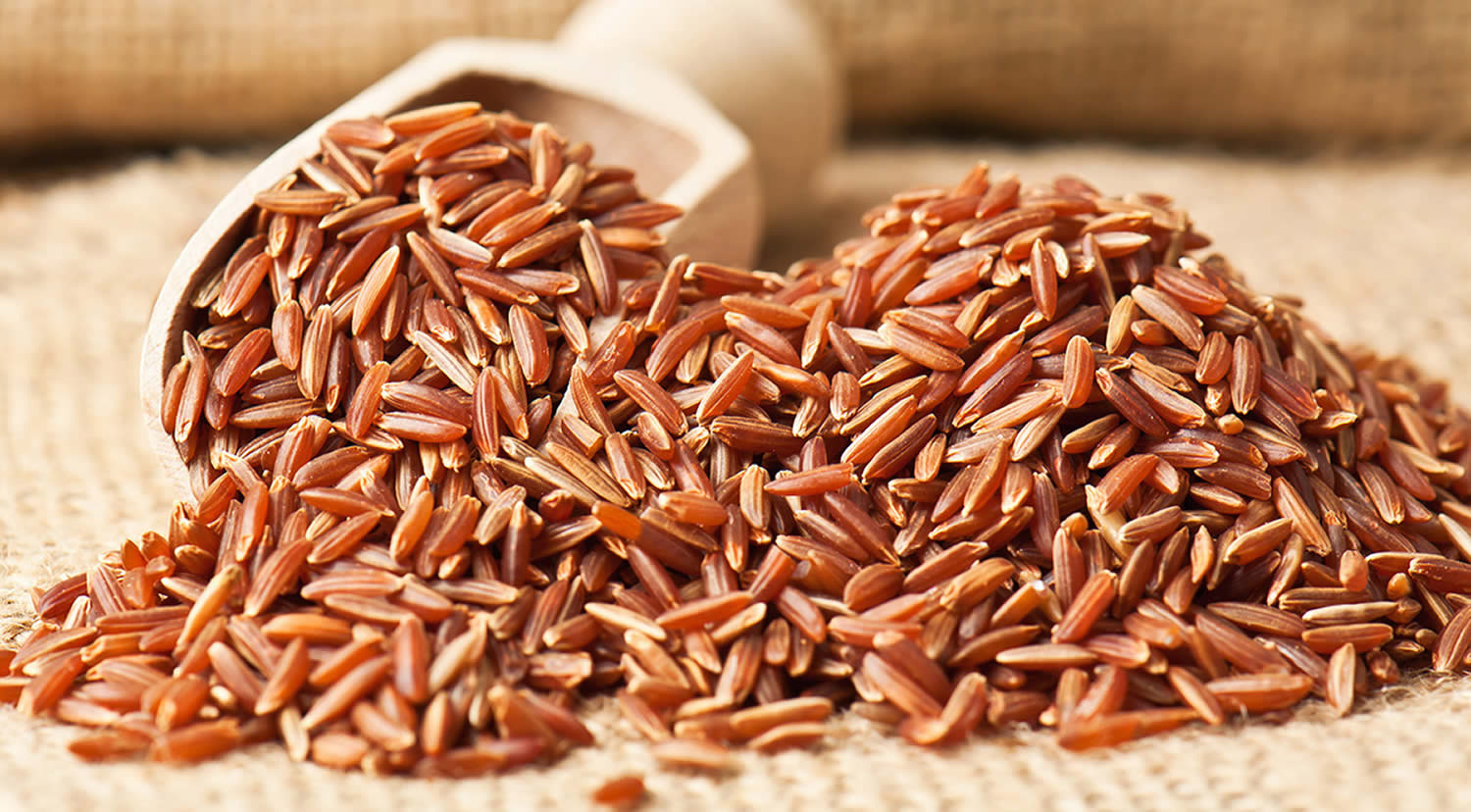
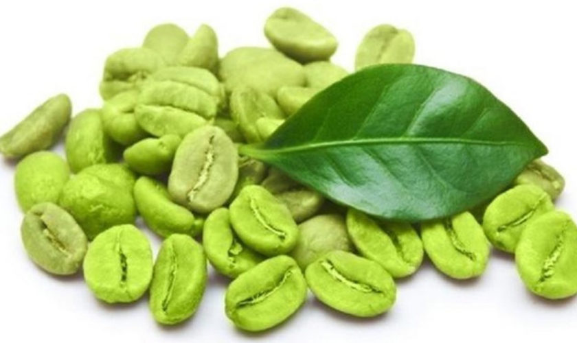
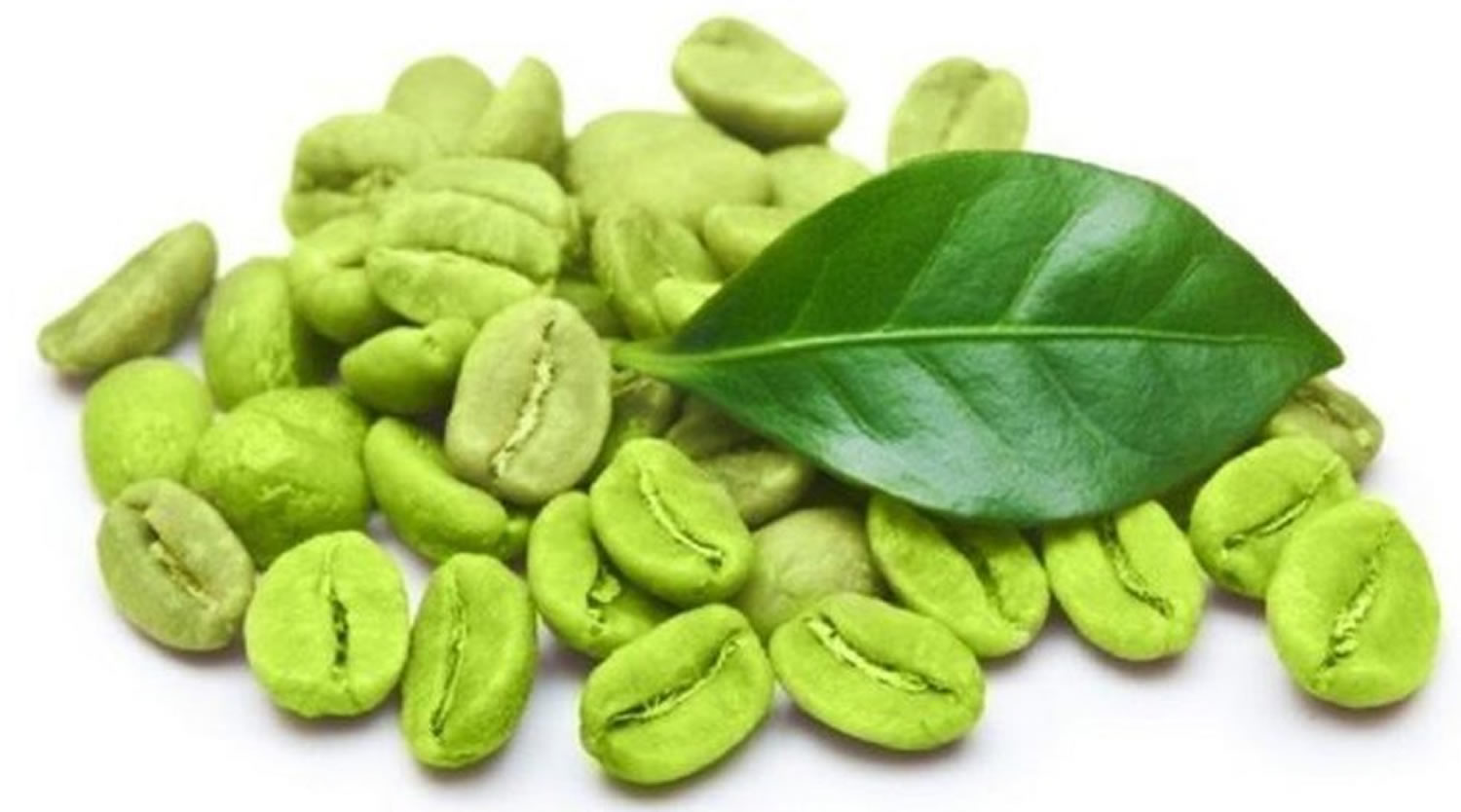
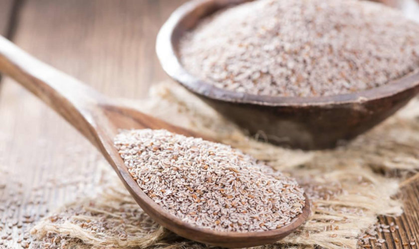
Figure 1. Red yeast rice

red yeast rice

Red yeast rice benefits

Red yeast rice products that contain substantial amounts of monacolin K can lower blood cholesterol levels. Researchers have not reported results of any studies of red yeast rice products that contain little or no monacolin K, so whether these products have any effect on blood cholesterol is unknown.

Red yeast rice for cholesterol

In clinical trials (studies in people) of red yeast rice products that contained substantial amounts of monacolin K, the products lowered blood levels of total cholesterol and low-density lipoprotein (LDL) cholesterol (the so-called bad cholesterol that is linked to increased heart disease risk). It is important to emphasize that all of these clinical trials used products that contained substantial amounts of monacolin K. A 2011 analysis showed that some of the red yeast rice products on the market contain very little monacolin K. These products may have little or no effect on blood cholesterol levels. Therefore, even though the participants in the clinical trials were able to lower their cholesterol levels by taking red yeast rice, you might not be able to achieve the same results.

In one of the clinical trials, the tested product produced a cholesterol-lowering effect greater than would be expected based on its monacolin K content. Further investigations, supported by the National Center for Complementary and Integrative Health, suggested that other monacolins or other substances present in the product may have contributed to its cholesterol-lowering effect.

This systematic review 13 assessed the efficacy and safety of red yeast rice versus simvastatin in the management of dyslipidemia. The evidence suggests that red yeast rice and simvastatin have similar effects in reducing total cholesterol, LDL “bad” cholesterol and triglyceride and increasing HDL “bad” cholesterol. The same adverse events reported between the red yeast rice and simvastatin groups were upset stomach 14, nausea 15, abdominal distention 15, aspartate aminotransferase (AST) increased 15, alanine aminotransferase (ALT) increased 16 and anorexia 17. One trial 18 reported ‘gastrointestinal symptoms’ as an adverse event in both treatment groups with no further description of symptoms. Dry mouth 16 and creatine kinase increased 19 were reported only in the simvastatin group. However, because the included trials in this review were of low quality, the review authors are unable to conclusively suggest replacing simvastatin with red yeast rice for the management of dyslipidaemia particularly as red yeast rice is not standardized in active drug content 13. Furthermore, all trial subjects were recruited from Chinese populations 13. Hence, the results of this review may not be applicable to other populations. Therefore, the beneficial effects of red yeast rice over simvastatin need to be established using trials with larger sample size, diverse population, improved methodology and longer duration of treatment 13.

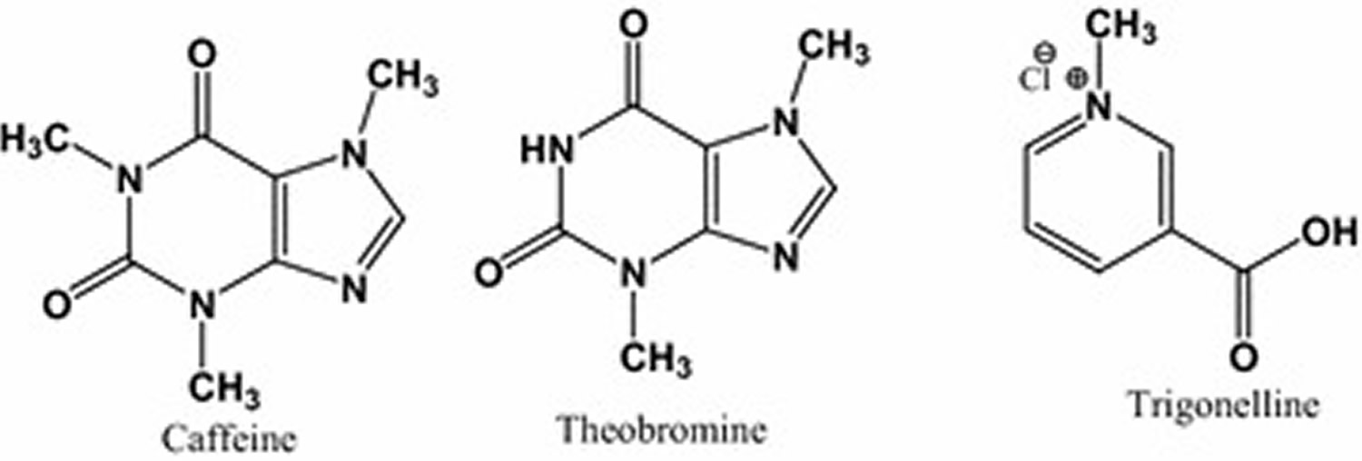
These properties of red yeast rice are due to its monacolins content, a family of naturally occurring substances that inhibit 3-hydroxy-3-methylglutaryl coenzyme A (HMG-CoA) reductase, the rate‐limiting step in cholesterol synthesis. Monacolins content in red yeast rice is usually around 0.4% w/w; of this about 90% consists of monacolin K (also known as Mevinolin) which is chemically identical to lovastatin. Other chemical components of red yeast rice are fatty acids and the pigments monascidin A, ankaflavin, monascorubrine and monascorubramine. A further pigment citrinin, a nephrotoxic mycotoxin, can be produced by some strains of Monascus 20. A dose of red yeast rice containing about 5–7 mg of monacolin K is considered as effective in lowering cholesterol as 20–40 mg of pure lovastatin, probably owing to the presence of other active monacolins or to an increased bioavailability of lovastatin when given as red yeast rice 21. Clinical studies suggest that red yeast rice has the potential to reduce serum LDL “bad” cholesterol levels by 10% to 33% 22, 23 and total cholesterol, triglyceride and as well as at increasing HDL “good” cholesterol in people with moderate hypercholesterolemia, but those indicators return to the baseline level when the treatment is discontinued 24. This phenomenon is similar to statins, which could be explained by the main components of red yeast rice (monacolins, which are capable of inhibiting the HMG-CoA reductase enzyme). In addition to lovastatin, most red yeast rice preparations contain other active substances such as Coenzyme Q10 and isoflavones 25. Moreover, a recent study showed that the oral bioavailability of lovastatin is significantly improved in red yeast rice products as a result of a higher dissolution rate and reduced crystallinity. This indicates that probably the activity of red yeast rice is much higher than predicted based on the very low doses generally given to patients 26. However, whether red yeast rice preparations should be used as drugs or dietary supplements is still inconclusive. In the US, the FDA recognizes these supplements as drugs when they contain a standardized, specific amount of lovastatin 27. Despite the lipid regulating benefits, some other positive effects of red yeast rice have also been found in studies, such as improving endothelial function and insulin resistance in patients with mild or moderate hypercholesterolemia, reducing high-sensitivity C-reactive Protein (hs-CRP) and markers of vascular remodeling in Italian subjects. Moreover, red yeast rice was proved effective, safe and well tolerated in nephrotic dyslipidemia both in adults and children and in familial hypercholesterolemia children 28.

Tolerability of Red Yeast Rice Products

Two studies supported by National Center for Complementary and Integrative Health have indicated that some people who had been unable to tolerate statin drugs because of side effects (muscle pain or weakness) were able to tolerate red yeast rice. It is uncertain whether the smaller amount of monacolin K in the red yeast rice products, as compared with the amounts of active ingredients in the drugs, accounted for the greater tolerability or whether other factors were responsible.

Red yeast rice dosage

Based on Chinese clinical trials, these are the dosage used: in 19 trials prescribed Xuezhikang 600 mg once daily, one trial used Xuezhikang 600 mg three times daily if the serum total cholesterol or triglyceride is still higher after having been prescribed for 6 weeks (600 mg twice daily in previous 6 weeks) 29, one trial 30 prescribed Xuezhikang 300 mg three times daily, and one trial 31 prescribed Xuezhikang 1200 mg at night. The duration of treatment ranged from 4 weeks to 7 years 32. According to the systematic review of Xuezhikang authors (Shang Q, Liu Z, Chen K, Xu H, Liu J. A Systematic Review of Xuezhikang, an Extract from Red Yeast Rice, for Coronary Heart Disease Complicated by Dyslipidemia. Evidence-based Complementary and Alternative Medicine : eCAM. 2012;2012:636547. doi:10.1155/2012/636547. https://www.ncbi.nlm.nih.gov/pmc/articles/PMC3332166/)), “before recommending Xuezhikang as an alternative to statins in coronary heart disease patients, more rigorous trials with high quality are needed to give high level of evidence, especially for comparing the effectiveness and safety between Xuezhikang and statins”.

Xuezhikang is a partially purified extract of fermented red yeast rice (Monascus purpureus). It is composed of 13 kinds of natural statins, unsaturated fatty acids, ergosterol, amino acids, flavonoids, alkaloid, trace element, and so forth 32.

In other clinical trials conducted outside of China, their results are listed in Table 1. The red yeast rice dose per day used range from 500 mg to 3600 mg 33. This meta-analysis included 13 random, placebo-controlled trials containing 804 participants. Red yeast rice showed significant lowering effects on total total cholesterol, triglyceride, and LDL “bad” cholesterol, but did not show a significant increasing effect on HDL “good” cholesterol compared with the placebo group which was different from the result of the last meta-analysis where most of the trials used Xuezhikang as intervention 34. That Chinese meta-analysis suggested that the lipid modification effects of red yeast rice preparations appeared to be similar to simvastatin, atorvastatin, pravastatin, lovastatin or fluvastatin 34.

Table 1. Basic characteristics of included red yeast rice trials

Referencesclinical trial sitesSample size(I/C)InterventionControlDoses of red yeast rice per dayDoses of lovastatinOther ingredientsDuration of treatment
Ogier 2013 35France19/20Compoundsplacebo500 mg2 mgSCdP, artichoke leaf extract16 weeks
Barrat 2013 36France50/50Compoundsplacebo500 mg2 mgpolicosanol, artichoke leaf extract16 weeks
Barrat 2012 37France15/15/15CompoundsplaceboI1:500 mg/I2:1000 mgI1:2 mg/I2:4 mgpolicosanol, artichoke leaf extract4 weeks
Lee 2012 38Taiwan54/52Compoundsplacebo1110 mgNo mentionbitter gourd, chlorella, soybean, licorice12 weeks
Karl 2012 39USA26/25Compoundsplacebo1200 mg4.8 mgniacin, phytosterol esters, L-carnitine, vitamin C,CoQ-108 weeks
Higashikawa 2012 40Japan28/27Compoundsplacebo900 mg2 mgGarlic12 weeks
Marazzi 2011 41Italy40/40Compoundsplacebo200 mgNo mentionberberine, policosanol, folic acid, CoQ-10, astaxanthin12 months
Bogsrud 2010 42Norway22/20Red yeast riceplacebo1200 mg7.2 mgno16 weeks
Affuso 2010 43Italy25/25Compoundsplacebo200 mg3 mgberberine, policosanols6 weeks
Yang 2009 44Taiwan19/10Compoundsplacebo1200 mgNo mentionnattokinase6 months
Becker 2009 45America31/31Red yeast riceplacebo3600 mg6.12 mgno24 weeks
Huang 2007 46Taiwan39/40Red yeast riceplacebo1200 mg11.4 mgno8 weeks
Heber 1999 47USA42/41Red yeast riceplacebo2400 mg7.2 mgno12 weeks

Abbreviations: RYR = Red yeast rice, I = Intervention group, C = Control group, I1 = high dose group, I2 = low dose group.

[Source 33]

Red yeast rice side effects

Statin drugs such as lovastatin are associated with various side effects such as headache, dizziness, rash, upset stomach, and hepatic dysfunction 3. The most common adverse effect is muscle weakness, which can be a sign of more serious myopathy or, in rare cases, rhabdomyolysis. Double-blind, controlled clinical trials have demonstrated that red yeast rice is effective and well tolerated in a wide range of patients 48; however, case reports have linked it to muscular myopathy and rhabdomyolysis.

Red yeast rice safety

  • The same types of side effects that can occur in patients taking lovastatin as a drug can also occur in patients who take red yeast rice products that contain monacolin K. Potential side effects include myopathy (muscle symptoms such as pain and weakness), rhabdomyolysis (a condition in which muscle fibers break down, releasing substances into the bloodstream that can harm the kidneys), and liver toxicity. Each of these three side effects has been reported in people who were taking red yeast rice.
  • Red yeast rice supplements should not be used while pregnant or breastfeeding.
  • Lovastatin can interact with a variety of drugs to increase the risk of rhabdomyolysis; these drugs include other cholesterol-lowering agents, certain antibiotics, the antidepressant nefazodone, drugs used to treat fungal infections, and drugs used to treat HIV infection. Red yeast rice containing monacolin K could interact with drugs in the same way.
  • If the process of culturing red yeast rice is not carefully controlled, a substance called citrinin can form. Citrinin has been shown to cause kidney failure in experimental animals and genetic damage in human cells. In a 2011 analysis of red yeast rice products sold as dietary supplements, 4 of 11 products were found to contain this contaminant.

Red yeast rice caused or exacerbated myopathy marked by elevated serum creatine kinase levels. Rhabdomyolysis, the most severe adverse effect associated with statins, occurred in a renal transplant patient who used red yeast rice while concomitantly taking cyclosporine 49. Lovastatin as a prescription drug is contraindicated in pregnancy and is a Category X agent. Category X means studies in animals or humans have demonstrated fetal abnormalities and/or there is positive evidence of human fetal risk based on adverse reaction data from investigational or marketing experience, and the risks involved in use of the drug in pregnant women clearly outweigh potential benefits. This labeling is a main reason for the FDA’s rejection of the application submitted by Merck to sell lovastatin over the counter 50. Red yeast rice, when used by pregnant women, places the fetus at unnecessary risk of central nervous system defects during the first trimester. Although red yeast rice contains a lower dose of lovastatin compared with the FDA-approved product, the risk posed may be similar.

Overall, the findings from Adverse Reactions Surveillance System of Natural Health Products raise the hypothesis that the safety profile of red yeast rice is similar to synthetic statins:

  • Myopathies (muscle pain, creatine kinase and transaminases elevation) 51,
  • Cutaneous reactions, gastrointestinal and liver reactions emerged as a potential safety signals of red yeast rice and have been reported as not uncommon adverse reactions associated with statins 52.

These data are also in line with cases collected from WHO‐Vigibase and reports received by the ANSES French Agency, where muscle‐ and liver‐related adverse reactions were largely reported with red yeast rice 30. In 14 cases (27%), the reactions were serious and mostly required hospitalization, especially those related to liver injury. Most of the reactions involved women, probably because they use more dietary supplements than men 53. Notably, some patients who showed muscular adverse reactions associated with statin switched to red yeast rice supplements as an alternative and experienced similar adverse effects 54. The time to onset of muscle‐ and liver‐related side effects was relatively rapid: one third of cases occurred during the first month of treatment, and two‐thirds within 60 days.

Therefore, you should know that monacolin K contained in red yeast rice is identical to lovastatin, and you should consider early monitoring of your liver function and become aware of signs of muscle injury. Furthermore, consumers should be discouraged from using red yeast rice preparations as self‐medication, particularly if they have experienced previous adverse reactions to statins 54. When self‐prescribed, without medical advice and monitoring and possibly for the long term, patients should be aware they are consuming an active substance, with both therapeutic and toxic effects. In fact, even serious adverse reactions, such as hepatitis or rhabdomyolysis, can remain asymptomatic for long periods with the risk of organ failure progression. In contrast, when statins are taken under medical control, blood tests to check creatine kinase and organ function are performed periodically so that statin use can be stopped as soon as abnormal results are shown.

The potential safety signals of myopathies and liver injury raise the hypothesis that the safety profile of red yeast rice is similar to that of statins.

Red yeast rice complications

Musculoskeletal and connective tissue disorders

Muscular pain, with or without indication of creatine phosphokinase (creatine kinase) increase, occurred in 19 patients, one case of rhabdomyolysis was also recorded. Muscle pain affected generally the lower extremities and cramps were sometimes present. The increase in creatine kinase values was mild in some cases (about 200 U/L or less), high in others (from 288 to 732 U/L) and very high (up to ten times the normal concentrations) in two patients; in the rhabdomyolysis case, the creatine kinase level reached 12,245 U/L. The latency of the reaction was variable, ranging between 9 days and more than one year; however, about one third of the reactions occurred within 30 days and 63% within two months 54. Several patients were taking medications, most of which are not known to be associated with muscular disorders, with exception of venlafaxine 55; this evidence was taken into account in the causality assessment. Noteworthy, some patients were using red yeast rice preparations as an alternative option, being intolerant to statins, while some patients previously experienced myopathy and creatine kinase increase associated with statin use; patient #15, who showed rhabdomyolysis, presented a previous identical reaction after taking statins; in these cases red yeast rice use was considered a positive rechallenge.

The causality assessment, according to the WHO scale, resulted as possible in 8 cases, probable in 11 cases and certain in case #15 54.

Gastrointestinal disorders

Gastrointestinal reactions occurred in 12 patients and consisted mostly in dyspepsia, nausea, vomiting and abdominal pain, sometimes in diarrhea 54. The reactions occurred within one or two days or after 3–4 weeks of treatment 54. The reactions were in general not serious, and only one case needed hospitalization 54. Dechallenge was always positive 54. Nevertheless, in two patients the reaction, even if improved, was persistent. Five patients had a positive rechallenge. Some cases were judged as possible because similar reactions were reported for concomitant drugs; for example, abdominal pain has been reported in more than 1% of hypertensive patients who received moexipril/hydrochlorotiazide. In causality assessment a conservative approach was used, taking in account that gastrointestinal symptoms such as nausea, vomiting and impaired digestion do not represent specific medical disorders and are common side‐effects of drugs, a positive response to withdrawal and a positive rechallenge.

Hepatobiliary disorders

Hepatic reactions occurred in ten patients and consisted in acute hepatitis (six patients), that required hospitalization, or in increase of hepatic enzymes. These six cases fulfilled criteria for drug‐induced liver injury, according to the international Expert Working Group 56. In one patient increase in aspartate aminotransferase (AST) occurred together with creatine phosphokinase (creatine kinase) increase. Nine cases were compatible with the definition of drug‐induced liver injury, as recommended by the International Drug Safety Department 57.

Time to onset was between two weeks and one year of treatment, 37% of the reactions occurred within one month and 75% within two months 54. Transaminases ranged from about twice to 80 times the upper normal level 54. In all cases, except for two in which it was not reported, dechallenge was positive. Patients were taking other drugs or dietary supplements, but generally these were not known to be associated with liver injury 54. Case 26 regarded a 49‐year‐old woman who had consumed Armolipid plus® for 50 days. She was hospitalized for suspected myocardial infarction (heart attack); the infarction was excluded, while an acute hepatitis was diagnosed with alanine aminotransferase (ALT) and aspartate aminotransferase (AST) values corresponding to 46 and 57 times the upper normal concentrations, respectively. Total bilirubin was normal, viral and autoimmune serology was negative and other drug and non‐drug causes of hepatitis were excluded; rhabdomyolysis was also excluded. The supplement consumption was suspended and the values decreased, returning to normal after 17 days. The causality assessment resulted in a score of 7 (probable).

Case 27 was a 68‐year‐old woman who had a history of hypercholesterolemia, for which she had been taking the supplement Armolipid plus® for 2 years. An increase of liver enzymes (AST = 87; ALT = 168) was discovered during a routine blood test, and an additional laboratory test reported total bilirubin 0.5, gamma-glutamyl transferase (GGT) 53, ALP 109. Creatine kinase was normal, thus suggesting that the increase in transaminases did not have a muscular aetiology. The ultrasound scan did not show clinically important signs of hepato‐biliary damage, except for moderate steatosis and lithiasis (previously documented). Major infective hepatitis was excluded, no recent flu‐like episodes or gastro‐intestinal symptoms were documented, no history of pre‐existing liver or biliary disease, blood transfusion or alcohol abuse was documented. Concomitant drugs included levothyroxine (for 7 years) and chenodeoxycholic acid (for 5 years). The product was suggested by healthcare professionals because of the patient’s complaints about symptomatic myopathy, probably related to the administration of statins. Notably, no elevation of creatine kinase or hepatic transaminases was previously documented when the woman was treated with statins. The supplement was suspended. Laboratory blood test, performed after less than 4 weeks, gave normal values for AST, ALT, gamma-glutamyl transferase (GGT) and ALP. Based on transaminases values, the liver injury was classified as hepatocellular and the application of the CIOMS/RUCAM scale resulted in a score of 5 (possible).

Skin and subcutaneous disorders

Nine cutaneous reactions associated with red yeast rice products were collected. Most of them occurred with a 1–4‐day latency, the others occurred after 10 days, 3 weeks or about 2 months of treatment 54. Four cases were serious and required hospitalization 54. In one case, which involved a 57‐year‐old woman, the reaction consisted in Pemphigus vulgaris, diagnosed by titration of anti‐desmoglein 1 and 3 antibodies and by biopsy. The reaction occurred 2 weeks after discontinuation of the supplement and needed a pharmacological therapy. The patient was not taking other drugs, but as she had experienced a previous episode of Pemphygus eight years before, then the reaction could be considered a relapse and the causality was judged as unlikely. Another patient a 75‐year‐old woman was admitted to hospital with severe urticaria, rash and edema of the lips. The reaction improved after discontinuation of the red yeast rice supplement but recurred after 6 days, without rechallenge. It is to be underlined that this patient, even without predisposing conditions, was taking various medications (cardioaspirin, amiloride plus hydrochlorothiazide, triazolam, metoprolol, lansoprazole, risedronate), but, most importantly, received flu vaccine 5 days before the onset of the reaction, so, also in this case, the causality was judged as unlikely. In other cases, the causality was judged as possible despite a positive outcome and dechallenge, because patients were taking other drugs, some of which (amiloride plus hydrochlorothiazide, lansoprazole) have been reported to induce cutaneous adverse reactions 58. Another patient was hospitalized for DRESS (drug reaction with eosinophilia and systemic symptoms) syndrome, a severe drug‐induced reaction 59. Laboratory tests suggested that the patient had a Herpes simples virus infection, a condition associated with DRESS 60. Cases of DRESS are described also for atorvastatine 61, a lipid‐lowering drug belonging to the statins class (as monacolin contained in red yeast rice), as well as for omeprazole 62, a proton pump inhibitor similar to pantoprazole that the patient was consuming. Based on these considerations, the causality between DRESS and red yeast rice preparation was judged as possible. In the other cutaneous reactions the causality was considered as probable on the basis of the positive dechallenge, outcome and absence of alternative causes.

Others miscellaneous reactions

A case involved a 78‐year‐old woman who was taking warfarin and showed an increase of INR (international normalized ratio) after consumption of the red yeast rice supplement Armolipid plus®. The dose of warfarin was reduced and the INR value returned to normal. In this case a pharmacodynamic interaction between the anticoagulant drug and red yeast rice supplement can be hypothesized, even if the dechallenge was not done 54. Furthermore, three patients showed nausea, vertigo and hazy vision, tingling of extremities and tachycardia; the latter required hospitalization 54. Dechallenge was always positive and other causes were excluded, so the causality was judged as probable in all of them 54. Nevertheless, it has to be pointed out that in one case the supplement consumed (Fisiostatin®) contained, besides red yeast rice, green tea [Camellia sinensis (L.) O. Kuntze], a source of caffeine that could be responsible for the adverse reaction 54.

  1. Smith DJ, Olive KE. Chinese red rice–induced myopathy. South Med J. 2003;96(12):1265–1267. https://www.ncbi.nlm.nih.gov/pubmed/14696880[]
  2. Lachenmeier DW, Monakhova YB, Kuballa T et al. NMR evaluation of total statin content and HMG‐CoA reductase inhibition in red yeast rice (Monascus spp.) food supplements. Chin Med, 2012;7:8.[]
  3. Klimek M, Wang S, Ogunkanmi A. Safety and Efficacy of Red Yeast Rice (Monascus purpureus) as an Alternative Therapy for Hyperlipidemia. Pharmacy and Therapeutics. 2009;34(6):313-327. https://www.ncbi.nlm.nih.gov/pmc/articles/PMC2697909/[][]
  4. Wang J, Lu Z, Chi J et al. Multicenter clinical trial of the serum lipid‐lowering effects of a Monascus purpureus (red yeast) rice preparation from traditional Chinese medicine. Curr Ther Res, 1997;58:964–978.[][]
  5. Huang CF, Li TC, Lin CC, Liu CS, Shih HC, Lai MM. Efficacy of Monascus purpureus Went rice on lowering lipid ratios in hypercholesterolemic patients. Eur J Cardiovasc Prev Rehabil, 2007;14:438–440.[]
  6. Heber D, Yip I, Ashley JM, Elashoff DA, Elashoff RM, Go VLW. Cholesterol‐lowering effects of a proprietary chinese red‐yeast‐rice dietary supplement. Am J Clin Nutr, 1999;69:231–236.[][]
  7. Ma J, Li Y, Ye Q, Li J, Hua Y, Ju D, et al. Constituents of red yeast rice, a traditional Chinese food and medicine. J Agric Food Chem 2000; 48: 5220–5225 https://www.ncbi.nlm.nih.gov/pubmed/11087463[]
  8. Moriarty PM, Roth EM, Karns A, Ye P, Zhao SP, Liao Y, et al. Effects of Xuezhikang in patients with dyslipidemia: a multicenter, randomized, placebo‐controlled study. J Clin Lipidol 2014; 8: 568–575.[]
  9. Gonnelli S, Caffarelli C, Stolakis K, Cuda C, Giordano N, Nuti R. Efficacy and tolerability of a nutraceutical combination (red yeast rice, policosanols, and berberine) in patients with low‐moderate risk hypercholesterolemia: a double‐blind, placebo‐controlled study. Curr Ther Res Clin Exp 2014; 77: 1–6.[]
  10. Marazzi G, Pelliccia F, Campolongo G, Quattrino S, Cacciotti L, Volterrani M, et al. Usefulness of nutraceuticals (Armolipid plus) versus ezetimibe and combination in statin‐intolerant patients with dyslipidemia with coronary heart disease. Am J Cardiol 2015; 116: 1798–1801[]
  11. Ong YC, Aziz Z. Systematic review of red yeast rice compared with simvastatin in dyslipidaemia. J Clin Pharm Ther 2016; 41: 170–179.[]
  12. Childress L, Gay A, Zargar A, Ito MK. Review of red yeast rice content and current Food and Drug Administration oversight. J Clin Lipidol 2013; 7: 117–122.[]
  13. Systematic review of red yeast rice compared with simvastatin in dyslipidaemia. Journal of Clinical Pharmacy and Therapeutics Volume 41, Issue 2, April 2016. https://doi.org/10.1111/jcpt.12374[][][][]
  14. Chen L, Liu J. The effects of xuezhikang on hypercholesterolemia. Herald Med, 2002;21:31–32. Chinese.[]
  15. Hua D. Comparative observation of simvastatin and xuezhikang for patients with hyperlipidemia. Hebei Med, 2008;14:879–881. Chinese.[][][]
  16. Li KL. Comparison study on the effect of xuezhikang and simvastatin for treatment of primary hypercholesterolemia. China Med Herald, 2006;3:22–23. Chinese.[][]
  17. Quan SL, Wang W, Qu XW. The effect of xuezhikang and simvastatin on lipid of hypercholesterolemia. China Clinical Pract Med, 2008;2:91–92. Chinese.[]
  18. Qi MY, Zhang J, Xiao JG. Clinical observation of xuezhikang on treatment of 112 cases of hypercholesterolemia. Pract Clin Med, 2004;5:20–22. Chinese.[]
  19. Li LH, Deng GL, Chen YZ. Clinical comparative study of simvastatin and xuezhikang and pravastatin in common doses on primary hyperlipidemia. J Chongqing Med University, 2005;30:278–284. Chinese.[]
  20. Flajs D, Peraica M. Toxicological properties of citrinin. Arh Hig Rada Toksikol 2009; 60: 457–464.[]
  21. Burke FM. Red yeast rice for the treatment of dyslipidemia. Curr Atheroscler Rep 2015; 17: 495.[]
  22. Ross SM (2012) Red yeast rice: efficacy and tolerability of Monascus purpureus yeast, for treatment of hyperlipidemia in patientswith statin-associated myalgias. Holist Nurs Pract 26: 173–175. https://www.ncbi.nlm.nih.gov/pubmed/22517353[]
  23. Venero CV, Venero JV, Wortham DC, Thompson PD (2010) Lipid-lowering efficacy of red yeast rice in a population intolerant to statins. Am J Cardiol 105: 664–666. https://www.ncbi.nlm.nih.gov/pubmed/20185013[]
  24. Lu JH, Bonovich K, Colfer H, Davidson M, Dujovne CA, et al. (2013) Clinical study on cholesterol-lowering effect of RYR Cholestin among Americans with moderate hy-percholesterolemia. Shanghai Journal of Preventive Medicine 25: 501–506.[]
  25. Yang CW, Mousa SA (2012) The effect of red yeast rice (Monascus purpureus) in dyslipidemia and other disorders. Complement Ther Med 20: 466–474.[]
  26. Chen CH1, Yang JC, Uang TD, Lin CJ (2013) Improved dissolution rate and oral bioavailability of lovastatin in red yeast rice products. Int J Pharm 444: 18–24[]
  27. Gordon RY, Cooperman T, Obermeyer W, Becker DJ (2010) Marked variability of monacolin levels in commercial red yeast rice products: buyer beware! Arch Intern Med. 170: 1722–1727.[]
  28. Guardamagna O, Abello F, Baracco V, Stasiowska B, Martino F (2011) The treatment of hypercholesterolemic children: efficacy and safety of a combination of red yeast rice extract andpolicosanols. Nutr Metab Cardiovasc Dis 21: 424–429 https://www.ncbi.nlm.nih.gov/pubmed/20153154[]
  29. Qi BL, Zhang GJ, Suo XX. Efficiency rate of Xuezhikang for angina pectoris. Chinese Journal of Primary Medicine and Pharmacy. 2001;8(6):p. 547.[]
  30. Zhang X. Clinical effect of atorvastatin calcium tablets for coronary heart disease complicated with hyperlipidemia. Hebei Medical Journal. 2011;33(6):882–883.[]
  31. Shang XB. Clinical observation of Xuezhikang and atorvastatin for patients with coronary heart disease complicated with dyslipidemia on serum lipid and hemorheology. Guangxi Medical Journal. 2007;29(8):1158–1159.[]
  32. Shang Q, Liu Z, Chen K, Xu H, Liu J. A Systematic Review of Xuezhikang, an Extract from Red Yeast Rice, for Coronary Heart Disease Complicated by Dyslipidemia. Evidence-based Complementary and Alternative Medicine : eCAM. 2012;2012:636547. doi:10.1155/2012/636547. https://www.ncbi.nlm.nih.gov/pmc/articles/PMC3332166/[][]
  33. Li Y, Jiang L, Jia Z, et al. A Meta-Analysis of Red Yeast Rice: An Effective and Relatively Safe Alternative Approach for Dyslipidemia. Calabresi L, ed. PLoS ONE. 2014;9(6):e98611. doi:10.1371/journal.pone.0098611. https://www.ncbi.nlm.nih.gov/pmc/articles/PMC4045580[][]
  34. Liu J, Zhang J, Shi Y, Grimsgaard S, Alraek T, et al. (2006) Chinese red yeast rice (Monascus purpureus) for primary hyperlipidemia: a meta-analysis of randomizedcontrolled trials. Chin Med 1: 4. https://www.ncbi.nlm.nih.gov/pmc/articles/PMC1761143/[][]
  35. Ogier N, Amiot MJ, Georgé S, Maillot M, Mallmann C, et al. (2013) LDL-cholesterol-lowering effect of a dietary supplement with plant extracts in subjects with moderatehypercholesterolemia. Eur J Nutr 52: 547–557.[]
  36. Barrat E, Zaïr Y, Ogier N, Housez B, Vergara C, et al. (2013) A combined natural supplement lowers LDL cholesterol in subjects with moderate untreatedhypercholesterolemia: a randomized placebo-controlled trial. Int J Food Sci Nutr 64: 882–889.[]
  37. Barrat E, Zaïr Y, Sirvent P, Chauveau P, Maudet C, et al. (2012) Effect on LDL-cholesterol of a large dose of a dietary supplement with plant extracts in subjects with untreatedmoderate hypercholesterolaemia: a randomised, double-blind, placebo-controlled study. Eur J Nutr 52: 1843–1852.[]
  38. Lee IT, Lee WJ, Tsai CM, Su IJ, Yen HT, et al. (2012) Combined extractives of red yeast rice, bitter gourd, chlorella, soy protein, and licorice improve total cholesterol, low-density lipoprotein cholesterol, and triglyceride in subjects with metabolic syndrome. Nutr Res 32: 85–92.[]
  39. Karl M, Rubenstein M, Rudnick C, Brejda J (2012) A multicenter study of nutraceutical drinks for cholesterol (evaluating effectiveness and tolerability). J Clin Lipidol 6: 150–158.[]
  40. Higashikawa F, Noda M, Awaya T, Ushijima M, Sugiyama M (2012) Reduction of serum lipids by the intake of the extract of garlic fermented with Monascus pilosus: a randomized,double-blind, placebo-controlled clinical trial. Clin Nutr 31: 261–266.[]
  41. Marazzi G, Cacciotti L, Pelliccia F, Iaia L, Volterrani M, et al. (2011) Long-term effects of nutraceuticals (berberine, red yeast rice, policosanol) in elderly hypercholesterolemicpatients. Adv Ther 28: 1105–1113.[]
  42. Bogsrud MP, Ose L, Langslet G, Ottestad I, Strøm EC, et al. (2010) HypoCol (red yeast rice) lowers plasma cholesterol – a randomized placebo controlled study. Scand Cardiovasc J 44: 197–200.[]
  43. Affuso F, Ruvolo A, Micillo F, Saccà L, Fazio S (2010) Effects of a nutraceutical combination (berberine, red yeast rice and policosanols) on lipid levels and endothelialfunction randomized, double-blind, placebo-controlled study. Nutr Metab Cardiovasc Dis 20: 656–661.[]
  44. Yang NC, Chou CW, Chen CY, Hwang KL, Yang YC (2009) Combined nattokinase with red yeast rice but not nattokinase alone has potent effects on blood lipids in humansubjects with hyperlipidemia. Asia Pac J Clin Nutr 18: 310–317[]
  45. Becker DJ, Gordon RY, Halbert SC, French B, Morris PB, et al. . (2009) Red yeast rice for dyslipidemia in statin-intolerant patients: a randomized trial.Ann Intern Med 150: : 830–839, W147–149.[]
  46. Huang CF, Li TC, Lin CC, Liu CS, Shih HC, et al. (2007) Efficacy of Monascus purpureus Went rice on lowering lipid ratios in hypercholesterolemic patients. Eur J Cardiovasc Prev Rehabil 14: 438–440.[]
  47. Heber D, Yip I, Ashley JM, Elashoff DA, Elashoff RM, et al. (1999) Cholesterol-lowering effects of a proprietary Chinese red-yeast-rice dietary supplement. Am J Clin Nutr 69: 231–236.[]
  48. Lu Z, Kou W, Du B, et al. Effect of Xuezhikang, an extract from red yeast Chinese rice, on coronary events in a Chinese population with previous myocardial infarction. Am J Cardiol. 2008;101(12):1689–1693.[]
  49. Prasad GV, Wong T, Meliton G, et al. Rhabdomyolysis due to red yeast rice (Monascus purpureus) in a renal transplant recipient. Transplantation. 2002;74(8):1200–1201. https://www.ncbi.nlm.nih.gov/pubmed/12438974[]
  50. Dyer O. FDA rejects sale of over the counter statins. BMJ. 2005;330(7484):164. https://www.ncbi.nlm.nih.gov/pmc/articles/PMC545018/[]
  51. Perdices EV, Medina‐Caliz I, Hernando S, Ortega A, Martin‐Ocana F, Navarro JM, et al. Hepatotoxicity associated with statin use: analysis of the cases included in the Spanish Hepatotoxicity Registry. Rev Esp Enferm Dig 2014; 106: 246–254.[]
  52. Wilinski J, Dabrowski M. Safety and tolerability of the use of atorvastatin 40 mg in common daily practice in short‐term observation in 3227 patients. Przegl Lek 2013; 70: 373–376.[]
  53. Schwab S, Heier M, Schneider A, Fischer B, Huth C, Peters A, et al. The use of dietary supplements among older persons in southern Germany – results from the KORA‐age study. J Nutr Health Aging 2014; 18: 510–519.[]
  54. Mazzanti G, Moro PA, Raschi E, Da Cas R, Menniti‐Ippolito F. Adverse reactions to dietary supplements containing red yeast rice: assessment of cases from the Italian surveillance system. British Journal of Clinical Pharmacology. 2017;83(4):894-908. doi:10.1111/bcp.13171. https://www.ncbi.nlm.nih.gov/pmc/articles/PMC5346868/[][][][][][][][][][][][][][][][][]
  55. Huang SS, Yang HY, Lin YC, Chan CH. Low‐dose venlafaxine‐induced severe rhabdomyolysis: a case report. Gen Hosp Psychiatry 2012; 34: 436–437.[]
  56. Aithal GP, Watkins PB, Andrade RJ, Larrey D, Molokhia M, Takikawa H, et al. Case definition and phenotype standardization in drug‐induced liver injury. Clin Pharmacol Ther 2011; 89: 806–815.[]
  57. Danan G, Benichou C. Causality assessment of adverse reactions to drugs – I. A novel method based on the conclusions of international consensus meetings: application to drug‐induced liver injuries. J Clin Epidemiol 1993; 46: 1323–1330. https://www.ncbi.nlm.nih.gov/pubmed/8229110[]
  58. Thestrup‐Pedersen K. Adverse reactions in the skin from anti‐hypertensive drugs. Dan Med Bull 1987; 34 (Suppl 1): 3–5.[]
  59. Avancini J, Maragno L, Santi CG, Criado PR. Drug reaction with eosinophilia and systemic symptoms/drug‐induced hypersensitivity syndrome: clinical features of 27 patients. Clin Exp Dermatol 2015; 40: 851–859.[]
  60. Descamps V, Ranger‐Rogez S. DRESS syndrome. Joint Bone Spine 2014; 81: 15–21.[]
  61. Gressier L, Pruvost‐Balland C, Dubertret L, Viguier M. Atorvastatin‐induced drug reaction with eosinophilia and systemic symptoms (DRESS). Ann Dermatol Venereol 2009; 136: 50–53.[]
  62. Bourneau‐Martin D, Leclech C, Jamet A, Drablier G, Trenque T, Juengel K, et al. Omeprazole‐induced drug reaction with eosinophilia and systemic symptoms (DRESS). Eur J Dermatol 2014; 24: 413–415.[]
read more

Inositol

Inositol

What is inositol

Inositol (1,2,3,4,5,6‐hexahydroxycyclohexane) is a naturally occurring sugar alcohol, an isomer of glucose, found in cell membrane phospholipids, plasma lipoproteins, and as the phosphate form in the cell nucleus with potential chemopreventive properties 1. Inositol is an essential nutrient required by human cells in culture for growth and survival 2. There are nine stereoisomers of inositol, of which myo‐, D‐chiro‐, scyllo‐, epi‐, muco‐, and neo‐inositol occur naturally, with the predominant form being myo‐inositol 3. It’s been further observed that the human body (namely liver and kidney) could produce up to 4 g/day on inositol 4. Inositol can be synthesized from glucose by means of conversion of glucose 6‐phosphate to inositol 1‐phosphate (Figure 2). Adults typically consume approximately 1 g of myo‐inositol per day which is present in a variety of foods including nuts, whole-grain cereals, plants, vegetables, citrus fruits, fruits and meats 5. Plants, particularly legumes, oil seeds, and grains, are particularly rich in myo‐inositol hexakisphosphate (IP6; phytic acid); this is mostly hydrolyzed to free inositol before absorption from the gut 6. Given that myo-inositol can be obtained through biosynthetic mechanisms as well as from dietary source, one would surmise that inositol deficiency is unlikely to happen. However, owing to its ion‐chelating properties, an excess of phytate from dietary sources could theoretically hinder absorption of cations, such as Ca2+, from the gut 7. Myo‐inositol in the nonphosphorylated form, typically available in vitamin supplements, does not have this property. Although less abundant, D‐chiro‐inositol is also obtained from dietary sources, principally in the methylated form 3‐O‐methyl‐D‐chiro‐inositol (pinitol) 8.

Inositol is not a prescription medication, but is widely available as a dietary supplement. Within the body, inositol is used for the production of inositol triphosphate (IP3) and diacylglycerol (DAG), important ‘second messengers’ allowing cell surface receptors for neurotransmitters, including serotonin (5 HT), to affect intracellular processes 9. As one of a number of intracellular phosphate compounds, inositol is involved in cell signaling and may stimulate tumor cell differentiation. Inositol has traditionally been considered to be a pseudo‐vitamin B (vitamin Bh or B8) although it has an uncertain status as a vitamin and a deficiency syndrome has not been identified in man 10. Inositol phospholipids are important in signal transduction. Scyllo-inositol (Scyllitol) has been investigated for the treatment of Alzheimer Disease.

Depletion of cellular content of inositol and of its isomers and phosphate derivatives, has been reported in both diabetic and cancerous tissues 11, while deregulation of myo-inositol metabolism has been found in a number of conditions (PCOS, metabolic syndrome), mechanistically and epidemiologically associated with high-glucose diet or altered glucose metabolism. Inositol deficit may first arise from low nutritional intake of phytate-rich foods, the principal alimentary source of myo-inositol 12. High glucose content inhibits myo-inositol uptake by cells, the gut, and the kidney. Moreover, through the activation of the polyol pathway, glucose extrudes myo-inositol from cells, thus “buffering” the increased osmolarity due to the augmented sorbitol levels. Biosynthesis of myo-inositol begins with G-6P enzymatic transformation into inositol-1-phosphate. However, high glucose levels indirectly inhibit myo-inositol biosynthesis, probably by increasing intracellular phosphatidic acid and consequently activating inositol hexakisphosphate kinase (IP6K1), the principal negative regulator of myo-inositol de novo synthesis. Ultimately, glucose increases catabolism and urinary loss of inositol through myo-inositol oxygenase (MIOX) activation and overexpression. To sum up: high glucose levels hinder inositol availability by increasing its degradation and by inhibiting both myo-inositol biosynthesis and absorption 12. These underappreciated mechanisms may likely account for acquired, metabolic deficiency in inositol bioavailability. How these effects could participate in the pathogenesis of different degenerative diseases still necessitates further studies.

While a few different hypotheses have been proposed to explain the protective role exerted by diets with high-fiber content 13, some early reports pointed out the presence of inositol(s) as causative protective agents. Such studies demonstrated that only fibers with high phytate content, such as cereals and legumes, show negative correlation with colon cancer, indicating that phytate, and not fiber, suppressed colon carcinogenesis 14. Moreover, it was showed that phytic acid exerts some effects originally attributed to fibers. Phytate increases the weights of the cecum and cecal digesta and reduces pH of cecal digesta 15. In addition, phytate improves the composition of cecal organic acids, microflora, and mucins, and decreases the levels of serum proinflammatory cytokines in rats fed a high-fat, mineral-sufficient diet 16.

Indeed, myo-Inositol and its phosphate derivatives (namely, inositol-hexaphosphate (IP6) also called phytate, or phytic acid) [inositol pentaphosphate (IP5), tetraphosphate (IP4), and triphosphate (IP3) are also called “phytates”] have been demonstrated to exert a plethora of valuable health effects—including anti-diabetic, anti-oxidant, anti-inflammatory, and anticancer effects 17. Hence, in recent decades many attempts have been conducted to deepen the understanding of inositol involvement in different physiological and pathological conditions, including fertility 18, regenerative processes 19, oogenesis 20 and sperm function 21, glucose 22 and fat metabolism 23, morphogenesis 24, neurological disorders 25, and respiratory function in newborn 26. For instance, in a very preliminary study, F344 rats were injected with azoxymethane to induce colon tumors. Colon cancer development was heralded by the appearance of aberrant crypts in colon epithelium. In the absence of surgical excision, these inflammatory-like lesions progressed towards full neoplastic transformation. Adding Inositol-hexaphosphate (IP6) to the drinking water significantly reduced the number and depth of the colonic crypts, as well as the incidence of colon tumors [83% in controls versus 25% in rats treated with inositol-hexaphosphate (phytate) 27. These preliminary results have been further strengthened by additional investigations, which evidenced that both inositol-hexaphosphate (phytate) and inositol play some appreciable anticancer effects in a number of in vitro and in vivo studies 28.

Inositol polyphosphates are important as second messengers in signal transduction, act as anti-oxidants, and mediate calcium regulation in membrane signaling. Nuclear inositol signaling may also play a role in DNA repair and chromatin remodeling 29. Due to these effects, inositol has been tested in a wide range of human clinical trials, including cancer prevention, autism, and psychiatric disorders. Pre-clinical studies show myo-inositol inhibits carcinogenesis by 40% to 50% in both the induction and post-initiation phase 30. In a Randomized phase IIb trial 31 of myo-Inositol in smokers with bronchial dysplasia were randomly assigned to receive oral placebo or myo-inositol, 9 g once/day for two weeks, and then twice/day for 6 months. Seventy four (n=38 myo-inositol and n=36 placebo) participants with a baseline and 6-month bronchoscopy were included in all efficacy analyses. The complete response and the progressive disease rates were 26.3% versus 13.9% and 47.4% versus 33.3%, respectively, in the myo-inositol and placebo arms. Compared with placebo, myo-inositol intervention significantly reduced IL-6 levels in bronchoalveolar lavage fluid over 6 months 31. Among those with a complete response in the myo-inositol arm, there was a significant decrease in a gene-expression signature reflective of phosphatidylinositol 3-kinase activation within the cytologically-normal bronchial airway epithelium. The heterogeneous response to myo-inositol suggests a targeted therapy approach based on molecular alterations is needed in future clinical trials to determine the efficacy of myo-inositol as a chemopreventive agent 31.

Lower than normal levels of inositol are found in the cerebrospinal fluid of people with depression 32. Post mortem studies have shown that levels of inositol in particular areas of the cerebral cortex of suicide victims, and people with bipolar disorder are also lowered 33. It has also been reported that the anti-manic effect of lithium treatment is associated with a reduction in inositol levels 34. These findings raised the possibility that increasing inositol levels in depression might be therapeutic. Treatment with inositol has been effective in so-called ‘animal models’ of depression 35. Inositol taken orally has been shown to increase inositol levels within the central nervous system in humans 36. However, based on a Cochrane Systematic Review 9, the currently available evidence does not indicate a clear benefit from the use of inositol in depression. It is currently unclear whether or not inositol is of benefit in the treatment of depression. Ongoing studies should reduce this uncertainty. People with depression and their clinicians may wish to await further evidence before using inositol in a widespread fashion.

Figure 1. Inositol (myo-inositol)

Figure 2. Inositol synthesis

Inositol synthesis

[Source 37]

Figure 3. Myo-inositol synthesis

Myo-inositol synthesisFootnotes: (A) Pathways related to glucose 6-phosphate metabolism. Enzyme reactions responsible for the conversion of glucose to myo-inositol are indicated with bold arrows. For simplicity, cofactors are not shown for some of the reactions. (B) The three enzyme reactions responsible for the conversion of glucose to myo-inositol. In the MIPS reaction, NAD+ is once reduced to NADH in the first half of the MIPS reaction but is regenerated in the second half of the reaction. (C) The designed pathway for the in vitro conversion of starch or amylose to myo-inositol. Abbreviations: GK, ATP- or ADP-dependent glucokinase; MIPS, myo-inositol-3-phosphate synthase; MalP: maltodextrin phosphorylase; PGM, phosphoglucomutase; IMPase, inositol monophosphatase.

[Source 38]

Inositol metabolism

Inositol is chiefly catabolized in the kidney, where the enzyme myo-inositol oxygenase specifically metabolizes myo-inositol through the glucuronate-xylulose pathway. Myo-inositol oxygenase is exclusively expressed in the renal cortical tubules and is markedly downregulated in acute kidney injury 39. Myo-inositol oxygenase catalyzes myo-inositol transformation into d-glucuronic acid, which is further metabolized into l-gulonate by aldehyde reductase. The latter is hence converted into xylulose and ribulose, which finally enter into the glycolytic pathway 40.

Nephrectomy in animals impairs myo-inositol degradation, while renal failure is associated with significant abnormalities in myo-inositol metabolism and both inositol plasma and urinary levels 41.

Furthermore, elevated inositol excretion (including both myo-inositol and d-Chiro-inositol) have been observed in diabetic animal models characterized by hyperglycemia and glycosuria, regardless of circulating insulin and body weight 42. Previous studies on animal models of diabetes documented an increased urinary loss involving only one isomer (myo-inositol or d-Chiro-inositol) 43, or both 42. Similarly, in humans, increased urinary myo-inositol has been consistently demonstrated in both type 2 diabetes 44, and type 1 diabetes 45. On the contrary, d-chiro-inositol has been alternatively found increased 46 or reduced 45. It is noticeable that, despite increased expression of the inositol transporter systems (SMT1/2), inositol reabsorption is significantly inhibited at the tubular level 47. Other pathophysiological processes should therefore explain increased urinary loss of inositol isomers in diabetic kidneys 48.

Increased urinary excretion significantly contributes to depleting inositol, and may represent an independent relevant cause of inositol deficiency during both renal failure and diabetes. However, renal depletion of myo-inositol persisted despite normalization of sorbitol levels in diabetic rats treated with an aldose reductase inhibitor, strongly suggesting that inositol depletion occurs independently of the polyol pathway 49.

Indeed, it has recently been observed that myo-inositol depletion happens even in insulin-resistant or hypertensive animals, without involvement of the inositol biosynthetic capability, while myo-inositol oxygenase activity steadily increases 50. Furthermore, myo-inositol oxygenase overexpression has been already observed in other models of animal diabetes. It is argued that, at least in diabetic, hyperglycemic animals, myo-inositol oxygenase overexpression may be fostered by xylose, an intermediate metabolite of the glucuronate-xylulose pathway (through a positive feedback), and further reinforced by xylulose-5-phosphate, which activates carbohydrate-responsive element binding protein 51. In turn, it is worth noting that a sustained activation of the glucuronate-xylulose pathway would be a significant source of oxidative stress 40, thus contributing to fibrosis and progressive impairment of renal function 52. Conversely, inhibition of myo-inositol oxygenase expression reduces renal (tubular) damage, improving renal function in diabetic animals. Myo-inositol oxygenase overexpression is modulated by glucose-induced transcription factors such as NFAT-5, ChREBP, Nrf-2, AP1, and cAMP-response element-binding protein. Post-translational modifications, chiefly controlled by PKA, PDK1 and PKC, activate myo-inositol oxygenase through phosphorylation of serine and threonine residues, mostly clustered in the N-terminal segment of the myo-inositol oxygenase enzyme 53. Aldehyde reductase is also strongly transcriptionally upregulated by high glucose ambience and oxidative stress stemming from the advanced glycation products 54. Thereby, high glucose levels enhance myo-inositol catabolism by overexpressing and post-translationally activating both myo-inositol oxygenase and aldehyde reductase. It is worth noting that the administration of antioxidants can strongly counteracts these effects, and improves the renal function 50.

These studies suggest a pivotal role of myo-inositol oxygenase in diabetes-associated renal damage. Moreover, as myo-inositol oxygenase overexpression is independent from the glucose-sorbitol pathway, that finding explain why by inhibiting aldose reductase (which catalyzes the main enzymatic step of that pathway) renal function could be only minimally improved in diabetic animals 49.

Diabetic nephropathy may directly arise from myo-inositol oxygenase deregulation, as suggested by studies on renal mitochondria from diabetic animals. Indeed, both in vivo and in vitro investigations revealed that myo-inositol oxygenase upregulation in the presence of high glucose levels induces mitochondrial fragmentation and depolarization, while inhibiting autophagic removal of damaged mitochondria through Pink1-dependent Mfn2–Parkin interaction 55. The net effect of these events would be disruption in the surveillance of mitochondrial quality control, accumulation of dysfunctional organelles, generation of reactive oxygen species, and increased apoptosis, leading to tubular injury.

These findings suggest that deregulation of glucose metabolism will trigger abnormalities in myo-inositol metabolism and can increase the production of inositol-dependent toxic metabolites, which ultimately will damage renal function and promote inositol urinary loss. Therefore, it is likely that during hyperglycemia, insulin resistance and/or hypertension, changes in kidney metabolism are directly responsible for increased inositol loss, including both myo-inositol and d-Chiro-inositol isomers 56.

What is inositol used for

Inositol and PCOS

There have been a few small clinical trials involving myo-inositol and d-chiro-inositol in PCOS (polycystic ovary syndrome) etiology and therapy. In tissue such as the liver both myo-inositol and d-chiro-inositol are involved in the insulin signaling, i.e. myo-inositol promotes glucose uptake and d-chiro-inositol glycogen synthesis. In reproductive tissue such as the ovary, myo-inositol regulates glucose uptake and follicle stimulating hormone (FSH) signaling, whereas d-chiro-inositol is devoted to the insulin-mediated androgen production. Unlike other tissues, ovary is not insulin resistant, indeed because the epimerase enzyme, which converts myo-inositol to d-chiro-inositol, is insulin dependent, the “d-chiro-inositol paradox” hypothesis suggests that in the ovary of PCOS women, an increased epimerase activity leads to a d-chiro-inositol overproduction and myo-inositol depletion 57. This imbalance could be the cause of the poor oocyte quality and the impairment in the FSH signaling. Owing to this situation, the focal point is the administration of both myo-inositol and d-chiro-inositol in a proper ratio for treating PCOS. Insulin resistance plays a pivotal role in PCOS. Insulin-sensitizer agents such as metformin and inositols have been shown to improve the endocrine and metabolic aspects of PCOS 58. This study compare the effects of metformin and inositols on the clinical and metabolic features of the women with PCOS. Fifty PCOS women with insulin resistance and/or hyperinsulinemia were randomized to treatment with metformin (1500 mg/day) or myo-inositol (4 g/day). Insulin resistance was defined as HOMA-insulin resistance >2.5, while hyperinsulinemia was defined as a value of area under the curve for insulin after a glucose load over the cutoff of our laboratory obtained in normal women. The women have been evaluated for insulin secretion, BMI, menstrual cycle length, acne and hirsutism, at baseline and after 6 months of therapy. The results obtained in both groups were similar 58. The insulin sensitivity improved in both treatment groups. The BMI significantly decreased and the menstrual cycle was normalized in about 50% of the women 58. No significant changes in acne and hirsutism were observed. The two insulin-sensitizers, metformin and myo-inositol, show to be useful in PCOS women in lowering BMI and ameliorating insulin sensitivity, and improving menstrual cycle without significant differences between the two treatments 58.

Inositol in preterm infants at risk for or having respiratory distress syndrome

In human infants with respiratory distress syndrome (RDS), a premature drop in serum inositol levels predicts a more severe course of the syndrome 59. Inositol supplementation increases the amount of saturated phosphatidylcholine in surfactant in newborns and produces a rise in serum inositol concentration 60. Inositol promotes maturation of the surfactant phospholipids phosphatidylcholine and phosphatidylinositol, and the synthesis of phosphatidylinositol in type II pneumocytes appears to be dependent on extracellular inositol concentrations 61. Compositional changes in fetal rat lung surfactant correlate with changes in plasma inositol levels, and supplementation increases phospholipid levels to normal in the deprived rat pup 62. In humans, free inositol levels in sera from preterm neonates are 2 to 20 times higher than are levels in maternal or adult sera (Bromberger 1986; Burton 1974; Lewin 1978). Studies in newborns suggest an endogenous synthesis of inositol during fetal life (Bromberger 1986; Pereira 1990). Human milk has a high concentration of inositol, with preterm milk being the richest source. Infants who are breast fed have higher serum inositol levels compared to those that are not breastfed at one to two weeks of life 63. These facts suggest a critical role for inositol in fetal and early neonatal life. Several studies have been published assessing serum inositol levels in the preterm human infant 63, 64 as well as the effects of inositol supplementation. As additional evidence has become available, another critical overview of the use of inositol supplementation that includes all known trials to date was warranted. Maintaining inositol concentrations similar to those occurring naturally in utero may reduce the rates of retinopathy of prematurity and bronchopulmonary dysplasia in preterm infants.

Inositol is effective in the management of preterm babies who have or are at a risk of infant respiratory distress syndrome 65. Inositol is administered intravenously as long as the infant is not on full oral feeds. When the infant progresses to full feeds inositol is given orally or via an oro-gastric tube. Inositol supplementation results in statistically significant and clinically important reductions in important short-term adverse neonatal outcomes. A large size multi-center randomized controlled trial is currently ongoing and the trial will likely confirm or refute the findings from this Cochrane systematic review.

Inositol and depression

Current scientific evidence does not support the use of inositol for the treatment of depression.

The current evidence for efficacy of inositol for depression consists of inadequate, small, randomized controlled trials and a single meta-analysis, as well as inclusion in a few systematic reviews along with other nutrient-based therapies for depressive disorders.

A 2014 meta-analysis 66 of seven randomized controlled trials (two bipolar studies, one bipolar and major depressive disorder study, two major depressive disorder studies, and two premenstrual dysphoric disorder studies) involving 242 participants found no significant treatment effect of inositol for depressed patients. However, inositol showed a trend of efficacy of depressive symptoms over placebo in patients with premenstrual dysphoric disorder.

A 2004 Cochrane review 9 of four double-blind, randomized controlled trials involving a total of 141 participants found no clear evidence of a therapeutic benefit.

Inositol dosage

For PCOS the myo-inositol dose used in a clinical trial was 4 g/day 58.

The myo-inositol through the diet has rarely been investigated, and researchers have to rely on indirect estimations, based on the consumption of phytate-rich aliments. Consuming mostly a vegetarian diet will give you mean daily inositol-hexaphosphate (IP6) intake of 1487 ± 791 mg/day) 67. Moreover, available data evidence shows huge differences among different countries, and an even more relevant variability among different studies performed in the same country. At a first glance, the daily myo-inositol intake does not exceed 500–700 mg/day for western countries, while higher consumption data have been recorded in Africa and Asia. Yet, in both cases, a wide variability has been recorded, probably reflecting hidden diversities (of societal, educational, nutritional origins) among the population under observation. For example, in a study conducted in India, huge differences have been tracked depending on the age and gender of subjects 68. Similar results have been reported among Egyptians 69 as well as in many other countries. Moreover, in Europe, contrary to what is generally assumed, inositol-hexaphosphate (phytate) intake is usually far below the supposed daily doses. For instance, in Italy, an early report has documented a broad range of 112–1367 mg phytic acid intake per day with an estimated mean value of 219 mg/day 70. A further study stated the mean phytic acid intake of the national Italian diet approximates 293 mg, defined as the average of typical diets from the north-west (288 mg), from the northeast (320 mg), and from the south of Italy (265 mg) 71.

Inositol side effects

There is a paucity of data on the safety and side effects of inositol. In a study on myo-inositol some paticipants reported syncope, dyspnea, dizziness, pain, arthralgia and gastrointestinal side effects 72. A 2014 meta-analysis of inositol for depression and anxiety disorders found that inositol marginally caused gastrointestinal upset compared with placebo. A 2011 European review on the safety of inositol had similar findings in that inositol induced gastrointestinal side effects such as nausea, flatus, and diarrhea.

  1. Inositol. https://pubchem.ncbi.nlm.nih.gov/compound/myo-inositol[]
  2. Eagle H, Oyama VI, Levy M, Freeman AE. Myo-inositol as an essential growth factor for normal and malignant human cells in tissue culture. The Journal of Biological Chemistry 1957;266(1):191-205.[]
  3. Leung KY, Mills K, Burren KA, et al. 2011. Quantitative analysis of myo‐inositol in urine, blood and nutritional supplements by high‐performance liquid chromatography tandem mass spectrometry. J Chromatogr B Analyt Technol Biomed Life Sci 879:2759–2763 https://www.ncbi.nlm.nih.gov/pmc/articles/PMC3632838/[]
  4. Involvement of inositol in reproduction. Beemster P, Groenen P, Steegers-Theunissen R. Nutr Rev. 2002 Mar; 60(3):80-7. https://www.ncbi.nlm.nih.gov/pubmed/11908744/[]
  5. Croze ML, Soulage CO. 2013. Potential role and therapeutic interests of myo‐inositol in metabolic diseases. Biochimie 95:1811–1827.[]
  6. Schlemmer U, Frolich W, Prieto RM, Grases F. 2009. Phytate in foods and significance for humans: food sources, intake, processing, bioavailability, protective role and analysis. Mol Nutr Food Res 53(Suppl 2):S330–S375.[]
  7. Wilson MS, Bulley SJ, Pisani F, et al. 2015. A novel method for the purification of inositol phosphates from biological samples reveals that no phytate is present in human plasma or urine. Open Biol 5:150014.[]
  8. Negishi O, Mun’im A, Negishi Y. 2015. Content of methylated inositols in familiar edible plants. J Agric Food Chem 63:2683–2688.[]
  9. Taylor MJ, Wilder H, Bhagwagar Z, Geddes J. Inositol for depressive disorders. Cochrane Database of Systematic Reviews 2004, Issue 1. Art. No.: CD004049. DOI: 10.1002/14651858.CD004049.pub2. http://cochranelibrary-wiley.com/doi/10.1002/14651858.CD004049.pub2/full[][][]
  10. Martindale, The Extra Pharmacopoeia, 30th ed, p1379[]
  11. Deja S., Dawiskiba T., Balcerzak W., Orczyk-Pawiłowicz M., Głód M., Pawełka D., Młynarz P. Follicular adenomas exhibit a unique metabolic profile. ¹H NMR studies of thyroid lesions. PLoS ONE. 2013;8:e84637 doi: 10.1371/journal.pone.0084637[]
  12. Dinicola S, Minini M, Unfer V, Verna R, Cucina A, Bizzarri M. Nutritional and Acquired Deficiencies in Inositol Bioavailability. Correlations with Metabolic Disorders. International Journal of Molecular Sciences. 2017;18(10):2187. doi:10.3390/ijms18102187 https://www.ncbi.nlm.nih.gov/pmc/articles/PMC5666868/[][]
  13. Cohen LA: Dietary fiber and breast cancer. Anticancer Res. 1999;19:3685–3688.[]
  14. Graf E., Eaton J.W. Dietary suppression of colonic cancer: Fiber or phytate? Cancer. 1985;56:717–718. doi: 10.1002/1097-0142(19850815)56:4<717::AID-CNCR2820560402>3.0.CO;2-4[]
  15. Nakahara N., Okazaki Y., Katayama T. Hypertrophic effect of dietary phytate on cecum in rats. Trace Nutr. Res. 2000;17:47–52.[]
  16. Okazaki Y., Katayama T. Dietary phytic acid modulates characteristics of the colonic luminal environment and reduces serum levels of proinflammatory cytokines in rats fed a high-fat diet. Nutr. Res. 2014;34:1085–1091. doi: 10.1016/j.nutres.2014.09.012[]
  17. Tantivejkul K., Vucenik I., Shamsuddin A.M. Inositol hexaphosphate (IP6) inhibits key events of cancer metastasis: I. In vitro studies of adhesion, migration and invasion of MDA-MB 231 human breast cancer cells. Anticancer Res. 2003;23:3671–3679. https://www.ncbi.nlm.nih.gov/pubmed/14666663[]
  18. Facchinetti F., Bizzarri M., Benvenga S., D’Anna R., Lanzone A., Soulage C., Di Renzo G.C., Hod M., Cavalli P., Chiu T.T., et al. Results from the international consensus conference on myo-inositol and d-chiro-inositol in obstetrics and gynecology: The link between metabolic syndrome and PCOS. Eur. J. Obstet. Gynecol. Reprod. Biol. 2015;195:72–76. doi: 10.1016/j.ejogrb.2015.09.024[]
  19. Tsui M.M., York J.D. Roles of inositol phosphates and inositol pyrophosphates in development, cell signaling and nuclear processes. Adv. Enzyme Regul. 2010;50:324–337. doi: 10.1016/j.advenzreg.2009.12.002[]
  20. Chiu T.T., Rogers M.S., Law E.L., Briton-Jones C.M., Cheung L.P., Haines C.J. Follicular fluid and serum concentrations of myo-inositol in patients undergoing IVF: Relationship with oocyte quality. Hum. Reprod. 2002;17:1591–1596. doi: 10.1093/humrep/17.6.1591[]
  21. Condorelli R.A., La Vignera S., Di Bari F., Unfer V., Calogero A.E. Effects of myo-inositol on sperm mitochondrial function in-vitro. Eur. Rev. Med. Pharmacol. Sci. 2011;15:129–134.[]
  22. Croze M.L., Soulage C.O. Potential role and therapeutic interests of myoinositol in metabolic diseases. Biochimie. 2013;95:1811–1827. doi: 10.1016/j.biochi.2013.05.011[]
  23. Santamaria A., Giordano D., Corrado F., Pintaudi B., Interdonato M.L., Vieste G.D., Benedetto A.D., D’Anna R. One-year effects of myo-inositol supplementation in postmenopausal women with metabolic syndrome. Climacteric. 2012;15:490–495. doi: 10.3109/13697137.2011.631063[]
  24. Colazingari S., Fiorenza M.T., Carlomagno G., Najjar R., Bevilacqua A. Improvement of mouse embryo quality by myo-inositol supplementation of IVF media. J. Assist. Reprod. Genet. 2014;31:463–469. doi: 10.1007/s10815-014-0188-1[]
  25. Forlenza O.V., De-Paula V.J., Diniz B.S. Neuroprotective effects of lithium: Implications for the treatment of Alzheimer’s disease and related neurodegenerative disorders. ACS Chem. Neurosci. 2014;5:443–450. doi: 10.1021/cn5000309[]
  26. Hallman M., Epstein B.L. Role of myo-inositol in the synthesis of phosphatidylglycerol and phosphatidylinositol in the lung. Bioch. Biophys. Res. Comm. 1980;92:1151–1159. doi: 10.1016/0006-291X(80)90407-6[]
  27. Pretlow T.P., O’Riordan M.A., Somich G.A., Amini S.B., Pretlow T.G. Aberrant crypts correlate with tumor incidence in F344 rats treated with azoxymethane and phytate. Carcinogenesis. 1992;13:1509–1512. doi: 10.1093/carcin/13.9.1509[]
  28. Bizzarri M., Dinicola S., Bevilacqua A., Cucina A. Broad spectrum anti-cancer activity of myo-inositol and inositol hexakisphosphate. Int. J. Endocrinol. 2016;2016:5616807. doi: 10.1155/2016/5616807[]
  29. Signal transduction. Unexpected mediators of protein phosphorylation. York JD, Hunter T. Science. 2004 Dec 17; 306(5704):2053-5. https://www.ncbi.nlm.nih.gov/pubmed/15604398/[]
  30. Chemopreventive effects of myo-inositol and dexamethasone on benzo[a]pyrene and 4-(methylnitrosoamino)-1-(3-pyridyl)-1-butanone-induced pulmonary carcinogenesis in female A/J mice. Wattenberg LW, Estensen RD. Cancer Res. 1996 Nov 15; 56(22):5132-5. http://cancerres.aacrjournals.org/content/56/22/5132.full-text.pdf[]
  31. Lam S, Mandrekar SJ, Gesthalter Y, et al. A Randomized Phase IIb Trial of myo-Inositol in Smokers with Bronchial Dysplasia. Cancer prevention research (Philadelphia, Pa). 2016;9(12):906-914. doi:10.1158/1940-6207.CAPR-15-0254 https://www.ncbi.nlm.nih.gov/pmc/articles/PMC5136333/[][][]
  32. Barkai AI, Dunner DL, Gross HA, Mayo P, Fieve RR. Reduced myo-inositol levels in cerebrospinal fluid from patients with affective disorder. Biological Psychiatry 1978;13(1):65-72.[]
  33. Shimon H, Agam G, Belmaker RH, Hyde TM, Kleinman JE. Reduced frontal cortex inositol levels in postmortem brain of suicide victims and patients with bipolar disorder. American Jornal of Psychiatry 1997;154(8):1148-50.[]
  34. Kofman O, Belmaker RH. Ziskind-Somerfeld Research Award 1993. Biochemical, behavioral, and clinical studies of the role of inositol in lithium treatment and depression. Biol Psychiatry 1993;34(12):839-52.[]
  35. Einat H, Belmaker RH. The effects of inositol treatment in animal models of psychiatric disorders. Journal of Affective Disorders 2001;62(1-2):113-21.[]
  36. Levine J, Rapaport A, Lev L, Bersudsky Y, Kofman O, Belmaker RH, et al. Inositol treatment raises CSF inositol levels. Brain Research 1993;627(1):168-70.[]
  37. Greene NDE, Leung K, Copp AJ. Inositol, neural tube closure and the prevention of neural tube defects. Lupo PJ, Zohn I, eds. Birth Defects Research. 2017;109(2):68-80. doi:10.1002/bdra.23533 https://www.ncbi.nlm.nih.gov/pmc/articles/PMC5353661/[]
  38. Fujisawa T, Fujinaga S, Atomi H. An In Vitro Enzyme System for the Production of myo-Inositol from Starch. Zhou N-Y, ed. Applied and Environmental Microbiology. 2017;83(16):e00550-17. doi:10.1128/AEM.00550-17. https://www.ncbi.nlm.nih.gov/pmc/articles/PMC5541223/[]
  39. Yang Q., Dixit B., Wada J., Tian Y., Wallner E.I., Srivastva S.K., Kanwar Y.S. Identification of a renal-specific oxido-reductase in newborn diabetic mice. Proc. Natl. Acad. Sci. USA. 2000;97:9896–9901. doi: 10.1073/pnas.160266197[]
  40. Hankes L.V., Politzer W.M., Touster O., Anderson L. Myo-inositol catabolism in human pentosurics: The predominant role of the glucuronate-xylulosepentose phosphate pathway. Ann. N. Y. Acad. Sci. 1969;165:564–576[][]
  41. Pitkänen E. Changes in serum and urinary myo-inositol levels in chronic glomerulonephritis. Clin. Chim. Acta. 1976;71:461–468. doi: 10.1016/0009-8981(76)90097-8[]
  42. Kawa J.M., Przybylski R., Taylor C.G. Urinary chiro-inositol and myoinositol excretion is elevated in the diabetic db/db mouse and streptozotocin diabetic rat. Exp. Biol. Med. 2003;228:907–914. doi: 10.1177/153537020322800806[][]
  43. Portha B., Serradas P., Bailbé D., Suzuki K.-I., Goto Y., Giroix M.-H. Beta-cell insensitivity to glucose in the GK rat, a spontaneous nonobese model for type II diabetes. Diabetes. 1991;40:486–491. doi: 10.2337/diab.40.4.486[]
  44. Kunjara S., Wang D.Y., Greenbaum A.L., McLean P., Kurtz A., Rademacher T.W. Inositol phosphoglycans in diabetes and obesity: Urinary levels of IPG A-Type and IPG-P Type, and relationship to pathophysiological change. Mol. Genet. Metab. 1999;68:488–502. doi: 10.1006/mgme.1999.2936[]
  45. Ostlund R.E., Jr., McGill J.B., Herskowitz I., Kipnis D.M., Santiago J.V., Sherman W. D-chiro-inositol metabolism in diabetes mellitus. Proc. Natl. Acad. Sci. USA. 1993;90:9988–9992. doi: 10.1073/pnas.90.21.9988[][]
  46. Suzuki S., Kawasaki H., Satoh Y., Ohtomo M., Hirai M., Hirai A., Hirai S., Onoda M., Matsumoto M., Hinokio Y., et al. Urinary chiro-inositol excretion is an index marker of insulin sensitivity in Japanese Type II Diabetes. Diabetes Care. 1994;17:1465–1468. doi: 10.2337/diacare.17.12.1465[]
  47. Lahjouji K., Aouameur R., Bissonnette P., Coady M.J., Bichet D.G., Lapointe J.Y. Expression and functionality of the Na+/myo-inositol cotransporter SMIT2 in rabbit kidney. Biochim. Biophys. Acta. 2007;1768:1154–1159. doi: 10.1016/j.bbamem.2007.01.007[]
  48. Chang H.H., Choong B., Phillips A.R., Loomes K.M. The diabetic rat kidney mediates inosituria and selective urinary partitioning of d-chiroinositol. Exp. Biol. Med. 2015;240:8–14. doi: 10.1177/1535370214543064[]
  49. Raccah D., Coste T., Cameron N., Dufayet D., Vague P., Hohman T. Effect of the aldose reductase inhibitor tolrestat on nerve conduction velocity, Na/K ATPase activity, and polyols in red blood cells, sciatic nerve, kidney cortex, and kidney medulla of diabetic rats. J. Diabetes Compl. 1998;12:154–162. doi: 10.1016/S1056-8727(97)00093-7[][]
  50. Chang H.H., Chao H.N., Walker C.S., Choong S.Y., Phillips A., Loomes K.M. Renal depletion of myo-inositol is associated with its increased degradation in animal models of metabolic disease. Am. J. Physiol. Renal. Physiol. 2015;309:755–763. doi: 10.1152/ajprenal.00164.2015[][]
  51. Prabhu K.S., Arner R.J., Vunta H., Reddy C.C. Up-regulation of human myo-inositol oxygenase by hyperosmotic stress in renal proximal tubular epithelial cells. J. Biol. Chem. 2005;280:19895–19901. doi: 10.1074/jbc.M502621200[]
  52. Vallon V., Thomson S. Renal function in diabetic disease models: The tubular system in the pathophysiology of the diabetic kidney. Annu. Rev. Physiol. 2012;74:351–375. doi: 10.1146/annurev-physiol-020911-153333[]
  53. Nayak B., Kondeti V.K., Xie P., Lin S., Viswakarma N., Raparia K., Kanwar Y.S. Transcriptional and post-translational modulation of myo-inositol oxygenase by high glucose and related pathobiological stresses. J. Biol. Chem. 2011;286:27594–27611. doi: 10.1074/jbc.M110.217141[]
  54. Nakamura N., Obayashi H., Fujii M., Fukui M., Yoshimori K., Ogata M., Hasegawa G., Shigeta H., Kitagawa Y., Yoshikawa T., et al. Induction of aldose reductase in cultured human microvascular endothelial cells by advanced glycation end products. Free Radic. Biol. Med. 2000;29:17–25. doi: 10.1016/S0891-5849(00)00286-0[]
  55. Zhan M., Usman I.M., Sun L., Kanwar Y.S. Disruption of renal tubular mitochondrial quality control by myo-inositol oxygenase in diabetic kidney disease. J. Am. Soc. Nephrol. 2015;26:1304–1321. doi: 10.1681/ASN.2014050457[]
  56. Dinicola S, Minini M, Unfer V, Verna R, Cucina A, Bizzarri M. Nutritional and Acquired Deficiencies in Inositol Bioavailability. Correlations with Metabolic Disorders. International Journal of Molecular Sciences. 2017;18(10):2187. doi:10.3390/ijms18102187. https://www.ncbi.nlm.nih.gov/pmc/articles/PMC5666868/[]
  57. Reflections on inositol(s) for PCOS therapy: steps toward success. Gynecological Endocrinology Volume 31, 2015 – Issue 7; 501-505. https://doi.org/10.3109/09513590.2015.1054802[]
  58. Comparison of two insulin sensitizers, metformin and myo-inositol, in women with polycystic ovary syndrome (PCOS). Gynecological Endocrinology Volume 33, 2017 – Issue 1, 39-42. https://doi.org/10.1080/09513590.2016.1236078[][][][][]
  59. Hallman M, Saugstad OD, Porreco RP, Epstein BL, Gluck L. Role of myo-inositol in regulation of surfactant phospholipids in the newborn. Early Human Devevelopment 1985;10(3-4):245-54.[]
  60. Hallman M, Arjomaa P, Hoppu K. Inositol supplementation in respiratory distress syndrome: Relationship between serum concentration, renal excretion, and lung effluent phospholipids. Journal of Pediatrics 1987;110(4):604-10.[]
  61. Hallman M. Effect of intracellular myo-inositol on the surfactant phospholipid synthesis in the fetal rabbit lung. Biochimica et Biophysica Acta 1984;795(1):67-78.[]
  62. Egberts J, Noort WA. Gestational age-dependent changes in plasma inositol levels and surfactant composition in the fetal rat. Pediatric Research 1986;20(1):24-7.[]
  63. Bromberger P, Hallman M. Myoinositol in small preterm infants: Relationship between intake and serum concentration. Journal of Pediatric Gastroenterology and Nutrition 1986;5(3):455-8.[][]
  64. Pereira GR, Baker L, Egler J, Corcoran L, Chiavacci R. Serum myoinositol concentrations in premature infants fed human milk, formula for infants, and parenteral nutrition. American Journal of Clinical Nutrition 1990;51(4):589-93.[]
  65. Howlett A, Ohlsson A, Plakkal N. Inositol in preterm infants at risk for or having respiratory distress syndrome. Cochrane Database of Systematic Reviews 2015, Issue 2. Art. No.: CD000366. DOI: 10.1002/14651858.CD000366.pub3. http://cochranelibrary-wiley.com/doi/10.1002/14651858.CD000366.pub3/full[]
  66. A meta‐analysis of inositol for depression and anxiety disorders. Human Psychopharmacology Volume 29, Issue 1, January 2014, pages 55-63. https://doi.org/10.1002/hup.2369[]
  67. Bindra G.S., Gibson R.S., Thompson L.U. [Phytate][calcium][zinc] ratios in asian immigrant lacto-ovo vegetarian diets and their relationship to zinc nutriture. Nutr. Res. 1986;6:475–483. doi: 10.1016/S0271-5317(86)80101-4[]
  68. Khokhar S., Fenwick G.R. Phytate content of Indian foods and intakes by vegetarian Indians of Hisar Region, Haryana State. J. Agric. Food Chem. 1994;42:2440–2444. doi: 10.1021/jf00047a014[]
  69. Murphy S.P., Calloway D.H., Beaton G.H. Schoolchildren have similar predicted prevalences of inadequate intakes as toddlers in village populations in Egypt, Kenya, and Mexico. Eur. J. Clin. Nutr. 1995;49:647–657[]
  70. Carnovale E., Lombardi-Boccia G., Lugaro E. Phytate and zinc content of italian diets. Hum. Nutr. Appl. Nutr. 1987;41:180–186[]
  71. Ruggeri S., De Santis N., Carnovale E. Intake and sources of phytic acid in Itanlian diets. In: Kozlowska H., Fornal J., Zdunczyk Z., editors. Bioactive Substances in Food of Plant Origin. Centre for Agrotechnology and Veterinary Sciences; Olsztyn, Poland: 1994. pp. 355–359.[]
  72. Lam S, Mandrekar SJ, Gesthalter Y, et al. A Randomized Phase IIb Trial of myo-Inositol in Smokers with Bronchial Dysplasia. Cancer prevention research (Philadelphia, Pa). 2016;9(12):906-914. doi:10.1158/1940-6207.CAPR-15-0254. https://www.ncbi.nlm.nih.gov/pmc/articles/PMC5136333/[]
read more

Green coffee

green coffee

What is green coffee

Coffee plants consist of several species such as Coffea arabica, Coffea canephora, Coffea liberica, Coffea excelsa, and Coffea stenophylla 1. Coffea arabica (Arabica coffee) and Coffea canephora (Robusta coffee) are two most commercially important coffee species 1. Green coffee beans are coffee seeds (beans) of Coffea fruits that have not yet been roasted. The roasting process of coffee beans reduces amounts of the chemical chlorogenic acid (a polyphenol). Therefore, green coffee beans have a higher level of chlorogenic acid compared to regular, roasted coffee beans. Chlorogenic acid (a polyphenol) in green coffee is thought to have health benefits. Of a variety of chlorogenic acids, 5-caffeoylquinic acid has been known to protect tissues from oxidative stress, modulate glucose metabolism, and mediate antiobesity effect 2. Based on an animal study involving mice fed high fat diet 3, decaffeinated green coffee bean extract has demonstrated a significant weight-lowering effect in high fat diet-fed mice. Among the green coffee bean extract dosages (0.1%, 0.3%, and 0.9%), 0.3% green coffee bean extract (300 mg green coffee bean extract/kg diet) was proved to be the minimum effective dose for preventing body weight gain, fat accumulation, and insulin resistance in mice fed the high fat diet for 11 weeks 3. The dose of 0.3% green coffee bean extract (300 mg green coffee bean extract/kg diet) in mice corresponds to approximately 1,460 mg/60 kg body weight in human when calculated on the basis of normalization to body surface area as recommended by Reagan-Shaw et al. 4. No further dose-dependent decreases in body weight gain, visceral fat-pad weights, and plasma lipids and glucose profiles were noted at 0.9% green coffee bean extract dosage. In order to obtain 1.460 mg of decaffeinated green coffee bean extract, 9.7 grams of decaffeinated green coffee beans is required as calculated from its extraction yield of 15%. According to Moon, the content of total chlorogenic acids is reduced by approximately 90% in dark roasted beans 5. This implies that 10 times more dark roasted beans (97 grams) are required to produce similar weight-reducing effects as the decaffeinated green coffee beans.

Green coffee beans are a rich source of polyphenols, especially chlorogenic acids. Of a variety of chlorogenic acids, 5-caffeoylquinic acid has been known to protect tissues from oxidative stress, modulate glucose metabolism, and mediate antiobesity effect 2. In the mice fed high fat diet study 3, 5-caffeoylquinic acid was the most abundant and active component contained in green coffee bean extract and exerted a significant weight-lowering effect in high fat diet-fed mice.

Green coffee extract is a powder extract made of unroasted green coffee beans with potential health effects 6. In general, green coffee extract can be made of any unroasted green coffee beans, but most of green coffee extract products found in the market are acclaimed to be made of Coffea arabica beans (Arabica coffee). Green coffee extract is present in green or raw coffee 7. Green coffee extract is also present in roasted coffee, but much of the green coffee extract is destroyed during the roasting process. Some green coffee extract constituents, such as chlorogenic acid can also be found in a variety of fruits and vegetables 8. The daily intake of chlorogenic acid in persons drinking coffee varies from 0.5 to 1 g 9. The traditional method of extraction of green coffee extract from green coffee bean, Coffea canephora robusta, involves the use of alcohol as a solvent 10. Extracted green coffee extract is marketed as a weight loss supplement under a variety of brand names as a weight loss supplement such as “Coffee Slender”, and “Svetol”.

In animals, green coffee extract has been reported to influence postprandrial glucose concentration and blood lipid concentration 10. This is thought to be via reduction in the absorption of glucose in the intestine; a mechanism achieved by promoting dispersal of the Na+ electrochemical gradient. This dispersal leads to an influx of glucose into the enterocytes 11. Green coffee extract is also thought to inhibit the enzymatic activity of hepatic glucose-6-phosphatase, which is involved in the homeostasis of glucose [20]. Reports from animal studies have suggested that green coffee extract mediates its antiobesity effect possibly by suppressing the accumulation of hepatic triglycerides 12. Some authors have also posited that the antiobesity effect of green coffee extract may be mediated via alteration of plasma adipokine level and body fat distribution and downregulating fatty acid and cholesterol biosynthesis, whereas upregulating fatty acid oxidation and peroxisome proliferator-activated receptor alpha (PPARα) expression in the liver 13.

Caffeic and quinic acid combine to form chlorogenic acid, which is found in many types of fruit and in high concentrations in coffee: a single cup may contain 70–350 mg chlorogenic acid 14. The types of fruit having the highest content (blueberries, kiwis, plums, cherries, apples) contain 0.5–2 g hydroxycinnamic acids/kg fresh weight (Table 1) 15. Diets rich in polyphenols may help to prevent various kinds of diseases associated with oxidative stress, including coronary heart disease and some forms of cancer 16. Green coffee extract has been reported to have antioxidant activity, demonstrated by its ability to scavenge free radicals in vitro, and to increase the antioxidant capacity of plasma in vivo 17. Green coffee became popular for weight loss after it was mentioned on the Dr. Oz show in 2012. The Dr. Oz show referred to it as “The green coffee bean that burns fat fast” and claims that no exercise or diet is needed. However, the Federal Trade Commission has sued a Florida-based operation that capitalized on the green coffee diet fad by using bogus weight loss claims and fake news websites to market the dietary supplement pure green coffee 18. “Not only did these defendants trick consumers with their phony weight loss claims, they also compounded the deception by advertising on pretend news sites, making it impossible for people to know whether they were seeing news or an ad,” said Jessica Rich, Director of the Federal Trade Commission’s Bureau of Consumer Protection 18.

The Federal Trade Commission charged the defendants with false and unsupported advertising claims, including 18:

  • that consumers using pure green coffee can lose 20 pounds in four weeks; 16 percent of body fat in twelve weeks; and 30 pounds and four-to-six inches of belly fat in three to five months.
  • that studies prove pure green coffee use can result in average weight loss of 17 pounds in 12 weeks or 22 weeks, weight loss of 10.5 percent, and body fat loss of 16 percent without diet or exercise.

The Federal Trade Commission also charged the defendants with deceptively failing to disclose that consumers who endorsed the supplement had received it for free and were paid to provide a video testimonial 18.

People take green coffee by mouth for obesity, diabetes, high blood pressure, Alzheimer’s disease, and bacterial infections. For high blood pressure green coffee might affect blood vessels so that blood pressure is reduced. However, the caffeine found in green coffee might increase blood pressure in people with high blood pressure.

Figure 1. Green coffee beans

green coffee beans

Table 1. Polyphenols in foods

Polyphenol content
Source (serving size)By wt or volBy serving
mg/kg fresh wt (or mg/L)mg/serving
Hydroxybenzoic acids (2, 6)Blackberry (100 g)80–2708–27
    Protocatechuic acidRaspberry (100 g)60–1006–10
    Gallic acidBlack currant (100 g)40–1304–13
    p-Hydroxybenzoic acidStrawberry (200 g)20–904–18
Hydroxycinnamic acids (2, 5–7)Blueberry (100 g)2000–2200200–220
    Caffeic acidKiwi (100 g)600–100060–100
    Chlorogenic acid Cherry (200 g)180–115036–230
    Coumaric acidPlum (200 g)140–115028–230
    Ferulic acidAubergine (200 g)600–660120–132
    Sinapic acidApple (200 g)50–60010–120
Pear (200 g)15–6003–120
Chicory (200 g)200–50040–100
Artichoke (100 g)45045
Potato (200 g)100–19020–38
Corn flour (75 g)31023
Flour: wheat, rice, oat (75 g)70–905–7
Cider (200 mL)10–5002–100
Coffee (200 mL)350–175070–350
Anthocyanins (8–10)Aubergine (200 g)75001500
    CyanidinBlackberry (100 g)1000–4000100–400
    PelargonidinBlack currant (100 g)1300–4000130–400
    PeonidinBlueberry (100 g)250–500025–500
    DelphinidinBlack grape (200 g)300–750060–1500
    MalvidinCherry (200 g)350–450070–900
Rhubarb (100 g)2000200
Strawberry (200 g)150–75030–150
Red wine (100 mL)200–35020–35
Plum (200 g)20–2504–50
Red cabbage (200 g)25050
Flavonols (11–18)Yellow onion (100 g)350–120035–120
    QuercetinCurly kale (200 g)300–60060–120
    KaempferolLeek (200 g)30–2256–45
    MyricetinCherry tomato (200 g)15–2003–40
Broccoli (200 g)40–1008–20
Blueberry (100 g)30–1603–16
Black currant (100 g)30–703–7
Apricot (200 g)25–505–10
Apple (200 g)20–404–8
Beans, green or white (200 g)10–502–10
Black grape (200 g)15–403–8
Tomato (200 g)2–150.4–3.0
Black tea infusion (200 mL)30–456–9
Green tea infusion (200 mL)20–354–7
Red wine (100 mL)2–300.2–3
Flavones (11–12, 14, 18)Parsley (5 g)240–18501.2–9.2
    ApigeninCelery (200 g)20–1404–28
    LuteolinCapsicum pepper (100 g)5–100.5–1
Flavanones (19–21)Orange juice (200 mL)215–68540–140
    HesperetinGrapefruit juice (200 mL)100–65020–130
    NaringeninLemon juice (200 mL)50–30010–60
    Eriodictyol
Isoflavones (22–25)Soy flour (75 g)800–180060–135
    DaidzeinSoybeans, boiled (200 g)200–90040–180
    GenisteinMiso (100 g)250–90025–90
    GlyciteinTofu (100 g)80–7008–70
Tempeh (100 g)430–53043–53
Soy milk (200 mL)30–1756–35
Monomeric flavanols (6, 17, 26, 27)Chocolate (50 g)460–61023–30
    CatechinBeans (200 g)350–55070–110
    EpicatechinApricot (200 g)100–25020–50
Cherry (200 g)50–22010–44
Grape (200 g)30–1756–35
Peach (200 g)50–14010–28
Blackberry (100 g)13013
Apple (200 g)20–1204–24
Green tea (200 mL)100–80020–160
Black tea (200 mL)60–50012–100
Red wine (100 mL)80–3008–30
Cider (200 mL)408
[Source 15]

Coffee composition

The main coffee constituents are carbohydrates (polysaccharides range from 34 to 44% in Arabica and 48–55% in Robusta coffees), followed by lipids, proteins and peptides, and free sugars 19. The lipid content of coffee is significantly different between Arabica and Robusta, with 15–17% and 7–10% coffee oil for the two species, respectively 20. The sucrose content is reported to have a wide range between batches, from 3.8 to 10.7% (dry weight basis) based on the analysis of 14 species and 6 new taxa, depending on the botanical and geographical origins 21. Green Arabica coffee beans have sucrose content ranging from 6.25 to 8.45%, whereas in Robusta it ranges from 0.9% to 4.85%, with Robusta also containing more reducing sugars 22. Other studies have reported sucrose content for Robusta coffee of 4.05–7.05% (dry weight basis) 23. The post-harvest processing of coffee beans can also dramatically affect composition; for example, ranges of 2.60–3.02% and 6.60–7.02% have been reported for sucrose in dry-processed and wet-processed green coffee 24.

Among the other compounds in coffee, acids and alkaloids both play a critical role in terms of coffee quality, as they influence the flavour of the beverage. Caffeine is a heat stable methylxanthine, with a distinctive bitter taste and a stimulating effect. Caffeine content in Arabica coffee beans is in the range of 0.90–1.3% 20, while it ranges from 1.51 to 3.33% (dry weight basis) for Robusta 25. Trigonelline is an alkaloid whose synthesis is carried out by enzymatic methylation of nicotinic acid. Its importance in coffee is mainly related to its degradation during roasting to give several volatile compounds; mainly pyrroles and pyridines. Its concentration in Robusta varies from 0.75 to 1.24% (dry weight basis) 25, which is considerably higher than Arabica 20.

Coffee composition is known to vary widely depending on the genotype (Arabica, Coffea arabica, or Robusta, Coffea canephora), environmental factors such as the geographical origin, and post-harvest processing 26. Remarkable differences within the same batch might also be observed, similarly to what has been reported for other crops; e.g. hazelnut plants, which showed significant differences in chemical composition even within the same plant 27.

How effective is green coffee?

Coffee is one of the most widely consumable beverages around the globe nowadays 28. The most commercialized coffee species universally are Coffea arabica and Coffea canephora commonly known as arabica and robusta varieties 29. The arabica variety which is higher in cost than robusta variety due to its lower bitterness, better aroma and flavor is more prized by consumers 30. Coffee is the principal source of bioactive compounds that mainly comprises alkaloids classified as methylxanthines (caffeine, theobromine and theophylline) and trigonelline 31 and the structures of these coffee alkaloids are shown in Figure 2. The two types of alkaloids are derived from nucleotides. These are purine alkaloids like caffeine (1,3,7-trimethylxanthine) and theobromine (3,7-dimethylxanthine) as well as pyridine alkaloid, trigonelline (1-methylnicotinic acid) 32. Caffeine and theobromine are essentially found in coffee beans, tea leaves, cacao beans, cola nuts and mate leaves 33. But caffeine is the predominant alkaloid in coffee. Even though trigonelline which is the second class of alkaloid occurs in coffee, barley, corn, onion, pea, soybean and tomato, it is the second most abundant alkaloid in coffee 34.

Trigonelline and sucrose content is dependent on the coffee genotype, with trigonelline content reported to range from 0.39 to 1.77% (dry weight basis) depending on the species 35. However, in contrast, some studies have found that sucrose, caffeine and trigonelline concentrations in green coffee seem not to be significantly affected by the country of origin, nor the genetic groups 25. Differences in sucrose content are also linked to the degree of ripening, pre and post-harvest processing 22, as well as post-harvest processing 36.

Figure 2. Coffee alkaloids structures

Coffee alkaloids structures

Methylxanthines (caffeine and theobromine) were reported to inhibit the elevation of body fat percentage in the developmental stage of rats, improve blood microcirculation and cardiovascular activities, use in the treatment of congestive heart failure and anginal syndrome, reduce the risk of coronary heart disease and stroke, decrease type 2 diabetes mellitus incidence and attribute relevant anti-cancer actions and potential 37. In addition, caffeine is recognized as a stimulant to the central nervous system and is generally related with enhancement of alertness, learning capacity, relaxation, recreation, providing energy, decrease fatigue, performance enhancement, muscle relaxant when reasonably consumed 38. Theobromine also stimulates the central nervous system to a lower degree than caffeine 39, usually used as smooth muscle relaxant and also causes diuresis 40. Trigonelline which is pyridine alkaloid derived from the methylation of the nitrogen atom of nicotinic acid does have hypoglycemic, hypolipidemic, sedative, anti-migraine, anti-bacterial, anti-viral, anti-tumor activities and is able to improve memory, hinder platelet aggregation 30 and anti-invasive activity against cancer cells 38.

The amount of alkaloids (caffeine, theobromine and trigonelline) in green coffee beans is influenced by numerous factors such as coffee variety, genetic properties of the cultivars, environmental factors (soil, altitude, sun exposure), climatic parameters (rainfall, temperature), maturity of the beans at harvest, harvesting method and agricultural practices (shade, pruning, fertilization) 41. For instance, the amount of alkaloids found in green arabica coffee beans were reported in the range 0.87–1.38% (w/w), 0.0048–0.0094% (w/w) and 0.98–1.32% (w/w) for caffeine, theobromine and trigonelline, respectively 34 and another report revealed, 0.8–1.4% (w/w), 0.6–1.2% (w/w) for caffeine and trigonelline, respectively, while on the other hand, 1.7–4.0% (w/w), 0.3–0.9% (w/w) for the respective alkaloids in green robusta coffee beans 42. However, the content of theobromine in coffee is considerably lower than caffeine and is hardly ever investigated 31.

Caffeine and caffeinated coffee have been shown to acutely increase blood pressure and thereby to pose a health threat to persons with cardiovascular disease risk. One short-term study found that ground decaffeinated coffee did not increase blood pressure 43. Decaffeinated coffee, therefore, may be the type of coffee that can safely help persons decrease diabetes risk. However, the ability of decaffeinated coffee to achieve these effects is based on a limited number of studies, and the underlying biological mechanisms have yet to be elucidated 43.

The effectiveness ratings for green coffee are as follows:

Insufficient evidence to rate effectiveness for:

  • High blood pressure. Early research suggests that taking green coffee extracts containing 50 mg to 140 mg of chlorogenic acids daily for 4 weeks to 12 weeks can reduce blood pressure in Japanese adults with mild and untreated high blood pressure. Systolic blood pressure (the top number) appears to be reduced by 5 mmHg to 10 mmHg. Diastolic blood pressure (the bottom number) appears to be reduced by 3 mmHg to 7 mmHg.
  • Obesity. Early research shows that adults with obesity who take a specific green coffee extract (Svetol, Naturex) five times daily for 8 weeks to 12 weeks, either alone or together with the regular coffee product Coffee Slender (Med-Eq Ltd., Tonsberg, Norway), lose an average of 2.5 to 3.7 kg more weight than people taking a placebo or regular coffee by itself.
  • Alzheimer’s disease.
  • Type 2 diabetes.
  • Other conditions.

More evidence is needed to rate green coffee for these uses.

Green coffee extract for weight loss

Green coffee extract is present in green or raw coffee 7. Green coffee extract is also present in roasted coffee, but much of the green coffee extract is destroyed during the roasting process. Some green coffee extract constituents, such as chlorogenic acid can also be found in a variety of fruits and vegetables 44. The types of fruit having the highest content (blueberries, kiwis, plums, cherries, apples) contain 0.5–2 g hydroxycinnamic acids/kg fresh weight (Table 1) 15. The daily intake of chlorogenic acid in persons drinking coffee varies from 0.5 to 1 g 9. The traditional method of extraction of green coffee extract from green coffee bean, Coffea canephora robusta, involves the use of alcohol as a solvent 10. Extracted green coffee extract is marketed as a weight loss supplement under a variety of brand names as a weight loss supplement such as “Coffee Slender”, and “Svetol”.

In human subjects, coffee intake has been reported to be inversely associated with weight gain 45. Consumption of coffee has also been shown to produce changes in several glycemic markers in older adults 46. Similarly, other research has indicated that the consumption of caffeinated coffee can lead to some reductions in long-term weight gain, an effect which is likely to be due to the known thermogenic effects of caffeine intake as well as effects of green coffee extract and other pharmacologically active substances present in coffee 47. Green coffee extract has also been postulated to modify hormone secretion and glucose tolerance in humans 48. This effect is accomplished by facilitating the absorption of glucose from the distal, rather than the proximal part of the gastrointestinal tract.

There is also evidence that certain dietary phenols, including green coffee extract, may modify intestinal glucose uptake in a number of ways 49. This activity might provide a basis for explaining its effects on body weight. The purported slimming effect of green coffee extract would have a protective effect against diabetes mellitus, via changes in gastrointestinal hormone secretion 50. A few questions, however, arise from the randomized clinical trials which involve the use of green coffee extract as a weight loss aid.

All the randomized clinical trials involving the use of green coffee extract which have been conducted so far have very small sample sizes, with the largest number of participants being 62 in one trial 51. These small sample sizes increase the possibility of spurious or false positive results. Two of the randomized clinical trials were unclear about drop-outs of participants from the trial; neither did they report on intention-to-treat analysis 51, 52. All of the trials so far identified have been of very short duration. This makes it difficult to assess the efficacy and safety of green coffee extract as a weight reduction agent on the medium to long-term. Although none of the randomized clinical trials identified reported any adverse events, this does not indicate that green coffee extract intake is “risk-free”. Two participants in a study report dropped out due to adverse events associated with the intake of green coffee extract 53. These included headache and urinary tract infection. Thus, the safety of this weight loss aid is not established.

The effective dosage of green coffee extract for use as a weight loss supplement is also not established. The dosages of green coffee extract reported in most of the human trials identified were estimated, as the green coffee extract was a component of coffee. While 2 of the randomized clinical trials identified enriched their green coffee extract with chlorogenic acid 10, the third trial did not report that the green coffee extract used was fortified with chlorogenic acid 52. This warrants further investigation.

The randomized clinical trials identified from a systematic review and meta-analysis of randomized clinical trials 54 were not also clear on blinding issues. None of the randomized clinical trials reported on how randomization was carried out, and none provided information regarding blinding of outcome assessors. This casts doubt on the internal validity of these trials. Future trials involving the use of green coffee extract as a weight loss supplement should be conducted in line with the CONSORT (Consolidated Standards of Reporting Trials) guidelines (http://www.consort-statement.org/). This will ensure the validity and applicability of study results. Two authors in one study were affiliated to a company which markets Svetol 52 but did not specify whether or not they had any conflicts of interest.

Green coffee bean extract and type 2 diabetes

Several studies have identified specific noncaffeine compounds that could affect diabetes risk. Johnston et al 55 reported that 5-caffeoxylquinic acid, the major chlorogenic acid in coffee, may help explain coffee’s ability to decrease diabetes risk in human subjects. They found that the ingestion of either caffeinated or decaffeinated coffee containing equal amounts of chlorogenic acid and glucose caused acute changes in gastrointestinal hormone concentrations. They concluded that chlorogenic acid attenuated the rate of glucose uptake in the proximal small intestine and moved it to more distal regions of the small intestine. Their findings suggest that chlorogenic acid or some other noncaffeine coffee constituents antagonizes caffeine’s stimulation of glucose uptake in the small intestine. Using a sugar absorption test of intestinal permeability, Nieuwenhoven et al 56 found that the addition of caffeine to a sports drink expedited glucose uptake in the small bowel in 10 athletes. The implication is that chlorogenic acid slows the absorption of glucose from the gut, whereas caffeine accelerates it. Rodriguez de Sotillo and Hadley 57 found that 3 weeks of intravenous infusion of chlorogenic acid significantly lowered the postprandial peak response to a glucose challenge in insulin-resistant Zucker rats. Chlorogenic acid may have other positive effects on glucose metabolism, including enhancing the antioxidant effects of coffee 58, decreasing glucose output in the liver 59, and helping preserve β-cell function by promoting the synthesis of the homeodomain transcription factor IDX-1, which helps beta cells respond to increases in plasma glucose 60.

It is not known whether tolerance develops to the effects of chlorogenic acid, antioxidants, or other coffee compounds that have the ability to enhance insulin sensitivity. It may be that such tolerance does not develop, even though tolerance to caffeine’s ability to depress insulin sensitivity does develop 61. If tolerance to the noncaffeine compounds does not develop, that could help explain the apparent contradiction between the long-term epidemiologic finding that coffee enhances glucose tolerance and the short-term finding that coffee impairs glucose tolerance 61.

Green coffee side effects

Green coffee is possibly safe when taken by mouth appropriately. Green coffee extracts taken in doses up to 480 mg daily have been used safely for up to 12 weeks. Also, a specific green coffee extract (Svetol, Naturex, South Hackensack, NJ) has been used safely in doses up to 200 mg five times daily for up to 12 weeks.

It is important to understand that green coffee contains caffeine, similar to regular coffee. Therefore, green coffee can cause caffeine-related side effects similar to coffee.

Caffeine can cause insomnia, nervousness and restlessness, stomach upset, nausea and vomiting, increased heart and breathing rate, and other side effects. Consuming large amounts of coffee might also cause headache, anxiety, agitation, ringing in the ears, and irregular heartbeats.

Special precautions and warnings

Pregnancy and breast-feeding: There is not enough reliable information about the safety of taking green coffee if you are pregnant or breast feeding. Stay on the safe side and avoid use..

Abnormally high levels of homocysteine: Consuming a high dose of chlorogenic acid for a short duration has caused increased plasma homocysteine levels, which may be associated with conditions such as heart disease.

Anxiety disorders: The caffeine in green coffee might make anxiety worse.

Bleeding disorders: There is some concern that the caffeine in green coffee might make bleeding disorders worse.

Diabetes: Some research suggests that caffeine contained in green coffee might change the way people with diabetes process sugar. Caffeine has been reported to cause increases as well as decreases in blood sugar. Use caffeine with caution if you have diabetes and monitor your blood sugar carefully.

Diarrhea: Green coffee contains caffeine. The caffeine in coffee, especially when taken in large amounts, can worsen diarrhea.

Glaucoma: Taking caffeine which is contained in green coffee can increases pressure inside the eye. The increase starts within 30 minutes and lasts for at least 90 minutes.

High blood pressure: Taking caffeine found in green coffee might increase blood pressure in people with high blood pressure. However, this effect might be less in people who consume caffeine from coffee or other sources regularly.

High cholesterol: Certain components of unfiltered coffee have been shown to increase cholesterol levels. These components can be found in green coffee as well. However, it is unclear if green coffee can also cause increased cholesterol levels.

Irritable bowel syndrome (IBS): Green coffee contains caffeine. The caffeine in coffee, especially when taken in large amounts, can worsen diarrhea and might worsen symptoms of IBS.

Thinning bones (osteoporosis): Caffeine from green coffee and other sources can increase the amount of calcium that is flushed out in the urine. This might weaken bones. If you have osteoporosis, limit caffeine consumption to less than 300 mg per day (approximately 2-3 cups of regular coffee). Taking calcium supplements may help to make up for calcium that is lost. Postmenopausal women who have an inherited condition that keeps them from processing vitamin D normally, should be especially cautious when using caffeine.

Green coffee interactions with medications

Moderate interactions

Be cautious with this combination:

Adenosine (Adenocard)

The caffeine in green coffee might block the effects of adenosine (Adenocard). Adenosine (Adenocard) is often used by doctors to do a test on the heart. This test is called a cardiac stress test. Stop consuming green coffee or other caffeine-containing products at least 24 hours before a cardiac stress test.

Alcohol

The body breaks down the caffeine in green coffee to get rid of it. Alcohol can decrease how quickly the body breaks down caffeine. Taking green coffee along with alcohol might cause too much caffeine in the bloodstream and caffeine side effects including jitteriness, headache, and fast heartbeat.

Alendronate (Fosamax)

Green coffee might decrease how much alendronate (Fosamax) the body absorbs. Taking coffee and alendronate (Fosamax) at the same time can decrease the effectiveness of alendronate (Fosamax). Don’t take green coffee within two hours of taking alendronate (Fosamax).

Antibiotics (Quinolone antibiotics)

The body breaks down caffeine from green coffee and other sources to get rid of it. Some antibiotics might decrease how quickly the body breaks down caffeine. Taking these antibiotics along with green coffee can increase the risk of side effects including jitteriness, headache, increased heart rate, and other side effects.

Some antibiotics that decrease how quickly the body breaks down caffeine include ciprofloxacin (Cipro), enoxacin (Penetrex), norfloxacin (Chibroxin, Noroxin), sparfloxacin (Zagam), trovafloxacin (Trovan), and grepafloxacin (Raxar).

Clozapine (Clozaril)

The body breaks down clozapine (Clozaril) to get rid of it. The caffeine in green coffee might decrease how fast the body breaks down clozapine (Clozaril). Taking green coffee along with clozapine (Clozaril) can increase the effects and side effects of clozapine (Clozaril).

Dipyridamole (Persantine)

The caffeine in green coffee might block the effects of dipyridamole (Persantine). Dipyridamole (Persantine) is often used by doctors to do a test on the heart. This test is called a cardiac stress test. Stop taking green coffee or other caffeine-containing products at least 24 hours before a cardiac stress test.

Disulfiram (Antabuse)

The body breaks down the caffeine in green coffee to get rid of it. Disulfiram (Antabuse) can decrease how quickly the body gets rid of caffeine. Taking green coffee along with disulfiram (Antabuse) might increase the effects and side effects of coffee including jitteriness, hyperactivity, irritability, and others.

Ephedrine

Stimulant drugs speed up the nervous system. The caffeine in green coffee and ephedrine are both stimulant drugs. Taking green coffee and ephedrine might cause too much stimulation and sometimes serious side effects and heart problems. Do not take caffeine-containing products and ephedrine at the same time.

Estrogens

The body breaks down the caffeine in green coffee to get rid of it. Estrogens can decrease how quickly the body breaks down caffeine. Taking estrogen pills and green coffee might cause jitteriness, headache, fast heartbeat, and other side effects. If you take estrogen pills limit your caffeine intake.

Some estrogen pills include conjugated equine estrogens (Premarin), ethinyl estradiol, estradiol, and others.

Fluvoxamine (Luvox)

The body breaks down the caffeine in green coffee to get rid of it. Fluvoxamine (Luvox) can decrease how quickly the body breaks down caffeine. Taking caffeine along with fluvoxamine (Luvox) might cause too much caffeine in the body, and increase the effects and side effects of caffeine.

Lithium

Your body naturally gets rid of lithium. The caffeine in green coffee can increase how quickly your body gets rid of lithium. If you take products that contain caffeine and you take lithium, stop taking caffeine products slowly. Stopping caffeine too quickly can increase the side effects of lithium.

Medications for asthma (Beta-adrenergic agonists)

Green coffee contains caffeine. Caffeine can stimulate the heart. Some medications for asthma can also stimulate the heart. Taking caffeine with some medications for asthma might cause too much stimulation and cause heart problems.

Some medications for asthma include albuterol (Proventil, Ventolin, Volmax), metaproterenol (Alupent), terbutaline (Bricanyl, Brethine), and isoproterenol (Isuprel).

Medications for depression (MAOIs)

The caffeine in green coffee can stimulate the body. Some medications used for depression can also stimulate the body. Taking green coffee and taking some medications for depression might cause too much stimulation and serious side effects including fast heartbeat, high blood pressure, nervousness, and others.

Some of these medications used for depression include phenelzine (Nardil), tranylcypromine (Parnate), and others.

Medications that slow blood clotting (Anticoagulant / Antiplatelet drugs)

Caffeine in green coffee might slow blood clotting. Taking green coffee along with medications that also slow clotting might increase the chances of bruising and bleeding.

Some medications that slow blood clotting include aspirin, clopidogrel (Plavix), diclofenac (Voltaren, Cataflam, others), ibuprofen (Advil, Motrin, others), naproxen (Anaprox, Naprosyn, others), dalteparin (Fragmin), enoxaparin (Lovenox), heparin, warfarin (Coumadin), and others.

Pentobarbital (Nembutal)

The stimulant effects of the caffeine in green coffee can block the sleep-producing effects of pentobarbital.

Phenylpropanolamine

The caffeine in green coffee can stimulate the body. Phenylpropanolamine can also stimulate the body. Taking caffeine and phenylpropanolamine together might cause too much stimulation and increase heartbeat, blood pressure, and cause nervousness.

Riluzole (Rilutek)

The body breaks down riluzole (Rilutek) to get rid of it. Taking green coffee can decrease how fast the body breaks down riluzole (Rilutek). In theory, combined use might increase the effects and side effects of riluzole.

Stimulant drugs

Stimulant drugs speed up the nervous system. By speeding up the nervous system, stimulant medications can make you feel jittery and speed up your heartbeat. The caffeine in green coffee can also speed up the nervous system. Taking green coffee along with stimulant drugs might cause serious problems including increased heart rate and high blood pressure. Avoid taking stimulant drugs along with green coffee.

Some stimulant drugs include diethylpropion (Tenuate), epinephrine, phentermine (Ionamin), pseudoephedrine (Sudafed), and many others.

Theophylline

The caffeine in green coffee works similarly to theophylline. Caffeine can also decrease how quickly the body gets rid of theophylline. Taking green coffee and taking theophylline might increase the effects and side effects of theophylline.

Verapamil (Calan, Covera, Isoptin, Verelan)

The body breaks down the caffeine in green coffee to get rid of it. Verapamil (Calan, Covera, Isoptin, Verelan) can decrease how quickly the body gets rid of caffeine. Drinking coffee and taking verapamil (Calan, Covera, Isoptin, Verelan) can increase the risk of side effects for green coffee including jitteriness, headache, and an increased heartbeat.

Minor interactions

Be watchful with this combination:

Birth control pills (Contraceptive drugs)

The body breaks down the caffeine in green coffee to get rid of it. Birth control pills can decrease how quickly the body breaks down caffeine. Taking green coffee along with birth control pills can cause jitteriness, headache, fast heartbeat, and other side effects.

Some birth control pills include ethinyl estradiol and levonorgestrel (Triphasil), ethinyl estradiol and norethindrone (Ortho-Novum 1/35, Ortho-Novum 7/7/7), and others.

Cimetidine (Tagamet)

The body breaks down the caffeine in green coffee to get rid of it. Cimetidine (Tagamet) can decrease how quickly your body breaks down caffeine. Taking cimetidine (Tagamet) along with green coffee might increase the chance of caffeine side effects including jitteriness, headache, fast heartbeat, and others.

Fluconazole (Diflucan)

The body breaks down the caffeine in green coffee to get rid of it. Fluconazole (Diflucan) might decrease how quickly the body gets rid of caffeine. Taking fluconazole (Diflucan) and green coffee might increase the effects and side effects of coffee including nervousness, anxiety, and insomnia.

Medications for diabetes (Antidiabetes drugs)

Caffeine in green coffee might increase blood sugar. Diabetes medications are used to lower blood sugar. By increasing blood sugar, green coffee might decrease the effectiveness of diabetes medications. Monitor your blood sugar closely. The dose of your diabetes medication might need to be changed.

Some medications used for diabetes include glimepiride (Amaryl), glyburide (DiaBeta, Glynase PresTab, Micronase), insulin, pioglitazone (Actos), rosiglitazone (Avandia), chlorpropamide (Diabinese), glipizide (Glucotrol), tolbutamide (Orinase), and others.

Medications for high blood pressure (Antihypertensive drugs)

Green coffee might decrease blood pressure. Taking green coffee along with medications for high blood pressure might cause your blood pressure to go too low.

Some medications for high blood pressure include captopril (Capoten), enalapril (Vasotec), losartan (Cozaar), valsartan (Diovan), diltiazem (Cardizem), Amlodipine (Norvasc), hydrochlorothiazide (HydroDIURIL), furosemide (Lasix), and many others.

Mexiletine (Mexitil)

Green coffee contains caffeine. The body breaks down caffeine to get rid of it. Mexiletine (Mexitil) can decrease how quickly the body breaks down caffeine. Taking Mexiletine (Mexitil) along with green coffee might increase the caffeine effects and side effects of coffee.

Terbinafine (Lamisil)

The body breaks down the caffeine in green coffee to get rid of it. Terbinafine (Lamisil) can decrease how fast the body gets rid of caffeine and increase the risk of side effects including jitteriness, headache, increased heartbeat, and other effects.

Are there interactions with herbs and supplements?

Bitter orange

Bitter orange in combination with caffeine or caffeine-containing herbs can increase blood pressure and heart rate in otherwise healthy adults with normal blood pressure. This might increase the risk of developing serious heart problems. Avoid this combination.

Caffeine-containing herbs and supplements

Using green coffee along with other caffeine-containing herbs and supplements increases exposure to caffeine and increases the risk of developing caffeine-related side effects. Other natural medicines that contain caffeine include black tea, cocoa, cola nut, green tea, oolong tea, guarana, and mate.

Calcium

High caffeine intake from foods and beverages including green coffee increases the amount of calcium that is flushed out in the urine.

Cyclodextrin

The dietary fiber cyclodextrin has been shown to complex with certain components of green coffee that are responsible for its blood pressure-lowering effects. Theoretically, consuming cyclodextrin and green coffee may reduce the absorption of this component and reduce its beneficial effects on blood pressure.

Ephedra (Ma huang)

Green coffee contains caffeine, which is a stimulant. Using green coffee with ephedra, which is also a stimulant, might increase the risk of experiencing serious or life-threatening side effects such as high blood pressure, heart attack, stroke, seizures, and death. Avoid taking coffee with ephedra and other stimulants.

Herbs and supplements that might lower blood pressure

Green coffee decreases blood pressure. When used with other herbs and supplements that reduce blood pressure, green coffee may have additive blood pressure-lowering effects. Other natural medicines with blood pressure-lowering effects include alpha-linolenic acid, blond psyllium, calcium, cocoa, cod liver oil, coenzyme Q-10, garlic, olives, potassium, pycnogenol, sweet orange, vitamin C, wheat bran, and others.

Herbs and supplements that might lower blood sugar

Green coffee extract can lower blood glucose levels. Using it with other herbs or supplements that have the same effect might cause blood sugar levels to drop too low. Some herbs and supplements that can lower blood sugar include alpha-lipoic acid, chromium, devil’s claw, fenugreek, garlic, guar gum, horse chestnut, Panax ginseng, psyllium, Siberian ginseng, and others.

Herbs and supplements that slow blood clotting

The caffeine in green coffee might slow blood clotting. Taking green coffee and using herbs that might also slow blood clotting could increase the risk of bleeding in some people. Some of these herbs include angelica, clove, danshen, garlic, ginger, ginkgo, Panax ginseng, and others.

Iron

Certain components of green coffee may prevent iron from being absorbed from food. Theoretically, this may result in levels of iron in the body becoming too low.

Magnesium

Taking large amounts of green coffee can increase the amount of magnesium that is flushed out in the urine.

  1. Farah A., Dos Santo T. F. The coffee plant and beans. In: Victor Preedy V., editor. Coffee in Health and Disease Prevention. New York, NY, USA: Academic Press; 2015. pp. 5–10.[][]
  2. Chlorogenic acid exhibits anti-obesity property and improves lipid metabolism in high-fat diet-induced-obese mice. Cho AS, Jeon SM, Kim MJ, Yeo J, Seo KI, Choi MS, Lee MK. Food Chem Toxicol. 2010 Mar; 48(3):937-43. https://www.ncbi.nlm.nih.gov/pubmed/20064576/[][]
  3. Song SJ, Choi S, Park T. Decaffeinated Green Coffee Bean Extract Attenuates Diet-Induced Obesity and Insulin Resistance in Mice. Evidence-based Complementary and Alternative Medicine : eCAM. 2014;2014:718379. doi:10.1155/2014/718379. https://www.ncbi.nlm.nih.gov/pmc/articles/PMC4003760/[][][]
  4. Dose translation from animal to human studies revisited. Reagan-Shaw S, Nihal M, Ahmad N. FASEB J. 2008 Mar; 22(3):659-61.[]
  5. Role of roasting conditions in the level of chlorogenic acid content in coffee beans: correlation with coffee acidity. Moon JK, Yoo HS, Shibamoto T. J Agric Food Chem. 2009 Jun 24; 57(12):5365-9.[]
  6. Babova O., Occhipinti A., Maffei M. E. Chemical partitioning and antioxidant capacity of green coffee (Coffea arabica and Coffea canephora) of different geographical origin. Phytochemistry. 2016;123:33–39. doi: 10.1016/j.phytochem.2016.01.016[]
  7. Farah A, Monteiro M, Donangelo CM, Lafay S. Chlorogenic acids from green coffee extract are highly bioavailable in humans. Journal of Nutrition. 2008;138(12):2309–2315.[][]
  8. Manach C, Scalbert A, Morand C, Rémésy C, Jiménez L. Polyphenols: food sources and bioavailability. American Journal of Clinical Nutrition. 2004;79(5):727–747 https://academic.oup.com/ajcn/article/79/5/727/4690182[]
  9. Clifford MN. Chlorogenic acids and other cinnamates: nature, occurrence, dietary burden, absorption and metabolism. Journal of the Science of Food and Agriculture. 2000;80(7):1033–1043.[][]
  10. Thom E. The effect of chlorogenic acid enriched coffee on glucose absorption in healthy volunteers and its effect on body mass when used long-term in overweight and obese people. Journal of International Medical Research. 2007;35(6):900–908.[][][][]
  11. Welsch CA, Lachance PA, Wasserman BP. Dietary phenolic compounds: inhibition of Na+-dependent D-glucose uptake in rat intestinal brush border membrane vesicles. Journal of Nutrition. 1989;119(11):1698–1704.[]
  12. Hemmerle H, Burger H-J, Below P, et al. Chlorogenic acid and synthetic chlorogenic acid derivatives: novel inhibitors of hepatic glucose-6-phosphate translocase. Journal of Medicinal Chemistry. 1997;40(2):137–145.[]
  13. Cho A-S, Jeon S-M, Kim M-J, et al. Chlorogenic acid exhibits anti-obesity property and improves lipid metabolism in high-fat diet-induced-obese mice. Food and Chemical Toxicology. 2010;48(3):937–943. https://www.ncbi.nlm.nih.gov/pubmed/20064576[]
  14. Clifford MN. Chlorogenic acids and other cinnamates—nature, occurence and dietary burden. J Sci Food Agric 1999;79:362–72.[]
  15. Macheix J-J, Fleuriet A, Billot J. Fruit phenolics. Boca Raton, FL: CRC Press, 1990.[][][]
  16. Tijburg LBM, Mattern T, Folts JD, Weisgerber UM, Katan MB. Tea flavonoids and cardiovascular diseases: a review. Critical Reviews in Food Science and Nutrition. 1997;37(8):771–785.[]
  17. Monteiro M, Farah A, Perrone D, Trugo LC, Donangelo C. Chlorogenic acid compounds from coffee are differentially absorbed and metabolized in humans. Journal of Nutrition. 2007;137(10):2196–2201.[]
  18. FTC Charges Green Coffee Bean Sellers with Deceiving Consumers through Fake News Sites and Bogus Weight Loss Claims. https://www.ftc.gov/news-events/press-releases/2014/05/ftc-charges-green-coffee-bean-sellers-deceiving-consumers-through?utm[][][][]
  19. Caporaso N, Whitworth MB, Grebby S, Fisk ID. Non-destructive analysis of sucrose, caffeine and trigonelline on single green coffee beans by hyperspectral imaging. Food Research International (Ottawa, Ont). 2018;106:193-203. doi:10.1016/j.foodres.2017.12.031. https://www.ncbi.nlm.nih.gov/pmc/articles/PMC5886291/[]
  20. Farah A. Coffee: emerging health effects and disease prevention. Vol. 1. 2012. Coffee constituents; pp. 22–58.[][][]
  21. Campa C., Ballester J., Doulbeau S., Dussert S., Hamon S., Noirot M. Trigonelline and sucrose diversity in wild Coffea species. Food Chemistry. 2004;88(1):39–43.[]
  22. Clarke R., Vitzthum O. John Wiley & Sons; 2008. Coffee: Recent developments.[][]
  23. Ky C.-L., Louarn J., Dussert S., Guyot B., Hamon S., Noirot M. Caffeine, trigonelline, chlorogenic acids and sucrose diversity in wild Coffea arabica L. and C. canephora P. accessions. Food Chemistry. 2001;75(2):223–230[]
  24. Clarke R., Vitzthum O. John Wiley & Sons; 2008. Coffee: Recent developments[]
  25. Ky C.-L., Louarn J., Dussert S., Guyot B., Hamon S., Noirot M. Caffeine, trigonelline, chlorogenic acids and sucrose diversity in wild Coffea arabica L. and C. canephora P. accessions. Food Chemistry. 2001;75(2):223–230.[][][]
  26. Joët T., Laffargue A., Descroix F., Doulbeau S., Bertrand B., Dussert S. Influence of environmental factors, wet processing and their interactions on the biochemical composition of green Arabica coffee beans. Food Chemistry. 2010;118(3):693–701.[]
  27. Pannico A., Cirillo C., Giaccone M., Scognamiglio P., Romano R., Caporaso N., Basile B. Fruit position within the canopy affects kernel lipid composition of hazelnuts. Journal of the Science of Food and Agriculture. 2017;97(14):4790–4799.[]
  28. Nuhu AA. Bioactive micronutrients in coffee: recent analytical approaches for characterization and quantification. ISRN Nutr. 2014;2014:384230. doi: 10.1155/2014/384230[]
  29. Gebeyehu BT, Bikila SL. Determination of caffeine content and antioxidant activity of coffee. Am J Appl Chem. 2015;3:69–76. doi: 10.11648/j.ajac.20150302.16[]
  30. Arai K, Terashima H, Aizawa S, Taga A, Yamamoto A, Tsutsumiuchi K, Kodama S. Simultaneous determination of trigonelline, caffeine, chlorogenic acid and their related compounds in instant coffee samples by HPLC using an acidic mobile phase containing octane sulfonate. Anal Sci. 2015;31:831–835. doi: 10.2116/analsci.31.831[][]
  31. Rodrigues NP, Bragagnolo N. Identification and quantification of bioactive compounds in coffee brews by HPLC–DAD–MS. J Food Compos Anal. 2013;32:105–115. doi: 10.1016/j.jfca.2013.09.002[][]
  32. Ashihara H. Metabolism of alkaloids in coffee plants. Braz J Plant Physiol. 2006;18:1–8. doi: 10.1590/S1677-04202006000100001[]
  33. Gerald I, Arthur DE, Adedayo A. Determination of caffeine in beverages: a review. Am J Eng Res. 2014;3:124–137[]
  34. Mehari B, Redi-Abshiro M, Chandravanshi BS, Atlabachew M, Combrink S, McCrindle R. Simultaneous determination of alkaloids in green coffee beans from Ethiopia: chemometric evaluation of geographical origin. Food Anal Methods. 2016;9:1627–1637. doi: 10.1007/s12161-015-0340-2[][]
  35. Campa C., Ballester J., Doulbeau S., Dussert S., Hamon S., Noirot M. Trigonelline and sucrose diversity in wild Coffea species. Food Chemistry. 2004;88(1):39–43[]
  36. Casal S., Oliveira M., Alves M., Ferreira M. Discriminate analysis of roasted coffee varieties for trigonelline, nicotinic acid, and caffeine content. Journal of Agricultural and Food Chemistry. 2000;48(8):3420–3424[]
  37. Monteiro J, Alves MG, Oliveira PF, Silva BM. Structure bioactivity relationships of methylxanthines: trying to make sense of all the promises and the drawbacks. Molecules. 2016;21:974. doi: 10.3390/molecules21080974[]
  38. Perrone D, Donangelo CM, Farah A. Fast simultaneous analysis of caffeine, trigonelline, nicotinic acid and sucrose in coffee by liquid chromatography–mass spectrometry. Food Chem. 2008;110:1030–1035. doi: 10.1016/j.foodchem.2008.03.012[][]
  39. Czech K, Johnson A, Rodeberg N. Simultaneous determination of caffeine and theobromine in local area coffee brews. Concordia Coll J Anal Chem. 2011;2:17–22.[]
  40. Srdjenovic B, Djordievic-Mellic V, Grujic N, Injac R, Lepojevic Z. Simultaneous HPLC determination of caffeine, theobromine, and theophylline in food, drinks, and herbal products. J Chromatogr Sci. 2008;46:144–149. doi: 10.1093/chromsci/46.2.144[]
  41. Alonso-Salces RM, Serra F, Reniero F, Haberger K. Botanical and geographical characterization of green coffee (Coffea arabica and Coffea canephora): chemometric evaluation of phenolic and methylxanthine contents. J Agric Food Chem. 2009;57:4224–4235. doi: 10.1021/jf8037117[]
  42. Belitz HD, Grosch W, Schieberle P. Food chemistry. 4. Berlin: Springer; 2009[]
  43. Coffee, diabetes, and weight control. Am J Clin Nutr. 2006 Oct;84(4):682-93. https://academic.oup.com/ajcn/article/84/4/682/4633022[][]
  44. Manach C, Scalbert A, Morand C, Rémésy C, Jiménez L. Polyphenols: food sources and bioavailability. American Journal of Clinical Nutrition. 2004;79(5):727–747. https://academic.oup.com/ajcn/article/79/5/727/4690182[]
  45. Lopez-Garcia E, Van Dam RM, Rajpathak S, Willett WC, Manson JE, Hu FB. Changes in caffeine intake and long-term weight change in men and women. American Journal of Clinical Nutrition. 2006;83(3):674–680. https://www.ncbi.nlm.nih.gov/pubmed/16522916[]
  46. Hiltunen LA. Are there associations between coffee consumption and glucose tolerance in elderly subjects? European Journal of Clinical Nutrition. 2006;60(10):1222–1225. https://www.nature.com/articles/1602441[]
  47. Greenberg JA, Boozer CN, Geliebter A. Coffee, diabetes, and weight control. American Journal of Clinical Nutrition. 2006;84(4):682–693. https://www.ncbi.nlm.nih.gov/pubmed/17023692[]
  48. Johnston KL, Clifford MN, Morgan LM. Coffee acutely modifies gastrointestinal hormone secretion and glucose tolerance in humans: glycemic effects of chlorogenic acid and caffeine. American Journal of Clinical Nutrition. 2003;78(4):728–733.[]
  49. Bidel S, Hu G, Sundvall J, Kaprio J, Tuomilehto J. Effects of coffee consumption on glucose tolerance, serum glucose and insulin levels: a cross-sectional analysis. Hormone and Metabolic Research. 2006;38(1):38–43.[]
  50. Greenberg JA, Boozer CN, Geliebter A. Coffee, diabetes, and weight control. American Journal of Clinical Nutrition. 2006;84(4):682–693.[]
  51. Ayton Global Research. Independent market study on the effect of coffee shape on weight loss—the effect of chlorogenic acid enriched coffee (coffee chape) on weight when used in overweight people. June 2009[][]
  52. Dellalibera O, Lemaire B, Lafay S. Svetol®, green coffee extract, induces weight loss and increases the lean to fat mass ratio in volunteers with overweight problem. Phytotherapie. 2006;4(4):194–197.[][][]
  53. Blum J, Lemaire B, Lafay S, et al. Effect of a green decaffeinated coffee extract on glycemia: a pilot prospective clinical study. Nutrafoods. 2007;6(3):13–17.[]
  54. Onakpoya I, Terry R, Ernst E. The Use of Green Coffee Extract as a Weight Loss Supplement: A Systematic Review and Meta-Analysis of Randomised Clinical Trials. Gastroenterology Research and Practice. 2011;2011:382852. doi:10.1155/2011/382852. https://www.ncbi.nlm.nih.gov/pmc/articles/PMC2943088/[]
  55. Johnston KL, Clifford MN, Morgan LM. Coffee acutely modifies gastrointestinal hormone secretion and glucose tolerance in humans: glycemic effects of chlorogenic acid and caffeine. Am J Clin Nutr 2003;78:728–33.[]
  56. Nieuwenhoven MA, Brummer RJM, Brouns F. Gastrointestinal function during exercise: comparison of water, sports drink and sports drink with caffeine. J Appl Physiol 2000;89:1079–85.[]
  57. Rodriguez de Sotillo DV, Hadley M. Chlorogenic acid modifies plasma and liver concentrations of: cholesterol, triacylglycerol, and minerals in (fa/fa) Zucker rats. J Nutr Biochem 2002;13:717–26.[]
  58. Clifford MN. Chlorogenic acid and other cinnamates—nature, occurrence, dietary burden, absorption and metabolism. J Sci Food Agric 2000;80:1033–43.[]
  59. Arion WJ, Canfield WK, Ramos FC, et al. Chlorogenic acid and hydroxynitrobenzaldehyde: new inhibitors of hepatic glucose 6-phosphatase. Arch Biochem Biophys 1997;339:315–22.[]
  60. McCarty MF. A chlorogenic acid-induced increase in GLP-1 production may mediate the impact of heavy coffee consumption on diabetes risk. Med Hypoth 2004;64:848–53.[]
  61. James A Greenberg, Carol N Boozer, Allan Geliebter; Coffee, diabetes, and weight control, The American Journal of Clinical Nutrition, Volume 84, Issue 4, 1 October 2006, Pages 682–693, https://doi.org/10.1093/ajcn/84.4.682[][]
read more

Mucuna pruriens

Mucuna pruriens

What is mucuna pruriens

Mucuna pruriens (Fabaceae) is a tropical legume native to Africa and tropical Asia and widely naturalized and cultivated. Mucuna pruriens other common names include velvet bean, Bengal velvet bean, Florida velvet bean, Mauritius velvet bean, Yokohama velvet bean, cowhage, cowitch, lacuna bean, and Lyon bean 1. Since Mucuna pruriens is a legume, it fixes nitrogen and fertilizes soil. In Indonesia, particularly Java, the Mucuna pruriens beans are eaten and widely known as ‘Benguk’. The beans can also be fermented to form a food similar to tempe and known as Benguk tempe or ‘tempe Benguk’.

Mucuna pruriens is also a widespread fodder plant in the tropics. To that end, the whole Mucuna pruriens plant is fed to animals as silage, dried hay or dried seeds. Mucuna pruriens silage contains 11-23% crude protein, 35-40% crude fiber, and the dried beans 20-35% crude protein. Mucuna pruriens also has use in the countries of Benin and Vietnam as a biological control for problematic Imperata cylindrica grass 2. Mucuna pruriens is said to not be invasive outside its cultivated area 2. However, the plant is invasive within conservation areas of South Florida, where it frequently invades disturbed land and rockland hammock edge habitats. Mucuna pruriens is sometimes used as a coffee substitute. Cooked fresh shoots or beans can also be eaten.

The hairs lining the Mucuna pruriens seed pods contain serotonin and the protein mucunain which cause severe itching when the pods are touched 3. The calyx below the flowers is also a source of itchy spicules and the stinging hairs on the outside of the seed pods are used in itching powder 3. Scratching the exposed area can spread the itching to other areas touched. Once this happens, the subject tends to scratch vigorously and uncontrollably and for this reason the local populace in northern Mozambique refer to the beans as “mad beans” (feijões malucos).

Figure 1. Mucuna pruriens

Mucuna pruriens

Figure 2. Mucuna pruriens seeds

Mucuna pruriens seeds

In addition to the low levels of sulfur-containing amino acids in Mucuna pruriens seeds, the presence of anti-physiological and toxic factors may contribute to a decrease in their overall nutritional quality 4. These factors include polyphenols, trypsin inhibitors, phytate, cyanogenic glycosides, oligosaccharides, saponins, lectins, and alkaloids 4. Polyphenols (or tannins) are able to bind to proteins, thus lowering their digestibility. Phenolic compounds inhibit the activity of digestive as well as hydrolytic enzymes such as amylase, trypsin, chymotrypsin, and lipase. Recently, phenolics have been suggested to exhibit health related functional properties such as anti-carcinogenic, anti-viral, anti-microbial, anti-inflammatory, hypotensive, and anti-oxidant activities.

Trypsin inhibitors belong to the group of proteinase inhibitors that include polypeptides or proteins that inhibit trypsin activity. Tannins exhibit weak interactions with trypsin, and thus also inhibit trypsin activity. Phytic acid [myoinositol-1,2,3,4,5,6-hexa(dihydrogen phosphate)] is a major component of all plant seeds, which can reduce the bioavailability of certain minerals such as zinc, calcium, magnesium, iron, and phosphorus, as well as trace minerals, via the formation of insoluble complexes at intestinal pH. Phytate-protein complexes may also result in the reduced solubility of proteins, which can affect the functional properties of proteins.

Cyanogenic glycosides are plant toxins that upon hydrolysis, liberate hydrogen cyanide. The toxic effects of the free cyanide are well documented and affect a wide spectrum of organisms since their mode of action is inhibition of the cytochromes of the electron transport system 5. Hydrogen cyanide is known to cause both acute and chronic toxicity, but the hydrogen cyanide content of Mucuna pruriens seeds is far below the lethal level. Janardhan et al. 6 have investigated the concentration of oligosaccharides in Mucuna pruriens seeds, and verbascose is reportedly the principal oligosaccharide therein 7. Fatty acid profiles reveal that lipids are a good source of the nutritionally essential linoleic and oleic acids. Linoleic acid is evidently the predominant fatty acid, followed by palmitic, oleic, and linolenic acids 8. The nutritional value of linoleic acid is due to its metabolism at tissue levels that produce the hormone-like prostaglandins. The activity of these prostaglandins includes lowering of blood pressure and constriction of smooth muscle. Phytohemagglutinins (lectins) are substances possessing the ability to agglutinate human erythrocytes.

The major phenolic constituent of Mucuna pruriens beans was found to be levodopa (L-DOPA) (5%), along with minor amounts of methylated and non-methylated tetrahydroisoquinolines (0.25%) 9. However, in addition to L-dopa, 5-indole compounds, two of which were identified as tryptamine and 5-hydroxytryptamine, were also reported in Mucuna pruriens seed extracts 10. Mucunine, mucunadine, prurienine, and prurieninine are four alkaloids that have been isolated from such extracts 11. The chemical structures of some of these compounds are shown in Figure 3.

Figure 3. Mucuna pruriens bioactive compounds

Mucuna pruriens bioactive compounds

[Source 4]

Pharmacological effects of Mucuna pruriens extracts

All parts of the Mucuna plant possess medicinal properties 12. In vitro and in vivo studies on Mucuna pruriens extracts have revealed the presence of substances that exhibit a wide variety of pharmacological effects, including anti-diabetic, anti-inflammatory, neuroprotective and anti-oxidant properties, probably due to the presence of L-dopa, a precursor of the neurotransmitter dopamine (Misra and Wagner, 2007). It is known that the main phenolic compound of Mucuna seeds is L-dopa (approximately 5%) (Vadivel and Pugalenthi, 2008). Nowadays, Mucuna is widely studied because L-dopa is a substance used as a first-line treatment for Parkinson’s disease. Some studies indicate that L-dopa derived from Mucuna pruriens has many advantages over synthetic L-dopa when administered to Parkinson’s patients, as synthetic L-dopa can have several side effects when used for many years.

In small amounts (approximately 0.25%) L-dopa corresponds to methylated and non-methylated tetrahydroisoquinoline 9. These substances are present in the Mucuna roots, stems, leaves, and seeds. Other substances are present in different parts of the plant, among which are N,N-dimethyl tryptamine and some indole compounds 10. Alcoholic extracts of the seeds were shown to have potential anti-oxidant activity in in vivo models of lipid peroxidation induced by stress 10. On the other hand, Spencer et al. 13 have reported that the pro-oxidant and anti-oxidant actions of levodopa (L-DOPA) and its metabolites promote oxidative DNA damage and could also be harmful to tissues damaged by neurodegenerative diseases, namely parkinsonism.

The Mucuna pruriens plant contains relatively high (3–7% dry weight) levels of levodopa (L-DOPA). The levodopa (L-DOPA) content of the raw local velvet bean varieties was 3.75, 3.90, and 4.36% for the white, speckled, and black beans, respectively 14. Some people who are sensitive to levodopa (L-DOPA) and may experience nausea, vomiting, cramping, arrhythmias, and hypotension. Up to 99% of the levodopa (L-DOPA) can be leached out of Mucuna pruriens by repeated soaking in boiling water and then cold water. Acidic water significantly increases the rate at which levodopa (L-DOPA) is leached out. Pre-boiling also contributes to better decomposition of anti-nutrients found in Mucuna pruriens through cooking 14. Soaking Mucuna pruriens grits in 1.5 L, boiling in 1.5 L and then soaking in 1.5 L water for 24 h, all in the presence of sodium bicarbonate (0.25%), extracted the most L-Dopa (90%; from 4.02 to 0.39 %) (Table 1) 14.

There is some limited evidence that Mucuna pruriens may have beneficial effects on some symptoms of Parkinson’s disease such as motor function because Mucuna pruriens seeds contain levodopa (L-DOPA) 15.

Mucuna pruriens and Parkinson disease research

  • A 2018 non-inferiority, randomized, crossover, pilot study 16 with 14 Parkinson’s disease patients received Mucuna pruriens powder and levodopa+carbidopa. Daily intake of Mucuna pruriens resulted in 50% of patients discontinuing uses due to either gastrointestinal side-effects (n = 4) or worsening of motor performance (n = 3). During the levodopa+carbidopa phase no one discontinued use. For patients who tolerated Mucuna pruriens, clinical response was similar to levodopa+carbidopa.
  • A 2017 randomized controlled trial 17 of 18 patients with advanced Parkinson’s disease found that Mucuna pruriens powder, at both high and low doses, is as effective and safe as levodopa/benserazide. The clinical response to Mucuna pruriens was similar to the effects of a pharmaceutical preparation of levodopa alone at similar doses, with less side effects. Mucuna pruriens could be a sustainable alternative to marketed levodopa for indigent individuals with Parkinson disease in low-income countries, provided that it is tolerated in the long term.
  • A 2004 preliminary pilot study 18 of eight participants with Parkinson’s disease with a short duration L-dopa response and disabling peak dose dyskinesias found that the seed powder formulation of Mucuna pruriens contains a considerable quantity of L-dopa and has a rapid onset of action with a slightly longer duration of therapeutic response compared with standard L-dopa. Mucuna pruriens’s long-term efficacy and safety has not yet been established.

Safety

  • The long-term safety of Mucuna pruriens has not yet been established.

Table 1. Levodopa (L-Dopa) content of processed Mucuna pruriens beans, whole and grits

Mucuna pruriens beans levodopa content

[Source 14]

Mucuna pruriens benefits

Mucuna pruriens use in traditional medicine

Mucuna pruriens is a popular Indian medicinal plant, which has long been used in traditional Ayurvedic Indian medicine, for diseases including parkinsonism 12 and as a powerful aphrodisiac 19. Mucuna pruriens have been used to treat nervous disorders and arthritis 20. The Mucuna pruriens bean, if applied as a paste on scorpion stings, is thought to absorb the poison 20.

Anti-epileptic and anti-neoplastic activity of methanol extract of Mucuna pruriens has been reported 21. A methanol extract of Mucuna pruriens seeds has demonstrated significant in vitro anti-oxidant activity, and there are also indications that methanol extracts of Mucuna pruriens may be a potential source of natural anti-oxidants and anti-microbial agents 22.

All parts of Mucuna pruriens possess valuable medicinal properties and it has been investigated in various contexts, including for its anti-diabetic, aphrodisiac, anti-neoplastic, anti-epileptic, and anti-microbial activities 12. Its anti-venom activities have been investigated by Guerranti et al. 23 and its anti-helminthic activity has been demonstrated by Jalalpure 24. Mucuna pruriens has also been shown to be neuroprotective 25, and has demonstrated analgesic and anti-inflammatory activity 26.

Mucuna pruriens seeds against snake venom poisoning

Mucuna pruriens is one of the plants that have been shown to be active against snake venom and, indeed, its seeds are used in traditional medicine to prevent the toxic effects of snake bites, which are mainly triggered by potent toxins such as neurotoxins, cardiotoxins, cytotoxins, phospholipase A2 (PLA2), and proteases 23. In Plateau State, Nigeria, the seed is prescribed as a prophylactic oral anti-snakebite remedy by traditional practitioners, and it is claimed that when the seeds are swallowed intact, the individual is protected for one full year against the effects of any snake bite 27. The mechanisms of the protective effects exerted by Mucuna pruriens seed aqueous extract (MPE), were investigated in detail, in a study involving the effects of Echis carinatus venom (EV) 23. In vivo experiments on mice showed that protection against the poison is evident at 24 hours (short-term), and 1 month (long term) after injection of Mucuna pruriens seed aqueous extract 28. Mucuna pruriens seed aqueous extract protects mice against the toxic effects of Echis carinatus venom via an immune mechanism 29. Mucuna pruriens seed aqueous extract contains an immunogenic component, a multiform glycoprotein, which stimulates the production of antibodies that cross-react with (bind to) certain venom proteins 23. This glycoprotein, called gpMuc, is composed of seven different isoforms with molecular weights between 20.3 and 28.7 kDa, and pI between 4.8 and 6.5 30.

It is likely that one or more gpMuc isoform is analogous in primary structure to venom PLA2. The presence of at least one shared epitope has been demonstrated with regard to Mucuna pruriens seeds and snake venom. These cross-reactivity data explain the mechanism of the long-term protection conferred by Mucuna pruriens, and confirm that certain plant species contain PLA2-like proteins, which are beneficial for plant growth, and are involved in important processes 31. In addition, Mucuna pruriens seeds contain protein and non-protein components that are able to directly inhibit the activity of proteases and PLA2, and are responsible for short-term protection. In fact, Mucuna pruriens seed aqueous extract contains protease inhibitors that are active against snake venom, in particular a gpMuc isoform sequence also found in a “Kunitz type” trypsin inhibitor contained in soy. Two-dimensional gel electrophoresis has been used to separate the seven gpMuc isoforms, in order to perform N-terminal analysis of each individual isoform. On the other hand, the direct inhibitory action of Mucuna pruriens seed aqueous extract is probably caused by L-dopa, the main bioactive component, which acts in synergy with other compounds.

Anti-microbial properties of Mucuna pruriens leaves

Various parts of certain plants are known to contain substances that can be used for therapeutic purposes or as precursors for the production of useful drugs 32. Plant-based anti-microbials represent a vast untapped source of medicines and further investigation of plant anti-microbials is needed. Anti-microbials of plant origin have enormous therapeutic potential. Phytochemical compounds are reportedly responsible for the anti-microbial properties of certain plants 33. While bioactive compounds are often extracted from whole plants, the concentration of such compounds within the different parts of the plant varies. Parts known to contain the highest concentration of the compounds are preferred for therapeutic purposes. Some of these active components operate individually, others in combination, to inhibit the life processes of microbes, particularly pathogens. Crude methanolic extracts of Mucuna pruriens leaves have been shown to have mild activity against some bacteria in experimental settings, probably due to the presence of phenols and tannins 34. Further studies are required in order to isolate the bioactive components responsible for the observed anti-microbial activity.

Neuroprotective effect of Mucuna pruriens seeds

In India, the seeds of Mucuna pruriens have traditionally been used as a nervine tonic, and as an aphrodisiac for male virility. The pods are anthelmintic, and the seeds are anti-inflammatory. Powdered seeds possess anti-parkinsonism properties, possibly due to the presence of L-dopa (a precursor of neurotransmitter dopamine). It is well known that dopamine is a neurotransmitter. The dopamine content in brain tissue is reduced when the conversion of tyrosine to L-dopa is blocked. L-Dopa, the precursor of dopamine, can cross the blood-brain barrier and undergo conversion to dopamine, restoring neurotransmission 35. Good yields of L-dopa can be extracted from Mucuna pruriens seeds with EtOH-H2O (1:1), using ascorbic acid as a protector 25. An n-propanol extract of Mucuna pruriens seeds yields the highest response in neuroprotective testing involving the growth and survival of DA neurons in culture. Interestingly, n-propanol extracts, which contain a negligible amount of L-dopa, have shown significant neuroprotective activity, suggesting that a whole extract of Mucuna pruriens seeds could be superior to pure L-dopa with regard to the treatment of parkinsonism.

Anti-diabetic effect of Mucuna pruriens seeds

Using a combination of chromatographic and NMR techniques, the presence of d-chiro-inositol and its two galacto-derivatives, O-α-d-galactopyranosil-(1→2)-d-chiro-inositol (FP1) and O-α-d-galactopyranosil-(1→6)-O-α-d-galactopyranosil-(1→2)-D-chiro-inositol (FP2), was demonstrated in Mucuna pruriens seeds 36. Galactopyranosyl d-chiro-inositols are relatively rare and have been isolated recently from the seeds of certain plants; they constitute a minor component of the sucrose fraction of Glycine max (Fabaceae) and lupins, and a major component of Fagopyrum esculentum (Polygonaceae) 37. Although usually ignored in phytochemical analyses conducted for dietary purposes, the presence of these cyclitols is of interest due to the insulin-mimetic effect of d-chiro-inositol, which constitutes a novel signaling system for the control of glucose metabolism 38. According to Akhtar et al., 39, Mucuna pruriens seeds used at a dose of 500 mg/kg reduced plasma glucose levels. These and other data demonstrated that the amount of seeds necessary to obtain a significant anti-diabetic effect contain a total of approximately 7 mg of d-chiro-inositol (including both free, and that derived from the hydrolysis of FP1 and FP2). The anti-diabetic properties of Mucuna pruriens seed EtOH/H2O 1:1 extract are most likely due to d-chiro-inositol and its galacto-derivatives.

Anti-oxidant activity of Mucuna pruriens

Free radicals that have one or more unpaired electrons are produced during normal and pathological cell metabolism. Reactive oxygen species (ROS) react readily with free radicals to become radicals themselves. Anti-oxidants provide protection to living organisms from damage caused by uncontrolled production of reactive oxygen species (ROS) and concomitant lipid peroxidation, protein damage and DNA strand breakage. Several substances from natural sources have been shown to contain anti-oxidants and are under study. Anti-oxidant compounds such as phenolic acids, polyphenols, and flavonoids, scavenge free radicals such as peroxide, hydroperoxide or lipid peroxyl, and thus inhibit oxidative mechanisms. Polyphenols are important phytochemicals due to their free radical scavenging and in vivo biological activities 40; the total polyphenolic content has been tested using Folin-Ciocalteau reagent. Flavonoids are simple phenolic compounds that have been reported to possess a wide spectrum of biochemical properties, including anti-oxidant, anti-mutagenic and anti-carcinogenic activity 41. The hydrogen donating ability of the methanol extract of Mucuna pruriens was measured in the presence of 1,1-diphenyl-2-picryl-hydrazyl (DPPH) radical. In a recent study, Kottai Muthu et al. 42 found that ethylacetate and methanolic extract of whole Mucuna pruriens plant (MEMP), which contains large amounts of phenolic compounds, exhibits high anti-oxidant and free radical scavenging activities. These in vitro assays indicate that this plant extract is a significant source of natural anti-oxidant, which may be useful in preventing various oxidative stresses. It has been reported 43 that methanolic extracts of Mucuna pruriens leaves have numerous biochemical and physiological activities, and contain pharmaceutically valuable compounds.

Mucuna pruriens for skin treatments

The skin is one of the main targets of several exogenous insults such as UV radiation, O3, and cigarette smoke, and all of these exert toxicity via the induction of oxidative stress 44. Several skin pathologies, such as psoriasis, dermatitis, and eczema, are related to increased oxidative stress and reactive oxygen species (ROS) production 45 and research investigating novel natural compounds with anti-oxidant proprieties is an expanding field. As mentioned above, certain plant-derived compounds have been an important source of traditional treatments for various diseases, and have received considerable attention in more recent years due to their numerous pharmacological proprieties.

Recent preliminary studies from our group have shown that human keratinocytes treated with a methanolic extract from Mucuna pruriens leaves exhibit downregulation of total protein expression. In addition, treatment with Mucuna pruriens significantly decreased the baseline levels of 4HNE present in human keratinocytes 46. This preliminary study suggests that evaluating the effect that topical Mucuna pruriens methanolic extract treatment may have on skin diseases would be worthwhile, as would further work aimed at clarifying the mechanisms involved in such effects.

Mucuna pruriens dosage

Based on the small data from a 2017 randomized controlled trial 17 of 18 patients with advanced Parkinson’s disease found that Mucuna pruriens powder, at both high dose at 17.5 mg/kg body weight and low dose at 12.5 mg/kg body weight, is as effective and safe as levodopa/benserazide.

When compared to levodopa+carbidopa, Mucuna pruriens powder low dose at 12.5 mg/kg body weight showed similar motor response with fewer dyskinesias and side effects, while Mucuna pruriens powder at high dose at 17.5 mg/kg body weight induced greater motor improvement at 90 and 180 minutes, longer ON duration, and fewer dyskinesias. Mucuna pruriens powder at high dose at 17.5 mg/kg body weight induced less side effects than levodopa+carbidopa and levodopa. No differences in cardiovascular response were recorded.

Mucuna pruriens side effects

Side effects most commonly occurred within 30–45 minutes after treatment administration and lasted <15 minutes, except for 4 patients, who reported side effects lasting >90 minutes after levodopa 17. Despite similar levodopa dose, levodopa was associated with more frequent side effects than Mucuna pruriens powder at high dose at 17.5 mg/kg body weight, and it was the only treatment associated with prolonged side effects. There was no difference among the active treatments in terms of change in blood pressure and heart rate between off and on state.

Levodopa side effects

Along with its needed effects, a medicine may cause some unwanted effects. Although not all of these side effects may occur, if they do occur they may need medical attention.

Check with your doctor as soon as possible if any of the following side effects occur:

More common

  • abnormal thinking: holding false beliefs that cannot be changed by fact
  • agitation
  • anxiety
  • clenching or grinding of teeth
  • clumsiness or unsteadiness
  • confusion
  • difficulty swallowing
  • dizziness
  • excessive watering of mouth
  • false sense of well being
  • feeling faint
  • general feeling of discomfort or illness
  • hallucinations (seeing, hearing, or feeling things that are not there)
  • hand tremor, increased
  • nausea or vomiting
  • numbness
  • unusual and uncontrolled movements of the body, including the face, tongue, arms, hands, head, and upper body
  • unusual tiredness or weakness

Less common

  • blurred vision
  • difficult urination
  • difficulty opening mouth
  • dilated (large) pupils
  • dizziness or lightheadedness when getting up from a lying or sitting position
  • double vision
  • fast, irregular, or pounding heartbeat
  • hot flashes
  • increased blinking or spasm of eyelids
  • loss of bladder control
  • mental depression
  • other mood or mental changes
  • skin rash
  • unusual weight gain or loss

Rare

  • back or leg pain
  • bloody or black tarry stools
  • chills
  • convulsions (seizures)
  • fever
  • high blood pressure
  • inability to move eyes
  • loss of appetite
  • pain, tenderness, or swelling of foot or leg
  • pale skin
  • prolonged, painful, inappropriate penile erection
  • sore throat
  • stomach pain
  • swelling of face
  • swelling of feet or lower legs
  • vomiting of blood or material that looks like coffee grounds

Some side effects may occur that usually do not need medical attention. These side effects may go away during treatment as your body adjusts to the medicine. Also, your health care professional may be able to tell you about ways to prevent or reduce some of these side effects. Check with your health care professional if any of the following side effects continue or are bothersome or if you have any questions about them:

More common

  • abdominal pain
  • dryness of mouth
  • loss of appetite
  • nightmares
  • passing gas

Less common

  • constipation
  • diarrhea
  • flushing of skin
  • headache
  • hiccups
  • increased sweating
  • muscle twitching
  • trouble in sleeping

Levodopa may sometimes cause the urine, saliva, and sweat to be darker in color than usual. The urine may at first be reddish, then turn to nearly black after being exposed to air. Some bathroom cleaning products will produce a similar effect when in contact with urine containing levodopa. This is to be expected during treatment with levodopa. Also, levodopa may cause a bitter taste, or a burning sensation of the tongue.

Other side effects not listed may also occur in some patients. If you notice any other effects, check with your healthcare professional.

Call your doctor for medical advice about side effects.

Nervous system side effects

Nervous system side effects most frequently reported have included involuntary movements and mental status changes (in as many as 50% of treated patients on long-term therapy). The types of involuntary movements due to levodopa have been characterized as choreiform, dystonic and dyskinetic. Fluctuations in motor function occur frequently and often increase as the duration of therapy increases.

Choreiform movements due to levodopa therapy may occur in as many as 80% of patients treated for one year and frequently involve facial grimacing, exaggerated chewing, and twisting and protrusion of the tongue.

Several types of motor fluctuations may occur and result in “bradykinetic episodes”. Some motor fluctuations are related to the timing of levodopa dosage administration. For example, patients may experience “peak of the dose dyskinesia” and a wearing-off effect called “end of the dose akinesia”. The “wearing-off effect may result in early morning dystonia. Such motor fluctuations may be managed by increasing the frequency of dosage administration and decreasing the dose administered to achieve a smoother therapeutic effect.

Other motor fluctuations are not related to the timing of dose administration. Such fluctuations are characterized by sudden loss of levodopa effect which may last for minutes to hours and result in akinesia followed by a sudden return of levodopa effect. These “on-off” fluctuations may occur many times per day. “On-off” fluctuations may respond to more frequent dose administration.

Finally, akinesia paridoxica is a sudden episode of akinesia which occurs as patients begin to walk. Akinesia paridoxica frequently results in falls and often responds to levodopa dose reductions.

Other adverse nervous system effects due to levodopa include myoclonus, sleep disturbances (including insomnia, daytime somnolence, altered dreams and episodic nocturnal myoclonus), Meige’s syndrome (blepharospasm-oromandibular dystonia) and ocular dyskinesia. In addition, the orofacial movements induced by levodopa have occasionally been reported to cause severe dental erosion.

Some investigators have suggested that levodopa may cause brain dysfunction and may have negative effects on cognitive performance. Levodopa “drug holidays” have been proposed by some investigators as potentially beneficial (perhaps by causing dopamine receptor resensitization). However, the therapeutic value of these drug holidays is controversial.

Gastrointestinal side effects

Exacerbation of preexisting stomach ulcer disease with severe upper gastrointestinal bleeding has been reported.

Gastrointestinal side effects most commonly reported have included nausea and vomiting. Anorexia and gastrointestinal hemorrhage have been reported rarely.

Psychiatric side effects

Some authors have suggested that clozapine may be useful in the management of levodopa-induced psychotic symptoms.

Other investigators have suggested that levodopa may induce alterations in the noradrenergic systems of the CNS which may lead to panic attacks.

Psychiatric side effects have included hallucinations (particularly visual hallucinations), psychosis, confusion, anxiety, mania, hypomania, depression, rapid mood cycling, nightmares, and hypersexuality.

Neuroleptic malignant syndrome

Fever, altered consciousness, autonomic dysfunction and muscle rigidity are the hallmarks of the neuroleptic malignant syndrome. The neuroleptic malignant syndrome (NMS) is associated with a case fatality rate of about 20%. If withdrawal of dopaminergic therapy is suspected as the cause of NMS (neuroleptic malignant syndrome), dopaminergic therapy should be restarted. If a neuroleptic agent is suspected as the cause, the neuroleptic agent should be immediately discontinued. For patients with NMS (neuroleptic malignant syndrome) suspected to be due to neuroleptic therapy, consideration should be given to dantrolene (or bromocriptine) administration. Intensive monitoring and supportive care are indicated for all patients with NMS (neuroleptic malignant syndrome).

Sudden discontinuation or rapid tapering of levodopa therapy may result in acute worsening of parkinsonism or less frequently, in a syndrome resembling the neuroleptic malignant syndrome. Cases of psychologic levodopa addiction have also been reported rarely.

Cardiovascular side effects

Some authors have reported marked hemodynamic and clinical improvements in patients with congestive heart failure treated with oral levodopa. However, at least one author has reported marked hemodynamic deterioration following such treatment.

Cardiovascular side effects have included hypotension and syncope. Arrhythmias have also been reported rarely.

Dermatologic side effects

Dermatologic side effects have included a number of cases of malignant melanoma in patients taking levodopa for Parkinson’s Disease. Additionally, several cases of maculopapular skin rashes have been reported in patients taking levodopa-containing drugs.

Despite reports of melanoma occurring in levodopa-treated patients, some authors have suggested that a causal association is tenuous and other authors have suggested that levodopa may have an antitumor effect on melanoma. Nevertheless, the manufacturers of levodopa-containing drugs report that either the history of melanoma or the presence of suspicious skin lesions is a contraindication for the use of levodopa-containing drugs.

Immunologic side effects

Immunologic side effects have included rare reports of a lupus-like syndrome.

Hematologic side effects

Hematologic side effects reported rarely have included severe hemolytic and nonhemolytic anemias.

Respiratory side effects

Respiratory side effects have included dyskinesias (occasionally of life-threatening severity).

Liver side effects

Hepatic side effects have included rare cases of asterixis (without abnormalities of liver function tests). The manufacturer of levodopa-containing products reports that abnormal liver function tests may occur.

Endocrine side effects

Endocrine side effects have included elevated urinary vanillylmandelic acid levels which have occasionally led to confusion concerning the diagnosis of pheochromocytoma.

Kidney side effects

Renal side effects have included hypokalemia and hyponatremia. Chronic administration of levodopa may also slightly but significantly increase BUN without changes in the glomerular filtration rate.

  1. Mucuna pruriens (L.) DC. https://npgsweb.ars-grin.gov/gringlobal/taxonomydetail.aspx?id=24652[]
  2. Mucuna pruriens http://www.tropicalforages.info/key/forages/Media/Html/entities/mucuna_pruriens.htm[][]
  3. Human Surrogate Models of Histaminergic and Non-histaminergic Itch. Acta Derm Venereol. 2015 Sep;95(7):771-7. doi: 10.2340/00015555-2146. https://www.medicaljournals.se/acta/content/html/10.2340/00015555-2146[][]
  4. Lampariello LR, Cortelazzo A, Guerranti R, Sticozzi C, Valacchi G. The Magic Velvet Bean of Mucuna pruriens. Journal of Traditional and Complementary Medicine. 2012;2(4):331-339. https://www.ncbi.nlm.nih.gov/pmc/articles/PMC3942911/[][][]
  5. Laurena A.C, Revilleza M.J.R, Mendoza E.M.T. Polyphenols phytate, cyanogenic glycosides and trypsin inhibitor activity of several Philippine indigenous food legumes. J. of Food Comp. and Analys. 1994;7:194–202.[]
  6. Janardhanan K, Gurumoorthi P, Pugalenthi M. Nutritional potential of five accessions of a South Indian tribal pulse, Mucuna pruriens var. utilis. Part I. The effect of processing methods on the contents of L-Dopa phytic acid, and oligosaccharides. Journal of Tropical and Subtropical Agro-ecosystems. 2003;1:141–152.[]
  7. Siddhuraju P, Becker K, Makkar H.P.S. Studies on the nutritional composition and antinutritional factors of three different seed material of an under-utilised tropical legume, Mucuna pruriens var. utilis. J. Agric. Food Chem. 2000;48:6048–6060.[]
  8. Mohan V.R, Janardhanan K. Chemical analysis and nutritional assessment of lesser-known pulses of the genus. Mucuna Food Chemistry. 1995;52:275–280.[]
  9. Siddhuraju P, Becker K. Rapid reversed-phase high performance liquid chromatographic method for the quantification of L-Dopa (L-3,4-dihydroxyphenylalanine), non-methylated and methylated tetrahydroisoquinoline compounds from Mucuna beans. Food Chem. 2001;72:389–394.[][]
  10. Tripathi Y.B, Updhyay A.K. Antioxidant property of Mucuna pruriens. Linn. Curr. Sci. 2001;80:1377–1378.[][][]
  11. Mehta J.C, Majumdar D.N. Indian medicinal plants-V Mucuna pruriens bark (N.O; Papilionaceae) Ind J Pharm. 1994;6:92–94.[]
  12. Sathiyanarayanan L, Arulmozhi S. Mucuna pruriens A comprehensive review. Pharmacognosy Rev. 2007;1:157–162.[][][]
  13. Spencer J.P.E, Jenner P, Halliwell B. Superoxide-dependent GSH depletion by L-Dopa and dopamine Relevance to Parkinson’s disease. Neuroreport. 1995;6:1480–1484[]
  14. The effects of different processing methods of velvet beans (Mucuna pruriens) on L-dopa content, proximate composition and broiler chicken performance. Tropical and Subtropical Agroecosystems, 1 (2003): 253-260. http://www.redalyc.org/pdf/939/93911288026.pdf[][][][]
  15. Parkinson’s Disease and Complementary Health Approaches: What the Science Says. https://nccih.nih.gov/health/providers/digest/parkinson-disease-science[]
  16. Daily intake of Mucuna pruriens in advanced Parkinson’s disease: A 16-week, noninferiority, randomized, crossover, pilot study. Parkinsonism Relat Disord. 2018 Apr;49:60-66. doi: 10.1016/j.parkreldis.2018.01.014. Epub 2018 Jan 11. https://www.prd-journal.com/article/S1353-8020(18)30014-2/fulltext[]
  17. Cilia R, Laguna J, Cassani E, et al. Mucuna pruriens in Parkinson disease: A double-blind, randomized, controlled, crossover study. Neurology. 2017;89(5):432-438. doi:10.1212/WNL.0000000000004175. https://www.ncbi.nlm.nih.gov/pmc/articles/PMC5539737/[][][]
  18. Katzenschlager R, Evans A, Manson A, et al. Mucuna pruriens in Parkinson’s disease: a double blind clinical and pharmacological study. Journal of Neurology, Neurosurgery, and Psychiatry. 2004;75(12):1672-1677. doi:10.1136/jnnp.2003.028761. https://www.ncbi.nlm.nih.gov/pmc/articles/PMC1738871/pdf/v075p01672.pdf[]
  19. Amin K.M.Y, Khan M.N, Zillur-Rehman S, Khan N.A. Sexual function improving effect of Mucuna pruriens in sexually normal male rats. Fitoterapia Milano. 1996;67:53–56.[]
  20. Jeyaweera D.M.A. Sri Lanka: National Science Council of Sri Lanka; 1981. Madicinal plants used in Ceylon Colombo.[][]
  21. Gupta M, Mazumder U.K, Chakraborti S, Bhattacharya S, Rath N, Bhawal S.R. Antiepileptic and anticancer activity of some indigenous plants. Indian J. of Physiol. Allied Sci. 1997;51:53–56.[]
  22. Rajeshwar Y, Kumar S.G.P, Gupta M, Mazumder K.U. Studies on in vitro antioxidant activities of mhetanol extract of Mucuna pruriens (Fabaceae) seeds. European Bull of Drug Research. 2005;13:31–39.[]
  23. Guerranti R, Aguiyi J.C, Ogueli I.G, Onorati G, Neri S, Rosati F, Del Buono F, Lampariello R, Leoncini R, Pagani R, Marinello E. Protection of Mucuna pruriens seeds against Echis carinatus venom is exerted through a multiform glycoprotein whose oligosaccharide chains are functional in this role. BBRC. 2004;323:484–490.[][][][]
  24. Jalalpure S.S, Alagawadi K.R, Mahajanashell C.S. In vitro antihelmintic property of various seed oils against Pheritima posthuma. Ind Pharm Sci. 2007;69:158–160.[]
  25. Misra L, Wagner H. Extraction of bioactive principles from Mucuna pruriens seeds. Indian J. Biochem. Biophys. 2007;44:56–60.[][]
  26. Hishika R, Shastry S, Shinde S, Guptal S.S. Preliminary phytochemical and anti-inflammatory activity of seeds of Mucuna pruriens. Indian J pharmacol. 1981;13(1):97–98.[]
  27. Guerranti R, Aguiyi J.C, Errico E, Pagani R, Marinello E. Effects of Mucuna pruriens extract on activation of prothrombin by Echis carinatus venom. J Ethnopharmacol. 2001;75:175–180.[]
  28. Guerranti R, Ogueli I.G, Bertocci E, Muzzi C, Aguiyi J.C, Cianti R, Armini A, Bini L, Leoncini R, Marinello E, Pagani R. Proteomic analysis of the pathophysiological process involved in the antisnake venom effect of Mucuna pruriens extract. Proteomics. 2008;8:402–412.[]
  29. Guerranti R, Aguiyi J.C, Neri S, Leoncini R, Pagani R, Marinello E. Proteins from Mucuna pruriens and enzymes from Echis carinatus venom: characterization and cross-reactions. J Biol Chem. 277:17072–17078.[]
  30. Di Patrizi L, Rosati F, Guerranti R, Pagani R, Gerwig G.J, Kamerling J.P. Structural characterization of the N-glycans of gpMuc from Mucuna pruriens seeds. 2006[]
  31. Lee H.Y, Bahn S.C, Shin J.S, Hwang I, Back K, Doelling J.H, Ryu S.B. Multiple forms of secretory phospholipase A2 in plants. Prog. Lipid. Res. 2005;44:52–67[]
  32. Sofowora A. 1st Edn. London: John Wiley and Sons; 1982. Medicinal plants in traditional medicine in West Africa.[]
  33. Mandal P, Sinha Babu S.P, Mandal N.V. Antimicrobial activity of saponins from Acacia auriculiformis. Fitoterapia. 2005;76:462–465.[]
  34. Ogundare A.O, Olorunfemi O.B. Antimicrobial efficacy of the leale of Dioclea reflexa, Mucana pruriens, Ficus asperifolia and Tragia spathulata. Res. J. of Microbiol. 2007;2:392–396.[]
  35. Kulhalli P. Heritage Healing. 1999 Jul;:29–30.[]
  36. Donati D, Lampariello L.R, Pagani R, Guerranti R, Cinci G, Marinello E. Antidiabetic oligocyclitols in seeds of Mucuna pruriens. Phytotherapy Res. 2005;19:1057–1060.[]
  37. Horbovitz M, Brenac P, Obendorf R.L. Fagopyritol B1, O-α- D-galactopyranosyl-(1→2)-D-chiro-inositol, a galactosylcyclitol in maturing buckwheat seeds associated with desiccation tolerance. Planta. 1998;205:1–11.[]
  38. Larner J, Allan G, Kessler C, Reamer P, Gunn R, Huang L.C. Phosphoinositol glycan derived mediators and insulin resistance Prospects for diagnosis and therapy. J. Basic Clin. Physiol. Pharmacol. 1998;9:127–137.[]
  39. Akhtar M.S, Qureshi A.Q, Iqbal J. Antidiabetic evaluation of Mucuna pruriens Linn seeds. JPMA. 1990;40:147–150.[]
  40. Bravo L, Siddhuraju P, Saura-Calixto F. Effect of various processing methods on the in vitrostarch digestibility and resistant starch content of Indian pulses. J. Agric. Food. Chem. 1998;46:4667–4674.[]
  41. Beta T, Nam S, Dexter J.E, Sapirstein H.D. Phenolic content and antioxidant activity of pearled wheat and roller-milled fractions. Cereal Chem. 2005;82:390–393.[]
  42. Kumar D.S, Muthu Kottai A, Smith A.A, Manavalan R. In vitro antioxidant activity of various extracts of whole plant of Mucuna pruriens (Linn) Int. J. Pharm. Tech. Res. 2010;2:2063–2070.[]
  43. Ujowundu C.O, Kalu F.N, Emejulu A.A, Okafor O.E, Nkwonta C.G, Nwosunjoku E. Afr. J. Pharm. Pharmacol. 2010;4:811–81.[]
  44. Valacchi G, Weber S.U, Luu C, Cross C.E, Packer L. Ozone potentiates vitamin E depletion by ultraviolet radiation in the murine stratum corneum. FEBS Lett. 2000;466:165–8.[]
  45. Briganti S, Picardo M. Antioxidant activity, lipid peroxidation and skin diseases What’s new. J Eur Acad Dermatol Venereol. 2003;17:663–9.[]
  46. Lampariello L.R, Cortelazzo A, Sticozzi C, Belmonte G, Guerranti R, Di Capua A, Anzini M, Valacchi G. Alba (Italy): 2012. Jun, Proeteomic profiling and post-trasductional modifications in human keratinocytes treated with Mucona Pruriens leale extract Oxygen Club of California World Congress 2012. Oxidants and Antioxidants in Biology Cell Signalling and Nutrient-Gene Interactions; pp. 20–23. 2012.[]
read more

Maral root

maral root

What is maral root

Maral root is also known as Rhaponticum carthamoides or Russian leuzea is a member of the Asteraceae family, is a perennial, herbaceous species naturally growing in the mountains of South Siberia, Middle Asia, and Mongolia 1. Maral root has been used for centuries in Siberian (eastern parts of Russia) folk medicine as a stimulant, mostly in the case of overstrain and weakness after illness 2. The root and rhizome extracts of Rhaponticum carthamoides possess a wide range of biological activities, including adaptogenic, antioxidant, cardioprotective, immunomodulatory, antihyperlipidemic, antihyperglycemic, and antimicrobial effects 2. These pharmacological properties are attributed to the presence of a variety of secondary metabolites including triterpenoids, polyacetylenes, sesquiterpene lactones, phenolic acids, flavonoids, and ecdysteroids with 20-hydroxyecdysone as the principal component 2.

Figure 1. Rhaponticum carthamoides (Maral root)

Rhaponticum carthamoides

Rhaponticum carthamoides extracts from roots and rhizomes of this species are used in various dietary supplements or nutraceutical preparations to increase energy level and eliminate physical weakness or for recovery after surgery 3. Rhaponticum carthamoides is also known to have adaptogenic, immunomodulatory, anticarcinogenic, antioxidant, and antimicrobial activities 4. The antiradical effects of the aerial part of Rhaponticum carthamoides have already been evaluated by the most common radical-scavenging assays based on 2,2-diphenyl-1-picrylhydrazyl (DPPH) 5, 2,2′-azinobis(3-ethylbenzothiazoline-6-sulfonic acid) (ABTS) assays 6 and ferric reducing antioxidant power (FRAP) tests 7. The important antioxidant compounds of this plant are polyphenolic compounds such as flavonoids (e.g., quercetin, quercetagetin, luteolin, patuletin, and kaempferol) and phenolic acids (e.g., caffeic, chlorogenic, and ferulic acids and caffeoylquinic acid derivatives) 8.

Table 1. Bioactive compounds from maral root extracts – quantification of polyphenols and 20-hydroxyecdysone

NumberCompoundt R (min)HR-LHR-DSR
mg g−1 DWmg g−1 DWmg g−1 DW
13-O-Caffeoylquinic acid4.00.06 (4.80)a0.70 (0.34)b
25-O-Caffeoylquinic acid7.45.12 (2.24)b1.96 (2.92)a18.26 (0.46)c
34-O-Caffeoylquinic acid8.10.12 (5.03)b0.06 (2.35)a0.42 (0.95)c
41,3-Di-O-caffeoylquinic acid10.20.22 (3.90)b0.12 (2.68)a1.21 (1.88)c
5Quercetagetin hexoside10.70.96 (1.65)
6Quercetin hexoside11.40.93 (1.71)
7Quercetin hexoside11.90.52 (0.61)
820-Hydroxyecdysone12.05.60 (1.36)
9Luteolin hexoside12.10.27 (1.43)
10Patuletin hexoside12.20.25 (1.24)
113,4-Di-O-caffeoylquinic acid13.10.22 (0.95)b0.15 (3.67)a0.52 (2.62)c
123,5-Di-O-caffeoylquinic acid13.43.08 (3.32)b1.92 (2.75)a8.47 (0.51)c
131,5-Di-O-caffeoylquinic acid13.90.28 (3.28)b0.12 (3.01)a1.44 (2.75)c
144,5-Di-O-caffeoylquinic acid14.11.97 (3.39)c1.23 (1.77)b1.04 (2.05)a
151,4,5-Tri-O-caffeoylquinic acid15.31.38 (4.67)b1.08 (2.86)a
16Tricaffeoylquinic acid derivative15.65.97 (4.88)c4.34 (2.09)b0.26 (2.18)a
17Tricaffeoylquinic acid derivative16.40.66 (0.24)b0.47 (4.58)a2.88 (1.38)c

Footnotes: Results are mean values of triplicate analyses calculated per DW of the plant material; the values in parentheses are relative standard deviations RSD (%); different superscript letter within the rows indicates significant differences in the mean values at P < 0.01 (one-way ANOVA by Tukey’s test).

HR-L: hairy roots cultured in the WPM liquid medium, in the presence of light.

HR-D: hairy roots cultured in the WPM liquid medium, in darkness.

SR: roots of 3-year-old soil-grown plants.

[Source 9]

Ecdysterones (also known as ectysterone, 20 Beta-Hydroxyecdysterone, turkesterone, ponasterone, ecdysone, or ecdystene) are naturally derived phytoecdysteroids (i.e., insect hormones) 10. They are typically extracted from the herbs Rhaponticum carthamoides, Leuza rhaptonticum sp., or Cyanotis vaga. They can also be found in high concentrations in the herb Suma (also known as Brazilian Ginseng or Pfaffia). Research from Russia and Czechoslovakia conducted over the last 30 years indicates that ecdysterones may possess some potentially beneficial physiological effects in insects and animals 11. However, since most of the data on ecdysterones have been published in obscure journals, results are difficult to interpret. Wilborn and coworkers 12 gave resistance trained males 200 mg of 20-hydroxyecdysone per day during 8-weeks of resistance training. It was reported that the 20-hydroxyecdysone supplementation had no effect on fat free mass or anabolic/catabolic hormone status 12. Due to the findings of this well controlled study in humans, ecdysterone supplementation at a dosage of 200 mg per day appears to be ineffective in terms of improving lean muscle mass 10. While future studies may find some ergogenic value of ecdysterones, it is the view of the International Society of Sports Nutrition that it is too early to tell whether phytoecdysteroids serve as a safe and effective nutritional supplement for athletes.

The principal bioactive constituents of the whole Rhaponticum carthamoides plant are ecdysteroids, flavonoids, and phenolic acids. The aerial parts also contain sesquiterpene lactones of the guaianolide type, while the roots contain thiophene-based polyines 13. There have been two recent studies about antioxidant properties of Rhaponticum carthamoides. The first study dealt with the antioxidant screening of 12 medical plants and concludes that the extract of Rhaponticum carthamoides possesses high radical scavenging activity 14. The second study identifies 7 natural compounds of Rhaponticum carthamoides by means of on-line LC-DAD-SPE-NMR system. Nevertheless, incomplete evaluation of the radical scavenging or antioxidant activity of Rhaponticum carthamoides extracts or pure compounds has occured 15. The results of DPPH and FRAP tests evaluated 6-hydroxykaempferol-7-O-(6″-O-acetyl-β-D-glucopyranoside) as the most antioxidant active compound. The antioxidant activity of maral roots is currently under investigation.

  1. Skała E, Kicel A, Olszewska MA, Kiss AK, Wysokińska H. Establishment of Hairy Root Cultures of Rhaponticum carthamoides (Willd.) Iljin for the Production of Biomass and Caffeic Acid Derivatives. BioMed Research International. 2015;2015:181098. doi:10.1155/2015/181098. https://www.ncbi.nlm.nih.gov/pmc/articles/PMC4354965/[]
  2. Chemistry and pharmacology of Rhaponticum carthamoides: a review. Kokoska L, Janovska D. Phytochemistry. 2009 May; 70(7):842-55. https://www.ncbi.nlm.nih.gov/pubmed/19457517/[][][]
  3. Skała E, Sitarek P, Różalski M, et al. Antioxidant and DNA Repair Stimulating Effect of Extracts from Transformed and Normal Roots of Rhaponticum carthamoides against Induced Oxidative Stress and DNA Damage in CHO Cells. Oxidative Medicine and Cellular Longevity. 2016;2016:5753139. doi:10.1155/2016/5753139. https://www.ncbi.nlm.nih.gov/pmc/articles/PMC4789442/[]
  4. Kokoska L., Janovska D. Chemistry and pharmacology of Rhaponticum carthamoides: a review. Phytochemistry. 2009;70(7):842–855. doi: 10.1016/j.phytochem.2009.04.008 https://www.ncbi.nlm.nih.gov/pubmed/19457517[]
  5. Miliauskas G., van Beek T. A., de Waard P., Venskutonis R. P., Sudhölter E. J. R. Identification of radical scavenging compounds in Rhaponticum carthamoides by means of LC-DAD-SPE-NMR. Journal of Natural Products. 2005;68(2):168–172. doi: 10.1021/np0496901 https://www.ncbi.nlm.nih.gov/pubmed/15730237[]
  6. Miliauskas G., Venskutonis P. R., van Beek T. A. Screening of radical scavenging activity of some medicinal and aromatic plant extracts. Food Chemistry. 2004;85(2):231–237. doi: 10.1016/j.foodchem.2003.05.007[]
  7. Biskup E., Szynklarz B., Golebiowski M., Borsuk K., Stepnowski P., Lojkowska E. Composition and biological activity of Rhaponticum carthamoides extracts obtained from plants collected in Poland and Russia. Journal of Medicinal Plants Research. 2013;7(11):687–695.[]
  8. Skała E., Kicel A., Olszewska M. A., Kiss A. K., Wysokinska H. Establishment of hairy root cultures of Rhaponticum carthamoides (Willd.) Iljin for the production of biomass and caffeic acid derivatives. BioMed Research International. 2015;2015:11. doi: 10.1155/2015/181098.181098 https://www.ncbi.nlm.nih.gov/pmc/articles/PMC4354965/[]
  9. Skała E, Kicel A, Olszewska MA, Kiss AK, Wysokińska H. Establishment of Hairy Root Cultures of Rhaponticum carthamoides (Willd.) Iljin for the Production of Biomass and Caffeic Acid Derivatives. BioMed Research International. 2015;2015:181098. doi:10.1155/2015/181098 https://www.ncbi.nlm.nih.gov/pmc/articles/PMC4354965/[]
  10. Kreider RB, Wilborn CD, Taylor L, et al. ISSN exercise & sport nutrition review: research & recommendations. Journal of the International Society of Sports Nutrition. 2010;7:7. doi:10.1186/1550-2783-7-7. https://www.ncbi.nlm.nih.gov/pmc/articles/PMC2853497/[][]
  11. Toth N, Szabo A, Kacsala P, Heger J, Zador E. 20-Hydroxyecdysone increases fiber size in a muscle-specific fashion in rat. Phytomedicine. 2008;15(9):691–8. doi: 10.1016/j.phymed.2008.04.015[]
  12. Wilborn C, Taylor L, Campbell B, Kerksick C, Rasmussen C, Greenwood M, Kreider R. Effects of methoxyisoflavone, ecdysterone, and sulfo-polysaccharide supplementation on training adaptations in resistance-trained males. Journal of the International Society of Sports Nutrition. 2006;3(2) doi: 10.1186/1550-2783-3-2-19 https://www.ncbi.nlm.nih.gov/pmc/articles/PMC2129166/[][]
  13. Opletal L, Sovova M, Dittrich M, Solich P, Dvorak J, Kratky F, Cerovsky J, Hofbauer J. Phytotherapeutic aspects of diseases of the circulatory system. 6. Leuzea carthamoides (WILLD.) DC: the status of research and possible use of the taxon. Ceska Slov Farm 1997; 46: 247–255[]
  14. Miliauskas G, Venskutonis PR, van Beek TA. Screening of radical scavenging activity of some medicinal and aromatic plant extracts. Food Chem 2004; 85: 231–237[]
  15. Miliauskas G, van Beek TA, de Waard P, Venskutonis RP, Sudhoolter EJR. Identification of radical scavenging compounds in Rhaponticum carthamoides by means of LC-DAD-SPE-NMR. J Nat Prod 2005; 68: 168–172[]
read more

GABA

GABA

What is GABA

GABA is short for Gamma-Aminobutyric Acid, which is a naturally occurring inhibitory neurotransmitter with central nervous system (CNS) inhibitory activity. GABA acts by binding to specific transmembrane receptors in the plasma membrane of both presynaptic and postsynaptic neurons in your brain. This binding causes the opening of ion channels to allow either the flow of negatively-charged chloride ions into the cell or positively-charged potassium ions out of the cell. This will typically result in a negative change in the transmembrane potential, usually causing hyperpolarization. Three general classes of GABA receptor are known 1. These include GABA-A and GABA-C ionotropic receptors, which are ion channels themselves, and GABA-B metabotropic receptors, which are G protein-coupled receptors that open ion channels via intermediaries known as G proteins 1. Activation of the GABA-B receptor by GABA causes neuronal membrane hyperpolarization and a resultant inhibition of neurotransmitter release. In addition to binding sites for GABA, the GABA-A receptor has binding sites for benzodiazepines, barbiturates, and neurosteroids. GABA-A receptors are coupled to chloride ion channels. Therefore, activation of the GABA-A receptor induces increased inward chloride ion flux, resulting in membrane hyperpolarization and neuronal inhibition 1. After release into the synapse, free GABA that does not bind to either the GABA-A or GABA-B receptor complexes can be taken up by neurons and glial cells. Four different GABA membrane transporter proteins (GAT-1, GAT-2, GAT-3, and BGT-1), which differ in their distribution in the central nervous system (CNS), are believed to mediate the uptake of synaptic GABA into neurons and glial cells. The GABA-A receptor subtype regulates neuronal excitability and rapid changes in fear arousal, such as anxiety, panic, and the acute stress response 1. Drugs that stimulate GABA-A receptors, such as the benzodiazepines and barbiturates, have anxiolytic and anti-seizure effects via GABA-A-mediated reduction of neuronal excitability, which effectively raises the seizure threshold. GABA-A antagonists (blockers) produce convulsions in animals and there is decreased GABA-A receptor binding in a positron emission tomography (PET) study of patients with panic disorder. Neurons that produce GABA as their output are called GABAergic neurons and have chiefly inhibitory action at receptors in the vertebrate. Medium spiny neurons are a typical example of inhibitory CNS GABAergic cells. GABA is known for its analgesic effects, anti-anxiety, and hypotensive activity. GABA has also been shown to have excitatory roles in the vertebrate, most notably in the developing cortex.

Although GABA usually induces hyperpolarization in adult neurons, GABA has also been shown to exert depolarizing responses in the immature CNS structures and CNS tumors 2. In particular, GABA increased the proliferation of immature cerebellar granule cells through the activation of GABA-A receptors and voltage-dependent calcium channels 3. Takehara et al. 4 reported that GABA stimulated pancreatic cancer growth through GABRP (GABA-A receptor pi subunit) by increasing intracellular Ca2+ levels and activating the mitogen-activated protein kinase/extracellular signal-regulated kinase cascade. Also, Minuk et al. 5 reported that human hepatocellular carcinoma tissues were depolarized compared with adjacent non-tumor tissues. From the results above, it was deduced that GABA may promote the malignant liver cell lines HepG2 cell proliferation through gamma-aminobutyric acid A receptor θ subunit (GABRQ) by voltage-dependent calcium channels. Interestingly, GABA inhibited the growth of the gamma-aminobutyric acid A receptor θ subunit (GABRQ)-knockdown malignant liver cell lines HepG2 cells. This indicates that GABA activates some other receptors to inhibit the proliferation without gamma-aminobutyric acid A receptor θ subunit (GABRQ), which is identical to some previous reports 6. Hepatocellular carcinoma tissues have increased gamma-aminobutyric acid A receptor θ subunit (GABRQ) receptor expression 7. Knockdown of gamma-aminobutyric acid A receptor θ subunit (GABRQ) expression in receptor-expressing malignant hepatocytes results in attenuated in vitro and in vivo tumor growth 7. Moreover, GABA promotes hepatocyte proliferation through GABRQ. These findings highlight the importance of elucidating the role of GABAergic activity in the pathogenesis of hepatocellular carcinoma. They also raise the potential for new therapeutic and diagnostic approaches to human hepatocellular carcinoma.

Figure 1. GABA (Gamma-Aminobutyric Acid)

GABAGABA dietary sources

GABA is found ubiquitously among plants (Figure 2), where GABA can be primarily synthesized from glutamic acid via glutamate decarboxylase enzyme. Levels of GABA were demonstrated to increase in response to biotic and abiotic stresses, such as drought, the presence of salt, wounds, hypoxia, infection, soaking, and germination 8. In particular, sprouts of Lupinus angustifolius L. (that is, lupin) 9, Vigna angularis W. (that is, adzuki bean) 10 and other germinating edible beans, such as Glycine max L. (that is, soya bean) 11, common bean, and pea 12, were reported to increase GABA content when compared to their raw beans. Furthermore, grains of the Gramineae family, such as Avena nuda L. (that is, oat) 13, Triticum aestivum L. (that is, wheat) 14, Hordeum vulgare L. (that is, barley) 15, and many species of the Oryza genus (for example, white, black, brown, and red rice) 8 can also significantly accumulate GABA. Sprouts of Fagopyrum esculentum M. (that is, buckwheat) 16 and the fruits of tomato also contain a substantial amount of this amino acid during the mature green stage 17.

Food technologies and molecular engineering are employed to synthesize GABA through enzymatic or whole-cell biocatalysis, microbial fermentation (for example, GABA soya yogurt 18, black raspberry juice 19, and chemical synthesis 20. Some authors found one of the highest contents on GABA to be 414 nmol/g of dry weight in raw spinach, followed by Solanum tuberosum L. (that is, potato), Ipomoea batatas L. (that is, sweet potato), and Brassica oleracea L. (that is, cruciferous such as kale and broccoli). Mushrooms, such as Lentinula edodes B. (that is, shiitake), and nuts of Castanea genus (that is, chestnut) also showed a significant amount of GABA 15. Among the many types of Chinese teas, the highest content was found in white tea 21. GABA content was found in mistletoe 22, but also in Phytolacca americana L. (that is, pokeroot) 23, Valeriana officinalis L. (that is, valerian), Angelica archangelica L.(that is, wild celery), Hypericum perforatum L. (that is, St John’s wort), Hieracium pilosella L. (that is, mouse-ear hawkweed), and Passiflora incarnata L. (that is, maypop) 24, the latter being used for the relief of mild symptoms of mental stress and as a sleep aid.

Moreover, the benefit from the consumption of GABA-containing vegetables showed the importance of dietary GABA on the sympathetic nerve activity 25. Conversely, there is still discordance over the alleged GABA capacity to cross the blood-brain barrier 26.

Figure 2. GABA dietary sources

GABA dietary sources

[Source 27]

GABA Synthesis

GABA synthesis occurs from glutamate or L-glutamate (the principal excitatory neurotransmitter in the brain) via the decarboxylation of L-glutamate by the enzyme L-glutamic acid decarboxylase (GAD)—a single-step, irreversible reaction dependent on the availability of the cofactor pyridoxal-50-phosphate (a vitamer of vitamin B6) 28. It is worth noting that this involves converting the principal excitatory neurotransmitter glutamate into GABA the principal inhibitory one. Gamma-aminobutyric acid (GABA) plays a role in regulating neuronal excitability by binding to its receptors, GABA-A and GABA-B, and thereby causing ion channel opening, hyperpolarization and eventually inhibition of neurotransmission.

Two major isoforms of the enzyme L-glutamic acid decarboxylase (GAD) exist—a 65 kDa isoform (GAD65) and a 67 kDa isoform (GAD67); GAD65 is the major isoform of GAD expressed in the mammalian brain 29, localized primarily to the axon terminals of synaptosomes and interacting readily with the plasma membrane, whereas GAD67 is more widely distributed in the cytosol of cells 30. The differential distribution of the two GAD isoforms corresponds well with the presence of two pools of intracellular GABA—one of which is found in vesicles and the other in the cytosol, and which are released by different mechanisms 31. It has thus been suggested that GAD65, being localized in the synaptic bouton, plays a role in the synthesis of GABA released via a vesicular mechanism 32, whereas GAD67 likely mediates the synthesis of the cytoplasmic pool of GABA 31.

What does GABA do?

GABA (gamma-aminobutyric acid) is the major inhibitory neurotransmitter in the brain 33 and is produced from glutamate by l-glutamic acid decarboxylase (GAD) within GABAergic neurons 34. GABA is then metabolized to succinic acid semialdehyde by GABA transaminase and then to succinate, mainly within astrocytic mitochondria 34. GABA is needed for normal brain function, synaptic plasticity, cortical adaptation and reorganization 35.

GABA is crucially important in the regulation of responsiveness and excitability in human cortical networks 36 and in the synchronization of cortical neuronal signaling activity by networks of cortical interneurons 37. The ubiquity of GABAergic regulation in the CNS gives this neurotransmitter a central role in a very wide range of physiological and biochemical processes—GABAergic control is involved in the regulation of cognition 38, memory and learning 39, motor function 40, circadian rhythms 41, neural development 42, adult neurogenesis 43, and sexual maturation44. Dysfunction in GABAergic signaling is known to be a central factor in the pathogenesis of several neurological disorders, with the role of GABA in epilepsy and the maintenance of the inhibitory-excitatory (E/I) balance in the human cortex being the subject of significant topic in research 45. The contribution of GABAergic dysfunction to
disorders such as epilepsy [genetic epilepsies] 46, Alzheimer’s disease 47, major depressive disorder 48, anxiety 48, autism 49, schizophrenia 50 and bipolar disorder 51 is also known or suspected, with many lines of evidence pointing to the underlying contribution of defects in the signaling system 49. GABAergic inhibition may be one of the mechanisms involved in use-dependent plasticity in the intact human motor cortex 52. Changes in GABA concentration in the sensorimotor cortex during motor learning have been demonstrated 53. One pilot study in relapsing-remitting multiple sclerosis (MS) 54 found that reduced motor performance correlated with increased GABA levels in the sensorimotor cortex. The increased GABA concentration was also associated with increased motor activation on functional MRI. Despite limitations, these in vivo results suggest that cortical reorganization occurring in the sensorimotor cortex in patients with relapsing-remitting multiple sclerosis, as reflected by increased functional MRI response, is linked with increased GABA levels, and is a possible compensatory mechanism that maintains motor function 54. The observed reduced GABA levels in the hippocampus and sensorimotor cortex in patients with secondary progressive multiple sclerosis when compared with healthy controls raises the possibility that GABA may be a marker of neurodegeneration in the brain. The reduction in GABA levels may reflect a combination of reduced GABA receptor levels and decreased density of inhibitory interneuron processes in the motor cortex in patients with progressive multiple sclerosis, which have been described by a previous histological study 55. Lower GABA levels in the sensorimotor cortex of multiple sclerosis patients are associated with reduced motor performance. These findings raise the possibility that altered GABA neurotransmission may be a marker of neurodegeneration, but it may also suggest that GABA is a mechanism of neurodegeneration in progressive multiple sclerosis patients. Thus, the GABAergic system has long been a major target in the development of treatment strategies for these conditions. Following is a brief description of the major components of the GABAergic system (summarized in Figure 3).

Figure 3. GABA signaling system

GABA signaling system

Footnotes: An overview of the γ-aminobutyric acid (GABA) signaling system. The schematic diagram represents a GABAergic synapse and depicts the key aspects of GABAergic signal transduction. GABA is synthesized in the pre-synaptic terminal from glutamate by glutamic acid decarboxylase (GAD). GABA is then recruited into synaptic vesicles via the action of vesicular GABA transporter (vGAT). Following membrane depolarization, GABA is released into the synapse and can bind to either ionotropic GABAA receptors (GABAAR) or metabotropic GABAB receptors (GABABR) on the postsynaptic membrane, resulting in inhibition of the post-synaptic neuron. Released GABA is cleared from the synapse by membrane-bound GABA transporters (GATs), localized to neurons and astrocytes. In astrocytes, GABA is recycled into synaptic vesicles or taken up by mitochondria, where it is metabolized by GABA transaminase (GABA-T) to glutamine for neuronal uptake.

Drugs that act as agonists of GABA receptors (known as GABA analogs or GABAergic drugs), or increase the available amount of GABA typically have relaxing, anti-anxiety, and anti-convulsive effects. GABA is found to be deficient in cerebrospinal fluid (CSF) and the brain in many studies of experimental and human epilepsy. Benzodiazepines (such as Valium) are useful in status epilepticus because they act on GABA receptors. GABA increases in the brain after administration of many seizure medications. Hence, GABA is clearly an antiepileptic nutrient.

Central nervous system (CNS) depressants, a category that includes tranquilizers, sedatives, and hypnotics, are substances that can slow brain activity. This property makes them useful for treating anxiety and sleep disorders. Most central nervous system (CNS) depressants act on the brain by increasing activity at receptors for the inhibitory neurotransmitter GABA (gamma-aminobutyric acid). Although the different classes of depressants work in unique ways, it is through their ability to increase GABA signaling—thereby increasing inhibition of brain activity—that they produce a drowsy or calming effect that is medically beneficial to those suffering from anxiety or sleep disorders 56.

The following are among the medications commonly prescribed for these purposes 56:

  • Benzodiazepines, such as diazepam (Valium), clonazepam (Klonopin), and alprazolam (Xanax), are sometimes prescribed to treat anxiety, acute stress reactions, and panic attacks. Clonazepam may also be prescribed to treat seizure disorders. The more sedating benzodiazepines, such as triazolam (Halcion) and estazolam (Prosom) are prescribed for short-term treatment of sleep disorders. Usually, benzodiazepines are not prescribed for long-term use because of the high risk for developing tolerance, dependence, or addiction.
  • Non-benzodiazepine sleep medications, such as zolpidem (Ambien), eszopiclone (Lunesta), and zaleplon (Sonata), known as z-drugs, have a different chemical structure but act on the same GABA type A receptors in the brain as benzodiazepines. They are thought to have fewer side effects and less risk of dependence than benzodiazepines.
  • Barbiturates, such as mephobarbital (Mebaral), phenobarbital (Luminal), and pentobarbital sodium (Nembutal), are used less frequently to reduce anxiety or to help with sleep problems because of their higher risk of overdose compared to benzodiazepines. However, they are still used in surgical procedures and to treat seizure disorders.

Inhibitors of GAM metabolism can also produce convulsions. Spasticity and involuntary movement syndromes, such as Parkinson’s, Friedreich’s ataxia, tardive dyskinesia, and Huntington’s chorea, are all marked by low GABA when amino acid levels are studied. Trials of 2 to 3 g of GABA given orally have been effective in various epilepsy and spasticity syndromes. Agents that elevate GABA are also useful in lowering hypertension. Three grams orally have been effective in controlling blood pressure. GABA is decreased in various encephalopathies. GABA can reduce appetite and is decreased in hypoglycemics. GABA reduces blood sugar in diabetics. Chronic brain syndromes can also be marked by deficiencies of GABA. Vitamin B6, manganese, taurine, and lysine can increase both GABA synthesis and effects, while aspartic acid and glutamic acid probably inhibit GABA effects.

Low plasma GABA has been reported in some depressed patients and may be a useful trait marker for mood disorders. GABA has an important role in embryonic development, especially facial development, as substantiated by the association of a cleft palate in transgenic mice deficient in GAD67 (glutamate decarboxylase). A recent Japanese population study reported linkage in patients with a nonsyndromic cleft lip with or without a cleft palate and specific GAD67 haplotypes 57.

Unusually high levels of GABA (especially in the brain) can be toxic and GABA can function as both a neurotoxin and a metabotoxin. A neurotoxin is a compound that damages the brain and/or nerve tissue. A metabotoxin is an endogenously produced metabolite that causes adverse health effects at chronically high levels. Chronically high levels of GABA are associated with at least five inborn errors of metabolism, including D-2-hydroxyglutaric aciduria, 4-hydroxybutyric aciduria/succinic semialdehyde dehydrogenase deficiency, GABA-transaminase deficiency, homocarnosinosis, and hyper beta-alaninemia 57. Nearly all of these conditions are associated with seizures, hypotonia, intellectual deficits, macrocephaly, encephalopathy, and other serious neurological or neuromuscular problems 57. Increased levels of GABA seem to alter the function of the GABA-B receptor, which may play a role in the tonic-clonic seizures that are often seen in patients with the above disorders.

GABA Metabolism and Homeostasis

GABA metabolism in the brain occurs through a highly compartmentalized set of enzymatic processes. Neurotransmitter levels at GABAergic and glutamatergic synapses are largely maintained by astrocytes, through their mediation of the glutamate/GABA-glutamine cycle (summarized in Figure 2) 58. Cortical synapses are tightly enveloped by highly specialized astroglial processes 59, where GABA is taken up following synaptic release 60 and catabolized to succinate in a two-step reaction catalyzed by the mitochondrial enzymes GABA transaminase (GABA-T) and succinate semialdehyde dehydrogenase (SSADH). Succinate, being an intermediate component of the tricarboxylic acid (TCA) cycle, is subsequently converted to glutamine (Gln) by glutamine synthase, and glutamine (Gln) is then transported into neurons where it undergoes conversion to glutamate (Glu) 61. It has been estimated that GABA metabolism accounts for ~8–10% of the total flow through the neuronal tricarboxylic acid cycle 61. Most of this glutamate then undergoes conversion to glutamine (Gln) through the action of the strictly astrocyte-specific enzyme glutamine synthetase (GS) 62]. Glutamine is released by astrocytes and taken up by the closely apposed presynaptic neurons, where it can be re-converted to glutamate through the action of the predominantly neuronally localized enzyme phosphate-activated glutaminase (PAG) 63; this process is regulated by the availability of phosphorylated species such as ATP and GTP 64, by tricarboxylic acid cycle intermediates 65, by cyclic nucleotides such as cAMP and cGMP [86], and by the products of the catalytic reaction (glutamate and NH4) 66, allowing for negative control of this process at a number of different levels. Glutamate is thus readily available for GABA synthesis by glutamic acid decarboxylase (GAD).

Figure 4. GABA metabolic pathway in the brain

GABA metabolic pathway

Footnotes: Disorders involving the GABA catabolic pathway are GABA-T deficiency, succinic semi-aldehyde dehydrogenase (SSADH) deficiency and homocarnosinosis; all of these entities invoke neurological dysfunction. Succinic semi-aldehyde dehydrogenase (SSADH) deficiency is the most common, but has a heterogeneous, nonspecific phenotype. Enzymatic deficiency can be documented in SSADH and GABA-T deficiency. Homocarnosine is a dipeptide compound consisting of GABA and histidine.

[Source 57]

Figure 5. GABA synthesis and catabolism in the brain

GABA synthesis and catabolism

Abbreviations: αKGDH = α-ketoglutarate dehydrogenase; AAT = aspartate aminotransferase; CoA = coenzyme A; Cys = cysteine; GABA = γ-aminobutyric acid; GDH = glutamate dehydrogenase; GABA-T = GABA transaminase; GAD = glutamic acid dehydrogenase; GCL = γ-glutamyl cysteine ligase; Gln = glutamine; Glu = glutamyl; Gly = glycine; GSH = glutathione; GHB = γ-hydroxybutyric acid; GGT = γ-glutamyl transferase; GGCT = γ-glutamyl cyclotransferase; OPLAH = 5-oxoprolinase (adenosine triphosphate-hydrolyzing); SSADH = succinic semialdehyde dehydrogenase; SSAR, succinic semialdehyde reductase.

[Source 67]

Mechanisms of GABA Transport and Synaptic Uptake

The GABAergic system plays an essential role in the fine temporal control of neuronal activity, at the level of individual neurons as well as larger neuronal populations. For this reason, the timing of receptor activation is important, and GABA levels in the extracellular compartment must be carefully regulated 68. The clearance of GABA from the synaptic cleft, and its reuptake into neurons and astrocytes following neurotransmission, occurs through high-affinity GABA uptake systems 69. GABA transport is mediated primarily by four GABA/Na+/Cl-symporters—in humans these are GABA transporter 1 (GAT1), GABA transporter 2 (GAT2), GABA transporter 3 (GAT3) and the betaine-GABA transporter (BGT1). Within neurons, GABA transport into synaptic vesicles is mediated by the vesicular GABA transporter (vGAT) 70. Functionally, the GABA transporters are responsible for the modulation of GABAergic inhibition by terminating the synaptic action of GABA and thus shaping the postsynaptic response to inhibitory presynaptic neurotransmitter release 71. The differing ionic and pharmacological sensitivities of the different GABA transporters, and their differing affinities for GABA transport, make this a very heterogeneous uptake system which can control inhibitory synaptic signaling in a number of ways 72. The GABA transporters are widely distributed throughout the mammalian central nervous system, with individual transporters having unique and sometimes overlapping regional distributions, mainly located on neuronal and glial cell membranes 73.

GABA transporter 1 (GAT1) is the most highly expressed GABA transporter in the mammalian cerebral cortex 74, and is generally considered to be the primary presynaptic neuronal GABA transporter. This transporter is also localized to astrocytic membranes at GABAergic synapses 75. The extensive nature of GABA transporter 1 (GAT1) expression reflects its importance in regulating cortical excitability and information processing at synapses 75. GABA transporter 2 (GAT2) is primarily found in various tissues outside of the CNS (in particular in the proximal tubules of the kidney, in the heart, and in liver hepatocytes), but has a limited distribution in some regions of the brain and in the retina 76. GABA transporter 3 (GAT3) is primarily found in the nervous system, and is localized almost entirely to the processes of astrocytes within the cerebral cortex, indicating that this transporter is responsible for the uptake of GABA into astrocytes rather than neurons 77. Betaine-GABA transporter (BGT1) is primarily a transporter for betaine, and has a lower affinity for GABA than the aforementioned transporters. The distribution of betaine-GABA transporter (BGT1) in the mammalian brain is contentious, but some studies report that it is expressed by astrocytes at extrasynaptic sites, possibly suggesting a role in the regulation of tonic extracellular GABA levels.

GABA, Diseases, and Treatment

Increasing GABA in the brain has for years been the focus of drug development aiming to alleviate the severity of epileptic seizures 78. Initial studies examined the efficacy of administering GABA directly. One study reported a reduction in the amount of seizures in epileptic patients who were administered a very high dose of GABA (0.8 g/kg daily) 79. However, this result was found only in four out of twelve patients. Additionally, the patients in whom the administration of GABA did have an effect were children below the age of 15. This finding is in line with the suggestion that the blood–brain barrier (BBB) permeability to GABA decreases with age 80. Perhaps more importantly, GABA’s half-life is about 17 min in mice 81. If the half-life has a similar short duration in humans, direct administration of GABA is unsuitable as pharmacological treatment of epilepsy.

The GABA analog gabapentin was developed as an anti-epileptic drug. Gabapentin functions by modulating enzymes involved in GABA synthesis. It differs in chemical structure from GABA and its half-life is much longer 82. One proton magnetic resonance spectroscopy (1H-MRS) study in humans has found that the administration of gabapentin increased brain GABA levels by 55.7% 83. Nonetheless, a study exploring the effects of gabapentin in both rat and human neocortical slice preparations suggests that there might be a considerable difference between rodents and humans in the effects on GABA levels: gabapentin was found to increase GABA concentrations by 13% in human neocortical slices, while having no significant effect in rat neocortical slices 84.

Patients with Huntington’s disease also have reduced GABA levels in the brain 85, but administration of GABA to remedy this deficiency has shown mixed results with regards to the reduction of symptoms 86. Of course, that the administration of GABA does not consistently alter the symptoms in complex and multifaceted disorders such as epilepsy and Huntington’s disease, does not necessarily mean that GABA is unable to affect the brain 87.

GABA and Alzheimer’s disease

In the past few decades, many studies have implicated the disruption of cholinergic and glutamatergic neurotransmission in Alzheimer’s disease. Now increasing attention is also being paid to the role of GABAergic dysfunction in Alzheimer’s disease 88. Despite some controversy in the field, there is much evidence to suggest that GABAergic remodeling is a feature of Alzheimer’s disease, being potentially initiated at early stages of disease pathogenesis. There is evidence that alterations in various components of the GABAergic system, including GABA levels, glutamic acid decarboxylase (GAD) activity, GABA currents, and the distribution and subunit composition of GABARs and GABA transporters, might not be occurring simply as a compensatory mechanism in response to glutamate excitotoxicity. Indeed, some alterations might also be caused by the direct effect of Aβ. Thus, the Aβ-induced disruption of GABAergic inhibitory neurotransmission could represent a key mechanism whereby network activity is impaired in Alzheimer’s disease. In this manner, GABAergic remodeling may be involved in E/I balance disruptions that lead to early cognitive deterioration in the Alzheimer’s disease brain. A novel idea emerging from this body of research is the suggestion that the GABAergic system is an important factor in both the early and later stages of disease progression, and is not simply altered as a secondary pathological response. It is thus important to consider both direct and compensatory alterations in GABAergic activity in Alzheimer’s disease. Due to the limitations of previous studies and the inconsistency of previous results, the consequences of these alterations on neural network activity and behavior/cognition are not yet well understood. It is also important to take into account the huge translational gap between animal and in vitro models of Alzheimer’s disease and human clinical trials, and to consider the possibility that currently available Alzheimer’s disease models fail to capture key characteristics of the human disease.

Currently, the only Food and Drug Administration (FDA) approved drugs available to Alzheimer’s disease patients are those targeting either the cholinergic system (e.g. donepezil, tacrine, rivastigmine and galantamine) or the glutamatergic system (e.g. memantine) 89. None of these drugs are curative or disease-modifying, providing only temporary, symptomatic relief, and only to a subset of Alzheimer’s disease patients. As these drugs are only marginally effective, unable to prevent or reverse cognitive decline, and produce many unwanted side effects, there is an ongoing search for novel therapeutic targets 90.

Therefore, there is an urgent need to pursue further research in this area, to enhance our understanding of Alzheimer’s disease-associated alterations in the GABAergic system. Evidence for Alzheimer’s disease-associated GABAergic remodeling along with the failure of anti-glutamatergic and acetylcholinesterase inhibitor therapies to halt the progression of the disease could point to the GABAergic system as a promising therapeutic target for Alzheimer’s disease.

GABA supplement

Gamma-aminobutyric acid (GABA) is the main inhibitory neurotransmitter in the human cortex. In recent years it has become widely available as a food supplement. In Europe and the United States, GABA is considered a “food constituent” and a “dietary supplement,” respectively. As such, manufacturers are not required to provide evidence supporting the efficacy of their products as long as they make no claims with regards to potential benefits in relation to specific diseases or conditions. The food supplement version of GABA is widely available online. Although many consumers claim that they experience benefits from the use of these products, it is unclear whether these supplements confer benefits beyond a placebo effect 87. In recent years researchers have reported a number of placebo-controlled studies in which GABA was administered as a food supplement to healthy participants and participants with a history of acrophobia. One study found an increase in alpha waves in healthy participants and reduced levels of immunoglobulin A (IgA; an indicator of immune system functioning) in participants with a history of acrophobia when they were exposed to heights 91. However, the sample size for the second finding was very small (four participants per group). Another study reported reduced heart rate variability and salivary chromogranin A (CgA) during an arithmetic task compared to a control group after the administration of GABA-enriched chocolate 92. A third study reported less salivary cortisol and CgA than a control group during a psychological stress-inducing arithmetic task. Additionally, participants who received 50 mg of GABA dissolved in a beverage reported less psychological fatigue after completion of the task 93. Finally, in a fourth study, participants were found to show a decrease in alpha waves over time while performing an arithmetic task. This decrease was smaller in the group that orally received GABA (100 mg) compared to a control group 94. By way of comparison, one would have to eat 2.34 kg of uncooked spinach in order to consume a similar amount of GABA, and spinach is relatively rich in GABA compared to other foods 95.

The results of these studies support the claims made by hundreds of consumers of GABA food supplement products and fit with a growing trend in which GABA is administered through everyday (natural) foods 96. However, there are some caveats to consider. First, at least one of the authors in each of these four studies was affiliated with the company that produces the GABA supplement in question. However, a declaration of conflicting interests is lacking in three out of four of these studies. Second, the reported studies used “pharma-GABA,” which is produced for the Asian market through a fermentation process using a strain of lactic acid bacteria, Lactobacillus hilgardii K-3 97. Pharma-GABA has been approved by the FDA as a food ingredient 98. While the manufacturer of pharma-GABA suggests that there are important differences with the synthetic GABA supplement sold online in Western countries, these differences refer to the production process and the occurrence of potentially harmful byproducts in synthetically produced GABA, and not to the chemical structure of the active compound GABA.

Currently, the mechanism of action behind these GABA supplements is unknown 87. It has long been thought that GABA is unable to cross the blood–brain barrier (BBB), but the studies that have assessed this issue are often contradictory and range widely in their employed methods. Accordingly, future research needs to establish the effects of oral GABA administration on GABA levels in the human brain, for example using magnetic resonance spectroscopy. There is some evidence in favor of a calming effect of GABA food supplements, but most of this evidence was reported by researchers with a potential conflict of interest 87.

A recent study by Steenbergen et al. (2015a) with human subjects has shown that the ingestion of synthetic GABA (800 mg) enhanced the ability of prioritized planned actions and inhibitory control 99. However, in view of the lack of evidence with regards to GABA’s blood–brain barrier (BBB) permeability in humans, the mechanism through which GABA might have exerted these effects remains unclear. The same holds for the pharma-GABA studies that were discussed above: none of these effects exclude an indirect of GABA on the brain. The oral intake of these supplements may have exerted these effects through indirect pathways, for example through the enteric nervous system.

Enteric Nervous System Effects of GABA

The bidirectional signaling between the brain and the enteric nervous system is vital in maintaining homeostasis 100. Even though most research thus far has focused on the signaling from the brain to the gut, an increasing number of studies has explored the influence of the gut’s microbiota on the brain. For example, gut microbiota have been shown to improve mood and reduce anxiety in patients with chronic fatigue 101. Similarly, oral intake of probiotics resulted in reduced urinary cortisol and perceived psychological stress 102 and reduced reactivity to sad mood 103 in healthy subjects.

It has been found that certain probiotic strains are able to produce GABA in vivo. Specifically, bacteria from the strains Lactobacillus and Bifidobacterium were effective at increasing GABA concentrations in the enteric nervous system 104. Indeed, both GABA and its receptors are widely distributed through the enteric nervous system 105. Additionally, there is considerable communication between the gut and the brain through the vagal nerve 106. This nerve consists, for the most part, of sensory nerve fibers that relay information about the state of bodily organs to the central nervous system 107.

A study in mice showed that the administration of Lactobacillus rhamnosus (JB-1) consistently modulated the mRNA expression of GABAAα2, GABAAα1, and GABAB1b receptor subunits 108, receptors commonly associated with anxiety-like behavior. Indeed, on a behavioral level the L. rhamnosus (JB-1)-fed mice were less anxious and displayed antidepressant-like behaviors in comparison with controls. Furthermore, the administration of these bacteria reduced the stress-induced elevation of corticosterone compared to the control mice. Importantly, none of these effects were present in mice that underwent vagotomy 108.

In humans, the stimulation of the vagus nerve through transcutaneous vagus nerve stimulation (tVNS) has been used to treat refractory epilepsy 109. This technique has been shown to affect norepinephrine, acetylcholine and GABA concentrations 110. With regards to GABA, VNS seems to increase the level of free GABA in the cerebrospinal fluid 111. Similarly to the administration of synthetic GABA 112, active tVNS was found to enhance the ability of prioritizing and cascading different actions when performing a stop-change paradigm 113.

To summarize, bacteria from the Lactobacillus spp. strain contribute to the formation of GABA in the enteric nervous system. The oral administration of bacteria from this strain can influence GABAergic firing in the mice brain through the vagus nerve. Furthermore, stimulation of the vagal nerve through transcutaneous vagus nerve stimulation (tVNS) has been shown to affect processes thought to be GABAergic in humans. Finally, a similar behavioral effect has been found both for the administration of synthetic GABA and tVNS with regards to action cascading. Even if GABA is unable to cross the blood–brain barrier (BBB) at all in humans, an indirect effect through the enteric nervous system might be a viable route for an effect of GABA food supplements. The link between the oral administration of GABA, the vagal nerve and GABA levels in the brain has not been established yet, but in view of the available evidence it is a promising candidate for future research.

GABA dosage

It is not clear whether GABA taken as a supplement reaches the brain in large enough quantities to have an effect. There isn’t a set dosage for GABA at this time.

In a a double-blind, randomized study involving thirty undergraduate students of the Leiden University (29 females, 1 male, mean age = 19.5 years, range 18–22) participated in the GABA experiment, GABA was proven to have central nervous system action after an oral administration of 800 mg synthetic GABA by modulating fronto-striatal networks 114. Results showed that the administration of GABA, compared to placebo, increased action selection when an interruption (stop) and a change towards an alternative response were required simultaneously, and when such a change had to occur after the completion of the stop process. Therefore, the outcome is consistent with, and further supports, previous findings suggesting that response inhibition processes are modulated by the GABA-ergic system 115. An important limitation of that study is the small sample size, including predominantly female participants. Therefore, further studies are needed in order to verify the reliability and repeatability of their findings in larger samples that are balanced for gender.

GABA supplement side effects

There has not been enough research to uncover the side effects of GABA supplements. Furthermore, there isn’t enough information to be sure about the safety of GABA. For this reason, it’s best to play it safe and not use GABA if you are pregnant or breastfeeding.

  1. The ‘ABC’ of GABA receptors: a brief review. Clin Exp Pharmacol Physiol. 1999 Nov;26(11):937-40. https://onlinelibrary.wiley.com/doi/pdf/10.1046/j.1440-1681.1999.03151.x[][][][]
  2. Ganguly K, Schinder AF, Wong ST, Poo M. GABA itself promotes the developmental switch of neuronal GABAergic responses from excitation to inhibition. Cell. 2001;105:521–532. https://www.cell.com/cell/fulltext/S0092-8674(01)00341-5[]
  3. Fiszman ML, Schousboe A. Role of calcium and kinases on the neurotrophic effect induced by gamma-aminobutyric acid. J Neurosci Res. 2004;76:435–441. https://www.ncbi.nlm.nih.gov/pubmed/15114615[]
  4. Takehara A, Hosokawa M, Eguchi H, Ohigashi H, Ishikawa O, Nakamura Y, Nakagawa H. Gamma-aminobutyric acid (GABA) stimulates pancreatic cancer growth through overexpressing GABAA receptor pi subunit. Cancer Res. 2007;67:9704–9712[]
  5. Minuk GY, Zhang M, Gong Y, Minuk L, Dienes H, Pettigrew N, Kew M, Lipschitz J, Sun D. Decreased hepatocyte membrane potential differences and GABAA-beta3 expression in human hepatocellular carcinoma. Hepatology. 2007;45:735–745.[]
  6. Zhang M, Gong Y, Assy N, Minuk GY. Increased GABAergic activity inhibits alpha-fetoprotein mRNA expression and the proliferative activity of the HepG2 human hepatocellular carcinoma cell line. J Hepatol. 2000;32:85–91. https://www.ncbi.nlm.nih.gov/pubmed/10673071[]
  7. Li Y-H, Liu Y, Li Y-D, et al. GABA stimulates human hepatocellular carcinoma growth through overexpressed GABAA receptor theta subunit. World Journal of Gastroenterology : WJG. 2012;18(21):2704-2711. doi:10.3748/wjg.v18.i21.2704. https://www.ncbi.nlm.nih.gov/pmc/articles/PMC3370009/[][]
  8. Gan R., Lui W., Wu K., Chan C., Dai S., Sui Z., Corke H. Bioactive compounds and bioactivities of germinated edible seeds and sprouts: An updated review. Trends Food Sci. Technol. 2017;59:1–14. doi: 10.1016/j.tifs.2016.11.010[][]
  9. Martinez-Villaluenga C., Kuo Y.-H., Lambein F., Frias J., Vidal-Valverde C. Kinetics of free protein amino acids, free non-protein amino acids and trigonelline in soybean (Glycine max L.) and lupin (Lupinus angustifolius L.) sprouts. Eur. Food Res. Technol. 2006;224:177–186. doi: 10.1007/s00217-006-0300-6[]
  10. Li L., Liu B., Zheng X. Bioactive ingredients in adzuki bean sprouts. J. Med. Plants Res. 2011;5:5894–5898.[]
  11. Xu J., Hu Q. Changes in γ-aminobutyric acid content and related enzyme activities in Jindou 25 soybean (Glycine max L.) seeds during germination. LWT Food Sci. Technol. 2014;55:341–346. doi: 10.1016/j.lwt.2013.08.008[]
  12. Kuo Y., Rozan P., Lambein F., Frias J., Vidal-Valverde C. Effects of different germination conditions on the contents of free protein and non-protein amino acids of commercial legumes. Food Chem. 2004;86:537–545. doi: 10.1016/j.foodchem.2003.09.042[]
  13. Xu J.G., Hu Q.P., Duan J.L., Tian C.R. Dynamic changes in gamma-aminobutyric acid and glutamate decarboxylase activity in oats (Avena nuda L.) during steeping and germination. J. Agric. Food Chem. 2010;58:9759–9763. doi: 10.1021/jf101268a[]
  14. Van Hung P., Maeda T., Morita N. Improvement of nutritional composition and antioxidant capacity of high-amylose wheat during germination. J. Food Sci. Technol. 2015;52:6756–6762. doi: 10.1007/s13197-015-1730-6[]
  15. Oh S., Moon Y., Oh C. γ-Aminobutyric Acid (GABA) Content of Selected Uncooked Foods. Prev. Nutr. Food Sci. 2003;8:75–78. doi: 10.3746/jfn.2003.8.1.075[][]
  16. Lin L.Y., Peng C.C., Yang Y.L., Peng R.Y. Optimization of bioactive compounds in buckwheat sprouts and their effect on blood cholesterol in hamsters. J. Agric. Food Chem. 2008;56:1216–1223. doi: 10.1021/jf072886x[]
  17. Akihiro T., Koike S., Tani R., Tominaga T., Watanabe S., Iijima Y., Aoki K., Shibata D., Ashihara H., Matsukura C., et al. Biochemical mechanism on GABA accumulation during fruit development in tomato. Plant Cell Physiol. 2008;49:1378–1389. doi: 10.1093/pcp/pcn113[]
  18. Park K.B., Oh S.H. Production of yogurt with enhanced levels of gamma-aminobutyric acid and valuable nutrients using lactic acid bacteria and germinated soybean extract. Bioresour. Technol. 2007;98:1675–1679. doi: 10.1016/j.biortech.2006.06.006[]
  19. Kim J.Y., Lee M.Y., Ji G.E., Lee Y.S., Hwang K.T. Production of gamma-aminobutyric acid in black raspberry juice during fermentation by Lactobacillus brevis GABA100. Int. J. Food Microbiol. 2009;130:12–16. doi: 10.1016/j.ijfoodmicro.2008.12.028[]
  20. Steenbergen L., Sellaro R., Stock A.K., Beste C., Colzato L.S. gamma-Aminobutyric acid (GABA) administration improves action selection processes: A randomised controlled trial. Sci. Rep. 2015;5:12770. doi: 10.1038/srep12770[]
  21. Zhao M., Ma Y., Wei Z.Z., Yuan W.X., Li Y.L., Zhang C.H., Xue X.T., Zhou H.J. Determination and comparison of gamma-aminobutyric acid (GABA) content in pu-erh and other types of Chinese tea. J. Agric. Food Chem. 2011;59:3641–3648. doi: 10.1021/jf104601v[]
  22. European Pharmacopoeia Commission . European Pharmacopoeia: Published under the Direction of the Council of Europe (Partial Agreement) in Accordance with the Convention on the Elaboration of a European Pharmacopoeia. 2nd ed. Maisonneuve; Sainte Ruffine, France: 1981.[]
  23. Funayama S., Hikino H. Hypotensive principles of Phytolacca roots. J. Nat. Prod. 1979;42:672–674. doi: 10.1021/np50006a015[]
  24. Carratu B., Boniglia C., Giammarioli S., Mosca M., Sanzini E. Free amino acids in botanicals and botanical preparations. J. Food Sci. 2008;73:C323–C328. doi: 10.1111/j.1750-3841.2008.00767.x[]
  25. Okita Y., Nakamura H., Kouda K., Takahashi I., Takaoka T., Kimura M., Sugiura T. Effects of vegetable containing gamma-aminobutyric acid on the cardiac autonomic nervous system in healthy young people. J. Physiol. Anthropol. 2009;28:101–107. doi: 10.2114/jpa2.28.101[]
  26. Takanaga H., Ohtsuki S., Hosoya K., Terasaki T. GAT2/BGT-1 as a system responsible for the transport of gamma-aminobutyric acid at the mouse blood-brain barrier. J. Cereb. Blood Flow Metab. 2001;21:1232–1239. doi: 10.1097/00004647-200110000-00012[]
  27. Briguglio M, Dell’Osso B, Panzica G, et al. Dietary Neurotransmitters: A Narrative Review on Current Knowledge. Nutrients. 2018;10(5):591. doi:10.3390/nu10050591. https://www.ncbi.nlm.nih.gov/pmc/articles/PMC5986471/[]
  28. Wu, J.-Y.; Matsuda, T.; Roberts, E. Purification and characterization of glutamate decarboxylase from mouse brain. J. Biol. Chem. 1973, 248, 3029–3034.[]
  29. Sheikh, S.; Martin, S.; Martin, D. Regional distribution and relative amounts of glutamate decarboxylase isoforms in rat and mouse brain. Neurochem. Int. 1999, 35, 73–80.[]
  30. Castañeda, M.T.; Sanabria, E.R.G.; Hernandez, S.; Ayala, A.; Reyna, T.A.; Wu, J.-Y.; Colom, L.V. Glutamic acid decarboxylase isoforms are differentially distributed in the septal region of the rat. Neurosci. Res. 2005, 52, 107–119.[]
  31. Soghomonian, J.J.; Martin, D.L. Two isoforms of glutamate decarboxylase: Why? Trends Pharmacol. Sci. 1998, 19, 500–505.[][]
  32. Jin, H.; Wu, H.; Osterhaus, G.; Wei, J.; Davis, K.; Sha, D.; Floor, E.; Hsu, C.-C.; Kopke, R.D.; Wu, J.-Y. Demonstration of functional coupling between aminobutyric acid (GABA) synthesis and vesicular GABA transport into synaptic vesicles. Proc. Natl. Acad. Sci. USA 2003, 100, 4293–4298.[]
  33. Neocortical neuronal diversity: chemical heterogeneity revealed by colocalization studies of classic neurotransmitters, neuropeptides, calcium-binding proteins, and cell surface molecules. DeFelipe J. Cereb Cortex. 1993 Jul-Aug; 3(4):273-89. https://www.ncbi.nlm.nih.gov/pubmed/8104567/[]
  34. Magnetic resonance spectroscopy studies of GABA in neuropsychiatric disorders. Chang L, Cloak CC, Ernst T. J Clin Psychiatry. 2003; 64 Suppl 3():7-14. https://www.ncbi.nlm.nih.gov/pubmed/12662128/[][]
  35. Magnetic Resonance Spectroscopy as a tool to study the role of GABA in motor-cortical plasticity. Stagg CJ. Neuroimage. 2014 Feb 1; 86():19-27. https://www.ncbi.nlm.nih.gov/pubmed/23333699/[]
  36. McCormick, D.A. GABA as an inhibitory neurotransmitter in human cerebral cortex. J. Neurophysiol. 1989, 62, 1018–1027.[]
  37. Szabadics, J.; Lorincz, A.; Tamas, G. Beta and gamma frequency synchronization by dendritic gabaergic synapses and gap junctions in a network of cortical interneurons. J. Neurosci. 2001,21, 5824–5831[]
  38. Möhler, H. Role of GABAA receptors in cognition. Biochem. Soc. Trans. 2009, 37, 1328–1333.[]
  39. Heaney, C.F.; Kinney, J.W. Role of GABAB receptors in learning and memory and neurological disorders. Neurosci. Biobehav. Rev. 2016, 63, 1–28.[]
  40. Hall, S.D.; Stanford, I.M.; Yamawaki, N.; McAllister, C.J.; Ronnqvist, K.C.; Woodhall, G.L.; Furlong, P.L. The role of GABAergic modulation in motor function related neuronal network activity. NeuroImage 2011, 56, 1506–1510.[]
  41. Wagner, S.; Castel, M.; Gainer, H.; Yarom, Y. GABA in the mammalian suprachiasmatic nucleus and its role in diurnal rhythmicity. Nature 1997, 387, 598–603.[]
  42. Luján, R.; Shigemoto, R.; López-Bendito, G. Glutamate and GABA receptor signalling in the developing brain. Neuroscience 2005, 130, 567–580.[]
  43. Pallotto, M.; Deprez, F. Regulation of adult neurogenesis by GABAergic transmission: Signaling beyond GABAA-receptors. Front. Cell. Neurosci. 2014, 8, 166.[]
  44. Clarkson, J.; Herbison, A.E. Development of GABA and glutamate signaling at the GnRH neuron in relation to puberty. Mol. Cell. Endocrinol. 2006, 254–255, 32–38.[]
  45. Macdonald, R.L.; Kang, J.Q.; Gallagher, M.J. Mutations in GABAA receptor subunits associated with genetic epilepsies. J. Physiol. 2010, 588, 1861–1869.[]
  46. Mutations in GABAA receptor subunits associated with genetic epilepsies. Macdonald RL, Kang JQ, Gallagher MJ. J Physiol. 2010 Jun 1; 588(Pt 11):1861-9. https://www.ncbi.nlm.nih.gov/pmc/articles/PMC2901974/[]
  47. Rissman, R.A.; De Blas, A.L.; Armstrong, D.M. GABAA receptors in aging and Alzheimer’s disease. J. Neurochem. 2007, 103, 1285–1292.[]
  48. Möhler, H. The GABA system in anxiety and depression and its therapeutic potential. Neuropharmacology 2012, 62, 42–53.[][]
  49. Coghlan, S.; Horder, J.; Inkster, B.; Mendez, M.A.; Murphy, D.G.; Nutt, D.J. GABA system dysfunction in autism and related disorders: From synapse to symptoms. Neurosci. Biobehav. Rev. 2012, 36, 2044–2055.[][]
  50. Nakazawa, K.; Zsiros, V.; Jiang, Z.; Nakao, K.; Kolata, S.; Zhang, S.; Belforte, J.E. GABAergic interneuron origin of schizophrenia pathophysiology. Neuropharmacology 2012, 62, 1574–1583.[]
  51. Guidotti, A.; Auta, J.; Chen, Y.; Davis, J.M.; Dong, E.; Gavin, D.P.; Grayson, D.R.; Matrisciano, F.; Pinna, G.; Satta, R.; et al. Epigenetic GABAergic targets in schizophrenia and bipolar disorder. Neuropharmacology 2011, 60, 1007–1016.[]
  52. The role of GABA in human motor learning. Stagg CJ, Bachtiar V, Johansen-Berg H. Curr Biol. 2011 Mar 22; 21(6):480-4. https://www.ncbi.nlm.nih.gov/pmc/articles/PMC3063350/[]
  53. Rapid modulation of GABA concentration in human sensorimotor cortex during motor learning. Floyer-Lea A, Wylezinska M, Kincses T, Matthews PM. J Neurophysiol. 2006 Mar; 95(3):1639-44. https://www.physiology.org/doi/full/10.1152/jn.00346.2005[]
  54. Sensorimotor cortex gamma-aminobutyric acid concentration correlates with impaired performance in patients with MS. Bhattacharyya PK, Phillips MD, Stone LA, Bermel RA, Lowe MJ. AJNR Am J Neuroradiol. 2013 Sep; 34(9):1733-9. http://www.ajnr.org/content/34/9/1733.long[][]
  55. Mitochondrial dysfunction as a cause of axonal degeneration in multiple sclerosis patients. Dutta R, McDonough J, Yin X, Peterson J, Chang A, Torres T, Gudz T, Macklin WB, Lewis DA, Fox RJ, Rudick R, Mirnics K, Trapp BD. Ann Neurol. 2006 Mar; 59(3):478-89. https://www.ncbi.nlm.nih.gov/pubmed/16392116/[]
  56. Hypnotics and Sedatives. In: Goodman & Gilman’s the Pharmacological Basis of Therapeutics. 11th ed. McGraw-Hill; 2006:401-427.[][]
  57. Pearl PL, Hartka TR, Cabalza JL, Taylor J, Gibson MK. Inherited disorders of GABA metabolism. Future neurology. 2006;1(5):631-636. doi:10.2217/14796708.1.5.631. https://www.ncbi.nlm.nih.gov/pmc/articles/PMC3675887/[][][][]
  58. Bak, L.K.; Schousboe, A.; Waagepetersen, H.S. The glutamate/GABA-glutamine cycle: Aspects of transport, neurotransmitter homeostasis and ammonia transfer. J. Neurochem. 2006, 98, 641–653.[]
  59. Witcher, M.R.; Park, Y.D.; Lee, M.R.; Sharma, S.; Harris, K.M.; Kirov, S.A. Three-dimensional relationships between perisynaptic astroglia and human hippocampal synapses. Glia 2010, 58, 572–587.[]
  60. Sonnewald, U.;Westergaard, N.; Schousboe, A. Glutamate transport and metabolism in astrocytes. Glia 1997, 21, 56–63.[]
  61. Patel, A.; Balazs, R.; Richter, D. Contribution of the GABA bypath to glucose oxidation, and the development of compartmentation in the brain. Nature 1970, 226, 1160–1161.[][]
  62. Norenberg, M.D.; Martinez-Hernandez, A. Fine structural localization of glutamine synthetase in astrocytes of rat brain. Brain Res. 1979, 161, 303–310.[]
  63. Kvamme, E.; Torgner, I.A.; Roberg, B. Kinetics and localization of brain phosphate activated glutaminase. J. Neurosci. Res. 2001, 66, 951–958.[]
  64. Svenneby, G. Time and temperature dependent activation of pig brain glutaminase. J. Neurochem. 1972, 19, 165–174.[]
  65. Kvamme, E.; Torgner, I.A. The effect of acetyl-coenzyme A on phosphate-activated glutaminase from pig kidney and brain. Biochem. J. 1974, 137, 525–530.[]
  66. Weil-Malherbe, H. Modulators of glutaminase activity. J. Neurochem. 1972, 19, 2257–2267.[]
  67. Pehrson AL, Sanchez C. Altered γ-aminobutyric acid neurotransmission in major depressive disorder: a critical review of the supporting evidence and the influence of serotonergic antidepressants. Drug Design, Development and Therapy. 2015;9:603-624. doi:10.2147/DDDT.S62912. https://www.ncbi.nlm.nih.gov/pmc/articles/PMC4307650[]
  68. Scimemi, A. Structure, function, and plasticity of GABA transporters. Front. Cell. Neurosci. 2014, 8, 161.[]
  69. Henn, F.A.; Hamberger, A. Glial cell function: Uptake of transmitter substances. Proc. Natl. Acad. Sci. USA 1971, 68, 2686–2690.[]
  70. McIntire, S.L.; Reimer, R.J.; Schuske, K.; Edwards, R.H.; Jorgensen, E.M. Identification and characterization of the vesicular GABA transporter. Nature 1997, 389, 870–876.[]
  71. Wu, Y.;Wang,W.; Richerson, G.B. Vigabatrin induces tonic inhibition via GABA transporter reversal without increasing vesicular GABA release. J. Neurophysiol. 2003, 89, 2021–2034.[]
  72. Grossman, T.R.; Nelson, N. Effect of sodium lithium and proton concentrations on the electrophysiological properties of the four mouse GABA transporters expressed in Xenopus oocytes. Neurochem. Int. 2003, 43, 431–443.[]
  73. Melone, M.; Ciappelloni, S.; Conti, F. A quantitative analysis of cellular and synaptic localization of GAT-1 and GAT-3 in rat neocortex. Brain Struct. Funct. 2015, 220, 885–897.[]
  74. Conti, F.; Minelli, A.; Melone, M. GABA transporters in the mammalian cerebral cortex: Localization, development and pathological implications. Brain Res. Rev. 2004, 45, 196–212.[]
  75. Minelli, A.; Brecha, N.C.; Karschin, C.; DeBiasi, S.; Conti, F. GAT-1, a high-affinity GABA plasma membrane transporter, is localized to neurons and astroglia in the cerebral cortex. J. Neurosci. 1995, 15, 7734–7746.[][]
  76. Ikegaki, N.; Saito, N.; Hashima, M.; Tanaka, C. Production of specific antibodies against GABA transporter subtypes (GAT1, GAT2, GAT3) and their application to immunocytochemistry. Mol. Brain Res. 1994, 26, 47–54.[]
  77. Minelli, A.; DeBiasi, S.; Brecha, N.C.; Vitellaro Zuccarello, L.; Conti, F. GAT-3, a high-affinity GABA plasma membrane transporter, Is localized to astrocytic processes, and it is not confined to the vicinity of GABAergic synapses in the cerebral cortex. J. Neurosci. 1996, 16, 6255–6264.[]
  78. Initial observations on effect of vigabatrin on in vivo 1H spectroscopic measurements of gamma-aminobutyric acid, glutamate, and glutamine in human brain. Petroff OA, Rothman DL, Behar KL, Mattson RH. Epilepsia. 1995 May; 36(5):457-64. https://www.ncbi.nlm.nih.gov/pubmed/7614922/[]
  79. Tower D. B. (1960). “The administration of gamma-aminobutyric acid to man: systemic effects and anticonvulsant action,” in Inhibition in the Nervous System and Gamma-Aminobutyric Acid, eds Roberts E., Baxter C. F., van Harreveld A., Wiersma C. A. G., Adey W. R., Killam K. F., editors. (New York: Pergamon Press; ), 562–578[]
  80. Transport of 14C-gamma-aminobutyric acid into brain, cerebrospinal fluid and choroid plexus in neonatal and adult rats. Al-Sarraf H. Brain Res Dev Brain Res. 2002 Dec 15; 139(2):121-9. https://www.ncbi.nlm.nih.gov/pubmed/12480126/[]
  81. Efflux of a suppressive neurotransmitter, GABA, across the blood-brain barrier. Kakee A, Takanaga H, Terasaki T, Naito M, Tsuruo T, Sugiyama Y. J Neurochem. 2001 Oct; 79(1):110-8. https://www.ncbi.nlm.nih.gov/pubmed/11595763/[]
  82. Clinical pharmacokinetics of gabapentin. McLean MJ. Neurology. 1994 Jun; 44(6 Suppl 5):S17-22; discussion S31-2. https://www.ncbi.nlm.nih.gov/pubmed/8022536/[]
  83. The impact of gabapentin administration on brain GABA and glutamate concentrations: a 7T ¹H-MRS study. Cai K, Nanga RP, Lamprou L, Schinstine C, Elliott M, Hariharan H, Reddy R, Epperson CN. Neuropsychopharmacology. 2012 Dec; 37(13):2764-71. https://www.ncbi.nlm.nih.gov/pmc/articles/PMC3499716/[]
  84. Gabapentin and vigabatrin increase GABA in the human neocortical slice. Errante LD, Williamson A, Spencer DD, Petroff OA. Epilepsy Res. 2002 May; 49(3):203-10. https://www.ncbi.nlm.nih.gov/pubmed/12076841/[]
  85. Huntington’s chorea. Deficiency of gamma-aminobutyric acid in brain. Perry TL, Hansen S, Kloster M. N Engl J Med. 1973 Feb 15; 288(7):337-42. https://www.nejm.org/doi/full/10.1056/NEJM197302152880703[]
  86. Huntington’s disease: treatment with dipropylacetic acid and gamma-aminobutyric acid. Shoulson I, Kartzinel R, Chase TN. Neurology. 1976 Jan; 26(1):61-3. https://www.ncbi.nlm.nih.gov/pubmed/128707/[]
  87. Boonstra E, de Kleijn R, Colzato LS, Alkemade A, Forstmann BU, Nieuwenhuis S. Neurotransmitters as food supplements: the effects of GABA on brain and behavior. Frontiers in Psychology. 2015;6:1520. doi:10.3389/fpsyg.2015.01520. https://www.ncbi.nlm.nih.gov/pmc/articles/PMC4594160/[][][][]
  88. Govindpani K, Calvo-Flores Guzmán B, Vinnakota C, Waldvogel HJ, Faull RL, Kwakowsky A. Towards a Better Understanding of GABAergic Remodeling in Alzheimer’s Disease. International Journal of Molecular Sciences. 2017;18(8):1813. doi:10.3390/ijms18081813. https://www.ncbi.nlm.nih.gov/pmc/articles/PMC5578199/[]
  89. Geldenhuys, W. J. and Darvesh, A. S. (2015) Pharmacotherapy of Alzheimer’s disease: current and future trends. Taylor & Francis.[]
  90. Greig, S. L. (2015) Memantine ER/donepezil: A review in Alzheimer’s disease. CNS Drugs, 29, 963-970.[]
  91. Relaxation and immunity enhancement effects of gamma-aminobutyric acid (GABA) administration in humans. Abdou AM, Higashiguchi S, Horie K, Kim M, Hatta H, Yokogoshi H. Biofactors. 2006; 26(3):201-8.[]
  92. Psychological stress-reducing effect of chocolate enriched with gamma-aminobutyric acid (GABA) in humans: assessment of stress using heart rate variability and salivary chromogranin A. Nakamura H, Takishima T, Kometani T, Yokogoshi H. Int J Food Sci Nutr. 2009; 60 Suppl 5():106-13.[]
  93. Relieving occupational fatigue by consumption of a beverage containing γ-amino butyric acid. Kanehira T, Nakamura Y, Nakamura K, Horie K, Horie N, Furugori K, Sauchi Y, Yokogoshi H. J Nutr Sci Vitaminol (Tokyo). 2011; 57(1):9-15.[]
  94. Oral intake of γ-aminobutyric acid affects mood and activities of central nervous system during stressed condition induced by mental tasks. Yoto A, Murao S, Motoki M, Yokoyama Y, Horie N, Takeshima K, Masuda K, Kim M, Yokogoshi H. Amino Acids. 2012 Sep; 43(3):1331-7.[]
  95. Oh S.-H., Moon Y.-J., Oh C.-H. (2003). γ-Aminobutyric acid content in selected uncooked foods. Nutr. Food 8, 75–78. 10.3746/jfn.2003.8.1.075[]
  96. Diana M., Quílez J., Rafecas M. (2014). Gamma-aminobutyric acid as a bioactive compound in foods: a review. J. Funct. Foods 10, 407–420. 10.1016/j.jff.2014.07.004[]
  97. Relieving occupational fatigue by consumption of a beverage containing γ-amino butyric acid. Kanehira T, Nakamura Y, Nakamura K, Horie K, Horie N, Furugori K, Sauchi Y, Yokogoshi H. J Nutr Sci Vitaminol (Tokyo). 2011; 57(1):9-15. https://www.ncbi.nlm.nih.gov/pubmed/21512285/[]
  98. Food and Drug Administration https://www.accessdata.fda.gov/scripts/fdcc/?set=GRASNotices&id=595[]
  99. Transcutaneous vagus nerve stimulation (tVNS) enhances response selection during action cascading processes. Steenbergen L, Sellaro R, Stock AK, Verkuil B, Beste C, Colzato LS. Eur Neuropsychopharmacol. 2015 Jun; 25(6):773-8. https://www.ncbi.nlm.nih.gov/pubmed/25869158/[]
  100. The microbiome-gut-brain axis: from bowel to behavior. Cryan JF, O’Mahony SM. Neurogastroenterol Motil. 2011 Mar; 23(3):187-92. https://www.ncbi.nlm.nih.gov/pubmed/21303428/[]
  101. A randomized, double-blind, placebo-controlled pilot study of a probiotic in emotional symptoms of chronic fatigue syndrome. Rao AV, Bested AC, Beaulne TM, Katzman MA, Iorio C, Berardi JM, Logan AC. Gut Pathog. 2009 Mar 19; 1(1):6.[]
  102. Assessment of psychotropic-like properties of a probiotic formulation (Lactobacillus helveticus R0052 and Bifidobacterium longum R0175) in rats and human subjects. Messaoudi M, Lalonde R, Violle N, Javelot H, Desor D, Nejdi A, Bisson JF, Rougeot C, Pichelin M, Cazaubiel M, Cazaubiel JM. Br J Nutr. 2011 Mar; 105(5):755-64.[]
  103. A randomized controlled trial to test the effect of multispecies probiotics on cognitive reactivity to sad mood. Steenbergen L, Sellaro R, van Hemert S, Bosch JA, Colzato LS. Brain Behav Immun. 2015 Aug; 48():258-64.[]
  104. γ-Aminobutyric acid production by culturable bacteria from the human intestine. Barrett E, Ross RP, O’Toole PW, Fitzgerald GF, Stanton C. J Appl Microbiol. 2012 Aug; 113(2):411-7. https://www.ncbi.nlm.nih.gov/pubmed/22612585/[]
  105. GABA and GABA receptors in the gastrointestinal tract: from motility to inflammation. Auteri M, Zizzo MG, Serio R. Pharmacol Res. 2015 Mar; 93():11-21. https://www.ncbi.nlm.nih.gov/pubmed/25526825/[]
  106. Mind-altering microorganisms: the impact of the gut microbiota on brain and behaviour. Cryan JF, Dinan TG. Nat Rev Neurosci. 2012 Oct; 13(10):701-12. https://www.ncbi.nlm.nih.gov/pubmed/22968153/[]
  107. Neural concomitants of immunity–focus on the vagus nerve. Thayer JF, Sternberg EM. Neuroimage. 2009 Sep; 47(3):908-10. https://www.ncbi.nlm.nih.gov/pmc/articles/PMC2860274/[]
  108. Ingestion of Lactobacillus strain regulates emotional behavior and central GABA receptor expression in a mouse via the vagus nerve. Bravo JA, Forsythe P, Chew MV, Escaravage E, Savignac HM, Dinan TG, Bienenstock J, Cryan JF. Proc Natl Acad Sci U S A. 2011 Sep 20; 108(38):16050-5.[][]
  109. Vagus nerve stimulation…25 years later! What do we know about the effects on cognition? Vonck K, Raedt R, Naulaerts J, De Vogelaere F, Thiery E, Van Roost D, Aldenkamp B, Miatton M, Boon P. Neurosci Biobehav Rev. 2014 Sep; 45():63-71.[]
  110. Transcutaneous Vagal Nerve Stimulation (tVNS): a new neuromodulation tool in healthy humans? Van Leusden JW, Sellaro R, Colzato LS. Front Psychol. 2015; 6():102.[]
  111. Effects of vagus nerve stimulation on amino acids and other metabolites in the CSF of patients with partial seizures. Ben-Menachem E, Hamberger A, Hedner T, Hammond EJ, Uthman BM, Slater J, Treig T, Stefan H, Ramsay RE, Wernicke JF. Epilepsy Res. 1995 Mar; 20(3):221-7.[]
  112. Transcutaneous vagus nerve stimulation (tVNS) enhances response selection during action cascading processes. Steenbergen L, Sellaro R, Stock AK, Verkuil B, Beste C, Colzato LS. Eur Neuropsychopharmacol. 2015 Jun; 25(6):773-8.[]
  113. γ-Aminobutyric acid (GABA) administration improves action selection processes: a randomised controlled trial. Steenbergen L, Sellaro R, Stock AK, Beste C, Colzato LS. Sci Rep. 2015 Jul 31; 5():12770.[]
  114. Steenbergen L, Sellaro R, Stock A-K, Beste C, Colzato LS. γ-Aminobutyric acid (GABA) administration improves action selection processes: a randomised controlled trial. Scientific Reports. 2015;5:12770. doi:10.1038/srep12770. https://www.ncbi.nlm.nih.gov/pmc/articles/PMC4521208/[]
  115. Individual differences in subconscious motor control predicted by GABA concentration in SMA. Boy F, Evans CJ, Edden RA, Singh KD, Husain M, Sumner P. Curr Biol. 2010 Oct 12; 20(19):1779-85. https://www.ncbi.nlm.nih.gov/pmc/articles/PMC3128986/[]
read more

Holy basil

holy basil

What is holy basil

Holy basil also known as Tulsi or Ocimum sanctum L. (Ocimum tenuiflorum L.) is an aromatic shrub in the basil family Lamiaceae (tribe ocimeae) that is thought to have originated in north central India and now grows native throughout the eastern world tropics 1. Holy basil has been used as a medicinal plant for thousands of years in Indian traditional medicine Ayurveda and its allied herbalism disciplines for its diverse healing properties 2. Holy basil plant is considered sacred and is worshipped in a sanctorum of its own in traditional Hindu temples, sacred groves, and households throughout the subcontinent and therefore its taxonomical synonym Ocimum sanctum L. is more popular in Indian scientific literature. Within Ayurveda, holy basil is known as “The Incomparable One,” “Mother Medicine of Nature” and “The Queen of Herbs,” and is revered as an “elixir of life” that is without equal for both its medicinal and spiritual properties 3. Within India, holy basil has been adopted into spiritual rituals and lifestyle practices that provide a vast array of health benefits. This emerging science on holy basil, which reinforces ancient Ayurvedic wisdom, suggests that holy basil is a tonic for the body, mind and spirit that offers solutions to many modern day health problems 4. A number of recent biochemical and physiological studies indicate that holy basil may possess antidiabetic 5, antimicrobial 6, anticancer 7, adaptogenic 8, and radioprotective 9 properties. Daily consumption of holy basil is said to prevent disease, promote general health, wellbeing and longevity and assist in dealing with the stresses of daily life 10. Holy basil is also credited with giving luster to the complexion, sweetness to the voice and fostering beauty, intelligence, stamina and a calm emotional disposition 11. In addition to these health-promoting properties, holy basil is recommended as a treatment for a range of conditions including anxiety, cough, asthma, diarrhea, fever, dysentery, arthritis, eye diseases, otalgia, indigestion, hiccups, vomiting, gastric, cardiac and genitourinary disorders, back pain, skin diseases, ringworm, insect, snake and scorpion bites and malaria 12.

Holy basil is an erect, much branched subshrub, 30–60 cm tall with hairy stems and simple, opposite, green leaves that are strongly scented. Leaves have petioles and are ovate up-to 5 cm long, usually slightly toothed 2. Recent molecular phylogenetic studies indicate that the tribe ocimeae is originated in tropical Asia and got introduced elsewhere 13.

Figure 1. Holy basil (Ocimum sanctum L)

Holy basil

Holy basil phytochemical constituents

The chemical composition of holy basil is highly complex, containing many nutrients and other biologically active compounds, the proportions of which may vary considerably between strains and even among plants within the same field. Furthermore, the quantity of many of these constituents is significantly affected by differing growing, harvesting, processing and storage conditions that are not yet well understood 14.

The nutritional and pharmacological properties of the whole herb in its natural form, as it has been traditionally used, result from synergistic interactions of many different active phytochemicals 14. Consequently, the overall effects of holy basil cannot be fully duplicated with isolated compounds or extracts 14. Because of its inherent botanical and biochemical complexity, holy basil standardization has, so far, eluded modern science. The holy basil leaf volatile oil 15 contains eugenol (1-hydroxy-2-methoxy-4-allylbenzene [Figure 2]), euginal (also called eugenic acid), urosolic acid, carvacrol (5-isopropyl-2-methylphenol), linalool (3,7-dimethylocta-1,6-dien-3-ol), limatrol, caryophyllene (4,11,11-trimethyl-8-methylene-bicyclo[7.2.0]undec-4-ene), methyl carvicol (also called Estragol: 1-allyl-4-methoxybenzene) while the seed volatile oil have fatty acids and sitosterol; in addition, the seed mucilage contains some levels of sugars and the anthocyans are present in green leaves. The sugars are composed of xylose and polysaccharides 14.

Although holy basil is known as a general vitalizer and increases physical endurance, it contains no caffeine or other stimulants 14. The stem and leaves of holy basil contain a variety of constituents that may have biological activity, including saponins, flavonoids, triterpenoids, and tannins 16. In addition, the following phenolic actives have been identified, which also exhibit antioxidant and antiinflammatory activities, Rosmarinic acid 17. Two water-soluble flavonoids: Orientin (8-C-beta-glucopyranosyl-3’,4’,5,7-tetrahydroxyflav-2-en-3-one) and Vicenin (6-C-beta-D-xylopyranosyl-8-C-beta-D-glucopyranosyl apigenin), have shown to provide protection against radiation-induced chromosomal damage in human blood lymphocytes 18.

Figure 2. Holy basil eugenol (1-hydroxy-2-methoxy-4-allylbenzene)

Holy basil eugenol


Holy basil uses in Ayurveda and traditional medicine

Anti-anxiety and anti-depressant

The psychotherapeutic properties of holy basil have been explored in various animal experiments that reveal that holy basil has anti-anxiety and anti-depressant properties 19, with effects comparable to diazepam and antidepressants drugs 20. Animal studies further reveal that holy basil enhances memory and cognitive function 21 and protects against aging-induced memory deficits 22. Similarly, in human studies, holy basil has been observed to reduce stress, anxiety and depression 23, with a 6-week, randomized, double-blind, placebo-controlled study reporting that holy basil significantly improves general stress scores, sexual and sleep problems and symptoms such as forgetfulness and exhaustion 24.

While modern scientific studies suggest that holy basil is effective in treating a range of stressful conditions, within Ayurveda, holy basil is more commonly recommended as a preventive measure to enhance the ability to adapt to both psychological and physical stress and therefore prevent the development of stress-related diseases. To this end, many Ayurvedic practitioners recommend the regular consumption of holy basil tea as an essential lifestyle practice.

Liquid yoga

Regular consumption of holy basil tea may be compared with the regular practice of yoga, which can be considered “adaptogenic” through nurturing and nourishing the body — mind — spirit while fostering a sense of relaxation and wellbeing. In contrast, regular consumption of caffeinated beverages such a black and green tea (Camellia sinensis L.) and coffee (Coffea arabica L.) may be compared with more aerobic exercise, which confers health benefits through stimulation and activation.

Like yoga, holy basil has a calming effect that leads to clarity of thought, along with a more relaxed and calm disposition. The cognitive and memory-enhancing properties of holy basil therefore differ from those of caffeine-containing beverages such as coffee and tea, which heightens arousal and may cause physical and mental agitation. Furthermore, holy basil does not produce the same physical dependence as caffeine and can be safely consumed on a regular basis without the fear of withdrawal effects.

The drinking of tea and coffee has become an integral part of modern life and has been ritualized in many cultures to guide social interactions, set social agendas and invoke spiritual awareness. For example, sophisticated Asian tea ceremonies involve a whole set of rituals, tools and gestures that serve to transcend normal consciousness, while in the west the ritual of “afternoon tea” or “high tea” emphasizes the surroundings, equipment, manners and social circle. In less-formal situations, many people ritualize their morning cup of coffee and use the “meet-up for coffee” to arrange their social agendas, while the “tea break” is often built into the modern-day work routine. Yet, while tea and coffee have infiltrated their way into modern living, they have not yet attained the status that holy basil has within traditional Indian life.

Infection protection

holy basil has also been shown to be active against many animal pathogens, and this has led to holy basil being used in animal rearing to reduce infections in cows, poultry, goats, fish and silkworms. Holy basil’s activity against water-borne and food-borne pathogens further suggests that it can be used in the preservation of food stuffs 25 and herbal raw materials as well as for water purification 26 and as a hand sanitizer 27.

Holy basil’s broad-spectrum activity, which includes activity against Streptococcus mutans, the organism responsible for tooth decay, further suggests that it can be used as a herbal mouth wash for treating bad breath, gum disease and mouth ulcers 28. This has been confirmed in clinical trials that have demonstrated that rinsing with holy basil is as effective as 0.2% Chlorhexidine and Listerine in reducing the levels of Streptococcus mutans 29 and that a herbal mouthwash that includes holy basil is preferred for its taste and convenience 30.

Holy basil’s unique combination of antibacterial antioxidant, anti-inflammatory and analgesic activities also makes it useful in wound healing 31. This is supported by experimental evidence that has shown that holy basil can increase wound-breaking strength and accelerate wound healing in laboratory animals. Holy basil has also been shown to have anti-ulcer and ulcer-healing activity that has been observed in many different animal models including aspirin-, indomethacin-, alcohol-, histamine-, reserpine-, serotonin-, acetic acid-, meloxicam-, cold restraint-, pyloric ligation- and stress-induced ulceration models 32. This anti-ulcer activity is attributed to multiple actions including the reduction of offensive factors such as acid-pepsin secretion and lipid peroxidation and the enhancement of gastric defensive factors such as mucin secretion, cellular mucus and longevity of mucosal cells 33.

Holy basil potential health benefits

Human clinical trials are lacking. In animal and in vitro experiments, effects of holy basil are largely attributed to antioxidant activity. Hypoglycemic activity and protective effects against noise stress have been studied, but clinical trials are lacking.

Holy basil dosage

Information is lacking. One clinical trial for hypoglycemic effect used 2.5 g leaves as dried powder in 200 mL water daily for 2 months.

Animal Studies (Pre-Clinical)

During the last two decades, holy basil has demonstrated various pre-clinical activities in animal models in vitro testing. Some such notable findings are reported here:

Antidiabetic

Ethanolic extract of holy basil significantly decreases the blood glucose, glycosylated hemoglobin and urea with a concomitant increase in glycogen, hemoglobin and protein in streptozotocin-induced diabetic rats 34. This extracts also resulted in an increase in insulin and peptide levels and glucose tolerance.

The constituents of holy basil leaf extracts have stimulatory effects on physiological pathways of insulin secretion, which may underlie its reported antidiabetic action 35.

Grover et al. 36 suggested that treatment with holy basil extract for 30 days to normal rats fed with fructose for 30 days significantly lowered serum glucose level in comparison with control group. However, holy basil extract has no significant effect on hyperinsulinemia.

Ghosap et al. 37 unravel the possible mechanism of glucose-lowering activity of holy basil in male mice. The study suggested that holy basil decreases the serum concentration of both cortisol and glucose and also exhibited antiperoxidative effect. Therefore holy basil may potentially regulate corticosteroid- induced diabetic mellitus.

In another study the effect of holy basil on three important enzymes of carbohydrate metabolism [glucokinase (gk), hexokinase (hk) and phosphofructokinase (PFK) along with glycogen content of insulin-dependent (skeletal muscle and liver) and insulin-independent tissues (kidneys and brain) was studied by Vats et al 38 in streptozotocin (STZ, 65 mg/kg)-induced model of diabetes for 30 days in rats. Administration of holy basil extracts 200 mg/kg for 30 days lead to decrease in plasma glucose levels by approximately 9.06 and 24.4% on 15th and 30th day. holy basil significantly decreased renal but not liver weight (expressed as % of body weight) holy basil glycogen content in any tissue; also holy basil partially corrected the activity of glucokinase (gk), hexokinase (hk) and phosphofructokinase (PFK) distributed in the diabetic control.

holy basil (holy basil) leaf powder was fed at the 1% level in normal and diabetic rats for a period of one month and the result indicated a significant reduction in fasting blood sugar urogenic acid, total amino acids level. This observation indicates the hypoglycemic effect of holy basil in diabetic rats 39.

Chattopadyay also reported that oral administration of alcoholic extract of leaves of holy basil led to marked lowering of blood sugar level in normal, glucose-fed hyperglycemic and streptozotocin-induced diabetic rats 40. Furthermore, the extract potentiates the action of exogenous insulin in normal rats. The activity of the extract was 91.55 and 70.43% of that of Tolbutamide in normal and diabetic rats, respectively.

Cardiac activity

Oral feeding of hydroalcoholic extract of holy basil (100 mg/kg) to male Wister rats subjected to chronic-resistant stress (6 h/day for 21 days) significantly prevented the chronic-resistant stress/induced rise in plasma cAMP level, myocardial superoxide dismutase and catalase activities as well as the light microscopic changes in the myocardium 41.

Wister rats fed with fresh leaf homogenate of holy basil (50 and 100 mg/kg body weight) daily 30 days inhibit isoproterenol-induced changes in myocardial superoxide dismutase, glutathione peroxidase and reduced glutathione 42.

In another study effect of pre- and co-treatment of hydroalcoholic extract of holy basil at different doses (25, 50, 75, 100, 200 and 400 mg/kg) was investigated against isoproterenol (ISO, 20 mg/kg, Sc) myocardial infarction in rats. holy basil at the dose of 25, 50, 75 and 100 mg/kg significantly reduced glutathione (GSH), superoxide dismutase and LDH levels. In this study 43, it was observed that holy basil at the dose of 50 mg/kg was found to demonstrate maximum cardioprotective effect.

The generation of drug-induced oxygen radicals in heart cells led to cardiac lipid membrane peroxidation 44. Urosolic acid(UA) isolated from holy basil have been identified as a protector against Adriamycin (ADR)-induced lipid peroxidation. Protection with UA was 13 and 17% in liver and heart microsomes, respectively. On combination with oleanolic acid (OA) isolated from Eugenia jumbolata , it increased to 69%.

Wound healing activity

Shetty et al. 45 evaluated the wound healing effect of aqueous extract of holy basil in rats. Wound-breaking strength in incision wound model, epithelization period and percent wound concentration in excision wound model were studied owing to increased per cent wound contraction. Holy basil may be useful in the management of abnormal healing such as keloids and hypertropic scars.

Ethanolic extract of leaves of holy basil was investigated for normal wound healing and dexamethasone-depressed healing 46.The extract significantly increased the wound breaking strength, wound epithelializes fast and wound contraction was significantly increased along with increase in wet and dry granulation tissue weight and granulation tissue breaking strength. The extract also significantly decreases the anti-healing activities of dexamethasone in all wound healing models.

Radio-protective effect

Radio-protective effect of aqueous extract of holy basil (40 mg/kg, for 15 days) in mice exposed to high doses (3.7 MBq) of oral 131 iodine was investigated by studying the organ weights, lipid peroxidation and antioxidant defense enzyme in various target organs like liver, kidney, salivary glands and stomach at 24 h after exposure 47. Pretreatment with holy basil in radioiodine-exposed group showed significant reduction in lipid peroxidation in both kidney and salivary glands. In liver, reduced glutathione (GSH) levels showed significant reduction after radiation exposure while pretreatment with holy basil exhibited less depletion in GSH level even after 131 iodine exposure. However, no such changes were observed in the stomach. The results indicate the possibility of using aqueous extract of holy basil for ameliorating 131 iodine induced damage to the salivary glands.

Two polysaccharides isolated from holy basil could prevent oxidative damage to liposomal lipids and plasmid DNA induced by various oxidants such as iron, AAPH and gamma radiation 48.

Vrinda et al. 49 reported that two water-soluble flavonoids, Orientin (Ot) and Vicenin (Vc), isolated from the leaves of holy basil provide significant protection against radiation, lethality and chromosomal aberration in vivo. In order to select the most effective drug concentration, fresh whole blood was exposed to 4 Gy of cobalt-60 gamma radiation with holy basil without a 30 min pretreatment with 6.25, 12.5, 15, 17.5 and 20 micron of Ot/Vc in micronucleus test. Radiation significantly increased the micronucleus (MN) frequently. Pretreatment with either Ot or Vc at all concentration-dependent manner, with optimum effect at 17.5 μm.

The effect of aqueous extract of leaves of holy basil against radiation lethality[30] and chromosome damage was studied by radiation-induced lipid peroxidation in liver. Adult Swiss mice were injected with 10 mg/kg of gamma radiation 30 min after last injection. Glutathione (GSH) and the antioxidant enzymes glutathione transferase (GST), reductase (GSRx), peroxidase (GSPx) and superoxide dismutase (SOD) as well as lipid peroxide (LPx) activity were estimated in the liver at 15 min, 30 min, 1, 2, 4 and 8 h post-treatment. Aqueous extract itself increased the GSH and enzymes significantly above normal level, whereas radiation significantly reduced all the values and significantly increased the lipid peroxidation rate, reaching a maximum value at 2 h after exposure (3.5 times of control). Aqueous extract significantly reduced the lipid peroxidation and accelerated recovery to normal levels.

In a comparative study of radioprotection by ocimum flavonoids and synthetic aminothiol protectors in mouse showed Ocimum flavonoids as promising human radiation protectant 50. In this study, adult Swiss mice were injected intraperitoneally with 50 μg/kg body weight of Orientin (OT) or vicenin (Vc) 20 mg/kg body weight of 2-ercaptopropionyl glycine (MPG) 150 mg/kg body weight of WR2721 and exposed to whole body irradiation of 2 Gy gamma radiation 30 min later. After 24 hours, chromosomal aberrations were studied in the bone marrow of the femur by routine metaphase preparation after colchicines treatment. Pretreatment with all the protective compounds resulted in a significant reduction in the percentage of aberrant metaphases. Vicenin produced the maximum reduction in per cent aberrant cells while MPG was the least effective; OT and WR-2721 showed an almost similar effect.

Ganasoundari et al. 51 investigated the radio-protective effect of the leaf extract of holy basil (OE) in combination with WR-2721 (WR) on mouse bone marrow. Adult Swiss mice were injected intraperitoneally with OE (10 mg/kg for five consecutive days) alone or 100-400 mg/kg WR (Single dose) holy basil combination of the two and whole body was exposed to 4.5Gy gamma irradiation (RT). Metaphase plates were prepared from femur bone marrow on days 1, 2, 7 and 14 post-treatment and chromosomal aberrations were scored. Pretreatment with leaf extract of holy basil or WR individually resulted in a significant decrease in aberrant cells as well as different types of aberrations. The combination of the two further enhanced this effect; resulting in a two-fold increase in the protection factors (PF = 6.68) compared to 400 mg/kg WR alone.

Genotoxicity

In vivo cytogenetic assay in Allium cepa root tip cells has been carried out to detect the modifying effect of holy basil aqueous leaf extract against chromium (Cr) and mercury (Hg)-induced genotoxicity 52. It was observed that the roots post-treated with the leaf extract showed highly significant recovery in mitotic index and chromosomal aberrations. When compared to pre-treated (Cr/Hg) samples, the lower doses of the leaf extract were found to be more effective than the higher doses.

Immu-21, a poly-herbal formulation containing holy basil and other herbal extracts when given at 100 mg per kg daily over 7 days and 300 mg/kg daily over 14 days inhibited both cyclophosphamide (40 mg/kg intraperitoneal)-induced classical and non-classical chromosomal aberration (40–60% of control) 53. This also reduces the increase in micronuclei in the bone marrow erythrocytes of mice treated with cyclophosphamide.

Antioxidant

The antioxidant capacity of essential oils obtained by steam hydrodistillation from holy basil was evaluated using a high-performance liquid chromatography (HPLC) based hypoxanthine xanthine oxidase and OPPH assays 54. In hypoxanthine xanthine oxidase assay, strong antioxidant capacity was evident from holy basil (IC50 = 0.46 μL/ml).

In another study the aqueous extract of holy basil significantly increases the activity of anti-oxidant enzymes such as superoxide dismutase, catalase level in extract-treated group compared to control 55.

Aqueous extract of holy basil inhibit the hypercholesterolemia-induced erythrocyte lipid peroxidation activity in a dose-dependent manner in male albino rabbits 56. Oral feeding also provides significant leaver and aortic tissue protection from hypercholestrolemia-induced peroxidative damage.

The effect of methanolic extract of holy basil leaves in cerebral reperfusion injury as well as long-term hypoperfusion was studied by Yanpallewar et al. 57. Holy basil pretreatment (200 mg/kg/day for 7 days) prevented reperfusion-induced rise in lipid peroxidation and superoxide dismutase. Holy basil pretreatment also stabilized the levels of tissue total sulfhydryl group during reperfusion.

Hypolipidemic

Administration of holy basil seed oil (0.8 gm/kg body weight/day) for four weeks, in cholesterol-fed (100 mg/kg body weight/day) rabbits significantly decreases serum cholesterol, triacylglycerol and LDL + VLDL cholesterol as compared to untreated cholesterol-fed group suggesting the hypo-cholesterolemic activity of holy basil 54.

Fresh leaves of holy basil mixed 1 and 2 g in 100 gm of diet given for four weeks brought about significant changes in the lipid of normal albino rabbits 58. This resulted in significant lowering in serum total cholesterol, triglyceride, phospholipids and LDL-cholesterol level and significant increase in the HDL-cholesterol and total fecal sterol contents.

Antimicrobial

Singh et al. 59 in his study suggested that higher content of linoleic acid in holy basil fixed oil could contribute towards its antibacterial activity. The holy basil oil show good antibacterial activity against Staphylococcus aureus, Bacillus pumius and Pseudomonas aeruginosa, where S. aureus was the most sensitive organism.

Geeta et al. 60 studied that the aqueous extract of holy basil (60 mg/kg) show wide zones of inhibition compared to alcoholic extract against Klebsiella, E. coli, Proteus, S. aureus and Candida albicans when studied by agar diffusion method. Alcoholic extract showed wider zone for Vibrio cholerae.

Effect on gene transcription

The genes that have direct role in artherogenesis include LDRL, LxRalpha, PPARs, CD-36 because these genes control lipid metabolism, cytotoxin production and cellular activity within the arterial wall. To know whether or not the polyphenols extracted from holy basil have any effect on the transcription of these genes, Kaul et al. 61 cultured human mononuclear cells in the presence of polyphenols extracted from holy basil Transcriptional expression of these genes was measured by using RT-PCR and SCION IMAGE analysis software. These polyphenolic extracts were found to have the inherent capacity to inhibit the transcriptional expression of these genes.

Gastroprotective

The standardized methanolic extract of leaves of holy basil given in doses of 50–200 mg/kg orally twice daily for five days showed dose-dependent ulcer protective effect against cold-restraint stress-induced gastric ulcers. Optimal effective dose (100 mg/kg) of holy basil extract showed significant ulcer protection against ethanol and pyloric ligation induced gastric ulcer but was ineffective against aspirin-induced ulcer 62. Holy basil extract (100 mg/kg) also inhibits the offensive acid pepsin secretion and lipid peroxidation and increases the gastric defensive factors like mucin secretion, cellular mucus and lifespan of mucosal cells.

Dharmani et al. 63 evaluated the anti-ulcerogenic activity in cold-restraint, aspirin, alcohol, pyloric ligation induced gastric ulcer models in rats, histamine-induced duodenal ulcer in guinea pigs and ulcer healing activity in acetic acid induced (AC) chronic ulcer model. Osimum sanctum L. at a dose of 100 mg/kg was found to be effective in cold-restraint (65.07%), aspirin (63.49%), alcohol (53.87%), pyloric ligation (62.06%) and histamine (61.76%) induced ulcer models and significantly reduced free, total acidity and peptic activity by 72.58, 58.63 and 57.6%, respectively, and increased mucin secretion by 34.61% conclusively holy basil could act as a potent therapeutic agent against peptic ulcer disease.

The antiulcerogenic property of holy basil was studied in pyloric-ligated and aspirin-treated rats 64. The holy basil extract of reduced ulcer index, free and total acidity on acute and chronic administration seven days pretreatment increased the mucus secretion also. So it may be concluded that holy basil extract has anti-ulcerogenic property against experimental ulcers and it is due to its ability to reduce acid secretion and increase mucus secretion.

Immunomodulatory effect

Immunotherapeutic potential of aqueous extract of holy basil leaf in bovine sub-clinical mastitis was investigated after intramammary infusion of aqueous extract 65. The results revealed that the aqueous extract of holy basil treatment reduced the total bacterial count and increased neutrophil and lymphocyte counts with enhanced phagocytic activity and phagocytic index.

In another study, the immunomodulatory effect of holy basil seed oil was evaluated in both non-stressed and stressed animals 66. Holy basil seed oil (3 ml/kg, intraperitoneal) produced a significant increase in anti-sheep red blood cells antibody titer and a decrease in percentage histamine release from peritoneal mast cell of sensitized rats (humoral immune responses) and decrease in food pad thickness and percentage leucocyte migration inhibition (cell-mediated immune responses). Co-administration of diazepam (1 mg/kg, subcutaneously), a benzodiazepine with holy basil seed oil (1 mg/kg, intraperitoneal) enhanced the effect of holy basil seed oil on resistant stress induced changes in both humoral and cell-mediated immune responses. Further, flumazenil (5 mg/kg, intraperitoneal) a central benzodiazepine receptor antagonist inhibited the immunomodulatory action of holy basil seed oil on resistant stress induced immune responsiveness. Thus, holy basil seed oil apparatus to modulate both humoral and cell-mediated immune responsiveness and these immunomodulatory effects may be mediated by GABAnergic pathway.

Godhwani et al. 67 investigated the immunoregulatory profile of methanolic extract and an aqueous suspension of holy basil leaves to antigenic challenge of Salmonella typhosa and sheep erythrocytes by quantifying agglutinating antibodies employing the Widal agglutination and sheep erythrocyte agglutination tests and E-rosette formation in albino rats. The data of the study indicate an immunostimulation of humoral immunogenic response as represented by an increase in antibody titer in both the Widal and sheep erythrocyte agglutination tests as well as by cellular immunologic response represented by E-rosette formation and lymphocytosis.

Sexually transmitted disease

Extract of holy basil caused inhibition of Neisseria gonorrhoeae clinical isolates and WHO organization strains 68. The activity is comparable to penicillin and ciprofloxacin.

Effect on central nervous system

Different extracts of stem, leaf and stem callus (induced on slightly modified Murashige and Skoog’s medium and supplemented with 2,4-dichlorophenonyacetic acid and kinetin) were tested for anticonvulsant activity by maximal electroshock model using Phenytoin as standard 16. It was observed that ethanol and chloroform extractives of stem, leaf and stem calli were effective in preventing tonic convulsions induced by transcorneal electroshock.

Ethanolic extract of leaves of holy basil prolonged the time of lost reflex in mice due to pentobarbital, decreased the recovery time and severity of electroshock and pentylenetetrazole-induced convulsions and decreased apomorphine-induced fighting time and ambulation in ‘open field’ studies 69. In the forced swimming behavioral despair model, the extract lowered immobility in a manner comparable to Imipramine. This action was blocked by haloperidol and sulpiride, indicating a possible action involving dopaminergic neurons. In similar studies, there was a synergistic action when the extract was combined with bromocriptine, a potent Dopamine 2-receptor agonist.

Nootropic agents are a new class of drugs used in situations where there is organic disorder in learning abilities. Joshi and Parle 70 assessed the potential of holy basil extract as a nootropic and anti-amensic agent in mice. Aqueous extract of derived whole plant of holy basil ameliorated the amensic effect of scopolamine (0.04 mg/kg), diazepam (1 mg/kg) and aging-induced memory deficits in mice. Elevated plus maze and passive avoidance paradigm served as the exteroceptive behavioral models. holy basil extract decreased transfer latency and increased step-down latency, when compared to control (piracetam-treated), scopolamine and aged groups of mice significantly. So holy basil preparation could be beneficial in the treatment of cognitive disorders such as dementia and Alzheimer’s disease.

Methanolic extract of holy basil root extract at a dose of 400 mg/kg intraperitoneal increases the swimming time of mouse in a despair swim test model, suggesting a central nervous system stimulant and/or anti-stress activity of holy basil 71.

Analgesic effect

The analgesic activity of alcoholic leaf extract of holy basil (50, 100 mg/kg, ip; 50, 100, 200 mg/kg, oral) was tested in mice using glacial acetic acid induced writhing test 72. Holy basil reduced the number of writhes. Holy basil (50, 100 mg/kg intraperitoneal) also increased the tail withdrawal latency in mice.

Anti-fertility

Benzene extract of holy basil leaves have a reversible anti-fertility effect, as holy basil extract (250 mg/kg body weight) for 48 days decreases the total sperm count, sperm motility and forward velocity 73. The percentage of abnormal sperm increased in caudal epididymal fluid and the fructose content decreased in the caudal plasma of the epididymis and the seminal vesicles. All these parameters returned to normal two week after the withdrawal of the treatment.

Anthelmintic activity

The anthelmintic activity of the essential oil from holy basil was evaluated by Caenorhabditis elegance model 74. Eugenol exhibited an ED50 of 62.1 μg/ml and being the predominant component of the essential oil, it was suggested as the putative anthelmintic principle.

Antiinflammatory

Compounds isolated from holy basil extract, Civsilineol, Civsimavatine , Isothymonin, Apigenin, Rosavinic acid and Eugenol were observed for their anti-inflammatory activity or cyclooxygenase inhibitory activity 75. Eugenol demonstrated 97% cyclooxygenase-1 inhibitory activity when assayed at 1000 μM concentration (pn). Civsilineol, Civsimavitin, Isothymonin, Apigenin and Rosavinic acid displayed 37, 50, 37, 65 and 58% cyclooxygenase-1 inhibitory activity, respectively, when assayed at 1000 μM concentrations. The activities of these compounds were comparable to Ibuprofen, Naproxen and aspirin at 10, 10 and 1000 μM concentrations.

Singh in his study 76 reported that linoleic acid present in different amount in the fixed oil of different species of holy basil has the capacity to block both the cyclooxygenase and lipoxygenase pathways of arachidonate metabolism and could be responsible for the anti-inflammatory activity.

A methanolic extract and an aqueous suspension of holy basil (500 mg/kg) inhibited acute as well as chronic inflammation in rats as tested by carrageenin-induced pedal edema and cratonoil -induced granuloma and exudates, respectively, and the response was comparable to the response observed with 300 mg/kg of sodium salicylate 77. Both the extract and suspension showed analgesic activity in mouse hot plate procedure, and the methanol extract caused an increase in tail withdrawal reaction time of a sub-analgesic dose of morphine. Both preparations reduced typhoid–paratyphoid A–B vaccine-induced pyrexia. The antipyretic action of methanol extract and aqueous suspension was weak and of shorter duration than that of 300 mg/kg sodium salicylate.

Anticancer

Fresh holy basil leaf paste (topically) aqueous and ethanolic extract (orally) for their chemopreventive activity against 7,12-dimethylbenzaanthracene (DMBA) induced (0.5%) hamster buccal pouch carcinogenesis 78. Incidence of papillomas and squamous cell carcinomas were significantly reduced and increased the survival rate in the topically applied leaf paste and orally administered extracts to animals. Histopathological observation made on the mucosa confirmed the profound effect of the orally administered aqueous extract than other.

Prasbar et al. 79 in their study reported that holy basil leaf extract blocks or suppresses the events associated with chemical carcinogenesis by inhibiting metabolic activation of the carcinogen. In this study, primary cultures of rat hepatocytes were treated with 0–500 μg of holy basil extract for 24 h and then with 7,12-dimethaylbenz[a] anthracene (DMBA, 10 or 50 μg) for 18 h. Cells were then harvested and their DNA was isolated and analyzed by 32p post-labeling. A significant reduction in the levels of DMBA/DNA adducts was observed in all cultures pretreated with holy basil extract. Hepatocytes that were treated with the highest dose of extract (500 μg) showed a maximum reduction of 93% in the mean values of DMBA/DNA adducts. This suggests the inhibition of metabolic activation of carcinogen.

The chemopreventive activity of holy basil seed oil of holy basil was evaluated against subsequently injected 20-methyl cholanthrene-induced fibrosarcoma tumors in the thigh region of Swiss albino mice 80. Supplementation of maximal-tolerated dose (100 μl/kg body wt.) of the oil significantly reduced 20-methaylcholathrene-induced tumor incidence and tumor volume. The enhanced survival rate and delay in tumor incidence was observed in seed oil supplemented mice. Liver enzymatic, non-enzymatic antioxidants and lipid peroxidation end product, malondialdehyde level were significantly modulated with oil treatment as compared to untreated 20-methylcholathrene injected mice. The chemopreventive efficacy of 100 μl/kg holy basil seed oil was comparable to that of 80 mg/kg vitamin E.

Thyroid activity

The extract of holy basil leaf extract on the changes in the concentrations of serum Triiodothyronine (T3), Thyronine (T4) and serum cholesterol were investigated 81. Holy basil leaf extract at the dose of 0.5 g/kg body weight for 15 days significantly decreased serum T4 concentration; however, no marked changes were observed in serum T3 level, T3/T4 ratio and in the concentration of serum cholesterol. It appears that holy basil leaf extract is antithyroidic in nature.

Miscellaneous activity

Graded dose of 100, 150, 200 and 400 mg/kg of holy basil leaves extract significantly decreases the sexual behavioral score in adult male Wistar rats 82. Broadband white noise exposure (100 dB) in Wistar strain male albino rats significantly increased the level of dopamine (DA), serotonin (5-HT) and 5-HT turnover in many of the discrete brain regions during sub-chronic noise exposure (4 h daily, 15 days). In acute (4 h for 1 day) and chronic noise exposures (4 h daily for 30 days) the levels were significantly altered in certain region 83. The intraperitoneal administration of 70% ethanolic extract of holy basil at dosage of 100 mg/kg body weight to animals subjected to noise exposure has prevented the noise-induced increase in neurotransmitter levels without affecting the normal levels. This suggests that holy basil can be a probable herbal remedy for noise-induced biogenic amine alterations. Gupta 84 studied the anticataract effect of holy basil extract on selenite-induced cataract (25 micromole/kg body weight) in 9 day old rat pups. Holy basil (5 and 10 mg/kg body wt.) injected intraperitoneally 4 h prior to selenite challenge reduces the incidence of selenite cataract by 20 and 60%, respectively, and prevented protein insolubilization as well.

Halder et al. 85 found aqueous extract of holy basil as the most effective aldose reductase inhibitor with a significant inhibition of 38.05% considering the aldose reductase activity of normal rat lenses as 100%. The IC50 value was found to be 20 μg/ml.

Holy basil extract (10 mg/kg body wt., oral) before and after mercury (HgCl2) intoxication (5 mg/kg body wt.) showed a significant decrease in lipid peroxidation 86. Serum glutamate pyruvate transaminase (SGPT) activities compared to HgCl2-induced values suggests that holy basil extract provides protection against HgCl2-induced toxicity in mice.

Holy basil fixed oil increases blood clotting time and percentage increase was comparable to aspirin and could be due to inhibition of platelet aggregation 87. Holy basil oil also increased pentobarbitone-induced sleeping time in rats indicating probable inhibitory effect of holy basil oil towards cytochromic enzyme responsible for hepatic metabolism of pentobarbitone.

Noise stress causes leucopenia, increased corticosterone level and enhances the neutrophil functions as indicated by increase in the candida phagocytosis and nitro blue tetrazolium reduction 88. Pre-treatment with the holy basil extract brought back the stress altered values to normal levels indicating the stress alleviating effect of holy basil.

Holy basil side effects

Information is limited. Few adverse reactions were noted in a Cochrane review of holy basil studies 89. Reversible inhibition of spermatogenesis, and decreased total sperm count and motility have been demonstrated in mice. Two animal studies suggested that large amounts of holy basil might negatively affect fertility 90, 91. Safety during pregnancy and lactation has not been investigated; until more is known, holy basil should probably be avoided at those times 92.

  1. Bast F, Rani P, Meena D. Chloroplast DNA phylogeography of holy basil (Ocimum tenuiflorum) in Indian subcontinent. ScientificWorldJournal. 2014;2014:847–482. https://www.ncbi.nlm.nih.gov/pmc/articles/PMC3910118/[]
  2. Bast F, Rani P, Meena D. Chloroplast DNA Phylogeography of Holy Basil (Ocimum tenuiflorum) in Indian Subcontinent. The Scientific World Journal. 2014;2014:847482. doi:10.1155/2014/847482. https://www.ncbi.nlm.nih.gov/pmc/articles/PMC3910118/[][]
  3. Singh N, Hoette Y, Miller R. Tulsi: The Mother Medicine of Nature. 2nd ed. Lucknow: International Institute of Herbal Medicine; 2010. pp. 28–47.[]
  4. Cohen MM. Tulsi – Ocimum sanctum: A herb for all reasons. Journal of Ayurveda and Integrative Medicine. 2014;5(4):251-259. doi:10.4103/0975-9476.146554 https://www.ncbi.nlm.nih.gov/pmc/articles/PMC4296439/[]
  5. Hannan JMA, Marenah L, Ali L, Rokeya B, Flatt PR, Abdel-Wahab YHA. Ocimum sanctum leaf extracts stimulate insulin secretion from perfused pancreas, isolated islets and clonal pancreatic β-cells. Journal of Endocrinology. 2006;189(1):127–136.[]
  6. Amber K, Aijaz A, Immaculata X, Luqman KA, Nikhat M. Anticandidal effect of Ocimum sanctum essential oil and its synergy with fluconazole and ketoconazole. Phytomedicine. 2010;17(12):921–925.[]
  7. Bhattacharyya P, Bishayee A. Ocimum sanctum Linn. (Tulsi): an ethnomedicinal plant for the prevention and treatment of cancer. Anticancer Drugs. 2013;24(7):659–666.[]
  8. Gupta P, Yadav DK, Siripurapu KB, Palit G, Maurya R. Constituents of Ocimum sanctum with antistress activity. Journal of Natural Products. 2007;70(9):1410–1416.[]
  9. Monga J, Sharma M, Tailor N, Ganesh N. Antimelanoma and radioprotective activity of alcoholic aqueous extract of different species of Ocimum in C57BL mice. Pharmaceutical Biology. 2011;49(4):428–436.[]
  10. Cohen MM. Tulsi – Ocimum sanctum: A herb for all reasons. Journal of Ayurveda and Integrative Medicine. 2014;5(4):251-259. doi:10.4103/0975-9476.146554. https://www.ncbi.nlm.nih.gov/pmc/articles/PMC4296439/[]
  11. Mahajan N, Rawal S, Verma M, Poddar M, Alok S. A phytopharmacological overview on Ocimum species with special emphasis on Ocimum sanctum. Biomed Prev Nutr. 2013;3:185–92.[]
  12. Pattanayak P, Behera P, Das D, Panda SK. Ocimum sanctum Linn. A reservoir plant for therapeutic applications: An overview. Pharmacogn Rev. 2010;4:95–105. https://www.ncbi.nlm.nih.gov/pmc/articles/PMC3249909/[]
  13. Paton AJ, Springate D, Suddee S, et al. Phylogeny and evolution of basils and allies (Ocimeae, Labiatae) based on three plastid DNA regions. Molecular Phylogenetics and Evolution. 2004;31(1):277–299.[]
  14. Pattanayak P, Behera P, Das D, Panda SK. Ocimum sanctum Linn. A reservoir plant for therapeutic applications: An overview. Pharmacognosy Reviews. 2010;4(7):95-105. doi:10.4103/0973-7847.65323. https://www.ncbi.nlm.nih.gov/pmc/articles/PMC3249909/[][][][][]
  15. Kelm MA, Nair MG, Strasburg GM, DeWitt DL. Antioxidant and cyclooxygenase inhibitory phenolic compounds from Ocimum sanctum Linn. Phytomedicine. 2000;7:7–13.[]
  16. Jaggi RK, Madaan R, Singh B. Anticonvulsant potential of holy basil, Ocimum sanctum Linn., and its cultures. Indian J Exp Biol. 2003;41:1329–33.[][]
  17. 2R)-2-[[(2E)-3-(3,4-Dihydroxyphenyl)-1-oxo-2-propenyl]]oxy]-3-(3,4-dihydroxyphenyl) propanoic acid), apigenin (5,7-dihydroxy-2-(4-hydroxyphenyl)-4H-1-benzopyran-4-one), cirsimaritin (5,4’-dihydroxy-6,7-dimethoxyflavone), isothymusin (6,7-dimethoxy-5,8,4’-trihydroxyflavone) and isothymonin ((Pattanayak P, Behera P, Das D, Panda SK. Ocimum sanctum Linn. A reservoir plant for therapeutic applications: An overview. Pharmacognosy Reviews. 2010;4(7):95-105. doi:10.4103/0973-7847.65323. https://www.ncbi.nlm.nih.gov/pmc/articles/PMC3249909/[]
  18. Uma Devi P, Ganasoundari A, Vrinda B, Srinivasan KK, Unnikrishnan MK. Radiation protection by the Ocimum flavonoids orientin and vicenin: Mechanisms of action. Radiat Res. 2000;154:455–60.[]
  19. Chatterjee M, Verma P, Maurya R, Palit G. Evaluation of ethanol leaf extract of Ocimum sanctum in experimental models of anxiety and depression. Pharm Biol. 2011;49:477–83.[]
  20. Pemminati S, Gopalakrishna HN, Venkatesh V, Rai A, Shetty S, Vinod A, et al. Anxiolytic effect of acute administration of ursolic acid in rats. Res J Pharm Biol Chem Sci. 2011;2:431–7.[]
  21. Giridharan VV, Thandavarayan RA, Mani V, Ashok Dundapa T, Watanabe K, Konishi T. Ocimum sanctum Linn. leaf extracts inhibit acetylcholinesterase and improve cognition in rats with experimentally induced dementia. J Med Food. 2011;14:912–9.[]
  22. Joshi H, Parle M. Cholinergic basis of memory improving effect of Ocimum tenuiflorum Linn. Indian J Pharm Sci. 2006;68:364–5.[]
  23. Bhattacharyya D, Sur TK, Jana U, Debnath PK. Controlled programmed trial of Ocimum sanctum leaf on generalized anxiety disorders. Nepal Med Coll J. 2008;10:176–9.[]
  24. Saxena RC, Singh R, Kumar P, Negi MP, Saxena VS, Geetharani P, et al. Efficacy of an extract of ocimum tenuiflorum (OciBest) in the management of general stress: A double-blind, placebo-controlled study. Evid Based Complement Alternat Med. 2012;2012:894509[]
  25. Mishra P, Mishra S. Study of antibacterial activity of ocimum sanctum extract against gram positive and gram negative bacteria. Am J Food Technol. 2011;6:336–41.[]
  26. Parag S, Vijyayshree N, Rami B, Patil B. Antibacterial activity of Ocimum sanctum Linn. and its application in water purification. Res J Chem Environ. 2010;14:46–50.[]
  27. Wani NS, Bhalerao AK, Ranaware VP, Zanje R. Formulation and evaluation of herbal sanitizer. Int J PharmTech Res. 2013;5:40–3.[]
  28. Kukreja BJ, Dodwad V. Herbal mouthwashes-A gift of nature. Int J Pharma Bio Sci. 2012;3:46–52.[]
  29. Agarwal P, Nagesh L. Comparative evaluation of efficacy of 0.2% Chlorhexidine, Listerine and Tulsi extract mouth rinses on salivary Streptococcus mutans count of high school children–RCT. Contemp Clin Trials. 2011;32:802–8.[]
  30. Malhotra R, Grover V, Kapoor A, Saxena D. Comparison of the effectiveness of a commercially available herbal mouthrinse with chlorhexidine gluconate at the clinical and patient level. J Indian Soc Periodontol. 2011;15:349–52.[]
  31. Singh S, Taneja M, Majumdar DK. Biological activities of Ocimum sanctum L. fixed oil–an overview. Indian J Exp Biol. 2007;45:403–12[]
  32. Kath RK, Gupta RK. Antioxidant activity of hydroalcoholic leaf extract of ocimum sanctum in animal models of peptic ulcer. Indian J Physiol Pharmacol. 2006;50:391–6[]
  33. Dharmani P, Kuchibhotla VK, Maurya R, Srivastava S, Sharma S, Palit G. Evaluation of anti-ulcerogenic and ulcer-healing properties of Ocimum sanctum Linn. J Ethnopharmacol. 2004;93:197–206[]
  34. Narendhirakannan RT, Subramanian S, Kandaswamy M. Biochemical evaluation of antidiabetogenic properties of some commonly used Indian plants on streptozotocin-induced diabetes in experimental rats. Clin Exp Pharmacol Physiol. 2006;33:1150–7[]
  35. Hannan JM, Marenah L, Ali L, Rokeya B, Flatt PR, Abdel-Wahab YH. Ocimum sanctum leaf extracts stimulate insulin secretion from perfusd pancreas, isolated islets and clonal pancreatic beta-cells. J Endocrinol. 2006;189:127–36.[]
  36. Grover JK, Vats V, Yadav SS. Pterocarpus marsupium extract (Vijayasar) prevented the alteration in metabolic patterns induced in the normal rat by feeding an adequate diet containing fructose as sole carbohydrate. Diabetes Obes Metab. 2005;7:414–20.[]
  37. Gholap S, Kar A. Hypoglycemic effects of some plant extracts are possibly mediated through inhibition in corticosteroid concentration. Pharmazie. 2004;59:876–8.[]
  38. Vats V, Yadav SP, Grover JK. Ethanolic extract of Ocimum sanctum leaves partially attenuates sterptozotocin-induced alterations in glycogen content and carbohydrate metabolism in rats. J Ethnopharmacol. 2004;90:155–60.[]
  39. Rai V, Iyer U, Mani UV. Effect of Tulasi (Ocimum sanctum) leaf power supplementation on blood sugar levels, serum lipids and tissue lipids in diabetic rats. Plant Foods Hum Nutr. 1997;50:9–16.[]
  40. Chattopadhyay RR. Hypoglycemic effect of Ocimum sanctum leaf extract in normal and streptozotocin diabetic rats. Indian J Exp Biol. 1993;31:891–3.[]
  41. Sood S, Narang D, Thomas MK, Gupta YK, Maulik SK. Effect of Ocimum sanctum Linn.on cardiac changes in rats subjected to chronic restraint stress. J Ethnopharmacol. 2006;108:423–7.[]
  42. Sood S, Narang D, Dinda AK, Maulik SK. Chronic oral administration of Ocimum sanctum Linn.augments cardiac endogenous antioxidants and prevents isoproterenol-induced myocardial necrosis in rats. J Pharm Pharmacol. 2005;57:127–33.[]
  43. Sharm M, Kishore K, Gupta SK, Joshi S, Arya DS. Cardiaprotective potential of Ocimum sanctum Linn in isoproterenol induced myocardial infraction in rats. Mol Cell Biochem. 2001;498:39–46.[]
  44. Balanehru S, Nagarajan B. Intervention of adriamycin induced free radical damage. Biochem Int. 1992;28:735–44.[]
  45. Shetty S, Udupa S, Udupa L, Somayaji N. Wound healing activity of Ocimum sanctum Linn with supportive role of antioxidant enzymes. Indian J Physiol Pharmacol. 2006;50:163–8.[]
  46. Udupa SL, Shetty S, Udupa AL, Somayaji SN. Effect of Ocimum sanctum Linn.on normal and dexamethasone suppressed wound healing. Indian J Exp Biol. 2006;44:49–54.[]
  47. Bhartiya US, Raut YS, Joseph LJ. Protective effect of Ocimum sanctum L after high-dose 131iodine exposure in mice: An in vivo study. Indian J Exp Biol. 2006;44:647–52.[]
  48. Subramanian M, Chintalwar GJ, Chattopadhyay S. Antioxidant and radioprotective properties of an Ocimum sanctum polysaccharide. Redox Rep. 2005;10:257–64.[]
  49. Vrinda B, Uma Devi P. Radiation protection of human lymphocyte chromosomes in vitro by orientin and vicenin. Mutat Res. 2001;498:39–46.[]
  50. Devi PU, Bisht KS, Vinitha M. A comparative study of radioprotection by Ocimum flavonoids and synthetic aminothiol protectors in the mouse. Br J Radiol. 1998;71:782–4.[]
  51. Ganasoundari A, Devi PU, Rao BS. Enhancement of bone marrow radioprotection and reduction of WR-2721 toxicity by Ocimum sanctum. Mutat Res. 1998;397:303–12.[]
  52. Babu K, Uma Maheswari KC. In vivo studies on the effect of Ocimum sanctum L. leaf extract in mordifying the genotoxicity induced by chromium and mercury in Allium root meristems. J Environ Biol. 2006;27:93–5.[]
  53. Jena GB, Nemmani KV, Kaul CL, Ramarao P. Protective effect of a polyherbal formulation (Immu-21) against cyclophosphamide-induced mutagenicity in mice. Phytother Res. 2003;17:306–10.[]
  54. Trevisan MT, Vasconcelos Silva MG, Pfundstein B, Spiegelhalder B, Owen RW. Characterization of the volatile pattern and antioxidant capacity of essential oils from different species of the genus Ocimum. J Agric Food Chem. 2006;54:4378–82.[][]
  55. Gupta S, Mediratta PK, Singh S, Sharma KK, Shukla R. Antidiabetic, antihypercholesterolaemic and antioxidant effect of Ocimum sanctum (Linn) seed oil. Indian J Exp Biol. 2006;44:300–4.[]
  56. Geetha RK, Vasudevan DM. Inhibition of lipid peroxidation by botanical extracts of Ocimum sanctum: In vivo and in vitro studies. Life Sci. 2004;76:21–8.[]
  57. Yanpallewar SU, Rai S, Kumar M, Acharya SB. Evaluation of antioxidant and neuroprotective effect of Ocimum sanctum on transient cerebral ischemia and long-term cerebral hypoperfusion. Pharmacol Biochem Behav. 2004;79:155–64.[]
  58. Sarkar A, Lavania SC, Pandey DN, Pant C. Changes in the blood lipid profile after administration of Ocimum sanctum (Tulsi) leaves in the normal albino rabbits. Indian J Physiol Pharmacol. 1994;38:311–2.[]
  59. Singh S, Malhotra M, Majumdar DK. Antibacterial activity of Ocimum sanctum L.fixed oil. Indian J Exp Biol. 2005;43:835–7.[]
  60. Geeta, Vasudevan DM, Kedlaya R, Deepa S, Ballal M. Activity of Ocimum sanctum (the traditional Indian medicinal plant) against the enteric pathogens. (472).Indian J Med Sci. 2001;55:434–8.[]
  61. Kaul D, Sukla AR, Sikand K, Dhawan V. Effect of herbal polyphenols on artherogenic transcriptome. Mol Cell Biochem. 2005;278:177–84.[]
  62. Goel RK, Sairam K, Dorababu M, Prabha T, Rao ChV. Effect of standardized extract of Ocimum sanctum Linn.on gastric mucosal offensive and defensive factors. Indian J Exp Biol. 2005;43:715–21.[]
  63. Dharmani P, Kuchibhotla VK, Maurya R, Srivastava S, Sharma S, Patil G. Evaluation of anti-ulcerogenic and ulcer-healing properties of Ocimum sanctum Linn. J Ethnophamacol. 2004;93:197–206.[]
  64. Mandal S, Das DN, De K, Ray K, Roy G, Chaudhuri SB, et al. Ocimum sanctum Linn: A study on gastric ulceration and gastric secretion in rats. Indian J Physiol Pharmacol. 1993;37:91–2.[]
  65. Mukherjee R, Dash PK, Ram GC. Immunotherapeutic potential of Ocimum sanctum (L) in bovine subclinical mastitis. Res Vet Sci. 2005;79:37–43.[]
  66. Mediratta PK, Sharma KK, Singh S. Evaluation of immunomodulatory potential of Ocimum sanctum seed oil and its possible mechanism of action. J Ethnopharmacol. 2002;80:15–20.[]
  67. Godhwani S, Godhwani JL, Vyas DS. Ocimum sanctum: A prelimnary study evaluating its immunoregulatory profile in albino rats. J Ethnopharmacol. 1988;24:193–8.[]
  68. Shokeen P, Ray K, Bala M, Tondon V. Prelimnary studies on activity of Ocimum sanctum, Drynaria quercifolia, and Annona squamosa against Neisseria gonorrohoeae. Sex Transm Dis. 2005;32:106–11.[]
  69. Sakina MR, Dandiya PC, Hamdard ME, Hameed A. Prelimnary psychopharmacological evaluation of Ocimum sanctum leaf extract. J Ethnopharmacol. 1990;28:143–50.[]
  70. Joshi H, Parle M. Evaluation of nootropic potential of Ocimum sanctum Linn.in mice. Indian J Exp Biol. 2006;44:133–6.[]
  71. Maity TK, Mandal SC, Saha BP, Pal M. Effect of Ocimum sanctum roots extract on swimming performance in mice. Phytother Res. 2000;14:120–1.[]
  72. Khanna N, Bhatia J. Antinociceptive action of Ocimum sanctum (Tulsi) in mice: Possible mechanisms involved. J Ethnopharmacol. 2003;88:293–6.[]
  73. Ahmed M, Ahamed RN, Aladakatti RH, Ghosesawar MG. Reversible anti-fertility effect of benzene extract of Ocimum sanctum leaves on sperm parameters and fructose content in rats. J Basic Clin Physiol Pharmacol. 2002;13:51–9.[]
  74. Asha MK, Prashanth D, Murali B, Padmaja R, Amit A. Anthelmintic activity of essential oil of Ocimum sanctum and eugenol. Fitoterapia. 2001;72:669–70.[]
  75. Kelm MA, Nair MG, Stasburg GM, DeWitt DL. Antioxidant and cyclooxygenase inhibitiory phenolic compounds from Ocimum sanctum Linn. Phytomedicine. 2000;7:7–13.[]
  76. Singh S. Comparative evalution of antiinflammatory potential of fixed oil of different species of Ocimum and its possible mechanism of action. Indian J Exp Biol. 1998;36:1028–31.[]
  77. Godhwani S, Godhwani JL, Vyas DS. Ocimum sanctum: An experimental study evaluating its anti-inflammatory, analgesic and antipyretic activity in animals. J Ethnopharmacol. 1987;21:153–63.[]
  78. Karthikeyan K, Ravichandran P, Govindasamy S. Chemopreventive effect of Ocimum sanctum on DMBA-induced hamster buccal pouch carcinogenesis. Oral Oncol. 1999;35:112–9.[]
  79. Prashar R, Kumar A, Hewer A, Cole KJ, Davis W, Phillips DH. Inhibition by an extract of Ocimum sanctum of DNA-binding activity of 7,12-dimethylbenz[a]anthracene in rat hepatocytes in vitro. Cancer Lett. 1998;128:155–60.[]
  80. Prakash J, Gupta SK. Chemopreventive activity of Ocimum sanctum seed oil. J Ethnopharmacol. 2000;72:29–34.[]
  81. Panda S, Kar A. Ocimum sanctum leaf extract in the regulation of thyroid function in the male mouse. Pharmacol Res. 1998;38:107–10.[]
  82. Kantak NM, Gogate MG. Effect of short term administration of Tulsi (Ocimum sanctum Linn.) on reproductive behavior of adult male rats. Indian J Physiol Pharmacol. 1992;36:109–11.[]
  83. Samson J, Sheela Devi R, Ravindran R, Senthilvelan M. Biogenic amine changes in brain regions and attenuating action of Ocimum sanctumin noise exposure. Pharmacol Biochem Behav. 2006;83:67–75.[]
  84. Gupta SK, Srivastava S, Trivedi D, Joshi S, Halder N. Ocimum sanctum modulates selenite-induced cataractogenic changes and prevents rat lens opaciification. Curr Eye Res. 2005;30:583–91.[]
  85. Halder N, Joshi S, Gupta SK. Lens aldose reductase inhibiting potential of some indigenous plants. J Ethnopharmacol. 2003;86:113–6.[]
  86. Sharma MK, Kumar M, Kumar A. Ocimum sanctum aqueous leaf extract provides protection against mercury induced toxicity in Swiss albino mice. Indian J Exp Biol. 2002;40:1079–82.[]
  87. Singh S, Rehan HM, Majumdar DK. Effect of Ocimum sanctum fixed oil on blood pressure, blood clotting time and pentobarbitone-induced sleeping time. J Ethnopharmacol. 2001;78:139–43.[]
  88. Archana R, Namasivayam A. Effect of Ocimum sanctum on noise induced changes in neutrophil functions. J Ethnopharmacol. 2000;73:81–5.[]
  89. Liu JP, Zhang M, Wang WY, Grimsgaard S. Chinese herbal medicines for type 2 diabetes mellitus. Cochrane Database Syst Rev . 2004;(3):CD003642.[]
  90. Seth SD, Johri N, Sundaram KR. Antispermatogenic effect of Ocimum sanctum. Indian J Exp Biol. 1981;19:975–6.[]
  91. Kasinathan S, Ramakrishnan S, Basu SL. Antifertility effect of Ocimum sanctum L. Indian J Exp Biol. 1972;10:23–5.[]
  92. Brinker F. NewYork: Eclectic Medical Publications; 1998. Herb Contraindications and Drug Interactions; p. 33.[]
read more

Vanadium

vanadium

What is vanadium

Vanadium is a metallic element with the atomic symbol V, atomic number 23, and atomic weight 50.94. Constituting 0.015% of the earth’s crust, vanadium is almost as abundant as zinc 1. Vanadium is omnipresent in the biosphere, another precondition for general availability for living organisms. Vanadium is the second most abundant transition element in seawater (45 nM or nanomolar), only below to molybdenum (100 nM) and more abundant than iron (0.02-1 nM) 2. Vanadium is used in the manufacture of vanadium steel 3. Prolonged exposure can lead to chronic intoxication caused by absorption usually via the lungs. Vanadium is a trace element that exists in multiple oxidation states and forms complexes with proteins. Vanadium has not been shown to be an essential element and indeed, is absorbed poorly. No deficiency state of vanadium has been demonstrated in humans 4. High doses of vanadium are toxic to animals and can cause neurologic, hematologic, renal and hepatic toxicity. Feeding of high doses to humans causes gastrointestinal upset, but vanadium has not been linked to hepatotoxicity due to dietary intake or environmental exposures in humans 4.

Vanadium is present in the human body tissues in smallest concentrations around 60 nM 5. Vanadium daily intake comes from eating food, drinking water, polluted air or industrially prepared nutrition supplements 6. Vanadium presence in the human body seems not to be essential and vanadium bearing coenzymes or enzymes have not been identified. Vanadium presence seems more a matter of tolerance. Vanadate (H2VO4ˉ in oxidation state +5) geometrically resembles the ubiquitous biological messenger phosphate (H2PO4ˉ or HPO42ˉ in formal state +5). The charges and structural match may explain its physiological role in analogy to phosphate ions in biochemical reactions 7. There is no strong evidence that supplements of the trace mineral vanadium improve blood sugar control in people with type 2 diabetes 8.

Vanadium has been suggested to have fetotoxicity and developmental toxicity in animal studies, and epidemiological studies have reported an association between a decrease in birthweight and vanadium exposure estimated from particulate matter 9. The results of this study 9 can enrich the biological monitoring data on urinary vanadium in pregnant women; and may be evidence that vanadium may affect fetal development.

Vanadium can cross the placental barrier, so the fetus is exposed and vanadium accumulates in the fetal skeleton 10. Animal studies have shown that exposure to vanadium during pregnancy induces reproductive toxic effects 10 and affects the development and behavior of offspring 11. In previous epidemiological studies, vanadium, as an oil-combustion-associated elemental constituent in air-borne particulate matter (PM2.5), has been found to be associated with a reduction in birthweight 12. Moreover, Bell et al. 13 analyzed the effect of exposure to PM2.5 vanadium during trimesters and found that the most significant association with reduced birthweight was observed during the third trimester. Low birthweight (LBW, <2500 g) continues to be a significant public health problem globally UNICEF 2009 14. An increasing number of studies have suggested that exposure to environmental heavy metals, such as arsenic, lead and cadmium, during pregnancy is a risk factor for low birthweight 15. A urinary vanadium concentration is considered as a biological indicator of exposure to vanadium 16. In this study, scientists conducted a nested case–control study in the province of Hubei, China to investigate the relationship between maternal urinary vanadium concentrations and the odds of delivering low birthweight infants.

The maternal urine samples were collected before labor (the median gestational age was 39 weeks, range 27–42 weeks) and stored in polypropylene tubes at −20°C until analysis. The frozen urine samples were thawed at room temperature, and then 1 ml of the urine sample (or reference standard) and 4 ml of 3% v/v HNO3 were added to a polypropylene tube for overnight nitrification and were further digested by ultrasound at 40°C for 1 h. The vanadium concentrations in the urine were measured by inductively coupled plasma mass spectrometry. The standard Reference Material Human Urine (SRM2670a Toxic Elements in Urine, National Institute of Standards and Technology, USA) was used as an external quality control in each batch to assess the instrument performance. The limit of detection for vanadium was 0.002 µg/l. The urinary vanadium concentrations in the Chinese study population were all above the limit of detection. The recovery of the quality control standard by using this procedure was 90%. The intra-day coefficient of variation (CV) was 1.57%, and the inter-day CV was 4.16%. Arsenic, lead, cadmium and nickel were measured simultaneously, and the detection rate of these metals was higher than 98%.

Urinary creatinine concentrations were measured to adjust for variability in the dilution of the urine and were determined by a creatinine kit. The vanadium concentration in the urine was reported as a ratio of the vanadium level to the creatinine level (μg/g creatinine).

Potential covariates were included in the models based on biologic plausibility or if they altered the parameter estimate of the main effect by more than 10%. Smoking and alcohol consumption during pregnancy were not included because few women smoke or drink during pregnancy in China 17, and there was only one woman who reported smoking and three women who reported drinking among the study participants. Previous studies suggested that gestational age is a major contributor to birthweight and should be considered a strong confounder when investigating associations with birthweight 18.

Table 1. Basic characteristics of the study population and urinary concentrations of vanadium in different demographic categories (μg/g creatinine).

CharacteristicsCases (n = 204)


Controls (n = 612)


N (%)Median (IQR)
Vanadium (μg/g creatinine)
N (%)Median (IQR)
Vanadium (μg/g creatinine)
Total2043.04 (1.76–5.77)6121.93 (1.19–4.28)
Infant gender
 Male101 (49.5)3.29 (1.74–8.17)303 (49.5)2.08 (1.31–5.61)
 Female103 (50.5)3.01 (1.76–4.95)309 (50.5)1.77 (1.09–3.45)
Maternal age
 <24 years33 (16.2)2.57 (1.67–5.48)95 (15.5)2.34 (1.30–5.27)
 24–34 years154 (75.5)3.28 (1.77–5.87)467 (76.3)1.85 (1.14–3.95)
 ≥35 years17 (8.3)2.21 (1.84–4.72)50 (8.2)2.07 (1.35–6.10)
Education
 Less than high school89 (43.6)2.69 (1.68–5.04)167 (27.3)2.29 (1.26–5.55)
 High school38 (18.6)3.13 (1.31–9.58)120 (19.6)2.02 (1.24–4.83)
 College and above77 (37.8)3.54 (1.98–5.78)322 (52.6)1.81 (1.14–3.54)
 Missing0 (0.0)/3 (0.5)1.28 (1.26–8.25)
Household income
 <50 000 Yuan per year116 (56.9)3.04 (1.76–5.47)275 (44.9)1.93 (1.19–3.79)
 ≥50 000 Yuan per year61 (29.9)3.37 (1.81–9.18)279 (45.6)1.77 (1.12–3.99)
 Missing27 (13.2)2.67 (1.32–4.51)58 (9.5)3.11 (1.63–7.13)
Parity
 1159 (77.9)3.09 (1.74–5.78)501 (81.9)1.97 (1.22–4.26)
 ≥245 (22.1)2.61 (1.84–5.48)111 (18.1)1.69 (0.99–4.54)
Pre-pregnancy BMI
 Underweight (<18.5 kg/m2)60 (29.4)3.39 (1.60–8.31)125 (20.4)1.97 (1.24–4.88)
 Normal (18.5–23.9 kg/m2)108 (52.9)3.31 (1.91–5.33)385 (62.9)1.93 (1.17–4.98)
 Overweight (≥24 kg/m2)26 (12.8)2.00 (1.23–4.42)85 (13.9)1.78 (1.31–2.54)
 Missing10 (4.9)2.42 (1.95–6.05)17 (2.8)2.02 (1.21–5.27)
Gestational weight gaina
 <14 kg83 (40.7)2.92 (1.62–4.72)145 (23.7)2.14 (1.43–4.60)
 14–19 kg66 (32.3)3.43 (1.89–5.58)269 (44.0)1.79 (1.15–3.65)
 >19 kg46 (22.6)3.12 (1.81–10.54)183 (30.0)2.00 (1.15–6.07)
 Missing9 (4.4)4.10 (1.95–7.69)15 (2.5)1.94 (1.24–3.40)
Passive smoking during pregnancy
 Yes47 (23.0)3.09 (1.76–9.84)129 (21.1)2.12 (1.41–5.25)
 No149 (73.0)3.24 (1.76–5.35)464 (75.8)1.91 (1.16–4.11)
 Missing8 (3.9)1.99 (1.74–2.77)19 (3.1)1.78 (0.94–7.13)
Gestational age
 <37 weeks108 (52.9)3.59 (1.79,5.92)14 (2.3)6.05 (2.52–7.66)
 ≥37 weeks96 (47.1)2.74 (1.68–5.55)598 (97.7)1.91 (1.19–4.14)

Footnotes: IQR = interquartile range.

aGestational weight gain was categorized into three levels based on the tertile distribution of the weight gain in the whole study population.

[Source 19]

Relationship between maternal vanadium exposure and low birthweight

Table 2 illustrates the relationship between maternal urinary vanadium levels and the odds of low birthweight. After adjusting for potential confounders, a significant positive trend was observed between low birthweight and increasing levels of maternal urinary vanadium concentrations relative to the lowest tertile [adjusted OR = 1.69 for the medium tertile; adjusted OR = 2.23 for the highest tertile]. The researchers also performed an analysis that excluded preterm births (96 pairs of case and control were included), and an adjusted OR of 2.15 was still observed for low birthweight in the medium tertile of vanadium and 2.40 for the highest tertile of term infants.

This study 19 is the first to examine the association between maternal urinary vanadium concentrations and infant low birthweight. The researchers found that higher levels of maternal urinary vanadium were associated with increased odds of delivering low birthweight infants. Specifically, compared with mothers in the lowest tertile (≤1.42 μg/g creatinine), mothers in the highest tertile (≥2.91 μg/g creatinine) and medium tertile of urinary vanadium levels had 2.23 and 1.69 times the likelihood of delivering low birthweight infants, respectively. Additionally, the association was not modified by maternal age and infant gender. As the causes of low birthweight in preterm and full-term births may be different 20, the study authors also conducted a sensitivity analysis excluding preterm births, but they still observed a positive association between maternal urinary vanadium and low birthweight in term infants.

In the same study, scientists found that maternal urinary vanadium concentrations were positively related to the odds of infant low birthweight. Several previous epidemiologic studies examined the association between vanadium exposure estimated from PM2.5 during pregnancy and infant birthweight and found that vanadium in PM2.5 was associated with reduced birthweight. A study conducted in four cities in Connecticut and Massachusetts, USA reported that exposure to an interquartile range (IQR, 0.004 μg/m³) increase in PM2.5 vanadium concentrations was associated with a 5 g decrease in birthweight, and the exposure that occurred in the third trimester showed the most significant effect 13, indicating that the third trimester may be the window period of vanadium exposure for the fetus. Basu et al. 12 also found a significant association between exposure to PM2.5 vanadium during pregnancy and a reduction in birthweight among residents in California. However, another study conducted in the north-eastern and mid-Atlantic USA by Ebisu and Bell 21 did not observe these effects. The inconsistent results may be attributed to differences in study design and to diverse vanadium levels in PM2.5.

Biological mechanisms explaining the impact of vanadium on birthweight are not clear. Vanadium is reported to be a potent inhibitor of DNA and protein synthesis and is able to affect several metabolic processes 22, which may lead to fetal intrauterine growth restriction after the vanadium compounds transfer from the mother to the fetus through the placenta. Additionally, some evidence suggested that vanadium triggers oxidative stress 23, and increased oxidative stress in uteroplacental tissues may influence placental maternal–fetal connection and affect fetal growth and development 24.

Exposure to arsenic, lead and cadmium have also been reported to be associated with decreased birthweight 15. Nickel has been explored together with vanadium as an indicator for oil combustion sources in PM2.5 13 and reported to be associated with term low birthweight 25.

Table 2. Odds Ratio of low birthweight associated with the levels of vanadium in maternal urine.

Vanadium (µg/g creatinine)CasesControlsORa (95% CI)ORb (95% CI)
Total (n = 816)
 ≤1.423920411
 1.42–2.91562041.43 (0.90, 2.26)1.69 (0.92, 3.10)
 ≥2.911092042.95 (1.93, 4.51)2.23 (1.23, 4.05)
 P for trend<0.010.02
Excluding preterm births (n = 384)
 ≤1.43199611
 1.43–3.1035961.89 (1.00, 3.54)2.15 (1.07, 4.34)
 ≥3.1042962.46 (1.27, 4.74)2.40 (1.16, 4.97)
 P for trend0.020.07

Footnotes: Gestational age was not adjusted after excluding preterm births.

Abbreviations: OR = odds ratio; CI = confidence interval.

aUnadjusted odds ratio.

bAdjusted for gestational age, education, pre-pregnancy body mass index, parity and passive smoking during pregnancy.

[Source 19]

Where is vanadium found?

Vanadium is a compound that occurs in nature as a white-to-gray metal, and is often found as crystals 3. Pure vanadium has no smell. Vanadium usually combines with other elements such as oxygen, sodium, sulfur, or chloride. Vanadium and vanadium compounds can be found in the earth’s crust and in rocks, some iron ores, and crude petroleum deposits 3. Vanadium is mostly combined with other metals to make special metal mixtures called alloys 3. Vanadium in the form of vanadium oxide is a component in special kinds of steel that is used for automobile parts, springs, and ball bearings. Most of the vanadium used in the United States is used to make steel 3. Vanadium oxide is a yellow-orange powder, dark-gray flakes, or yellow crystals. Vanadium is also mixed with iron to make important parts for aircraft engines. Small amounts of vanadium are used in making rubber, plastics, ceramics, and other chemicals.

What is vanadium used for?

As an important raw material, vanadium is extensively used in modern industry to produce steel and to manufacture automobiles, shipyards, fertilizers, etc. 26. Vanadium is used in many industries and applications, from automobiles, power generation, and hand tools, to ships, industrial tools and aeroplanes 27.

  • Aerospace: Vanadium is found in aircraft components including landing gear using ultra high strength steel 300M, and airframe and engine parts using titanium alloys such as Ti-6Al-4V.
  • Oil and Gas Pipelines: High strength, tough and weldable HSLA plate and coil steels containing vanadium are widely used for oil and gas transmission pipelines.
  • Power Stations: Power stations rely on vanadium in many high strength creep resistant components using carbon and stainless steels subjected to high temperatures and corrosive environments.
  • Rails: Vanadium can be added to improve the properties of high performance pearlitic and bainitic rail steels required for extreme service conditions.
  • Ships: In ship plate and bulb flats vanadium is used to achieve high strength and toughness, while maintaining excellent weldability.
  • Anti-Seismic Rebars: Vanadium microalloyed high strength rebar is a safe, reliable and cost effective solution for reinforced concrete construction in earthquake prone regions.
  • Automotive: Many applications of vanadium exist in modern automobiles including HSLA and AHSS for body structure, and microalloyed forging steels for engine and chassis.
  • Bridges: Steel bridges often use vanadium microalloyed HSLA steel due the excellent combination of high strength, toughness and weldability.
  • Construction: Vanadium plays an essential role in providing high strength and cost effective solutions for the construction sector.
  • Fusion Reactors: Vanadium alloys are being investigated as potential candidate materials for fusion reactors.
  • Wind Turbines: Wind turbine towers benefit from lighter weight and weldability when vanadium microalloyed HSLA steel plate is used.
  • Transmission Towers: Electricity transmission towers are lighter and higher performance due to high strength, tough and weldable vanadium microalloyed HSLA section steels.
  • Hand Tools: Vanadium and chromium are added to increase the surface hardness and resistance to distortion under load in many hand tools.
  • High Strength Bolts: Vanadium is often added to high strength bolts to improve their resistance to hydrogen induced delayed failure.
  • Knives: Many steels used for high quality knives contain vanadium to increase hardness and edge retention.
  • Tools & Dies: Tools and dies, used to manufacture engineering components and everyday articles, often contain vanadium for improved cutting edge hardness and wear resistance.
  • Machinery/Bearings: In high strength heat treated steels used in machinery vanadium is often an important component improving strength and toughness due to temper resistance.
  • Vanadium Redox Flow Battery: The Vanadium Redox Flow Battery uses vanadium electrolyte to store energy and enable wider use of renewable power generation such as wind and solar.

China is the world’s biggest vanadium-producing country, and it has the fastest growing and highest consumption of vanadium products in the world 28. Vanadium pollution is becoming an important environmental concern in China. General population exposure to vanadium is mainly via consuming food and drinking water. Contaminated air from the combustion of petroleum fuels is also a potential source of vanadium exposure 6.

  1. Scior T, Guevara-Garcia JA, Do Q-T, Bernard P, Laufer S. Why Antidiabetic Vanadium Complexes are Not in the Pipeline of “Big Pharma” Drug Research? A Critical Review. Current Medicinal Chemistry. 2016;23(25):2874-2891. doi:10.2174/0929867323666160321121138. https://www.ncbi.nlm.nih.gov/pmc/articles/PMC5068500/[]
  2. Guevara García J.A. Una Semblanza de la Química Bioinorgánica del Vanadio. Educ. Quim. 1996;7(4):185–189.[]
  3. Vanadium. https://www.atsdr.cdc.gov/substances/toxsubstance.asp?toxid=50[][][][][]
  4. Vanadium. https://livertox.nlm.nih.gov/TraceElementsAndMetals.htm[][]
  5. Sitprija V., Ong S.E. Vanadium and Metabolic Problems. In: Nriagu J.O., editor. Vanadium in the Environment, Part 2: Health Effects. New York: John Wiley & Sons; 1998. p. 91.[]
  6. Barceloux DG . Vanadium. J Toxicol Clin Toxicol 1999;37:265–278.[][]
  7. Brichard S.M., Henquin J.C. The role of vanadium in the management of diabetes. Trends Pharmacol. Sci. 1995;16(8):265–270.[]
  8. Diabetes and Dietary Supplements. https://nccih.nih.gov/sites/nccam.nih.gov/files/Diabetes_11-08-2015.pdf[]
  9. A nested case-control study of prenatal vanadium exposure and low birthweight. Hum Reprod. 2016 Sep;31(9):2135-41. doi: 10.1093/humrep/dew176. Epub 2016 Jul 4. https://academic.oup.com/humrep/article/31/9/2135/2914008[][]
  10. Morgan AM , El-Tawil OS. Effects of ammonium metavanadate on fertility and reproductive performance of adult male and female rats. Pharmacol Res 2003;47:75–85.[][]
  11. Poggioli R , Arletti R, Bertolini A, Frigeri C, Benelli A. Behavioral and developmental outcomes of prenatal and postnatal vanadium exposure in the rat. Pharmacol Res 2001;43:341–347[]
  12. Basu R , Harris M, Sie L, Malig B, Broadwin R, Green R. Effects of fine particulate matter and its constituents on low birth weight among full-term infants in California. Environ Res 2014;128:42–51.[][]
  13. Bell ML , Belanger K, Ebisu K, Gent JF, Lee HJ, Koutrakis P, Leaderer BP. Prenatal exposure to fine particulate matter and birth weight: variations by particulate constituents and sources. Epidemiology 2010;21:884–891.[][][]
  14. UNICEF (United Nations International Children’s Emergency Fund). The State of the World’s Children 2009: Maternal and Newborn Health . New York, DC: United Nations International Children’s Emergency Fund, 2009. https://www.unicef.org/health/index_47127.html[]
  15. Röllin HB , Kootbodien T, Channa K, Odland JØ. Prenatal exposure to cadmium, placental permeability and birth outcomes in coastal populations of South Africa. PLoS One 2015;10:e0142455.[][]
  16. Jergovic M , Miskulin M, Puntaric D, Gmajnic R, Milas J, Sipos L. Cross-sectional biomonitoring of metals in adult populations in post-war eastern Croatia: differences between areas of moderate and heavy combat. Croat Med J 2010;51:451–460.[]
  17. Cai M , Qian JC. Trends of smoking prevalence of Chinese women and associated factors. Chinese Hospital Statistics 2009;16:289–292.[]
  18. Wilcox AJ , Skjaerven R. Birth weight and perinatal mortality: the effect of gestational age. Am J Public Health 1992;82:378–382.[]
  19. Minmin Jiang, Yuanyuan Li, Bin Zhang, Aifen Zhou, Tongzhang Zheng, Zhengmin Qian, Xiaofu Du, Yanqiu Zhou, Xinyun Pan, Jie Hu, Chuansha Wu, Yang Peng, Wenyu Liu, Chuncao Zhang, Wei Xia, Shunqing Xu; A nested case–control study of prenatal vanadium exposure and low birthweight, Human Reproduction, Volume 31, Issue 9, 1 September 2016, Pages 2135–2141, https://academic.oup.com/humrep/article/31/9/2135/2914008[][][]
  20. Campbell J , Torres S, Ryan J, King C, Campbell DW, Stallings RY, Fuchs SC. Physical and nonphysical partner abuse and other risk factors for low birth weight among full term and preterm babies: a multiethnic case–control study. Am J Epidemiol 1999;150:714–726.[]
  21. Ebisu K , Bell ML. Airborne PM2.5 chemical components and low birth weight in the northeastern and mid-Atlantic regions of the United States. Environ Health Perspect 2012;120:1746–1752.[]
  22. Leonard A , Gerber GB. Mutagenicity, carcinogenicity and teratogenicity of vanadium compounds. Mutat Res 1994;317:81–88.[]
  23. Shi X , Jiang H, Mao Y, Ye J, Saffiotti U. Vanadium(IV)-mediated free radical generation and related 2’-deoxyguanosine hydroxylation and DNA damage. Toxicology 1996;106:27–38.[]
  24. Agarwal A , Aponte-Mellado A, Premkumar BJ, Shaman A, Gupta S. The effects of oxidative stress on female reproduction: a review. Reprod Biol Endocrinol 2012;10:49.[]
  25. Pedersen M , Gehring U, Beelen R, Wang M, Giorgis-Allemand L, Andersen AM, Basagaña X, Bernard C, Cirach M, Forastiere Fet al. . Elemental constituents of particulate matter and newborn’s size in eight European cohorts. Environ Health Perspect 2016;124:141–150.[]
  26. Imtiaz M , Rizwan MS, Xiong S, Li H, Ashraf M, Shahzad SM, Shahzad M, Rizwan M, Tu S. Vanadium, recent advancements and research prospects: a review. Environ Int 2015;80:79–88.[]
  27. Using Vanadium. http://vanitec.org/vanadium/using-vanadium[]
  28. Vanitec. Vanadium Production & Consumption Statistics. Vanitec, 2014. http://vanitec.org/[]
read more

Caralluma fimbriata

caralluma-fimbriata

What is caralluma fimbriata

Caralluma fimbriata is an edible succulent cactus, which belongs to the family Asclepiadaceae grows wild all over India 1. Caralluma fimbriata has been used by tribal Indians as a portable food and thirst quencher for hunting. Caralluma fimbriata is also used for its purported ability to suppress hunger and appetite and enhance endurance 2. Tribesmen on a day’s hunt will often only pack some Caralluma fimbriata to sustain themselves and hence it is commonly considered a “famine food” in India.

Caralluma fimbriata key ingredients are pregnane glycosides, flavone glycosides, megastigmane glycosides, bitter principles, saponins and various other flavonoids 3. The appetite suppressant action of Caralluma fimbriata could be mainly attributed to the pregnane glycosides. These compounds seem to have peripheral and central effects. In the adipose tissue, pregnane glycosides reduce lipogenesis 4. In the central structures regulating appetite, pregnane glycosides and its related molecules seem to share a similar mechanism of that of Hoodia gordonii where they act by amplifying the signalling of the energy sensing function in the hypothalamus 5.

Caralluma fimbriata is an erect branched herb, 20-30 cm tall. Stems are leafless, 4-angled, fleshy, green, tapering to a point. Leaves are minute, present only on young branches, soon falling off, leaving a tooth-like projection on the angles. Flowers are borne at the end of branches, singly or 2-3 together on short stalks. Flowers are like wheels, 2 cm across. Petals are narrow, purple with yellow marking, and margins frilly with hairs. Fruits are 10-12 cm long, cylindric with one of the pairs often suppressed. Caralluma is found in peninsular India. It has been eaten in rural India for centuries, raw, as a vegetable with spices, or preserved in chutneys and pickles, and is often found as a roadside shrub or boundary marker.

Figure 1. Caralluma fimbriata

caralluma fimbriata

The effect of Caralluma fimbriata extract was assessed in overweight individuals by a placebo controlled randomized trial. Fifty adult Indian men and women (25–60 years) with a body mass index (BMI) greater than 25 kg/m² were randomly assigned into a placebo or Caralluma fimbriata extract group who received 1 g of Caralluma fimbriata extract per day for 60 days. All subjects were given standard advice regarding a weight reducing diet and physical activity. At the end of 30 and 60 days of intervention, blood glucose and lipids, anthropometric measurements, dietary intake and assessment of appetite was performed. Waist circumference and hunger levels over the observation period showed a significant decline in the Caralluma fimbriata extract group when compared to the placebo group 2. While there was a trend towards a greater decrease in body weight, body mass index, hip circumference, body fat and energy intake between assessment time points in the experimental group, these were not significantly different between experimental and placebo groups 2. Caralluma fimbriata extract appears to suppress appetite, and reduce waist circumference when compared to placebo over a 2 month period 2.

In a randomized, double blind placebo controlled clinical trial, forty-three adults aged 29-59 years were recruited. The eligibility criteria included a Body Mass Index (BMI) >25 kg/m², or a waist circumference >94 cm (male), >80 cm (female) 6. Thirty-three participants completed the 12-week study at Victoria University Nutritional Therapy Clinic. Participants were randomly assigned into two groups. Caralluma fimbriata extract and placebo were orally administered as 500 mg capsules twice daily (1 g/day) and dietary intake and exercise were monitored weekly. The results of thirty-three participants (Caralluma fimbriata extract group, n = 17; placebo group n = 16) were analyzed. The primary outcome measure was the decline in waist circumference. By week 9, the Caralluma fimbriata extract group had lost 5.7 cm, compared to only 2.8 cm loss in the placebo group. Post intervention, the Caralluma fimbriata extract group had lost 6.5 cm compared to 2.6 cm loss in the placebo group. Waist to hip ratio also improved significantly after 12 weeks intervention in the experimental group, with a total reduction of 0.03 being recorded compared to 0.01 increase in the placebo group. There was also a significant decline in the palatability (visual appeal, smell, taste) of the test meal and sodium intake in the Caralluma fimbriata extract group at week 12. In addition a significant reduction in body weight, BMI, hip circumference, systolic blood pressure, heart rate, triglyceride levels, total fat and saturated fat intake within both groups was observed following the intervention period 6. Supplementation with caralluma fimbriata extract whilst controlling overall dietary intake and physical activity may potentially play a role in curbing central obesity, the key component of metabolic syndrome.

On the negative side, another randomized controlled study 7 involving 89 volunteers divided into two groups, to receive either Caralluma fimbriata extract (Cap S available commercially 500 mg) of the same batch of manufacturing (1 g daily)/oral for 12 weeks or matching placebo. Caralluma fimbriata extract dose selection of the test drug used was made from the available clinical study 8. The placebo group was similarly administered placebo in the form of 2 identical capsules to Caralluma fimbriata extract twice a day/oral for 12 weeks. All subjects were also provided with the same standard advice regarding diet and physical activity.

Patients were evaluated clinically and biochemically in current single blind study where patients were blind to treatment for a period of 3 months at 4, 8 and 12 weeks for anthropometric measurements (weight, waist circumference, hip circumference, waist hip ratio, BMI); appetite assessment using visual analog scales (VAS) for “hunger”, “thoughts of food”, “urge to eat” and “fullness of stomach” 9. Biochemical investigations like lipid profile (Total cholesterol, low-density lipoprotein cholesterol [LDL bad cholesterol], high-density lipoprotein cholesterol [HDL good cholesterol], very-low-density lipoprotein [VLDL], total triglycerides), blood glucose (fasting/postprandial), liver function tests (Total protein, Serum Bilirubin, aspartate aminotransferase [AST], alanine aminotransferase [ALT] and alkaline phosphatase [ALP]), renal function tests (serum creatinine and serum urea), complete blood counts (hemoglobin [Hb], total leukocyte count, platelet count) were undertaken. Other parameters evaluated include blood pressure, pulse rate, electrocardiography (ECG) and recording of any adriamycin during the study period. Compliance was assessed on basis of number of drop outs and capsule counts done 4 weekly.

Evaluation of the data generated from that current study 7 revealed that Caralluma fimbriata extract did not lead to any significant reduction in weight and BMI during the 12 weeks of the study. When compared with the placebo group no significant difference was observed at any time point during the study period.

Waist circumference is at least as good an indicator of total body fat as BMI and is also the best anthropometric predictor of visceral fat. Hip circumference is another important anthropometric parameter. Effect of Caralluma fimbriata extract on these anthropometric parameters (waist circumference and hip circumference) in that current study also did not lead to any significant reduction in waist circumference and hip circumference during the 12 weeks of the study 7. The WHR is a robust measure of risk in many population studies and has been suggested to be a superior predictor of cardiovascular disease risk. In the same study, evaluation of waist hip ratio revealed that it almost remained static over the entire period of the study 7. When compared with the placebo group no significant difference was observed at any time point during the study period.

Appetite assessment based on VAS in the current study failed to yield any positive results with Caralluma fimbriata extract. No significant differences were observed in the change of “hunger”, “thoughts of food”, “urge to eat” and “fullness of stomach” over a period of 12 weeks when compared to baseline both in the test and placebo group, however, a numerical improvement in all the parameters could be appreciated in both the groups 7. Furthermore, there were no significant differences observed in the change of appetite on comparative analysis between the test and placebo group 7. Kuriyan et al. 8 however, in their study reported a significant decline in the hunger levels in the experimental group when compared to the placebo group. A small sample size in their study could have led to disparity with the current study results 7.

The results of this clinical study 7 using commercially available extract of Caralluma fimbriata in an oral dose of 1 g/day for 12 weeks has failed to yield any positive results on anthropometry and appetite in overweight and obese patients. The current study underscores the need to carry more research before Caralluma fimbriata extract is recommended as an anti-obesity drug in the clinical practice. The negative anti-obesity results seen with Caralluma fimbriata extract in the current study open up a new debate about method of issuing regulatory approval to manufacture and sale any product without much of the existing scientific evidence in its favor 7.

Caralluma fimbriata side effects

Arora et al. 7 study explored product safety in the form of clinical evaluations and reported adverse events. The biochemical and clinical parameters of the subjects belonging to Caralluma fimbriata extract group showed no alterations at different assessment points of the study. Furthermore, no significant differences were observed on comparative analysis between the Caralluma fimbriata extract and placebo group. Most adverse events reported by patients in their study were mild in severity and transient in nature. The observed adverse events in their study were nausea, palpitation, glossitis, insomnia, generalized weakness and constipation 7. No significant differences were observed between placebo and Caralluma fimbriata extract groups in number of reported adverse events and no subjects were removed from the study for a treatment-related adverse event 7. However, strangely one known case of hypertension presented with exacerbation of blood pressure which was transient in nature and was controlled. The patient continued with Caralluma fimbriata extract with no recurrence of the adverse event during the entire period of the study. In a similar way studies by Kuriyan et al. 8 and Lawrence and Choudhary 10 also reported no serious adverse event with the use of Caralluma fimbriata extract.

Toxicological assessment evaluated the safety of a hydroethanolic extract prepared from Caralluma fimbriata (caralluma fimbriata extract), a dietary supplement marketed worldwide as an appetite suppressant. Studies included 2 in vitro genotoxicity assays, a repeated dose oral toxicity study, and a developmental study in rats. No evidence of in vitro mutagenicity or clastogenicity surfaced in the in vitro studies at concentrations up to 5000 μg of extract/plate (Ames test) or 5000 μg of extract/mL (chromosomal aberration test) 11. No deaths or treatment-related toxicity were seen in the 6-month chronic oral toxicity study in Sprague-Dawley rats conducted at 3 doses (100, 300, and 1000 mg/kg body weight/day) 11. The no observed effect level for caralluma fimbriata extract in this study was considered to be 1000 mg/kg body weight/day. A prenatal developmental toxicity study conducted at 3 doses (250, 500, and 1000 mg/kg body weight/day) in female Sprague-Dawley rats resulted in no treatment-related external, visceral, or skeletal fetal abnormalities, and no treatment-related maternal or pregnancy alterations were seen at and up to the maximum dose tested 11. Caralluma fimbriata extract was not associated with any toxicity or adverse events.

  1. Caralluma fimbriata – Caralluma-Flowers of India. http://www.flowersofindia.net/catalog/slides/Caralluma.html[]
  2. Effect of Caralluma Fimbriata extract on appetite, food intake and anthropometry in adult Indian men and women. Appetite Volume 48, Issue 3, May 2007, Pages 338-344. https://doi.org/10.1016/j.appet.2006.09.013[][][][]
  3. Bader A., Braca A., De Tommasi N., Morelli I. Further constituents from Caralluma negevensis. Phytochemistry. 2003;62:1277–1281. https://www.ncbi.nlm.nih.gov/pubmed/12648551[]
  4. Preuss H.G., Bagchi D., Bagchi M., Rao C.V., Dey D.K., Satyanarayana S. Effects of a natural extract of (-)-hydroxycitric acid (HCA-SX) and a combination of HCA-SX plus niacin-bound chromium and Gymnema sylvestre extract on weight loss. Diabetes Obes. Metab. 2004;6:171–180. https://www.ncbi.nlm.nih.gov/pubmed/15056124[]
  5. MacLean D.B., Luo L.G. Increased ATP content/production in the hypothalamus may be a signal for energy-sensing of satiety: Studies of the anorectic mechanism of a plant steroidal glycoside. Brain Res. 2004;1020:1–11. https://www.ncbi.nlm.nih.gov/pubmed/15312781[]
  6. A pilot study investigating the effect of Caralluma fimbriata extract on the risk factors of metabolic syndrome in overweight and obese subjects: a randomised controlled clinical trial. Complementary Therapies in Medicine Volume 21, Issue 3, June 2013, Pages 180-189. https://doi.org/10.1016/j.ctim.2013.01.004[][]
  7. Arora E, Khajuria V, Tandon VR, et al. To evaluate efficacy and safety of Caralluma fimbriata in overweight and obese patients: A randomized, single blinded, placebo control trial. Perspectives in Clinical Research. 2015;6(1):39-44. doi:10.4103/2229-3485.148812. https://www.ncbi.nlm.nih.gov/pmc/articles/PMC4314845/[][][][][][][][][][][][]
  8. Kuriyan R, Raj T, Srinivas SK, Vaz M, Rajendran R, Kurpad AV. Effect of Caralluma fimbriata extract on appetite, food intake and anthropometry in adult Indian men and women. Appetite. 2007;48:338–44. https://doi.org/10.1016/j.appet.2006.09.013[][][]
  9. Silverstone T. Bjorntorp P, Cairella M, Howard AN, editors. Techniques for evaluating antiobesity drugs in man. Recent Advances Obesity Research. 1981:173–9.[]
  10. Lawrence RM, Choudhary S. Caralluma fimbriata in the treatment of obesity. 12th Annual World Congress of Anti-Aging Medicine, Las Vegas. 2004 Dec 2-5[]
  11. Safety Assessment of a Hydroethanolic Extract of Caralluma fimbriata. International Journal of Toxicology Vol 32, Issue 5, 2013. http://journals.sagepub.com/doi/10.1177/1091581813492827[][][]
read more

Psyllium

psyllium

What is psyllium

Psyllium or psyllium husk also called isphagula is a bulk-forming laxative that is used to treat constipation. Psyllium husk is rich in soluble fiber and has a cholesterol-lowering effect. Psyllium husk consists of the ground husk of the dried ripe seeds of the Plantago psyllium, Plantago Indica, Plantago Ovata, and Plantago Arenaria plants with laxative and cholesterol lowering activities. Psyllium husk is a soluble fiber, which absorbs water in the intestines, swells, and forms a bulky stool, which is easy to pass 1. Psyllium husks contain mucilage that swells on exposure to water and provides an indigestible mucilaginous mass in the intestines, thereby causing lubrication, contraction of the smooth muscles of the intestinal walls, and thus stimulating bowel movement.

Psyllium husks are comprised of dietary fiber which, when mixed with water forms a gel-like mass that works as a mild laxative. This gel-like mass subsequently moves down a patient’s digestive system and makes stools softer by increasing their water contents. At the same time psyllium husk lubricates the intestine, which improves the transit of stools. Moreover, as the presence of the gel-like mass increases the stool bulk it also increases the tension and/or the stretch stimulus in the bowel wall which serves to trigger bowel movements. The dietary fiber of which psyllium husk is comprised is fermented to various degrees by bacteria in the colon, resulting in production of carbon dioxide, hydrogen, methane, water, and short chain fatty acids, which are absorbed and brought into the hepatic circulation. In humans, such fiber reaches the large bowel in a highly polymerized form that is fermented to a limited extent, resulting in increased fecal concentration and excretion of short chain fatty acids.

Psyllium and bran are the two best-studied dietary fibers. While bran was not shown to relieve IBS (irritable bowel syndrome) symptoms in studies, research suggests that psyllium can help 2. Symptoms improved in 14 out of 100 people who had about 20 grams of psyllium in their diet per day.

Few data are available on psyllium use during breastfeeding; however, it is unabsorbed orally. The polysaccharide dietary fibres of which psyllium husk is comprised of need to be hydrolyzed to monosaccharides before intestinal absorption can occur. The sugar residues of the xylan backbone and side chains are joined by beta-linkages however, which cannot be broken by human digestive enzymes. Less than 10% of the mucilage gets hydrolyzed in the stomach, with formation of free arabinose. Intestinal absorption of the free arabinose is about 85% to 93%. As a consequence, psyllium remains predominantly in the gastrointestinal tract as a ‘bulk’ agent that passes largely unchanged throughout the gut. Psyllium has remarkable water holding capacity because of its high hemicellulose content. Psyllium husk when administered as indicated is usually excreted in the feces. As psyllium remains largely in the gut as a ‘bulk’ agent that passes predominantly unchanged throughout the gastrointestinal tract, there is little opportunity for marked absorption into or metabolism by the body. Most authorities consider psyllium acceptable to use during breastfeeding 3. Twenty postpartum mothers were given a laxative containing 2.7 grams of psyllium and senna equivalent to 15 mg of sennosides a and b daily on days 2 to 4 postpartum. Of the 11 infants who were breastfed (extent not stated), none had any loose stools 4.

Table 1. Psyllium husk nutrition

NutrientUnitLEVEL TBSP 10 g Value per 100 g
Approximates
Energykcal35350
Proteing00
Total lipid (fat)g00
Carbohydrate, by differenceg880
Fiber, total dietaryg770
Fiber, solubleg660
Fiber, insolubleg110
Sugars, totalg00
Minerals
Calcium, Camg20200
Iron, Femg1.818
Sodium, Namg10100
Vitamins
Vitamin C, total ascorbic acidmg00
Vitamin A, IUIU00
Lipids
Fatty acids, total saturatedg00
Fatty acids, total transg00
Cholesterolmg00
[Source 5]

Psyllium husk benefits

Psyllium husk for constipation

Physicians often define constipation as less than 3 stools/week 6. A more complete definition of constipation includes two or more of the following complaints (in the absence of laxatives) for at least 12 months: straining on >25% of bowel movements, feeling of incomplete evacuation after> 25% of bowel movements, hard or pellety stools on >25% of bowel movements, fewer than 3 stools/week, or stools less frequent than 2/week with or without other symptoms of constipation 6.

Chronic idiopathic constipation is common in the general population, especially in women and the elderly 7. Constipation is the most common gastrointestinal complaint in the US, resulting in 2.5 million physician visits every year, and hard stool is a complaint often associated with constipation 8. This suggests that a significant stool softening effect would provide a major benefit in the treatment of chronic idiopathic constipation. A study 9 was conducted to compare the stool softening (stool water content) and laxative efficacy of psyllium vs. docusate sodium in subjects with chronic idiopathic constipation using objective and subjective measures associated with constipation. The study showed that psyllium is superior at softening stool compared to docusate 9. In contrast to the widely held perception that stool softeners work faster than bulk laxatives, this study showed that psyllium treatment resulted in softer stools for the 3 days after initiation of treatment. Furthermore, the superior stool softening effect of psyllium increased over the 2‐week treatment period, suggesting that the effect may increase with continued use 9. There were no significant differences in subjective measures of constipation between treatment groups in the first week of treatment. Stool consistency, straining with bowel movement, pain with bowel movement and evacuation completeness showed directional improvement of symptoms for both treatment groups. That study also demonstrated that psyllium is superior to docusate for stool softening, as measured by percentage water content, as well as the other objective measures of constipation, suggesting that psyllium is a more comprehensive treatment for the objective measures in subjects with a history of chronic idiopathic constipation. The use of a non‐systemic bulk fiber may also be preferable from a safety perspective, particularly for women of childbearing potential.

Psyllium fiber improves glycemic control for type 2 diabetes mellitus

More than 3 decades ago, a study established that gel-forming fibers were therapeutically useful in reducing postprandial blood glucose, which is a phenomenon that was highly correlated with the viscosity of the gel-forming fiber 10. In this 1978 study, raw guar gum (highly viscous and gel forming) showed a marked reduction in peak postprandial glucose 10. When the guar gum was hydrolyzed (e.g., partially hydrolyzed guar gum), viscosity was attenuated and the viscosity and gel-dependent effects on postprandial glucose were lost. The introduction of a gel-forming fiber significantly increases the viscosity of chyme in the upper intestine, which reduces the contact with digestive enzymes and delays absorption, thereby causing an increased fraction of nutrients to be delivered to distal regions of the small bowel 11. This effect is comparable to the effects of intestinal α-glucosidase inhibitors that reduce the digestion and absorption of carbohydrates and, thus, delay and blunt the delivery of glucose to the circulation. Moreover, the delivery of increased amounts of carbohydrate to the ileum has been associated with an increased release of the glucoregulatory factor glucagon-like peptide 1 (GLP-1), which may also contribute to better glycemic control in response to a gel-forming fiber 12. Insoluble fibers (e.g., wheat bran) and soluble nonviscous fibers (e.g., inulin and wheat dextrin) do not exhibit these viscous and gel-dependent effects 13. It was hypothesized that psyllium would have little to no effect on fasting blood glucose in euglycemic subjects, that a beneficial effect existed in subjects with prediabetes, and that this effect would be amplified with a progressive loss of glycemic control.

This systematic study 14 strengthens the existing clinical evidence, which was previously shown in numerous disparate studies 15, 16, that psyllium dosed before meals as a dietary supplement provides an effective modality for lowering elevated fasting blood glucose concentrations. This effect is both significant and clinically meaningful, with an ~1% (10.6-mmol/mol) lowering of HbA1c, which is comparable to the effect of many drugs that are used to treat diabetes. Moreover, the effect seems to be dependent on blood glucose concentrations, which were minimal in persons with euglycemia and most pronounced in patients who were being treated for type 2 diabetes mellitus.

The benefits of increased dietary fiber intake to mitigate metabolic disease have been broadly shown over the past 40 years 17. Conclusions from a large body of evidence have shown that diets with a higher fiber content from whole foods are associated with reduced rates of cardiac disease and stroke as well as lower concentrations of plasma lipids and glucose 18. In contrast with studies of dietary fiber from whole foods, an examination of the effects of isolated fiber sources present in fiber supplements did not provide a mechanistic insight with viscous, gel-forming fibers such as psyllium, guar gum, and β-glucan having been shown to reduce the absorption of bile (cholesterol) and delay the absorption of glucose from the gut 11. These effects are proportional to the degree of viscosity for gel-forming fibers 11, suggesting a significant component of mechanical interference with normal absorptive function of the small intestine.

A clinical study showed that the viscosity of a gel-forming fiber is actually a better predictor of cholesterol-lowering efficacy than is the quantity of fiber consumed 19. Focused studies of specific fibers with attributable actions on physiology have raised the potential for the use of these agents as nutriceuticals. Moreover, because the years of recommendations to increase the proportion of high fiber foods such as fruit, vegetables, and whole-grain cereals have had only a limited impact on the dietary practices of the American populace 20, supplements would seem to provide the most-efficacious application of the health benefits of ingested fiber. However, not all fiber supplements provide these measurable health benefits. Gel-dependent effects in the small bowel (e.g., cholesterol lowering and improved glycemic control) and in the large bowel (e.g., relief from constipation and diarrhea) are not provided by nongelling, nonviscous, fermentable, soluble supplements (e.g., wheat dextrin and inulin) 11.

On the basis of the experience in clinical trials 11, there is no attributable risk of clinically significant hypoglycemia that is due to psyllium. However, there have not been formal studies of the use of psyllium with glucose lowering drugs to examine this possibility. A possible interaction should be evaluated and would need to be monitored in specific patients with the use of the combination of psyllium and drugs that can cause hypoglycemia. Although additional studies are needed to determine how best to incorporate psyllium into clinical practice, because of the broad use of psyllium for numerous health benefits (e.g., cholesterol lowering, satiety, and treatment of constipation, diarrhea, and irritable bowel syndrome) 13, the glycemic data presented in the current article show that psyllium would be an effective addition to a lifestyle-intervention program.

Psyllium fiber and cholesterol

Dietary fiber is a collective term for a variety of plant substances that are resistant to digestion by human gastrointestinal enzymes 21. Dietary fibers can be classified in 2 major groups depending on their solubility in water. In humans, the structural or matrix fibers (lignins, cellulose, and some hemicelluloses) are insoluble, whereas the natural gel-forming fibers (pectins, gums, mucilages, psyllium and the remainder of the hemicelluloses) are soluble. Studies have focused on soluble fibers such as oats, psyllium, pectin, and guar gum, and qualitative reviews suggested that these fibers lower total and LDL “bad” cholesterol 22. Water-insoluble wheat fiber and cellulose have no effect unless they displace foods supplying saturated fats and cholesterol 23.

While a high saturated fat intake in the diet is believed to be a contributing factor towards elevated cholesterol levels, increased consumption of dietary fiber from cereals and fruits is inversely associated with the risk of coronary heart disease. In 1994, the Food and Drug Administration authorized a health claim that low-fat diets containing soluble gel-forming fibers such as psyllium and oat β-glucan lower plasma cholesterol and are thus associated with a lower risk of coronary heart disease. However, simply increasing the amount of gel-forming dietary fiber analogues in the diet results in a consistent but only modest effect on lowering lipids 24. For example, 3 g soluble fiber from oats (3 servings of oatmeal, 28 g each) can decrease total and LDL cholesterol by <0.13 mmol/L. Increasing soluble fiber can make only a small contribution to dietary therapy to lower cholesterol 25. Dietary fiber had a small HDL “good” cholesterol lowering effect at the borderline of statistical significance and did not affect triacylglycerol concentrations.

The mechanism by which fiber lowers blood cholesterol remains undefined 25. Evidence suggests that some soluble fibers bind bile acids or cholesterol during the intraluminal formation of micelles 26. The resulting reduction in the cholesterol content of liver cells leads to an up-regulation of the LDL receptors and thus increased clearance of LDL cholesterol. However, increased bile acid excretion may not be sufficient to account for the observed cholesterol reduction 27. Other suggested mechanisms include inhibition of hepatic fatty acid synthesis by products of fermentation (production of short-chain fatty acids such as acetate, butyrate, propionate) 28; changes in intestinal motility 29; fibers with high viscosity causing slowed absorption of macronutrients, leading to increased insulin sensitivity 30; and increased satiety, leading to lower overall energy intake 31.

Conflicting data do not support previous findings that patients with hypercholesterolemia are more responsive to dietary fiber than are healthy individuals 32. Subgroup analyses of initial cholesterol concentrations showed that persons with moderate or severe hypercholesterolemia (concentrations >6.20 mmol/L, or >240 mg/dL) showed only slightly larger decreases in total cholesterol than did those with lower cholesterol concentrations. Initial LDL cholesterol was a moderately significant predictor of LDL “bad” cholesterol changes, but the difference in responsiveness was small: 0.02 mmol/L (0.75 mg/dL) per gram of soluble fiber.

Most of the available epidemiologic studies suggest that dietary fiber is inversely related to coronary artery disease 33, 34, 35. Earlier studies suggested that the effects of fiber may be larger than those shown in this meta-analysis 25. However, methodologic problems including small sample sizes, incomplete dietary measures, and inadequate control of important confounders made it difficult to determine the effects of dietary fiber independently of other dietary components and, more specifically, the contribution of soluble compared with insoluble fiber. The modest reductions in cholesterol expected from intakes of soluble fiber within practical ranges may exert only a small effect on the risk of heart disease. For example, daily intake of 3 g soluble fiber from either 3 apples or 3 bowls (28-g servings) of oatmeal can decrease total cholesterol by ≈0.129 mmol/L (5 mg/dL), a ≈2% reduction. On the basis of estimates from clinical studies of cholesterol treatment 36, this could lower the incidence of coronary artery disease by ≈4%. These findings are consistent with an earlier summary of the cholesterol-lowering effects of oat products 37.

According to both the Food and Drug Administration and some fiber experts, viscosity is recognized as one of the major physico-chemical properties responsible for the physiological effects of consuming soluble fiber, including reduction in blood lipids 38. Viscosity is a physicochemical property associated with dietary fibers, particularly soluble dietary fibers. Viscous dietary fibers thicken when mixed with fluids and include polysaccharides such as gums, pectins, psyllium, and β-glucans 39. Although insoluble fiber particles may affect viscosity measurement, viscosity is not an issue regards insoluble dietary fibers. Viscous fibers have been credited for beneficial physiological responses in human, animal, and animal-alternative in vitro models.

A clinical study showed that the viscosity of a gel-forming fiber is actually a better predictor of cholesterol-lowering efficacy than is the quantity of fiber consumed 40. Focused studies of specific fibers with attributable actions on physiology have raised the potential for the use of these agents as nutriceuticals. Moreover, because the years of recommendations to increase the proportion of high fiber foods such as fruit, vegetables, and whole-grain cereals have had only a limited impact on the dietary practices of the American populace 20, supplements would seem to provide the most-efficacious application of the health benefits of ingested fiber. However, not all fiber supplements provide these measurable health benefits. Gel-dependent effects in the small bowel (e.g., cholesterol lowering and improved glycemic control) and in the large bowel (e.g., relief from constipation and diarrhea) are not provided by nongelling, nonviscous, fermentable, soluble supplements (e.g., wheat dextrin and inulin) 11.

Psyllium husk how to use

Psyllium comes as a powder, granules, capsule, liquid, and wafer to take by mouth. It usually is taken one to three times daily. Follow the directions on the package or on your prescription label carefully, and ask your doctor or pharmacist to explain any part you do not understand. Take psyllium exactly as directed. Do not take more or less of it or take it more often than prescribed by your doctor.

  • Take psyllium with a full glass (at least 8 ounces) of water or other liquid. Psyllium can swell in your throat and cause choking if you don’t take it with enough liquid. Drink plenty of fluids each day to help improve bowel regularity.
  • Swallow psyllium capsules one at a time. Do not take more than the recommended number of capsules per dose.
  • Psyllium powder must be mixed with liquid before you take it. Do not swallow the dry powder. Mix the powder with at least 8 ounces of liquid such as water or fruit juice. Stir and drink this mixture right away. To get the entire dose, add a little more water to the same glass, swirl gently and drink right away.
  • The psyllium wafer must be chewed before you swallow it.

Psyllium usually produces a bowel movement within 12 to 72 hours.

It may take up to 3 days before your symptoms improve. Do not take psyllium for longer than 7 days in a row without a doctor’s advice. Using a laxative too often or for too long may cause severe medical problems with your intestines.

Call your doctor if your symptoms do not improve, or if they get worse while using psyllium.

Psyllium may be only part of a complete program of treatment that also includes diet, exercise, and weight control. Follow your doctor’s instructions very closely.

Before taking psyllium:

  • tell your doctor and pharmacist if you are allergic to psyllium or any other drugs.
  • tell your doctor and pharmacist what prescription and nonprescription medications you are taking, including vitamins. Do not take digoxin (Lanoxin), salicylates (aspirin), or nitrofurantoin (Macrodantin, Furadantin, Macrobid) within 3 hours of taking psyllium.
  • tell your doctor if you have or have ever had diabetes mellitus, heart disease, high blood pressure, kidney disease, rectal bleeding, intestinal blockage, or difficulty swallowing.
  • tell your doctor if you are pregnant, plan to become pregnant, or are breast-feeding. If you become pregnant while taking psyllium, call your doctor.
  • tell your pharmacist or doctor if you are on a low-sugar or low-sodium diet.
  • be careful not to breathe in psyllium powder when mixing a dose. It can cause allergic reactions when accidentally inhaled.

What special dietary instructions should I follow?

To prevent constipation, drink plenty of fluids, exercise regularly, and eat a high-fiber diet, including whole-grain (e.g., bran) cereals, fruits, and vegetables.

psyllium

Psyllium husk dosage

Usual Adult Dose for Constipation

Daily fiber:

  • Adults 19 to 50 years: Male: 38 g/day; Female: 25 g/day
  • Pregnancy: 28 g/day
  • Lactation: 29 g/day

Dose:

  • 1 to 2 rounded teaspoonfuls, 1 to 2 packets, 1 to 2 wafers, or 5 to 6 capsules orally with 8 ounces of fluid 1 to 3 times a day

Usual Adult Dose for Irritable Bowel Syndrome

Daily fiber:

  • Adults 19 to 50 years: Male: 38 g/day; Female: 25 g/day
  • Pregnancy: 28 g/day
  • Lactation: 29 g/day

Dose:

  • 1 to 2 rounded teaspoonfuls, 1 to 2 packets, 1 to 2 wafers, or 5 to 6 capsules orally with 8 ounces of fluid 1 to 3 times a day

Usual Pediatric Dose for Constipation

Daily fiber:

  • Children 1 to 3 years: 19 g/day
  • Children 4 to 8 years: 25 g/day
  • Children 9 to 13 years: Male: 31 g/day; Female: 26 g/day
  • Children 14 to 18 years: Male: 38 g/day; Female: 26 g/day

Constipation:

  • Children 6 to 11 years: 1.25 to 15 g orally per day in divided doses
  • Children greater than or equal to 12 years and Adults: 2.5 to 30 g per day in divided doses

Renal Dose Adjustments

No adjustments recommended

Liver Dose Adjustments

No adjustments recommended

Dialysis

No adjustments recommended

What should I avoid while taking psyllium?

Avoid taking other oral (by mouth) medicines within 2 hours before or 2 hours after you take psyllium. Bulk-forming laxatives can make it harder for your body to absorb other medications, possibly making them less effective.

Avoid breathing in the dust from psyllium powder when mixing. Inhaling psyllium dust may cause an allergic reaction.

If you take psyllium as part of a cholesterol-lowering treatment plan, avoid eating foods that are high in fat or cholesterol. Your treatment will not be as effective in lowering your cholesterol if you do not follow a cholesterol-lowering diet plan.

What should I do if I forget a dose?

If you are taking scheduled doses of psyllium, take the missed dose as soon as you remember it. However, if it is almost time for the next dose, skip the missed dose and continue your dosing schedule. Do not take a double dose to make up for a missed one.

Psyllium side effects

Overdose with psyllium husk can cause abdominal discomfort, flatulence, and/or intestinal obstruction. The LD50 (lethal dose 50 where 50% of the test subjects die) of psyllium husk administered orally to mice has been observed in some studies to occur in doses up to 2940 mg/kg or 3360 mg/kg in rats.

Obstruction of the esophagus, stomach, small intestine, and colon has occurred when bulk-forming laxatives are administered without adequate fluids or in patients with intestinal stenosis.

Clinical studies have shown that 7 g per day of soluble fiber from psyllium husk may help reduce the risk of heart disease when used as part of a diet low in saturated fat and cholesterol.

Get emergency medical help if you have signs of an allergic reaction: hives; difficult breathing; swelling of your face, lips, tongue, or throat.

Stop using psyllium and call your doctor at once if you have:

  • choking or trouble swallowing;
  • severe stomach pain, cramping, nausea or vomiting;
  • constipation that lasts longer than 7 days;
  • rectal bleeding; or
  • itchy skin rash.

Common side effects may include:

  • bloating; or
  • minor change in your bowel habits.

This is not a complete list of side effects and others may occur.

Hypersensitivity

Hypersensitivity side effects have been reported rarely. They have included sensitization from inhalation of fine dust particles dispersed into the air as the product is mixed or poured.

While hypersensitivity reactions may be severe, they are most frequently reported by workers in the pharmaceutical firms that manufacture the drug. This is because when it is mixed or poured, fine dust particles are readily dispersed into the air and can then be inhaled and cause sensitization. Orally ingested psyllium seems less likely to induce sensitization.

Gastrointestinal

Gastrointestinal side effects have included several cases of esophageal impaction and duodenal bezoars in patients receiving a bulk laxative product containing 82% psyllium and 18% senna (commercially available as Perdiem Overnight Relief). Other gastrointestinal side effects have included nausea, intestinal gas, cramps, mild diarrhea, rectal pain, constipation, and irritation.

Hematologic

The case report of eosinophilia was believed to be a sole manifestation of an allergic reaction to psyllium.

Hematologic side effects have included a case of eosinophilia.

  1. Balch PA. Prescription for Nutritional Healing: a Practical A-to-Z Reference to Drug-free Remedies using Vitamins, Minerals, Herbs & Food Supplements. 5th Edition. New York: Avery, 2010.[]
  2. Irritable bowel syndrome: What helps – and what doesn’t. https://www.ncbi.nlm.nih.gov/pubmedhealth/PMH0072599[]
  3. Lewis JH, Weingold AB and the Committee of FDA-Related Matters, American College of Gastroenterology. The use of gastrointestinal drugs during pregnancy and lactation. Am J Gastroenterol. 1985;80:912-23. https://www.ncbi.nlm.nih.gov/pubmed/2864852[]
  4. Faber P, Strenge-Hesse A. Relevance of rhein excretion into breast milk. Pharmacology. 1988;36 (Suppl 1):212-20. https://www.ncbi.nlm.nih.gov/pubmed/336852[]
  5. United States Department of Agriculture Agricultural Research Service. USDA Branded Food Products Database. https://ndb.nal.usda.gov/ndb/search/list[]
  6. Kumar D, Bartolo D, Devroede G, et al. Symposium on constipation. Int J Colorect Dis 1992; 7: 47 67.[][]
  7. Johanson J, Sonnenberg A, Koch T. Clinical epidemiology of chronic constipation. J Clin Gastroenterol 1989; 11(5): 525 36.[]
  8. Goldfinger S. Constipation, Part 1: The hard facts. Harvard Health Lett 1991; 16(4): 1 3.[]
  9. Psyllium is superior to docusate sodium for treatment of chronic constipation. Alimentary Pharmacology and Therapeutics Volume 12, Issue 5, May 1998, Pages 491-497. https://onlinelibrary.wiley.com/doi/full/10.1046/j.1365-2036.1998.00336.x[][][]
  10. Jenkins DJ, Wolever T, Leeds A, Gassull M, Haisman P, Dilawari J. Dietary fibres, fibre analogues, and glucose tolerance: importance of viscosity. BMJ 1978;1:1392–4.[][]
  11. McRorie JW. Evidence-based approach to fiber supplements and clinically meaningful health benefits, part 1: what to look for and how to recommend an effective fiber therapy. Nutr Today 2015;50:82–9.[][][][][][]
  12. Göke B, Herrmann C, Göke R, Fehmann H, Berghöfer P, Richter G, Arnold R. Intestinal effects of alpha-glucosidase inhibitors: absorption of nutrients and enterohormonal changes. Eur J Clin Invest 1994;24(Suppl 3):25–30.[]
  13. McRorie J, Fahey G. Fiber supplements and clinically meaningful health benefits: identifying the physiochemical characteristics of fiber that drive specific physiologic effects. In: Wallace TC. editor. The CRC handbook on dietary supplements in health promotion. Boca Raton (FL): CRC Press, Tayor & Francis Group; 2015. p. 162–95.[][]
  14. Roger D Gibb, Johnson W McRorie, Darrell A Russell, Vic Hasselblad, David A D’Alessio; Psyllium fiber improves glycemic control proportional to loss of glycemic control: a meta-analysis of data in euglycemic subjects, patients at risk of type 2 diabetes mellitus, and patients being treated for type 2 diabetes mellitus, The American Journal of Clinical Nutrition, Volume 102, Issue 6, 1 December 2015, Pages 1604–1614, https://doi.org/10.3945/ajcn.115.106989[]
  15. Sartor G, Carlström S, Scherstén B. Dietary supplementation of fibre (Lunelax) as a means to reduce postprandial glucose in diabetics. Acta Med Scand Suppl 1981;656:51–3.[]
  16. Karhunen LJ, Juvonen K, Flander S. A psyllium fiber-enriched meal strongly attenuates postprandial gastrointestinal peptide release in healthy young adults. J Nutr 2010;140:737–44.[]
  17. Institute of Medicine (IOM), Food and Nutrition Board. Dietary Reference Intakes: energy, carbohydrates, fiber, fat, fatty acids cholesterol, protein and amino acids. Washington, DC: National Academies Press; 2002.[]
  18. McRorie J, Fahey G. A review of gastrointestinal physiology and the mechanisms underlying the health benefits of dietary fiber: matching an effective fiber with specific patient needs. Clinical Nursing Studies 2013;1:82–92.[]
  19. Vuksan V, Jenkins A, Rogovik A, Fairgrieve C, Jovanovski E, Leiter L. Viscosity rather than quantity of dietary fibre predicts cholesterol-lowering effect in healthy individuals. Br J Nutr 2011;106:1349–52. https://www.cambridge.org/core/journals/british-journal-of-nutrition/article/viscosity-rather-than-quantity-of-dietary-fibre-predicts-cholesterollowering-effect-in-healthy-individuals/66202DC009B026F379CFC5468F77A663/core-reader[]
  20. US Department of Health and Human Services, US Department of Agriculture. Dietary guidelines for Americans. Washington (DC): Government Printing Office; 2010.[][]
  21. Eastwood MA, Passmore R. Dietary fibre. Lancet 1983;2:202–6.[]
  22. Truswell AS. Dietary fibre and plasma lipids. Eur J Clin Nutr 1995;49(suppl):S105–9.[]
  23. Kris-Etherton PM, Krummel D, Russell ME, et al. The effect of diet on plasma lipids, lipoproteins, and coronary heart disease. J Am Diet Assoc 1988;88:1373–400.[]
  24. Cholesterol-lowering effects of dietary fiber: a meta-analysis. Am J Clin Nutr. 1999 Jan;69(1):30-42. https://academic.oup.com/ajcn/article/69/1/30/4694117[]
  25. Lisa Brown, Bernard Rosner, Walter W Willett, Frank M Sacks; Cholesterol-lowering effects of dietary fiber: a meta-analysis, The American Journal of Clinical Nutrition, Volume 69, Issue 1, 1 January 1999, Pages 30–42, https://doi.org/10.1093/ajcn/69.1.30[][][]
  26. Anderson JW, Tietyen-Clark JT. Dietary fiber: hyperlipidemia, hypertension and coronary artery disease. Am J Gastroenterol 1986;81:907–19.[]
  27. Federation of American Societies for Experimental Biology. Physiological effects and health consequences of dietary fiber. Washington, DC: US Department of Health and Human Services, 1987.[]
  28. Nishina PM, Freedland RA. The effects of dietary fiber feeding on cholesterol metabolism in rats. J Nutr 1990;120:800–5.[]
  29. Schneeman BO, Gallaher D. Effects of dietary fiber on digestive enzyme activity and bile acids in the small intestine. Proc Soc Exp Biol Med 1985;180:409–14.[]
  30. Schneeman BO. Dietary fiber and gastrointestinal function. Nutr Rev 1987;45:129–32.[]
  31. Blundell JE, Burley VJ. Satiation, satiety, and the action of fibre on food intake. Int J Obes 1987;11(suppl):9–25.[]
  32. Anderson JW. Dietary fibre, complex carbohydrate, and coronary artery disease. Can J Cardiol 1995;11(suppl G):55G–62G.[]
  33. Kushi LH, Lew RA, Stare FJ, et al. Diet and 20-year mortality from coronary heart disease: the Ireland-Boston Diet-Heart Study. N Engl J Med 1985;312:811–8.[]
  34. Kromhout D, Bosschieter EB, de Lezenne Coulander C. Dietary fiber and 10-year mortality from coronary heart disease, cancer and all causes: the Zutphen Study. Lancet 1982;2:518–21.[]
  35. Rimm EB, Ascherio A, Giovannucci E, Spiegelman D, Stampfer MJ, Willett WW. Vegetable, fruit, and cereal fiber intake and risk of coronary heart disease among men. JAMA 1996:275:447–51[]
  36. Lipid Research Clinics Program. The Lipid Research Clinics Coronary Primary Prevention Trial results. I. Reduction in the incidence of coronary heart disease. JAMA 1984;251:351–64.[]
  37. Ripsin CM, Keenan JM, Jacobs DR, et al. Oat products and lipid lowering: a meta-analysis. JAMA 1992;267:3317–25.[]
  38. Department of Health and Human Services & Food and Drug Administration (2002) Food labeling: health claims; soluble dietary fiber from certain foods and coronary heart disease. Interim final rule. Fed Regist 67, 61773–61783.[]
  39. Viscosity as related to dietary fiber: a review. Crit Rev Food Sci Nutr. 2006;46(8):649-63. https://www.tandfonline.com/doi/abs/10.1080/10408390500511862[]
  40. Vuksan V, Jenkins A, Rogovik A, Fairgrieve C, Jovanovski E, Leiter L. Viscosity rather than quantity of dietary fibre predicts cholesterol-lowering effect in healthy individuals. Br J Nutr 2011;106:1349–52.[]
read more
Health Jade